Article

Histochemical Study of Enzyme Activity in the Digestive Tract of the Small-Spotted Catshark (Scyliorhinus canicula) and the Smooth-Hound (Mustelus mustelus)

1
Faculty of Veterinary Medicine, University of Zagreb, Heinzelova 55, 10 000 Zagreb, Croatia
2
Ruđer Bošković Institute, Bijenička cesta 54, 10 000 Zagreb, Croatia
*
Author to whom correspondence should be addressed.
Fishes 2025, 10(8), 386; https://doi.org/10.3390/fishes10080386
Submission received: 29 May 2025 / Revised: 15 July 2025 / Accepted: 23 July 2025 / Published: 6 August 2025
(This article belongs to the Section Physiology and Biochemistry)

Abstract

The small-spotted catshark and the smooth-hound are cartilaginous, carnivorous fish with similar depth ranges in their habitats. These two species are among the most abundant elasmobranchs in the Adriatic Sea and are frequently caught by local fishermen using longline fishing. Despite their ecological similarities, little is known about the physiological differences in their digestive processes. The study of enzymatic digestion in these ecologically relevant species helps to fill the knowledge gap in the understanding of nutrient processing in cartilaginous fish. Therefore, the aim of this study was to determine, measure and compare the enzymatic activity of alkaline phosphatase, acid phosphatase, non-specific esterase and aminopeptidase. Fish were caught in the central part of the Adriatic Sea between 2021 and 2023. A total of 60 adult individuals were analyzed, with samples taken from six parts of the digestive tract. Histochemical analysis of 1440 slides revealed clear differences in enzyme activity between the two species. In the small-spotted catshark, cellular protein degradation was most pronounced in esophagus, posterior stomach and rectum, whereas in the smooth-hound, it was concentrated in posterior stomach and spiral intestine. Cellular digestion of lipids in the small-spotted catshark appears to occur primarily in the stomach. The results of this study provide new insights into the distribution of cellular digestive enzymes in cartilaginous fish and emphasize the importance of studying the entire digestive tract as an integrated system rather than focusing on individual parts. This study fills an important knowledge gap and contributes to a deeper understanding of digestive physiology, which in turn has implications for species conservation and biological variability.
Key Contribution: The article provides the first data on the cellular distribution and enzymatic activity of alkaline phosphatase, acid phosphatase, non-specific esterase, and aminopeptidase throughout the digestive tract of the small-spotted catshark (Scyliorhinus canicula) and the smooth-hound (Mustelus mustelus). Quantitative and reproducible assessment of histochemical staining was performed through digital analysis of microscopic images.

1. Introduction

The study of the digestive system of fish is particularly challenging due to the great diversity of species, their habitats and their feeding habits [1,2,3,4]. Consequently, there are significant variations in the morphology and functionality of the digestive tract. While the physiology of the digestive tract of commercially important fish species has been extensively studied, the digestive mechanisms of many wild fish species are not yet fully understood [5,6,7,8,9]. Among these wild fish, differences are expected between species that have different habitats and feeding habits. In contrast, differences in the digestive tract of fish that share similar characteristics could be of great importance for understanding biological variability and its influence on different species.
In general, the digestive system is involved in the digestion process through a series of activities that can be categorized as mechanical and biochemical [10,11]. Mechanical activities include food intake, processing in the oral cavity, swallowing, and the movements of the stomach and intestines. Biochemical activities include chemical, secretory and microbiological activities. Chemical digestion is mediated by the catalytic activity of enzymes. Based on the localization of enzyme activity, a distinction can be made between extracellular (luminal-phase) and cellular digestion [12]. Cellular digestion, as the final stage, includes membrane and intracellular digestion. The presence and quantity of specific enzymes in parts and layers of the digestive tract influence the digestion of nutrients [13,14,15]. In addition, the distribution of enzymes strongly depends on the dietary habits and the anatomical features of the digestive tract in fish [14,15,16,17,18,19]. The assessment of enzyme activity is crucial for understanding the physiological processes of digestion and thus contributes to a better understanding of fish metabolism [20,21].
Cartilaginous fish are currently experiencing considerable population declines, likely representing one of the most pronounced reductions in their evolutionary history [22,23,24]. The small-spotted catshark (Scyliorhinus canicula, Scyliorhinidae) and the smooth-hound (Mustelus mustelus, Triakidae) are cartilaginous fish species with a similar depth range in their benthic habitats [25,26,27,28]. They are carnivorous fish species that show a transition in their dietary preferences from crustaceans to larger prey such as cephalopods and teleosts as they grow [26,27,28,29,30]. The small-spotted catshark and the smooth-hound are among the most abundant elasmobranchs in the Adriatic Sea and are often caught by local fishermen using longline fishing. As apex predators, these species are not only threatened by overfishing but also serve as indicators of wider disturbances in the food web and their adaptations to environmental changes due to climate change. Recently, fishermen have reported a decline in their populations, raising concerns about their long-term conservation. Despite their ecological importance and accessibility, there is a lack of basic physiological knowledge about their digestive biology.
Based on the available literature, there are no studies to date that describe localization and distribution of digestive enzymes involved in cellular digestion in small-spotted catshark and smooth-hound. The reason behind focusing on digestive enzyme activity is due to the fundamental role that these enzymes play in nutrient processing and metabolism. While numerous studies have looked at enzyme activity in teleost fish, there is a notable lack of comparable data for cartilaginous fish [5,6,7,8,9]. This gap complicates the understanding of their digestive physiology and limits comparative studies between different taxonomic groups. Most available studies on digestive enzymes have focused on the intestine proper and used descriptive, semi-quantitative approaches based on visual scoring systems. By filling this methodological and informational gap, the present study contributes to a more detailed understanding of enzymatic digestion in cartilaginous fish and provides a basis for future comparative, ecological and physiological research. Increasing knowledge of cellular digestive processes provides valuable insights into the physiology of the digestive tract in marine cartilaginous fish, which has important implications for the conservation of their biodiversity. Therefore, the aim of this study was to define, measure and compare the enzymatic activity of alkaline phosphatase, acid phosphatase, non-specific esterase and aminopeptidase in the digestive tract of two selected cartilaginous fish species.

2. Materials and Methods

2.1. Sampling

This study included 30 adult specimens of small-spotted catshark and 30 smooth-hound (60 fish in total). In the Central Adriatic region off the Croatian coast, fish were obtained through longline fishing techniques. After measurements (total body length and weight), fish were opened, and the boundaries between different parts of the digestive tract were determined. In order to determine the localization and intensity of the digestive enzymes in cells, specimens were taken from different anatomical regions of the digestive tract (Figure 1). After 24 h fixation in 10% formal calcium at 4 °C, samples were placed in 30% gum sucrose solution [31]. The tissue was then embedded in Cryofix gel (Biognost, Zagreb, Croatia) and sectioned at 8 µm using a cryostat (Thermo Shandon (Thermo Fisher Scientific, Waltham, MA, USA), Tamiko Instruments (Tamiko Instruments d.o.o., Zagreb, Croatia)). For the demonstration of alkaline phosphatase, tissue sections were incubated in a medium composed of naphthol AS-MX phosphate disodium salt, 0.2 M Tris-HCl buffer at pH 8.7, and Fast Blue BB salt [32]. To detect acid phosphatase, a staining solution was composed of naphthol AS-TR phosphate disodium salt, 0.2 M acetate buffer at pH 5.2 and Fast Red Violet LB salt [32]. The incubation medium for the demonstration of non-specific esterase contained naphthol AS-acetate, 0.2 M Tris-HCl buffer at pH 7.2 and Fast Blue BB salt [33]. For the demonstration of aminopeptidase, the incubation solution consisted of L-leucyl-4-methoxy-2-naphthylamide hydrochloride, 0.1 M acetate buffer at pH 6.5, 0.85% sodium chloride, 0.02 M potassium cyanide and Fast Blue B salt [34]. The sites of enzyme activity were detected by different intensities of staining (alkaline phosphatase and non-specific esterase—blue, acid phosphatase—pink and aminopeptidase—red). Negative controls without substrate were also prepared. All samples were processed under uniform conditions to ensure consistency in staining results.

2.2. Microscopic Observation and Statistical Analysis

Microscopic examination was performed using a Digicyte DX50 microscope (Digicyte, Zagreb, Croatia), and images were captured with a Digicyte BigEye digital camera. The obtained images were processed with Digicyte Capture software (v2.40). For each enzyme with a visible reaction, images taken at 10× magnification were used to determine the optical density (OD) using ImageJ software (v.53u, v.53v, USA National Institutes of Health, Bethesda, MD, USA, www.imagej.net (accessed on 16 October 2022)). Prior to measurement, the calibration was performed using the program’s reference template (step_tablet_epson_8bit.tif). The mean gray values of the first 19 fields were measured. Under Analyze > Calibrate, the obtained gray values and corresponding OD values were entered. The “Rodbard” function was selected, OD was entered as the measurement unit, and “Global Calibration” was applied. Before measuring enzymatic OD, images were converted to grayscale (8-bit). To minimize background variability, OD was measured in background areas of each slide and subtracted from enzyme-positive regions to ensure accurate quantification of enzymatic activity.
Considering the study of cellular digestive processes, measurements were focused on the reactions observed within the epithelium and lamina propria. In each part where a positive reaction was detected, OD was measured in five randomly selected fields. Because the area of each measured field was limited (10.11 px × 3.96 px), individual reactions smaller than this threshold were described qualitatively as isolated reactions. Statistical significance was assigned to differences where p values were less than 0.05. Firstly, the significance of differences in mean optical densities (MODs) of alkaline phosphatase (ALP), acid phosphatase (AP), non-specific esterase (NSE) and aminopeptidase (A) within the same layer and species between different parts of the digestive tract was evaluated (e.g., MOD of AP in the epithelium of the esophagus and MOD of AP in the anterior part of the stomach in the small-spotted catshark). Secondly, the significance of differences in MOD in the same parts of the digestive tract between different layers was evaluated (e.g., MOD of AP in the epithelium and MOD of AP in the connective tissue of the esophagus in the small-spotted catshark). In both analyses, Mann–Whitney U-test was used for the comparison of two groups of data, and Kruskal–Wallis ANOVA with Tukey HSD post hoc test was used for the comparison of more than two groups of data. Subsequently, Student’s t-test was used to determine whether there were statistically significant differences in MOD within the same part of the digestive tract and the same layer in different fish species (e.g., MOD of AP in the epithelium of the esophagus in the small-spotted catshark and MOD of AP in the epithelium of the esophagus in the smooth-hound). Those differences were presented in figures. To determine the localization and OD of the enzymes, a total of 1440 slides (24 per fish) were analyzed.

3. Results

3.1. Enzyme Localization Among Digestive Tract

3.1.1. Esophagus

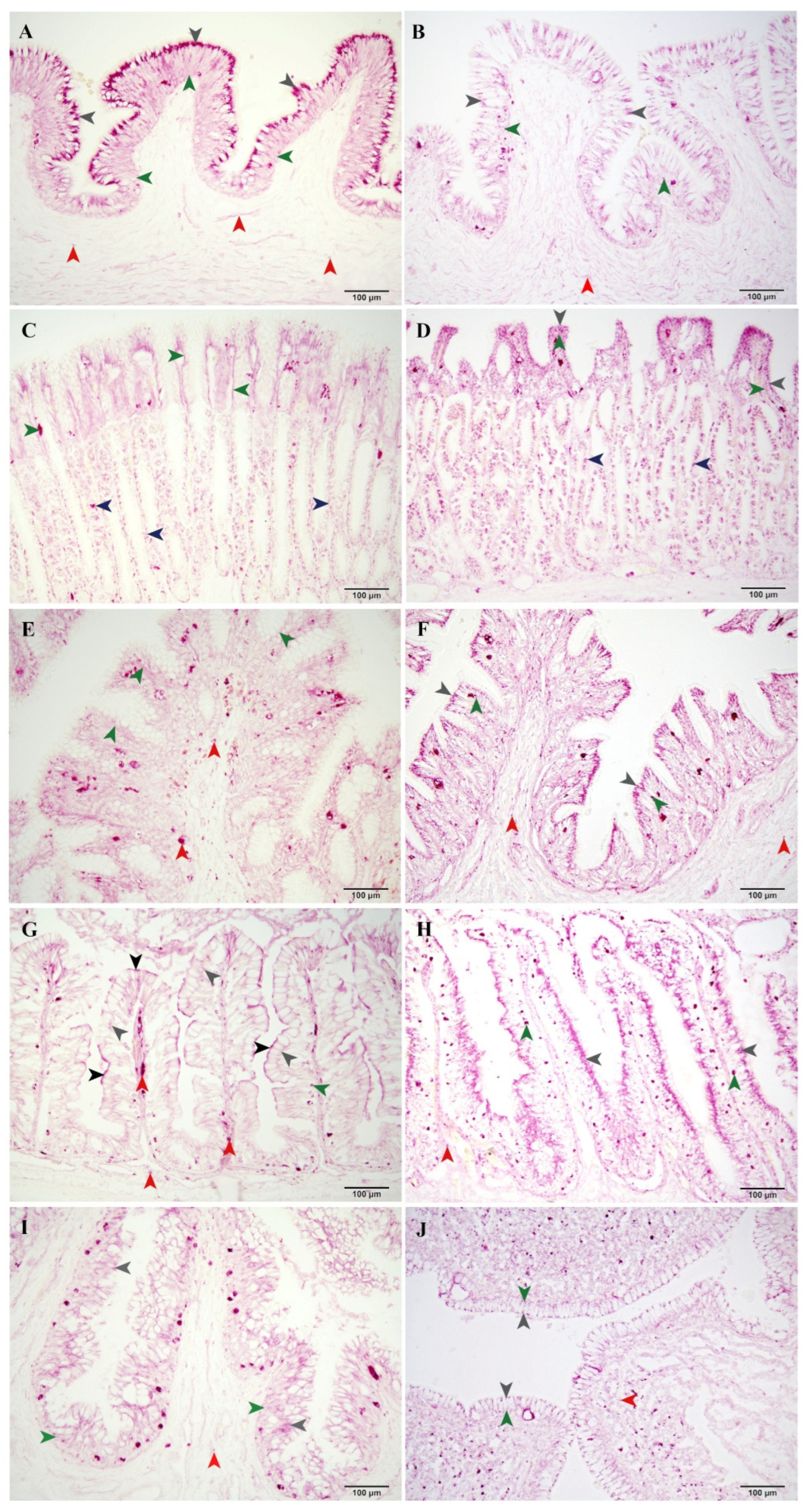
In the smooth-hound, ALP reactions were noted in the basal layer of the epithelium (Figure 2C). In both species, AP (Figure 3A,B) and NSE (isolated) reactions were observed in the cytoplasm of epithelial cells. Single granular positive reactions of AP (Figure 3A) and NSE were detected in the connective tissue layers. In the same layer in the smooth-hound, only AP reactions (Figure 3B) were found. The ALP reactions were localized around the blood capillaries (Figure 2C).

3.1.2. Stomach

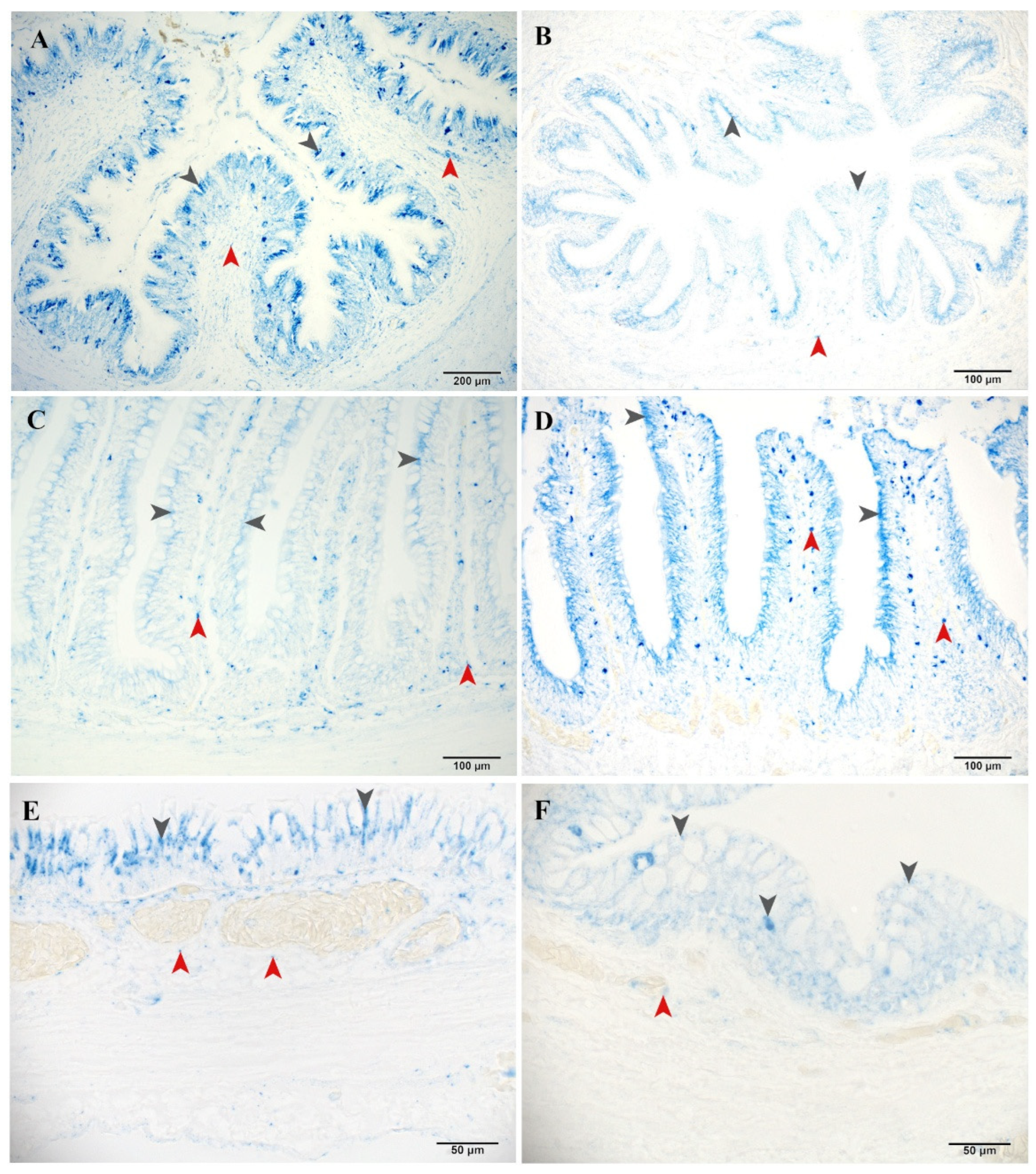
In the stomach of the small-spotted catshark, AP (Figure 3C,E) and NSE (Figure 5A) reactions were found in the perinuclear and basal part of the columnar epithelial cells. Positive reactions were also observed in the lamina propria. In the smooth-hound, positive reactions of these two enzymes were found at the same locations, except that they extended from the basal to the supranuclear part of the epithelium (Figure 3D,F and Figure 5B). In addition, positive reactions of the enzymes were found in the gastric glands in the anterior part of the stomach (Figure 3C,D). In the smooth-hound, ALP reactions were also found around the blood capillaries in the lamina propria.

3.1.3. Spiral Intestine

Positive reactions of ALP (Figure 2A,B) and A (Figure 7A,B) were detected in the brush border of the spiral intestine in both fish species. In the small-spotted catshark, reactions of AP were also observed in this part (Figure 3G). In both species, reactions of AP (Figure 3G,H) and NSE (Figure 5C,D) were found in epithelial cells. In the small-spotted catshark, the AP reactions were mainly localized in the apical part of the cells and rarely in the basal part (Figure 3G). In the smooth-hound, positive reactions were found in the supranuclear and rarely in the basal part of the cells (Figure 3H). In connective tissue of the lamina propria, positive granular reactions of AP (Figure 3G,H) and NSE (Figure 5C,D) were observed.

3.1.4. Rectum

Positive reactions of AP (Figure 3I,J) and NSE (Figure 5E,F) were observed in the cytoplasm of the epithelial cells and the connective tissue of propria-submucosa. In the smooth-hound, ALP reactions were also found around the blood capillaries in connective tissue.

3.2. Enzyme Activities in the Digestive Tract

3.2.1. Alkaline Phosphatase

In the small-spotted catshark, the ALP was only found in the brush border of the epithelial cells in the spiral intestine (0.261 ± 0.119) (Figure 2A). In the smooth-hound, the activity was also found in the same layer (0.188 ± 0.076) (Figure 2B) and additionally in the cytoplasm of epithelial cells in the esophagus (0.047 ± 0.012) (Figure 2C).

3.2.2. Acid Phosphatase

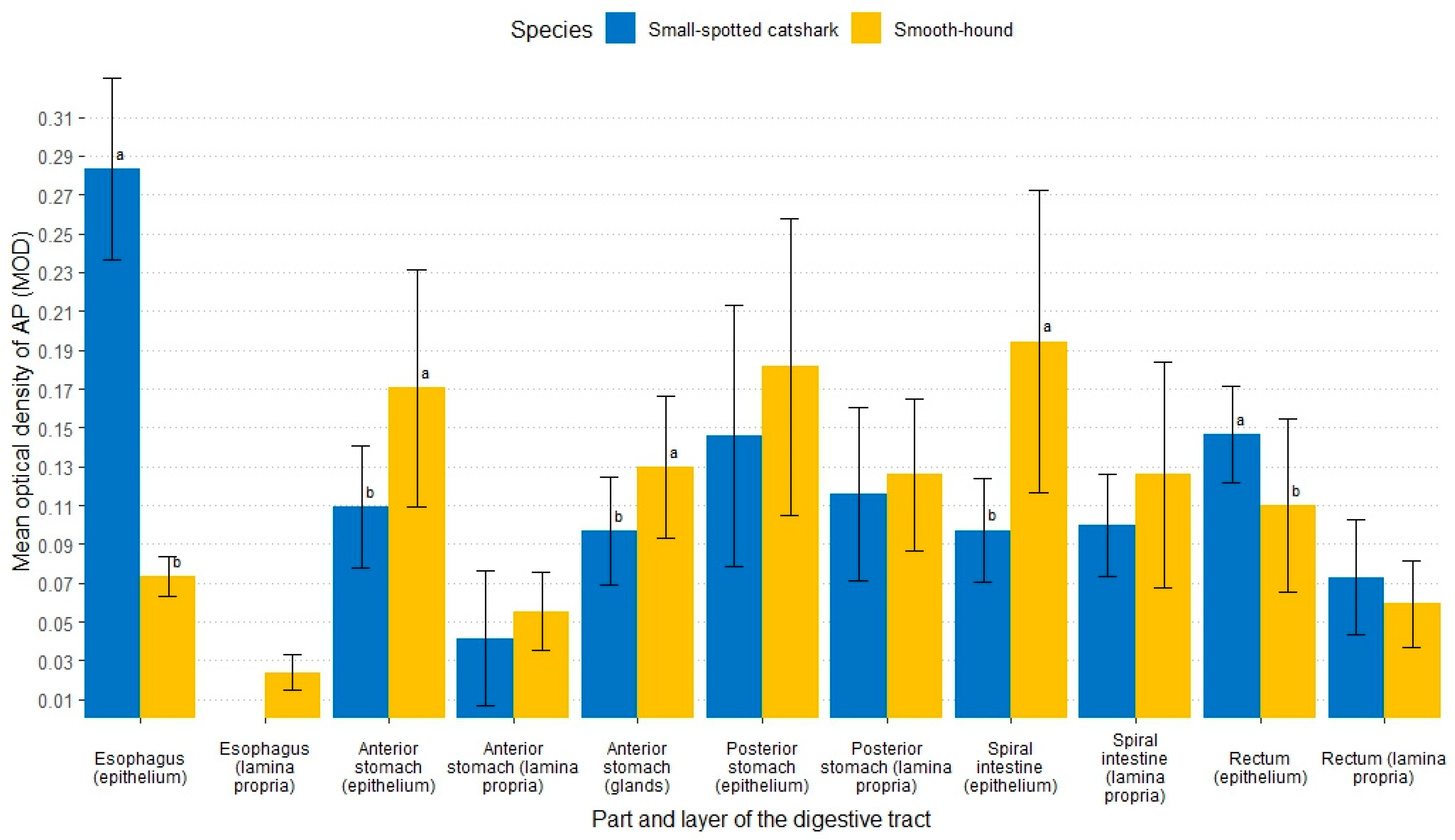
The values of statistical analysis of MOD of AP within fish species between the same layers in different parts of the digestive tract and in the same parts of the digestive tract between different layers were shown in Table 1. The statistical analysis of MOD within the same part and same layer of the digestive tract between the small-spotted catshark and smooth-hound was shown in Figure 4. In the brush border, the reaction of AP was only found in the spiral intestine of the small-spotted catshark (0.227 ± 0.117) (Figure 3G). In contrast, in the epithelium and connective tissue, AP activity was found in all parts of the digestive tract of both fish species (Figure 3). The highest MOD was measured in the cytoplasm of epithelium in the esophagus of the small-spotted catshark (0.283 ± 0.047) (Figure 3A), posterior part of the stomach (0.146 ± 0.067) (Figure 3E) and in the rectum (0.146 ± 0.025) (Figure 3I). In the smooth-hound the MOD increased up to the spiral intestine where it was highest (0.194 ± 0.078) (Figure 3H) and then decreased again. A significant difference between fish species was observed in the MOD of reactions in the epithelium of the esophagus, both parts of the stomach, the spiral intestine and the rectum (Figure 4).

3.2.3. Non-Specific Esterase

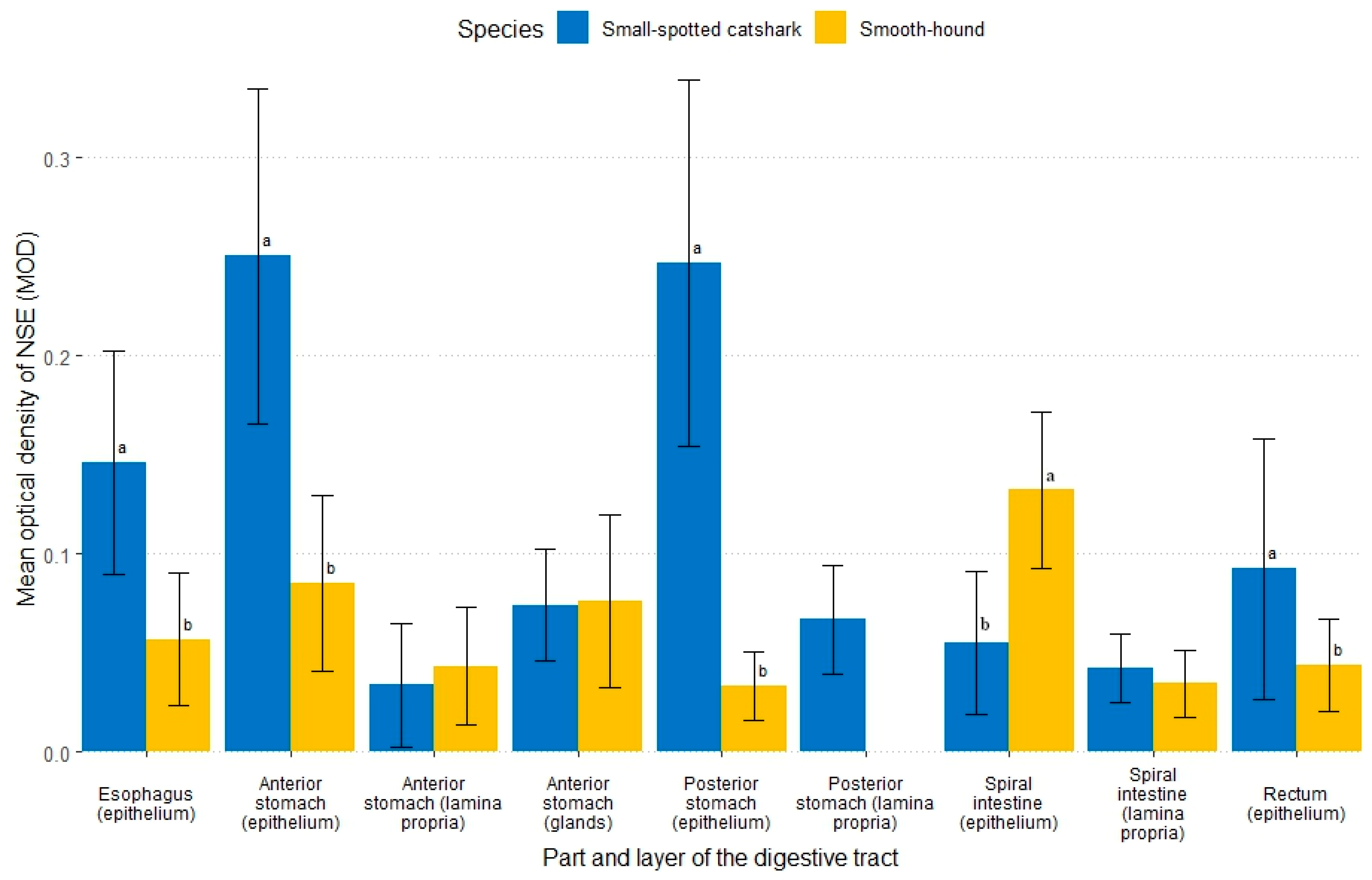
The values of statistical analysis of the MOD of NSE within fish species between the same layers in different parts of the digestive tract and in the same parts of the digestive tract between different layers were shown in Table 2. The statistical analysis of MOD within the same part and layer of the digestive tract between small-spotted catshark and smooth-hound was shown in Figure 6. The activity of NSE was found along the epithelium of the entire posterior part of the digestive tract. The highest value was measured in the stomach of the small-spotted catshark (0.250 ± 0.085 and 0.247 ± 0.092) (Figure 5A), while in the smooth-hound, it was measured in the epithelium of the spiral intestine (0.132 ± 0.039) (Figure 5D). The activity in the connective tissue was only found in the stomach and the spiral intestine. In gastric glands, similar values were observed in both investigated species. In general, higher values were measured in the small-spotted catshark than in the smooth-hound. A significant difference was found in the MOD of the reactions in the epithelium of the esophagus, both parts of the stomach, the spiral intestine and the rectum (Figure 6).
Figure 5. Distribution of non-specific esterase in the digestive tract of small-spotted catshark (A,C,E) and smooth-hound (B,D,F). Blue color indicates positive reactions in the posterior part of the stomach (A,B), spiral intestine (C,D) and rectum (E,F). Gray arrowheads show reactions in the epithelium and red in lamina propria.
Fishes 10 00386 g005
Figure 6. Mean optical density (MOD) of the non-specific esterase (NSE) within the same part and the same layer of the digestive tract between small-spotted catshark and smooth-hound. Letters (a, b) were used as indicators of statistically significant differences. Means within part and layer of digestive tract with different lowercase letters (a, b) were significantly different (p < 0.05).
Fishes 10 00386 g006

3.2.4. Aminopeptidase

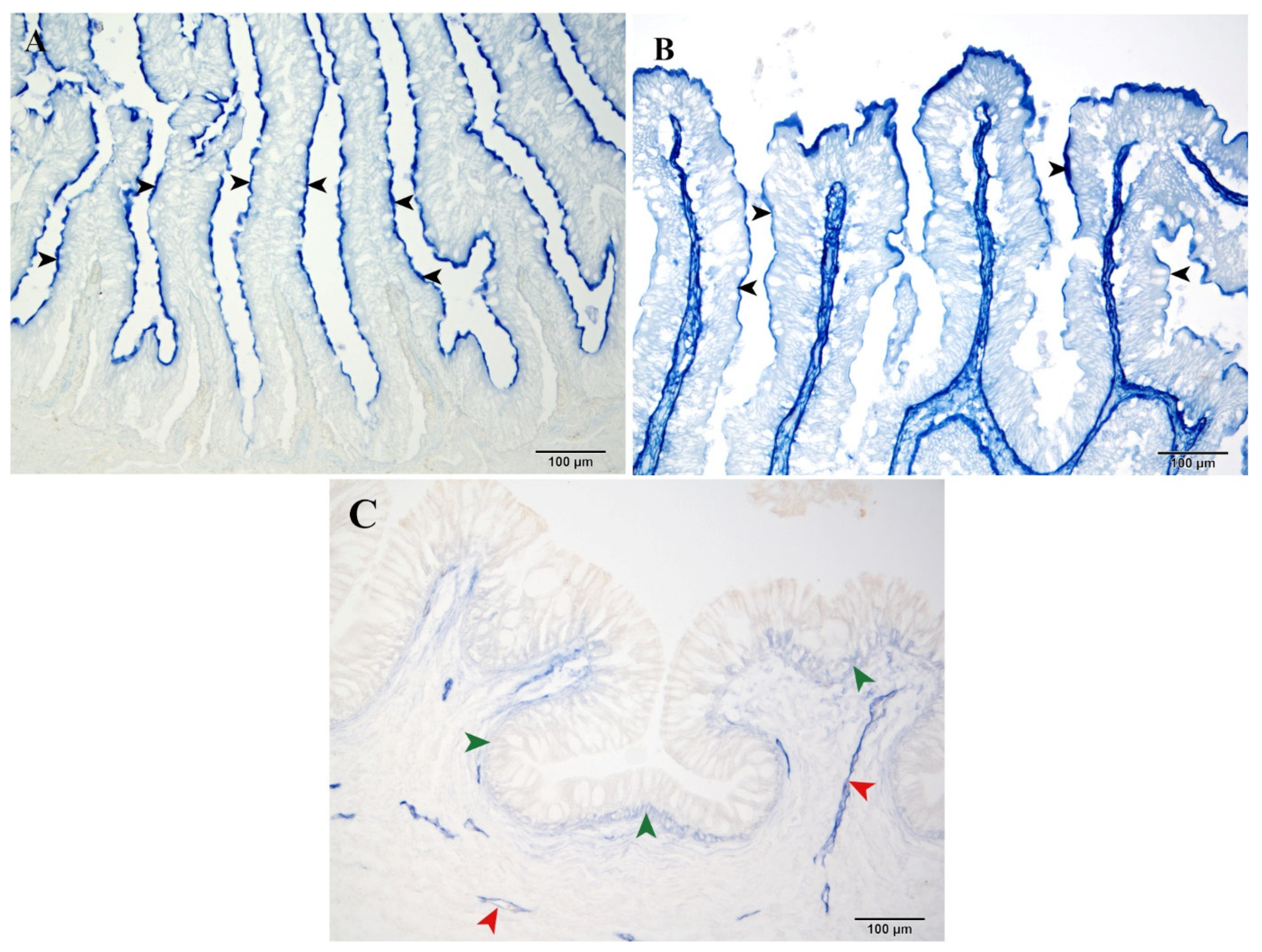
In both investigated species, the A reaction was found only in the brush border of the epithelium of the spiral intestine (Figure 7A,B). A higher MOD was measured in the smooth-hound than in the small-spotted catshark (0.128 ± 0.075 and 0.074 ± 0.011 respectively), and a significant difference was found between these two distributions.
Figure 7. Distribution of aminopeptidase in the spiral intestine of small-spotted catshark (A) and smooth-hound (B). Red color indicates positive reactions (black arrowheads).
Fishes 10 00386 g007

4. Discussion

In the available literature on cartilaginous fish, there are no data for the enzymes examined in this study, apart from the limited measurements of alkaline phosphatase [35] and aminopeptidase [36] in the brush border of the spiral intestine. This lack of data necessitates comparisons with bony fish to contextualize the results.
Alkaline phosphatase activity was found in the brush border of the spiral intestine in both investigated fish species. Similar results were reported for small-spotted catshark [35], European hake [6], large-scaled gurnard [13], European eel [14], porthole shovelnose catfish [20], half-smooth tongue sole [37], koi carp [38], tub gurnard [39], Jian carp [40], grass carp [41], rainbow trout [42], European barracuda [43] and many other fish species [43]. Alkaline phosphatase is an enzyme that catalyzes the hydrolysis of phosphoric acid monoesters at an alkaline pH. In the digestive tract, alkaline phosphatase is suggested to play a role in the metabolism of calcium, phosphorus, and fatty acids [44]. Therefore, its enzymatic activity was expected to be found in the brush border of the epithelium in the spiral intestine of cartilaginous fish. In the esophagus of the smooth-hound, additional activity was also found in the cytoplasm of epithelial cells. The enzyme activity observed in this part is consistent with findings reported in European hake [45]. Alkaline phosphatase is thought to act as a barrier and may be involved in the regulation of intestinal pH, the control of tight junctions, the detoxification of inflammatory microbial components and the modulation of the intestinal microbiota [44,45,46,47,48]. Therefore, additional enzyme activity in the basal part of the epithelium in the esophagus of the smooth-hound can be associated with a significant proportion of crustaceans in diet [27] and a location close to the body opening. This may suggest a protective function, where enzymatic activity found in the basal part of epithelium could serve to protect deeper layers from the mechanical impact of crustacean shells, potentially reflecting an adaptive digestive strategy in this species.
Acid phosphatase activity was detected throughout the digestive tract in both the small-spotted catshark and the smooth-hound. Data on acid phosphatase activity vary widely between different fish species [13,14,20,37,38,39]. Acid phosphatase is an enzyme that catalyzes the hydrolysis of phosphate monoesters in an acidic pH environment. After synthesis, it is mainly located in lysosomes but can also be found outside of them. Therefore, the intracellular distribution of this enzyme may vary depending on the metabolic state of the cell. The high MOD found in the esophagus and rectum of the small-spotted catshark could indicate a protective function. A similar finding was described in the tub gurnard [39]. Acid phosphatase activity is associated with cellular protein digestion [49]. In the small-spotted catshark, high MODs in the esophagus, posterior part of the stomach and rectum suggest that these segments may play an important role in protein metabolism. In contrast, in the smooth-hound, the posterior stomach and spiral intestine appear to be the main sites for intracellular protein degradation and absorption. Interestingly, the lower MOD observed in the spiral intestine of the small-spotted catshark compared to the smooth-hound presents an unexpected finding that necessitates further investigation.
Lipids are hydrophobic substances that do not mix with water. The primary function of emulsifiers, such as bile salts, is to facilitate emulsification, enabling the dispersion of lipid aggregates into smaller droplets and improving their interaction with digestive enzymes. This process enhances hydrolysis and nutrient absorption. In the digestive tract, bile secreted into the anterior part of the spiral intestine plays a key role in emulsification by increasing the surface area available for enzymatic degradation. The higher non-specific esterase activity observed in the intestine of the smooth-hound may be attributed to elevated enzymatic responses following lipid emulsification. While the intestine proper is frequently cited in the literature as the site of the highest enzymatic activity, most studies focus solely on this specific part [13,14,43]. However, the findings of this study suggest that cellular lipid digestion in the small-spotted catshark may occur primarily in the stomach, rather than the intestine. These results are particularly noteworthy as the enzymatic activity was detected before the entry point of bile into the digestive tract, implying a potential role for gastric enzymes in lipid processing. Such a finding highlights the need for broader investigations into fish digestion that extends beyond the intestine, considering the functional role of the other parts of the digestive tract and their potential influence on nutrient breakdown.
Aminopeptidase activity was measured in the brush border of the epithelium of the spiral intestine in the small-spotted catshark and smooth-hound. These results are in accordance with those previously reported in bonnethead shark [36] and in the intestine of many teleost fish such as European eel [14], perch [50] and Nile tilapia [51]. The hydrolysis of peptide bonds at the N-terminus of protein substrates is catalyzed by aminopeptidase. The extracellular phase of protein degradation is facilitated by pepsin and pancreatic enzymes, while cellular degradation is associated with brush border proteins [39]. Although this enzyme is typically associated with the brush border, its presence in the other parts layers and parts may indicate a broader or alternative functional role, potentially extending beyond membrane digestion [39]. Again, this highlights the importance of investigating the entire digestive tract as cellular protein degradation may occur not just in the brush border of the enterocytes but also in other parts of the digestive tract.

5. Conclusions

This study, based on histochemical analysis, provides valuable data about four important enzymes involved in cellular digestion. The small-spotted catshark and the smooth-hound are cartilaginous, carnivorous fish species with a similar depth range in their benthic habitats. Although their digestive tract morphology is similar, differences in enzymatic activity have been observed. While the intestines are the site of intense metabolism, the entire digestive tract is involved in the digestion of nutrients. In the small-spotted catshark, the stomach is the main location for cellular lipid digestion. In both species, the entire digestive tract is involved in protein metabolism. Given the importance of objective and reproducible assessment of histochemical staining, this study employed computer-assisted image analysis to specify OD. To the best of our knowledge, this is not only the first study to apply such a method in assessing the activity of key digestive enzymes in these species but also the first description of their cellular enzyme distribution within the digestive tract. This research underscores the necessity of investigating the entire digestive tract as a step toward a more comprehensive understanding of digestive processes in fish, recognizing that further studies are needed to fully elucidate their complexity. Moreover, it fills an important knowledge gap regarding the cellular digestive processes of these two cartilaginous fish species and contributes to a better understanding of their physiology and biology. The results provide a physiological basis that can support future conservation efforts by improving the understanding of digestive tract adaptations and interspecies variability in cartilaginous fish. This knowledge is important to assess the resilience of species and support strategies aimed at conserving marine biodiversity. Considering the ecological importance and reported decline of populations of these two elasmobranch species in the Adriatic Sea, the physiological findings of this study represent an important step towards better informed and targeted conservation and research efforts.

Author Contributions

Conceptualization, L.D., S.K., D.V. and M.L.; Methodology, L.D., S.K., D.V. and M.L.; Software, L.D. and I.V.; Formal Analysis, L.D., S.K. and I.V.; Investigation, L.D.; Resources, D.V. and L.D.; Data Curation, L.D.; Writing—Original Draft Preparation, L.D., S.K. and D.V.; Writing—Review and Editing, L.D., D.V., S.K., I.V., M.P., V.B., S.F. and M.L.; Visualization, V.B., I.V. and S.F.; Supervision, D.V. and S.K. All authors have read and agreed to the published version of the manuscript.

Funding

This research was conducted as part of the scientific project “Improvement of cooperation between fishermen and scientists for the purpose of introducing advanced technologies of marking of fishing tools, fish health and environmental protection” supported by the Operational Programme for Maritime Affairs and Fisheries of the Republic of Croatia for the programming period 2014–2020. No funding was received to assist with the preparation of this manuscript.

Institutional Review Board Statement

The study was conducted according to the guidelines of the Animal Protection Act by the Ministry of Agriculture and approved by the Committee for Ethics in Veterinary Medicine, Faculty of Veterinary Medicine (No. 251-61-01/139-20-32).

Informed Consent Statement

Not applicable.

Data Availability Statement

The raw data supporting the conclusions of this article will be made available by the authors on request due to the large size and complexity of the dataset.

Acknowledgments

The authors extend their gratitude to Nada Crnogaj, Department of Anatomy, Histology and Embryology, for her technical assistance.

Conflicts of Interest

The authors have no relevant financial or non-financial interests to disclose.

References

  1. Ray, A.K.; Ringø, E. The gastrointestinal tract of fish. In Aquacult Nutr: Gut Health, Probiotics and Prebiotics; Merrifield, D., Ringø, E., Eds.; John Wiley & Sons: Chichester, UK, 2014; pp. 1–10. [Google Scholar]
  2. Duque-Correa, M.J.; Clements, K.D.; Meloro, C.; Ronco, F.; Boila, A.; Indermaur, A.; Salzburger, W.; Clauss, M. Diet and habitat as determinants of intestine length in fishes. Rev. Fish Biol. Fish. 2024, 34, 1017–1034. [Google Scholar] [CrossRef]
  3. German, D.P.; Nagle, B.C.; Villeda, J.M.; Ruiz, A.M.; Thomson, A.W.; Contreras Balderas, S.; Evans, D.H. Evolution of herbivory in a carnivorous clade of minnows (Teleostei: Cyprinidae): Effects on gut size and digestive physiology. Physiol. Biochem. Zool. 2010, 83, 1–18. [Google Scholar] [CrossRef]
  4. Horn, M.H.; Gawlicka, A.; German, D.P.; Logothetis, E.A.; Cavanagh, J.W.; Boyle, K.S. Structure and function of the stomachless digestive system in three related species of New World silverside fishes (Atherinopsidae) representing herbivory, omnivory, and carnivory. Mar. Biol. 2006, 149, 1237–1245. [Google Scholar] [CrossRef]
  5. Denstadli, V.; Vegusdal, A.; Krogdahl, Å.; Bakke-McKellep, A.M.; Berge, G.M.; Holm, H.; Hillestad, M.; Ruyter, B. Lipid absorption in different segments of the gastrointestinal tract of Atlantic salmon (Salmo salar L.). Aquaculture 2004, 240, 385–398. [Google Scholar] [CrossRef]
  6. Kozarić, Z.; Kužir, S.; Nejedli, S.; Petrinec, Z.; Srebočan, E. Histochemical distribution of digestive enzymes in hake, Merluccius merluccius L. 1758. Vet. Arh. 2004, 74, 299–308. [Google Scholar]
  7. Refstie, S.; Bakke-McKellep, A.M.; Penn, M.H.; Sundby, A.; Shearer, K.D.; Krogdahl, A. Capacity for digestive hydrolysis and amino acid absorption in Atlantic salmon (Salmo salar) fed diets with soybean meal or inulin with or without addition of antibiotics. Aquaculture 2006, 261, 392–406. [Google Scholar] [CrossRef]
  8. Zhao, Y.; Hu, Y.; Zhou, X.Q.; Zeng, X.Y.; Feng, L.; Liu, Y.; Jiang, W.D.; Li, S.H.; Li, D.B.; Wu, X.Q.; et al. Effects of dietary glutamate supplementation on growth performance, digestive enzyme activities and antioxidant capacity in intestine of grass carp (Ctenopharyngodon idella). Aquacult. Nutr. 2015, 21, 935–941. [Google Scholar] [CrossRef]
  9. Cohen, S.; Diaz, M.V.; Díaz, A.O. Development of the digestive system of Argentine hake, Merluccius hubbsi, larvae. J. Morphol. 2020, 281, 578–590. [Google Scholar] [CrossRef] [PubMed]
  10. Ballantyne, J.S. Membranes and metabolism. In The Physiology of Fishes, 4th ed.; Evans, D.H., Claiborne, J.B., Currie, S., Eds.; CRC Press: Boca Raton, FL, USA; London, UK; New York, NY, USA, 2014; pp. 81–149. [Google Scholar]
  11. Cho, J.-H.; Park, J.W.; Ryu, Y.-W.; Kim, K.-W.; Hur, S.-W. Morphology, Histology, and Histochemistry of the Digestive Tract of the Marbled Flounder Pseudopleuronectes yokohamae. Animals 2023, 13, 936. [Google Scholar] [CrossRef]
  12. Ugolev, A.M. Membrane (contact) digestion. Physiol. Rev. 1965, 45, 555–595. [Google Scholar] [CrossRef]
  13. Kozarić, Z.; Petrinec, Z.; Kužir, S.; Gjurčević, E.; Baždarić, B. Histochemical analyses of digestive enzymes in the intestine of adult large-scaled gurnard (Lepidotrigla cavillone, Lacepède, 1801). Anat. Histol. Embryol. 2011, 40, 314–320. [Google Scholar] [CrossRef]
  14. Kužir, S.; Gjurčević, E.; Nejedli, S.; Baždarić, B.; Kozarić, Z. Morphological and histochemical study of intestine in wild and reared European eel (Anguilla anguilla L.). Fish Physiol. Biochem. 2012, 38, 625–633. [Google Scholar] [CrossRef] [PubMed]
  15. Weinrauch, A.M.; Schaefer, C.M.; Goss, G.G. Activity and post-prandial regulation of digestive enzyme activity along the Pacific hagfish (Eptatretus stoutii) alimentary canal. PLoS ONE 2019, 14, e0215027. [Google Scholar] [CrossRef] [PubMed]
  16. Kolkovski, S. Digestive enzymes in fish larvae and juveniles—Implications and applications to formulated diets. Aquaculture 2001, 200, 181–201. [Google Scholar] [CrossRef]
  17. Gawlicka, A.; Herold, M.A.; Barrows, F.T.; De La Noüe, J.; Hung, S.S.O. Effects of dietary lipids on growth, fatty acid composition, intestinal absorption and hepatic storage in white sturgeon (Acipenser transmontanus R.) larvae. J. Appl. Ichthyol. 2002, 18, 673–681. [Google Scholar] [CrossRef]
  18. Solovyev, M.M.; Kashinskaya, E.N.; Izvekova, G.I.; Glupov, V.V. pH Values and Activity of Digestive Enzymes in the Gastrointestinal Tract of Fish in Lake Chany (West Siberia). J. Ichthyol. 2015, 55, 251–258. [Google Scholar] [CrossRef]
  19. Solovyev, M.; Gisbert, E. Influence of time, storage temperature and freeze/thaw cycles on the activity of digestive enzymes from gilthead sea bream (Sparus aurata). Fish Physiol. Biochem. 2016, 42, 1383–1394. [Google Scholar] [CrossRef]
  20. Faccioli, C.K.; Chedid, R.A.; Mori, R.H.; Amaral, A.C.; Belmont, R.A.F.; Vicentini, I.B.F.; Vicentini, C.A. Organogenesis of the digestive system in Neotropical carnivorous freshwater catfish Hemisorubim platyrhynchos (Siluriformes: Pimelodidae). Aquaculture 2016, 451, 205–212. [Google Scholar] [CrossRef]
  21. Hani, Y.M.I.; Marchand, A.; Turiès, C.; Kerambrun, E.; Palluel, O.; Bado-Nilles, A.; Beaudouin, R.; Porcher, J.; Geffard, A.; Dedourge-Geffard, O. Digestive enzymes and gut morphometric parameters of threespine stickleback (Gasterosteus aculeatus): Influence of body size and temperature. PLoS ONE 2018, 13, e0194932. [Google Scholar] [CrossRef]
  22. Abdulla, A. Status and Conservation of Sharks in the Mediterranean Sea; IUCN Technical Paper, 2004; p. 7. Available online: https://www.researchgate.net/publication/237278296_Status_and_Conservation_of_Sharks_in_the_Mediterranean_Sea (accessed on 28 May 2025).
  23. Simpfendorfer, C.A.; Dulvy, N.K. Bright spots of sustainable shark fishing. Curr. Biol. 2017, 27, 97–98. [Google Scholar] [CrossRef]
  24. Nuez, I.; Giovos, I.; Tiralongo, F.; Penadés-Suay, J.; Cetkovic, I.; Di Lorenzo, M.; Kleitou, P.; Bakiu, R.; Bradai, M.N.; Almabruk, S.A.A.; et al. Assessing the current status of Hexanchus griseus in the Mediterranean Sea using local ecological knowledge. Mar. Policy 2023, 147, 105378. [Google Scholar] [CrossRef]
  25. Ebert, D.A.; Stehmann, M.F.W. Sharks, Batoids, and Chimaeras of the North Atlantic; FAO Species Catalogue for Fishery Purposes, No. 7; FAO: Rome, Italy, 2013; pp. 188–219. [Google Scholar]
  26. Jardas, I. Jadranska Ihtiofauna; Školska Knjiga: Zagreb, Croatia, 1996. [Google Scholar]
  27. Jardas, I.; Anti, M.; Nerlović, V.; Pallaoro, A. Diet of the smooth-hound, Mustelus mustelus (Chondrichthyes: Triakidae), in the eastern Adriatic Sea. Cybium 2007, 31, 459–464. [Google Scholar]
  28. Šantić, M.; Rađa, B.; Pallaoro, A. Feeding habits of small-spotted catshark (Scyliorhinus canicula Linnaeus, 1758) from the eastern central Adriatic Sea. Mar. Biol. Res. 2012, 8, 1003–1011. [Google Scholar] [CrossRef]
  29. Koutseni, V.; Karachle, P.K.; Megalofonou, P. Diet of the small-spotted catshark Scyliorhinus canicula in the Aegean Sea (eastern Mediterranean). Mar. Biol. Res. 2017, 13, 161–173. [Google Scholar] [CrossRef]
  30. Dulčić, J.; Kovačić, M. Ihtiofauna Jadranskoga Mora; Golden marketing—Tehnička Knjiga: Zagreb, Croatia, 2020. [Google Scholar]
  31. Suvarna, S.K.; Layton, C.; Bancroft, J.D. Bancroft’s Theory and Practice of Histological Techniques; Elsevier: Scotland, UK, 2019. [Google Scholar]
  32. Lojda, Z.; Gossrau, R.; Schiebler, T.H. Enzyme Histochemistry. A Laboratory Manual; Springer: Berlin/Heidelberg, Germany; New York, NY, USA, 1979. [Google Scholar]
  33. Pearse, A.G.E. Histochemistry, Theoretical and Applied, 3rd ed.; J. & A. Churchill Ltd.: London, UK, 1968. [Google Scholar]
  34. Sheehan, D.C.; Hrapchak, B.B. Theory and Practice of Histotechnology; Battelle Press: Columbus, OH, USA, 1980. [Google Scholar]
  35. Crane, R.K.; Boge, G.; Rigal, A. Isolation of brush border membranes in vesicular form from the intestinal spiral valve of the small dogfish (Scyliorhinus canicula). Biochim. Biophys. Acta 1979, 554, 264–267. [Google Scholar] [CrossRef]
  36. Jhaveri, P.; Papastamatiou, Y.P.; German, D.P. Digestive enzyme activities in the guts of bonnethead sharks (Sphyrna tiburo) provide insight into their digestive strategy and evidence for microbial digestion in their hindguts. Comp. Biochem. Physiol. A 2015, 189, 76–83. [Google Scholar] [CrossRef] [PubMed]
  37. Wang, Y.Z.; Sun, J.F.; Lv, A.J.; Zhang, S.L.; Sung, Y.Y.; Shi, H.Y.; Hu, X.C.; Chen, S.J.; Xing, K.Z. Histochemical distribution of four types of enzymes and mucous cells in the gastrointestinal tract of reared half-smooth tongue sole Cynoglossus semilaevis. J. Fish Biol. 2018, 92, 3–16. [Google Scholar] [CrossRef]
  38. Sun, J.; Wang, Y.; Lv, A.; Xian, J.-A.; Wang, Q.; Zhang, S.; Guo, Y.; Xing, K. Histochemical distribution of four types of enzymes and mucous cells in the intestine of koi carp (Cyprinus carpio var. koi). Fish Physiol. Biochem. 2019, 45, 1367–1376. [Google Scholar] [CrossRef] [PubMed]
  39. Bastiančić, L.; Vlahek, I.; Benko, V.; Lovrić, M.; Valić, D.; Kužir, S. Histochemical research of enzymes involved in cellular digestion in the digestive tract of tub gurnard, Chelidonichthys lucerna. Fish Physiol. Biochem. 2023, 50, 157–170. [Google Scholar] [CrossRef]
  40. Xiao, W.; Jiang, W.; Feng, L.; Liu, Y.; Wu, P.; Jiang, J.; Zhang, Y.; Zhou, X. Supplementation of enzyme-treated soy protein saves dietary protein and promotes digestive and absorptive ability referring to TOR signaling in juvenile fish. Fish Physiol. Biochem. 2017, 43, 1657–1675. [Google Scholar] [CrossRef]
  41. Wei, S.P.; Jiang, W.D.; Wu, P.; Liu, Y.; Zeng, Y.Y.; Jiang, J.; Kuang, S.Y.; Tang, L.; Zhang, Y.A.; Zhou, X.Q.; et al. Dietary magnesium deficiency impaired intestinal structural integrity in grass carp (Ctenopharyngodon idella). Sci. Rep. 2018, 8, 12705. [Google Scholar] [CrossRef]
  42. Pasquariello, R.; Pavlovic, R.; Chacon, M.A.; Camin, F.; Verdile, N.; Løkka, G.; Panseri, S.; Faustini, M.; Tandler, A.; Peggs, D.; et al. Development of a Rainbow Trout (Oncorhynchus mykiss) Intestinal In Vitro Platform for Profiling Amino Acid Digestion and Absorption of a Complete Diet. Animals 2023, 13, 2278. [Google Scholar] [CrossRef]
  43. Tlak Gajger, I.; Nejedli, S.; Kozarić, Z.; Vlainić, J. Histochemical analysis and distribution of digestive enzymes in the gastrointestinal system of the European barracuda Sphyraena sphyraena (Linnaeus, 1758). Animals 2024, 14, 2798. [Google Scholar] [CrossRef]
  44. Lallès, J.P. Intestinal alkaline phosphatase in the gastrointestinal tract of fish: Biology, ontogeny, and environmental and nutritional modulation. Rev. Aquacult. 2019, 12, 555–581. [Google Scholar] [CrossRef]
  45. Devčić, L.; Valić, D.; Lovrić, M.; Vlahek, I.; Benko, V.; Kužir, S. Comprehensive study of the digestive tract in the European hake (Merluccius merluccius). Slov. Vet. Res. 2024; in press. [Google Scholar] [CrossRef]
  46. Lallès, J.P. Intestinal alkaline phosphatase, inflammation and nutrition: Recent advances. Nutr. Rev. 2019, 77, 710–724. [Google Scholar] [CrossRef]
  47. Lallès, J.P. Intestinal alkaline phosphatase: Novel functions and protective effects. Nutr. Rev. 2014, 72, 82–94. [Google Scholar] [CrossRef] [PubMed]
  48. Rader, B.A. Alkaline phosphatase, an unconventional immune protein. Front. Immunol. 2017, 8, 897. [Google Scholar] [CrossRef]
  49. Ostaszewska, T.; Kamaszewski, M. Digestive system. In The Histology of Fishes; Kirschbaum, F., Formicki, K., Eds.; CRC Press: Boca Raton, FL, USA; London, UK; New York, NY, USA, 2019; pp. 88–100. [Google Scholar]
  50. Hirji, K.N.; Courtney, W.A.M. Leucine aminopeptidase activity in the digestive tract of perch, Perca fluviatilis L. J. Fish Biol. 1982, 21, 615–622. [Google Scholar] [CrossRef]
  51. Tengjaroenkul, B.; Smith, B.J.; Caceci, T.; Smith, S.A. Distribution of intestinal enzyme activities along the intestinal tract of cultured Nile tilapia, Oreochromis niloticus L. Aquaculture 2000, 182, 317–327. [Google Scholar] [CrossRef]
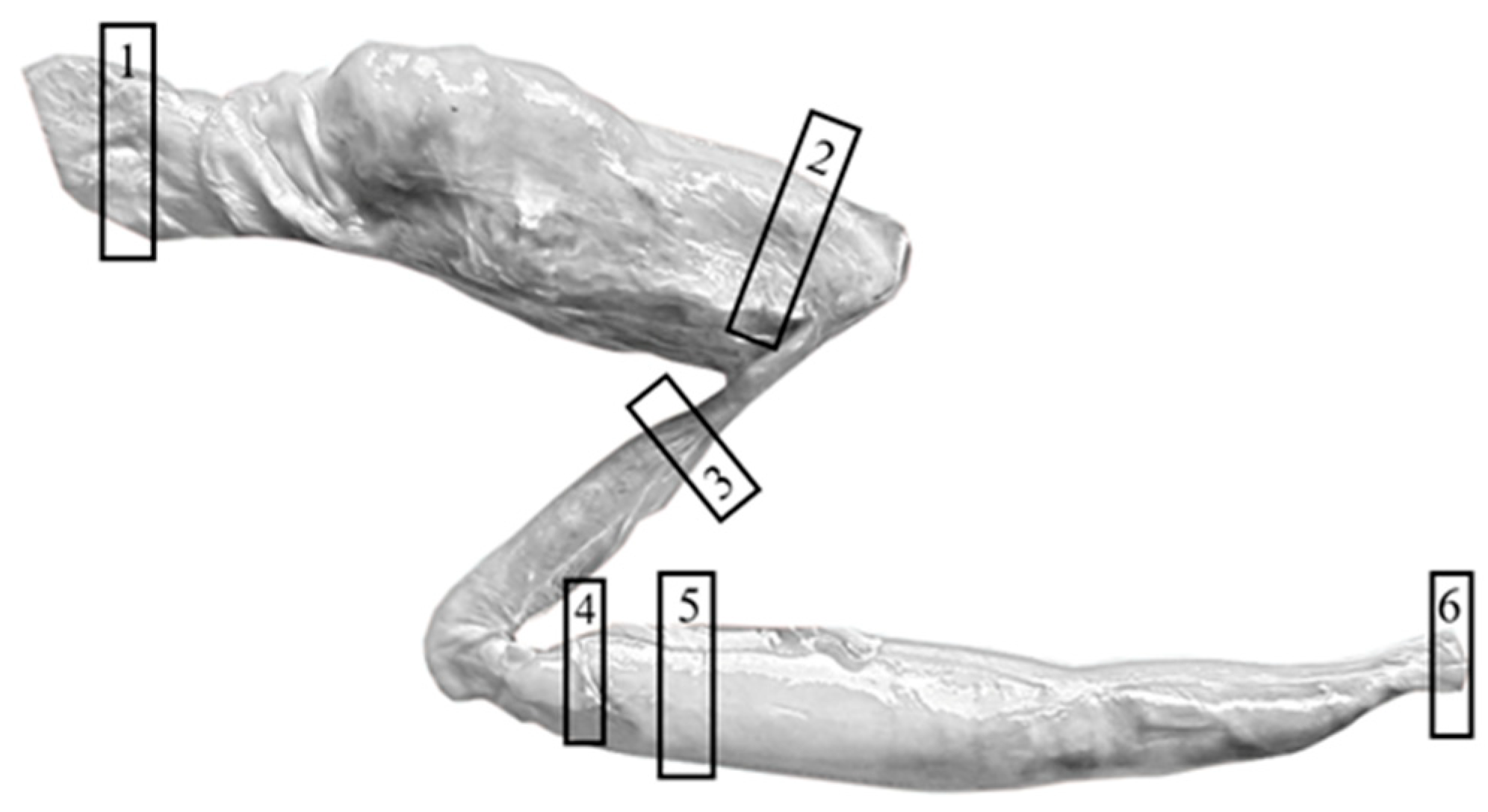
Figure 1. Sampling sites in the digestive tract for histochemical enzyme analysis. Black rectangles indicate sampling sites for the investigation of digestive enzymes in cells. The following areas are labeled: esophagus (1), the anterior (2) and posterior (3) part of the stomach, the anterior (4) and spiral (5) part of the intestine and the rectum (6).
Fishes 10 00386 g001
Figure 2. Distribution of alkaline phosphatase in the digestive tract of small-spotted catshark (A) and smooth-hound (B,C). Blue color indicates positive reactions in the brush border of the spiral intestine (A,B), in the basal part of the esophageal epithelium (C) and around blood vessels (C). Black arrowheads show positive reactions in the brush border, green in the basal part of the epithelium and red in the connective tissue.
Fishes 10 00386 g002
Figure 3. Distribution of acid phosphatase in the digestive tract of small-spotted catshark (A,C,E,G,I) and smooth-hound (B,D,F,H,J). Pink color indicates positive reactions in the esophagus (A,B), anterior (C,D) and posterior (E,F) part of the stomach, spiral intestine (G,H) and rectum (I,J). Black arrowheads show reactions in the brush border, gray in the apical and supranuclear part of the epithelium, green in perinuclear and basal part of the epithelium, red in the connective tissue and blue in the gastric glands.
Fishes 10 00386 g003
Figure 4. Mean optical density (MOD) of the acid phosphatase (AP) within the same part and the same layer of the digestive tract between small-spotted catshark and smooth-hound. Letters (a, b) were used as indicators of statistically significant differences. Means within part and layer of digestive tract with different lowercase letters (a, b) were significantly different (p < 0.05).
Fishes 10 00386 g004
Table 1. Distribution of acid phosphatase in the digestive tract measured by mean optical density (MOD).
Fish SpeciesDigestive TractEpithelium (MOD)Connective Tissue (MOD)Gastric Glands (MOD)
Brush BorderCytoplasm
Small-spotted catsharkEsophagus/0.283 a ± 0.047//
Stomach
Anterior part/0.109 bA ± 0.0320.042 cB ± 0.0340.097 A ± 0.028
Posterior part/0.146 b ± 0.0670.116 a ± 0.045/
Spiral intestine0.227 A ± 0.1170.097 bB ± 0.0270.100 abB ± 0.026/
Rectum/0.146 bA ± 0.0250.073 bcB ± 0.030/
Smooth-houndEsophagus/0.074 c ± 0.0100.024 ± 0.009/
Stomach
Anterior part/0.170 abA ± 0.0610.056 B ± 0.0200.130 A ± 0.037
Posterior part/0.181 ab ± 0.0760.126 ± 0.039/
Spiral intestine/0.194 a ± 0.0780.126 ± 0.058/
Rectum/0.110 bc ± 0.0440.059 ± 0.022/
The values were represented as mean ± SD. Superscript letters (a,b,c,A,B) were used as indicators of statistically significant differences. Means within columns and within fish species with different lowercase superscripts (a,b,c) were significantly different (p < 0.05). Means within rows and within fish species with different uppercase superscripts (A,B) were significantly different (p < 0.05). If positive reaction was not observed or it was isolated, it was marked with /. Significance of the differences were tested using Mann–Whitney U-test and Kruskal–Wallis ANOVA.
Table 2. Distribution of non-specific esterase in the digestive tract measured by mean optical density (MOD).
Fish SpeciesDigestive TractEpithelium
(MOD)
Connective Tissue (MOD)Gastric Glands (MOD)
Small-spotted catsharkEsophagus0.146 b ± 0.056//
Stomach
Anterior part0.250 aA ± 0.0850.034 bB ± 0.0310.074 B ± 0.028
Posterior part0.247 a ± 0.0920.067 a ± 0.027/
Spiral intestine0.055 c ± 0.0360.042 ab ± 0.017/
Rectum0.093 bc ± 0.066//
Smooth-houndEsophagus0.057 bc ± 0.033//
Stomach
Anterior part0.086 b ± 0.0440.043 ± 0.0290.076 ± 0.044
Posterior part0.033 c ± 0.017//
Spiral intestine0.132 aA ± 0.0390.034 B ± 0.017/
Rectum0.044 bc ± 0.023//
The values were represented as mean ± SD. Superscript letters (a,b,c,A,B) were used as indicators of statistically significant differences. Means within columns and within fish species with different lowercase superscripts (a,b,c) were significantly different (p < 0.05). Means within rows and within fish species with different uppercase superscripts (A,B) were significantly different (p < 0.05). If positive reaction was not observed or it was isolated, it was marked with /. Significance of the differences were tested using Mann–Whitney U-test and Kruskal–Wallis ANOVA.
Disclaimer/Publisher’s Note: The statements, opinions and data contained in all publications are solely those of the individual author(s) and contributor(s) and not of MDPI and/or the editor(s). MDPI and/or the editor(s) disclaim responsibility for any injury to people or property resulting from any ideas, methods, instructions or products referred to in the content.