Article

A Novel Approach to Perivitelline Fluid Extraction from Live Water-Activated Eggs from Zebrafish, Danio rerio

Department of Zoology, Division of Sciences, University of Otago, P.O. Box 56, Dunedin 9054, New Zealand
*
Author to whom correspondence should be addressed.
Fishes 2025, 10(8), 369; https://doi.org/10.3390/fishes10080369 (registering DOI)
Submission received: 17 June 2025 / Revised: 8 July 2025 / Accepted: 24 July 2025 / Published: 1 August 2025

Abstract

The collection of perivitelline fluid (PVF) from early-stage post-activation zebrafish (Danio rerio) eggs/embryos poses a significant challenge owing to the liability of the egg/embryo to sustain damage and rupture during handling. Rupture of the blastoderm and/or yolk presents a major risk of PVF sample contamination. Previous efforts to extract PVF at such early stages have employed formalin fixation to enhance the structural integrity of the blastoderm and yolk syncytial layer, thereby reducing the likelihood of contamination. While this approach successfully mitigates blastoderm and yolk rupture, formaldehyde fixation may cause issues with downstream proteomic analyses. Recent findings indicate that zebrafish PVF contains a range of maternally inherited proteins involved in innate immune defence. However, current extraction methods compromise the reliability of downstream protein analyses, raising concerns that fixation-induced protein crosslinking may obscure the presence of maternally inherited proteins during the earliest stages of development. The micro-aspiration technique described here allows for the precise extraction of PVF from living, water-activated eggs with minimal disruption to the blastodisc and yolk. This method reduces the risk of contamination from other non-target proteinaceous egg sources and eliminates the need for formalin fixation, thereby improving the integrity of PVF samples and enhancing the reliability of subsequent downstream analyses.
Key Contribution: This paper details a novel approach to perivitelline fluid extraction from live water-activated zebrafish eggs. This technique should facilitate future endeavours towards developing our understanding of maternal contributions to the perivitelline fluid proteome.

1. Introduction

The perivitelline fluid (PVF) is a biologically complex extraembryonic fluid, enclosed between the embryo and the chorion (Figure 1), which plays a vital role in the regulation of the local microenvironment throughout teleost embryonic development [1,2,3,4,5,6,7]. Interestingly, recent evidence suggests that the zebrafish (Danio rerio) PVF engulfs the embryo in a cocktail of maternally inherited proteins, many of which have been found to be broadly associated with innate immune defence [8]. The principal source of these immune-related proteins in the PVF are cortical alveoli (CA), which undergo a mass exocytosis into the perivitelline space (PVS) upon egg activation [9,10]. While the PVF is typically considered a sterile medium, both environmental and vertical transmission of bacteria into the PVS have been observed [11,12]. This transmission of bacteria primarily occurs via the passive entry of bacteria through the micropyle and into the PVS prior to the occlusion of the micropyle [13,14,15]. Moreover, in some fish, such as Oncorhynchus tshawytscha and Oncorhynchus kisutch, the plugs in the pore canals of the chorion are relatively “loosely set” and may become even looser in colder conditions [16]. This change in chorion permeability could provide access to the PVS for opportunistic bacteria or fungi [16]. Exacerbating this risk, pathogens such as Flexibacter spp. and Saprolegnia spp. can degrade and rupture the chorion, thereby compromising its protective functions [17,18].
The PVF also acts as an integral defensive barrier against environmental toxicants, which can penetrate the chorionic barrier and enter the PVS, where CA-derived glycoproteins are thought to act as a secondary barrier of innate immune defence [19]. The PVF therefore plays a major role in modulating the exposure of pathogens and other toxicants to the developing embryo, acting as a semi-permeable, immune-responsive barrier between the chorion and the embryo. These proposed interactions between the PVF and the surrounding microbial environment are particularly intriguing given recent findings that interactions with the external microbiome may influence early zebrafish embryogenesis [20]. The integral role of the PVF in modulating the relatively hostile environment in which fish embryos typically develop also makes it an attractive target for ecotoxicological studies. Acting as a non-destructive sampling medium, the PVF offers a potentially non-lethal means for assessing embryonic exposure to environmental toxicants [21,22].
The PVF is thought to be predominantly composed of maternally inherited proteins, which, at least in the zebrafish, seem to persist throughout the whole course of embryonic development [8]. An intuitive determinant of a maternally inherited protein, in the context of the zebrafish egg, is its presence prior to the initiation of the zygotic genome [23,24]. Unfortunately, PVF extraction during the earliest stages of embryogenesis is made difficult owing to the fragility of the blastoderm and yolk during this time [8,25]. The rupture of the yolk and/or blastoderm during PVF collection presents a significant risk of sample contamination from non-target proteinaceous sources within the egg. To alleviate this issue, current methods for PVF isolation utilise protein fixation in 4% formaldehyde to stabilise the egg structures prior to PVF extraction [8]. Unfortunately, protein crosslinking resulting from formalin fixation can hinder downstream mass spectrometry-based protein identification [26,27,28,29]. While workflows which reduce the impact of formalin fixation on downstream proteomics have been developed [30,31,32,33,34], these often require additional recovery protocols and still result in a reduction in protein recovery, the artificial enrichment of certain proteins, and reduced sensitivity [28]. This problem is of particular concern in the context of PVF, whose proteomic composition remains poorly characterised. In such exploratory studies, even subtle fixation-induced artefacts may lead to the underrepresentation of novel and/or biologically relevant proteins. Moreover, fixation does not fully mitigate the fragility of early-stage eggs, or the contamination risks posed by current extraction techniques. Therefore, the development of a fixation-free method for PVF extraction is essential to preserve protein integrity and ensure accurate proteomic profiling in zebrafish and other teleost models. Please see Steiner et al. (2014) for a thorough review of the effects of formalin fixation on downstream proteomics [29].
In the present study, perivitelline fluid was successfully micro-aspirated from unfertilised water-activated zebrafish eggs using standard equipment typically employed for routine microinjections. The method detailed herein offers a novel approach for the isolation of fresh zebrafish PVF, which improves sample integrity, eliminates the need for formalin-fixation, and thereby improves data reliability. This technique is expected to facilitate future research aimed at deepening our understanding of the importance of maternal contributions during teleost embryogenesis and the underlying biology that supports these processes.

2. Materials and Methods

A comprehensive list of all materials used for this experiment can be found in the Supplementary Materials (List S1).

2.1. Animals

Mixed-sex zebrafish (AB strain) were housed in a recirculating system within a temperature-controlled room maintained at 25 °C. Fish were subject to a 13.5 h light cycle, with a 30 min ramping up and down of light intensity to simulate dawn and dusk, respectively. Fish were fed once daily to satiation with protein-based granules (ZM-400; ZM-Fish Food and Equipment, Winchester, UK). All animal handling and euthanasia procedures were approved in accordance with the standards of the Animal Ethics Committee for the University of Otago (approval code, AUP-21-116; approval date, 15 October 2021).

2.2. Egg Collection

The day prior to egg collection, a single female and two males were netted from their stock tank and transferred into a spawning tank to stimulate ovulation. Fish were segregated by sex via an optically transparent partition. The following day, within approximately 1 h post-onset of the simulated dusk period, the female fish was netted from the spawning tank and euthanised via submersion in a lethal overdose of 250 mg/L of benzocaine, diluted in tank water. Following euthanasia, fish were blotted dry and the eggs were manually stripped. Only egg batches which exhibited characteristically healthy traits, such as optical translucency and a low prevalence of atretic oocytes, were used for this experiment [35]. Stripped eggs were immediately transferred into pre-warmed (23 °C) salmon ovarian fluid (collected from spawning chinook salmon and stored frozen, as per [36]), to prevent premature egg activation [37,38,39,40,41]. The eggs remained in salmon ovarian fluid at 23 °C until they were used for PVF extraction; this lag period was no longer than 1.5 h. Prior to PVF sample collection, ovulated eggs were exclusively manipulated with blunt plastic equipment to minimise the likelihood of physical damage [35].
Eggs were processed in subsamples of approximately 30 individual eggs to ensure the PVF could be collected within 30 min post-egg activation. Prior to PVF collection, the eggs were subjected to washing in five changes in Milli-Q water to remove any remaining salmon ovarian fluid. These wash steps also initiated the water activation of the egg, i.e., exocytosis of CA commenced. Following washing, individual eggs were placed into separate wells of a 72-well plate containing 100 μL of fresh Milli-Q water. Placing eggs into individual wells both improved stability for needle insertions into the perivitelline space (PVS) and ensured that ruptured eggs did not pose a contamination risk.

2.3. Extraction of Perivitelline Fluid

A Drummond Nanoject II (Drummond Scientific Company, Broomall, PA, USA) was used for PVF collection. The Nanoject II was secured within a micromanipulator to aid in precision and stability. Glass needles were pulled from Drummond Scientific glass capillaries (type: 3-000-203-G/X; Drummond Scientific Company, Broomall, PA, USA) using a P-87 Flaming/Brown Micropipette Puller (Sutter instrument company; Novato, CA, USA), with the following parameters: heat = 390; pull = 150; velocity = 80; time = 150 (variables given are Micropipette Puller units; no conversions to standard units are available).
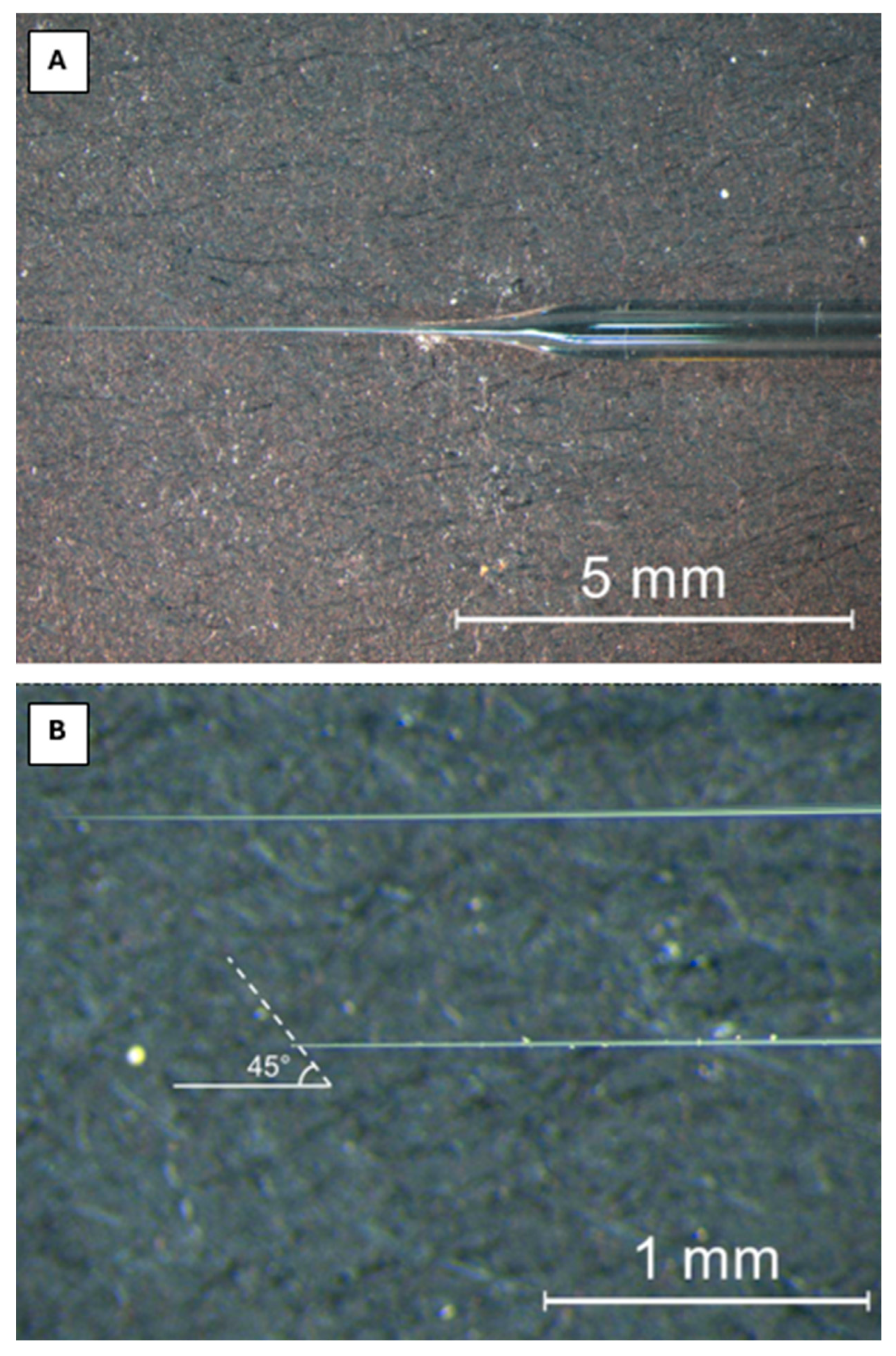
To ensure successful penetration of the needle into the PVS without excessive damage to the chorion and/or yolk, it was essential that needle tips were broken close to the tip and on an angle under a dissection microscope (Figure 2). Needle tips were broken by gently pinching the tip of the needle at a 45° angle with forceps, under a stereomicroscope. Needle tips that were broken too far up the needle caused excessive damage to the egg, which could result in disruption of the yolk and/or blastodisc/blastoderm and excessive leakage of PVF from the damaged chorion. Needles that were broken too far down the tip tended to not have the requisite strength to push through the chorion. Moreover, cortical alveolus exudate during these early stages of egg activation retained a thick and globular consistency, risking the blocking of needles that are broken too far down the tip.
Prior to breaking the tip, needles were backfilled with liquid paraffin, filling from the back (wide) opening of the glass needle with a syringe. To this end, the syringe was inserted into the needle as far as it would go. The syringe was then slowly removed from the glass needle while depositing liquid paraffin to reduce any trapped air. Following the breakage of the tip, approximately 3 μL of paraffin was dispensed from the tip of the needle to both remove any trapped air, and to make room for PVF collection.
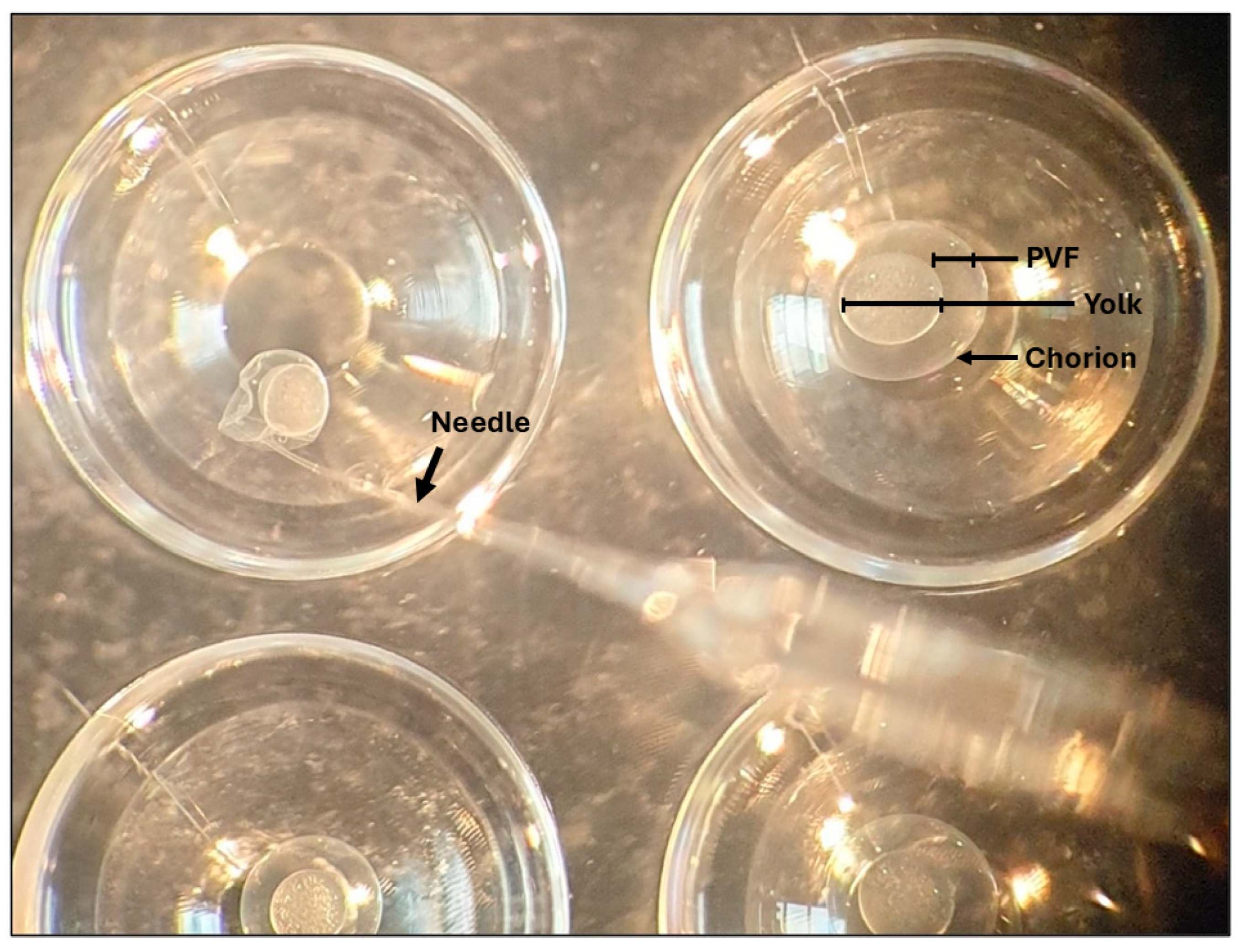
The needle was gently inserted into the PVS of the mature water-activated egg so as to not disturb the yolk. Perivitelline fluid was aspirated using the “fill” function of the Nanoject II, set to slow (23 nL/s). Approximately 60% of the PVF was aspirated from each egg to limit the risk of contamination of the sample with yolk (Figure 3). Needles were replaced as needed based on the ability of the needle tip to cleanly pass through the chorion. Fresh needles were also used between each fish. Perivitelline fluid was collected from several eggs of the same fish as a pooled sample in approximately 3 μL batches. Pooled batches were deposited on a clean prechilled Petri dish and collected with a 10 μL pipette. Perivitelline fluid was subsequently transferred into a prechilled microcentrifuge tube, snap frozen on dry ice, and stored at −80 °C. Between 30 and 50 μL of PVF was collected per fish.

2.4. Histological Validation of Perivitelline Fluid Extraction

Eggs from one zebrafish were collected for histological analysis before water activation, 6 min post-activation, and following PVF removal. Eggs were transferred into Baker’s fixative (4% formaldehyde, 0.1 M CaCl2) and fixed overnight at room temperature [42].
Fixed eggs were dehydrated through a graded series of ethanol and infiltrated with Technovit® 7100 resin, following the manufacturer’s instructions. Sections were cut at 2 µm using a Reichert-Jung 2050 microtome (Reichert-Jung, Vienna, Austria) and subsequently stained via a modified periodic acid–Schiff (PAS) staining protocol for resin-embedded tissues (S2). Images were taken with an Olympus DP80 using cellSens Dimension imaging software (version 4.4, Evident Scientific, Tokyo, Japan).

2.5. Visualisation of Sample Purity via SDS-PAGE

To gain initial insights into the potential cross-contamination of PVF samples from other major proteinaceous sources, samples of PVF, salmon ovarian fluid, zebrafish eggs prior to activation, and zebrafish eggs 8 min post-water activation were electrophoresed on a hand-cast sodium dodecyl–sulphate polyacrylamide gel electrophoresis (SDS-PAGE) gradient resolving gel (8–15%). Protein samples were derived from one adult female zebrafish. Samples were mixed with loading dye at a ratio of 1:4 v/v and Novex™ Sharp pre-stained protein standard was run as a reference protein ladder for molecular weight estimations. Sample protein concentrations were assessed via bicinchoninic acid assay (Pierce, Thermo Fisher, Waltham, MA, USA), following the manufacturer’s instructions; then, 20 μg of total protein was loaded per well. The gel was run continuously at 100 V until the dye front ran off the gel and was subsequently stained in Coomassie Brilliant Blue R-250 for protein visualisation.

3. Results

3.1. Histological Validation of Perivitelline Fluid Extraction

Figure 4 demonstrates histological validation of PVF removal from the PVS following PVF extraction (Figure 4). Additionally, it depicts the histological changes associated with zebrafish eggs following water activation and subsequent CA exocytosis (Figure 4). Prior to activation, CA were identified as peripherally located intracytoplasmic vesicles, exhibiting a dense periodic acid–Shiff-positive core. Following activation, CA underwent progressive exocytosis into the PVS in an ostensibly uniform manner along all points of the egg’s periphery. Exocytosis of CA was principally complete by 6 min post-contact with water.
Histological examination of zebrafish eggs which had undergone PVF extraction demonstrated that, relative to other observations throughout the egg activation time series, the PAS-positive cores of CA were substantially reduced in abundance in both the cytoplasmic periphery of the egg and in the PVS. This indicates that prior to PVF extraction, most CA had undergone exocytosis and were subsequently extracted during PVF isolation.

3.2. Visualisation of Sample Purity via SDS-PAGE

Figure 5 demonstrates the unique electrophoretic protein profile of the extracted PVF compared to that of both the pre-activation storage medium (salmon ovarian fluid) and whole egg samples. Notable differences include the diminished presence of presumed vitellogenin proteins (~100 kDa) in the PVF and the distinct PVF banding patterns relative to both salmon ovarian fluid and whole egg samples. This preliminary protein composition analysis highlights the enrichment of relatively low-molecular-weight proteins of between approximately 15 and 30 kDa in the PVF.

4. Discussion

As it is becoming increasingly evident that the PVF plays multifaceted and integral roles throughout the course of teleost embryogenesis, the development of sensitive and reliable techniques for PVF extractions is essential in order to improve sample integrity for downstream analyses. Moreover, it has recently come to light that many proteins constituting the zebrafish PVF are already present prior to the activation of the zygotic genome [8]. Thus, addressing the challenges associated with PVF collection during the earliest stages of embryonic development will provide a robust foundation for investigating the extent of maternal contributions to the PVF proteome.
The histological analysis demonstrated that PVF extractions were successful in isolating CA-derived material from the water-activated zebrafish eggs (Figure 4). Furthermore, analysis of the electrophoretic patterns of major proteinaceous sources from the zebrafish egg provides confidence that there was no substantial contamination of the PVF extracts with non-target egg proteins and that very few, if any, proteins from salmon ovarian fluid were able to pass through the chorion and contaminate the PVF. Smearing in the 8 min post-activation egg lane (Figure 5) may be explained by the presence of highly crosslinked insoluble oligomers produced during the restructuring of the chorion via the cleavage of ZP2 proteins and subsequent crosslinking with ZP3 proteins, which occurs shortly after egg activation [43]. The formation of these insoluble proteins appears to have impeded, or otherwise obscured, the migration of high-molecular-weight proteins in the 8 min post-activation egg samples. This is most notable for the presumptive heavy chain lipovitellin [44], which is more distinctly visible at approximately 80 kDa in the T0 samples (Figure 5).
It is acknowledged that the use of Milli-Q water as an activation medium does not accurately replicate the ionic conditions under which egg activation occurs in vivo. While its use in this study was intended to minimise the risk of protein contamination from environmental sources, future applications of this method, particularly those focused on characterising the PVF proteome, would benefit from the use of a more physiologically relevant media, such as E2 or similarly buffered solutions. Regardless of the media, this study demonstrated the value of micro-aspiration of PVF in live eggs as a valuable tool to study PVF composition and CA release dynamics.
It should be noted that this protocol can be technically demanding, introducing the potential for inter-operator variability. To minimise such variation, it is recommended that particular attention be given to maintaining methodological consistency across all sampling periods.
Minor cross-contamination, particularly from yolk proteins, may occur during PVF extraction. If a noticeable influx of yolk into the needle is observed during aspiration, it is advisable to discard the affected sub-sample and replace the needle before continuing. Collecting PVF in smaller sub-samples helps mitigate this risk, as contaminated aliquots can be identified and discarded without compromising the integrity of the remaining sample set. Future applications of this method could also utilise freshly fertilised eggs, as the current protocol is expected to be compatible with fertilised embryos with minimal modification. Investigating how fertilisation influences PVF composition and egg activation dynamics would provide valuable insights into the functions of the PVF.
While this technique is specifically developed with zebrafish in mind, there is no a priori reason why this protocol could not be applied to other species. However, further development of this method may be required to account for egg size and chorion penetrability, etc. Pursuing comparative studies of PVF composition among the diverse members of teleosts could greatly enhance our understanding of the functional properties of PVF. In equal measure, both the gain and loss of PVF-associated proteins throughout different teleost lineages would provide valuable insights into the potential functional roles of these proteins and how these systems may adapt to in response to diverse evolutionary pressures. Examining how the functional constituents of the PVF may vary to support the diverse evolutionary trajectories and life histories of different teleosts represents an exciting and promising direction for future research.

5. Conclusions

The technique described herein represents a valuable improvement over previously employed methods of PVF isolation, as it allows for the collection of fresh PVF from water-activated zebrafish eggs immediately following activation. By eliminating the need for formalin fixation prior to PVF isolation, this method enhances sample integrity for downstream proteomic analyses [26,27,28,29]. It is anticipated that the adoption of this technique will support the development of a more reliable characterisation of the PVF proteome during the earliest stages of zebrafish embryonic development. Crucially, the ability to retrieve high-quality, fresh PVF directly after egg activation will facilitate robust mass spectrometry-based analysis of the maternal contributions to the embryonic microenvironment.

Supplementary Materials

The following supporting information can be downloaded at https://www.mdpi.com/article/10.3390/fishes10080369/s1.

Author Contributions

Conceptualization, B.A.L.; methodology, B.A.L. and C.W.B.; validation, B.A.L.; formal analysis, B.A.L.; resources, C.W.B.; writing—original draft preparation, B.A.L.; writing—review and editing, B.A.L. and P.M.L.; supervision, P.M.L. and C.W.B. All authors have read and agreed to the published version of the manuscript.

Funding

This research received no external funding.

Institutional Review Board Statement

The animal study protocol was approved by the University of Otago Animal Ethics Committee (approval code, AUP-21-116; approval date, 15 October 2021).

Informed Consent Statement

Not applicable.

Data Availability Statement

The original contributions presented in this study are included in the article/Supplementary Material. Further inquiries can be directed to the corresponding author.

Conflicts of Interest

The authors declare no conflicts of interest.

Abbreviations

The following abbreviations are used in this manuscript:
CACortical alveoli
PAS Periodic acid–Schiff
PVFPerivitelline fluid
PVSPerivitelline space

References

  1. Shephard, K.L. Ion-exchange phenomena regulate the environment of embryos in the eggs of freshwater fish. Comp. Biochem. Physiol. Part A Physiol. 1987, 88, 659–662. [Google Scholar] [CrossRef]
  2. Shephard, K.L. Aluminium and calcium affect the perivitelline fluid in fish eggs. Comp. Biochem. Physiol. Part C Comp. Pharmacol. 1988, 91, 503–505. [Google Scholar] [CrossRef]
  3. Kudo, S.; Yazawa, S. Binding of antibiotics to glycoproteins of the vitelline and fertilization envelopes of cherry salmon eggs. Histochem. J. 1997, 29, 607–616. [Google Scholar] [CrossRef] [PubMed]
  4. Green, B.S. Embryogenesis and oxygen consumption in benthic egg clutches of a tropical clownfish, Amphiprion melanopus (Pomacentridae). Comp. Biochem. Physiol. Part A Mol. Integr. Physiol. 2004, 138, 33–38. [Google Scholar] [CrossRef]
  5. Osman, A.G.M.; Wuertz, S.; Mekkawy, I.A.; Exner, H.-J.; Kirschbaum, F. Lead induced malformations in embryos of the African catfish Clarias gariepinus (Burchell, 1822). Environ. Toxicol. 2007, 22, 375–389. [Google Scholar] [CrossRef]
  6. Chen, F.; Smith, C.; Wang, Y.; He, J.; Xia, W.; Xue, G.; Chen, J.; Xie, P. The evolution of alternative buoyancy mechanisms in freshwater fish eggs. Front. Ecol. Evol. 2021, 9. [Google Scholar] [CrossRef]
  7. Chen, F.; Wang, Y.; He, J.; Smith, C.; Xue, G.; Zhao, Y.; Peng, Y.; Zhang, J.; Liu, J.; Chen, J.; et al. Alternative signal pathways underly fertilization and egg activation in a fish with contrasting modes of spawning. BMC Genom. 2023, 24, 167. [Google Scholar] [CrossRef]
  8. De la Paz, J.F.; Anguita-Salinas, C.; Díaz-Celis, C.; Chávez, F.P.; Allende, M.L. The zebrafish perivitelline fluid provides maternally-inherited defensive immunity. Biomolecules 2020, 10, 1274. [Google Scholar] [CrossRef]
  9. Tateno Hiroaki Ogawa, T.; Muramoto, K.; Kamiya, H.; Saneyoshi, M. Rhamnose-binding lectins from steelhead trout (Oncorhynchus mykiss) eggs recognize bacterial lipopolysaccharides and lipoteichoic acid. Biosci. Biotechnol. Biochem. 2002, 66, 604–612. [Google Scholar] [CrossRef]
  10. Watanabe, Y.; Tateno, H.; Nakamura-Tsuruta, S.; Kominami, J.; Hirabayashi, J.; Nakamura, O.; Watanabe, T.; Kamiya, H.; Naganuma, T.; Ogawa, T.; et al. The function of rhamnose-binding lectin in innate immunity by restricted binding to Gb3. Dev. Comp. Immunol. 2009, 33, 187–197. [Google Scholar] [CrossRef]
  11. Mu, L.; Yin, X.; Bai, H.; Li, J.; Wu, H.; Qi, W.; Wang, B.; Ye, J. Expression and functional characterisation of an L-rhamnose-binding lectin from Nile tilapia (Oreochromis niloticus) in host defense against bacterial infection. Aquaculture 2021, 545, 737195. [Google Scholar] [CrossRef]
  12. Hansen, G.H.; Olafsen, J.A. Bacterial colonization of cod (Gadus morhua L.) and Halibut (Hippoglossus hippoglossus) eggs in marine aquaculture. Appl. Environ. Microbiol. 1989, 55, 1435–1446. [Google Scholar] [CrossRef] [PubMed]
  13. Kumagai, A.; Nawata, A. Mode of the intra-ovum infection of Flavobacterium psychrophilum in salmonid eggs. Fish Pathol. 2010, 45, 31–36. [Google Scholar] [CrossRef]
  14. Kumagai, A.; Yamaoka, S.; Takahashi, K.; Fukuda, H.; Wakabayashi, H. Waterborne transmission of Flavobacterium psychrophilum in coho salmon eggs. Fish Pathol. 2000, 35, 25–28. [Google Scholar] [CrossRef]
  15. Kohara, M.; Kasai, H.; Yoshimizu, M. Intra-ovum infection in salmonid eggs artificially contaminated with fish pathogenic bacteria: Flavobacterium psychrophilum, Renibacterium salmoninarum and Aeromonas salmonicida. Fish Pathol. 2012, 47, 49–55. [Google Scholar] [CrossRef]
  16. Cousins, K.L.; Jensen, J.O.T. The effects of temperature on external egg membranes in coho salmon (Oncorhynchus kisutch) and the occurrence of soft-shell disease. Can. J. Zool. 1994, 72, 1854–1857. [Google Scholar] [CrossRef]
  17. Bergh, Ø.; Hansen, G.H.; Jelmert, A. Bacterial Diseases of Eggs and Yolk Sac Larvae of Halibut (Hippoglossus hippoglossus L.): Characterisation and Experimental Infection. ICES: Copenhagen, Denmark, 1990. [Google Scholar]
  18. Liu, Y.; de Bruijn, I.; Jack, A.L.; Drynan, K.; van den Berg, A.H.; Thoen, E.; Sandoval-Sierra, V.; Skaar, I.; van West, P.; Diéguez-Uribeondo, J.; et al. Deciphering microbial landscapes of fish eggs to mitigate emerging diseases. ISME J. 2014, 8, 2002–2014. [Google Scholar] [CrossRef]
  19. Kudo, S.; Yazawa, S. Binding of bacterial toxins to glycoproteins in the envelopes of rainbow trout eggs. Histochem. J. 1995, 27, 300–308. [Google Scholar] [CrossRef]
  20. Green, E.; Harishchandra, A.; Ranasinghe, P.; Giulio, R.; Jayasundara, N. The external microbiome communicates with the developing zebrafish (Danio rerio) embryo through the protective chorion and influences developmental trajectory; bioRxiv: New York, NY, USA, 2024. [Google Scholar]
  21. Brox, S.; Ritter, A.P.; Küster, E.; Reemtsma, T. Influence of the perivitelline space on the quantification of internal concentrations of chemicals in eggs of zebrafish embryos (Danio rerio). Aquat. Toxicol. 2014, 157, 134–140. [Google Scholar] [CrossRef] [PubMed]
  22. Pereira, S.P.P.; Boyle, D.; Nogueira, A.; Handy, R.D. Differences in toxicity and accumulation of metal from copper oxide nanomaterials compared to copper sulphate in zebrafish embryos: Delayed hatching, the chorion barrier and physiological effects. Ecotoxicol. Environ. Saf. 2023, 253, 114613. [Google Scholar] [CrossRef] [PubMed]
  23. Pelegri, F. Maternal factors in zebrafish development. Dev. Dyn. 2003, 228, 535–554. [Google Scholar] [CrossRef] [PubMed]
  24. Żarski, D.; Le Cam, A.; Frohlich, T.; Kösters, M.; Klopp, C.; Nynca, J.; Ciesielski, S.; Sarosiek, B.; Dryl, K.; Montfort, J.; et al. Neurodevelopment vs. the immune system: Complementary contributions of maternally-inherited gene transcripts and proteins to successful embryonic development in fish. Genomics 2021, 113, 3811–3826. [Google Scholar] [CrossRef] [PubMed]
  25. Henn, K.; Braunbeck, T. Dechorionation as a tool to improve the fish embryo toxicity test (FET) with the zebrafish (Danio rerio). Comp. Biochem. Physiol. Part C Toxicol. Pharmacol. 2011, 153, 91–98. [Google Scholar] [CrossRef]
  26. Atwood, J.; Neumann, D.; Dammer, E.; Duong, D.; Dunn, C.; Seyfried, N. Evaluating the effect of formalin fixation on mass spectrometry-based proteomic profiling (984.1). FASEB J. 2014, 28 (Suppl. 1), 984.1. [Google Scholar] [CrossRef]
  27. Guo, T.; Wang, W.; Rudnick, P.A.; Song, T.; Li, J.; Zhuang, Z.; Weil, R.J.; DeVoe, D.L.; Lee, C.S.; Balgley, B.M. Proteome analysis of microdissected formalin-fixed and paraffin-embedded tissue specimens. J. Histochem. Cytochem. 2007, 55, 763–772. [Google Scholar] [CrossRef]
  28. Tanca, A.; Pagnozzi, D.; Burrai, G.P.; Polinas, M.; Uzzau, S.; Antuofermo, E.; Addis, M.F. Comparability of differential proteomics data generated from paired archival fresh-frozen and formalin-fixed samples by GeLC–MS/MS and spectral counting. J. Proteom. 2012, 77, 561–576. [Google Scholar] [CrossRef]
  29. Steiner, C.; Ducret, A.; Tille, J.-C.; Thomas, M.; McKee, T.A.; Rubbia-Brandt, L.A.; Scherl, A.; Lescuyer, P.; Cutler, P. Applications of mass spectrometry for quantitative protein analysis in formalin-fixed paraffin-embedded tissues. Proteomics 2014, 14, 441–451. [Google Scholar] [CrossRef] [PubMed]
  30. Tian, Y.; Zhang, H. Isolation of proteins by heat-induced extraction from formalin-fixed, paraffin-embedded tissue and preparation of tryptic peptides for mass spectrometric analysis. Curr. Protoc. Mol. Biol. 2010, 10, 1–7. [Google Scholar] [CrossRef]
  31. Hiratsuka, T.; Tsuruyama, T.; Hiratsuka, T.; Tsuruyama, T. Mass spectrometry analysis using formalin-fixed paraffin-embedded pathological samples. In Electron Microscopes, Spectroscopy and Their Applications; IntechOpen: London, UK, 2024. [Google Scholar]
  32. Hermann, J.; Noels, H.; Theelen, W.; Lellig, M.; Orth-Alampour, S.; Boor, P.; Jankowski, V.; Jankowski, J. Sample preparation of formalin-fixed paraffin-embedded tissue sections for MALDI-mass spectrometry imaging. Anal. Bioanal. Chem. 2020, 412, 1263–1275. [Google Scholar] [CrossRef]
  33. Nwosu, A.J.; Misal, S.A.; Truong, T.; Carson, R.H.; Webber, K.G.I.; Axtell, N.B.; Liang, Y.; Johnston, S.M.; Virgin, K.L.; Smith, E.G.; et al. In-depth mass spectrometry-based proteomics of formalin-fixed, paraffin-embedded tissues with a spatial resolution of 50-200 μm. J. Proteome Res. 2022, 21, 2237–2245. [Google Scholar] [CrossRef]
  34. Cho, A.; Ahn, J.; Kim, A.; Lee, J.H.; Ryu, H.S.; Kim, K.M.; Yi, E.C. Proteomics analysis of an individual formalin-fixed paraffin-embedded tissue section using isobaric-tag amplification. Rapid Commun. Mass Spectrom. 2023, 37, e9616. [Google Scholar] [CrossRef]
  35. Hagedorn, M.; Carter, V.L. Zebrafish reproduction: Revisiting in vitro fertilization to increase sperm cryopreservation success. PLoS ONE 2011, 6, e21059. [Google Scholar] [CrossRef]
  36. Johnson, S.L.; Villarroel, M.; Rosengrave, P.; Carne, A.; Kleffmann, T.; Lokman, P.M.; Gemmell, N.J. Proteomic analysis of chinook salmon (Oncorhynchus tshawytscha) ovarian fluid. PLoS ONE 2014, 9, e104155. [Google Scholar] [CrossRef]
  37. Ribeiro, D.; Chagas, J.; Bashiyo-Silva, C.; Costa, R.; Veríssimo-Silveira, R.; Ninhaus-Silveira, A. Oocyte viability and cortical activation under different salt solutions in Prochilodus lineatus (Teleostei: Prochilodontidae). Reprod. Domest. Anim. 2017, 52, 932–938. [Google Scholar] [CrossRef]
  38. Waghmare, S.G.; Samarin, A.M.; Franěk, R.; Pšenička, M.; Policar, T.; Linhart, O.; Samarin, A.M. Oocyte ageing in zebrafish Danio rerio (Hamilton, 1822) and its consequence on the viability and ploidy anomalies in the progeny. Animals 2021, 11, 912. [Google Scholar] [CrossRef] [PubMed]
  39. Corley-Smith, G.E.; Lim, C.J.; Brandhorst, B.P. Production of androgenetic zebrafish (Danio rerio). Genetics 1996, 142, 1265–1276. [Google Scholar] [CrossRef] [PubMed]
  40. Bahabadi, M.N.; Shoae, A.; Lokman, P.M. Effects of storage time, sex steroids and media composition on egg viability of rainbow trout (Oncorhynchus mykiss). Aquaculture 2011, 315, 306–311. [Google Scholar] [CrossRef]
  41. Gueho, A.; Żarski, D.; Rime, H.; Guével, B.; Com, E.; Lavigne, R.; Nguyen, T.; Montfort, J.; Pineau, C.; Bobe, J. Evolutionarily conserved ovarian fluid proteins are responsible for extending egg viability in salmonid fish. Sci. Rep. 2024, 14, 9651. [Google Scholar] [CrossRef]
  42. Rhodes, A. 4-Fixation of tissues. In Bancroft’s Theory and Practice of Histological Techniques, 7th ed.; Suvarna, S.K., Layton, C., Bancroft, J.D., Eds.; Churchill Livingstone: Oxford, UK, 2013; pp. 69–93. [Google Scholar]
  43. Fu, B.; Wu, D.; Yasumasu, S.; Hane, M.; Sato, C.; Kitajima, K. Critical role of the cortical alveolus protease alveolin in chorion hardening in vivo at medaka fertilization. Biomolecules 2023, 13, 146. [Google Scholar] [CrossRef]
  44. Yilmaz, O.; Patinote, A.; Nguyen, T.V.; Com, E.; Lavigne, R.; Pineau, C.; Sullivan, C.V.; Bobe, J. Scrambled eggs: Proteomic portraits and novel biomarkers of egg quality in zebrafish (Danio rerio). PLoS ONE 2017, 12, e0188084. [Google Scholar] [CrossRef] [PubMed]
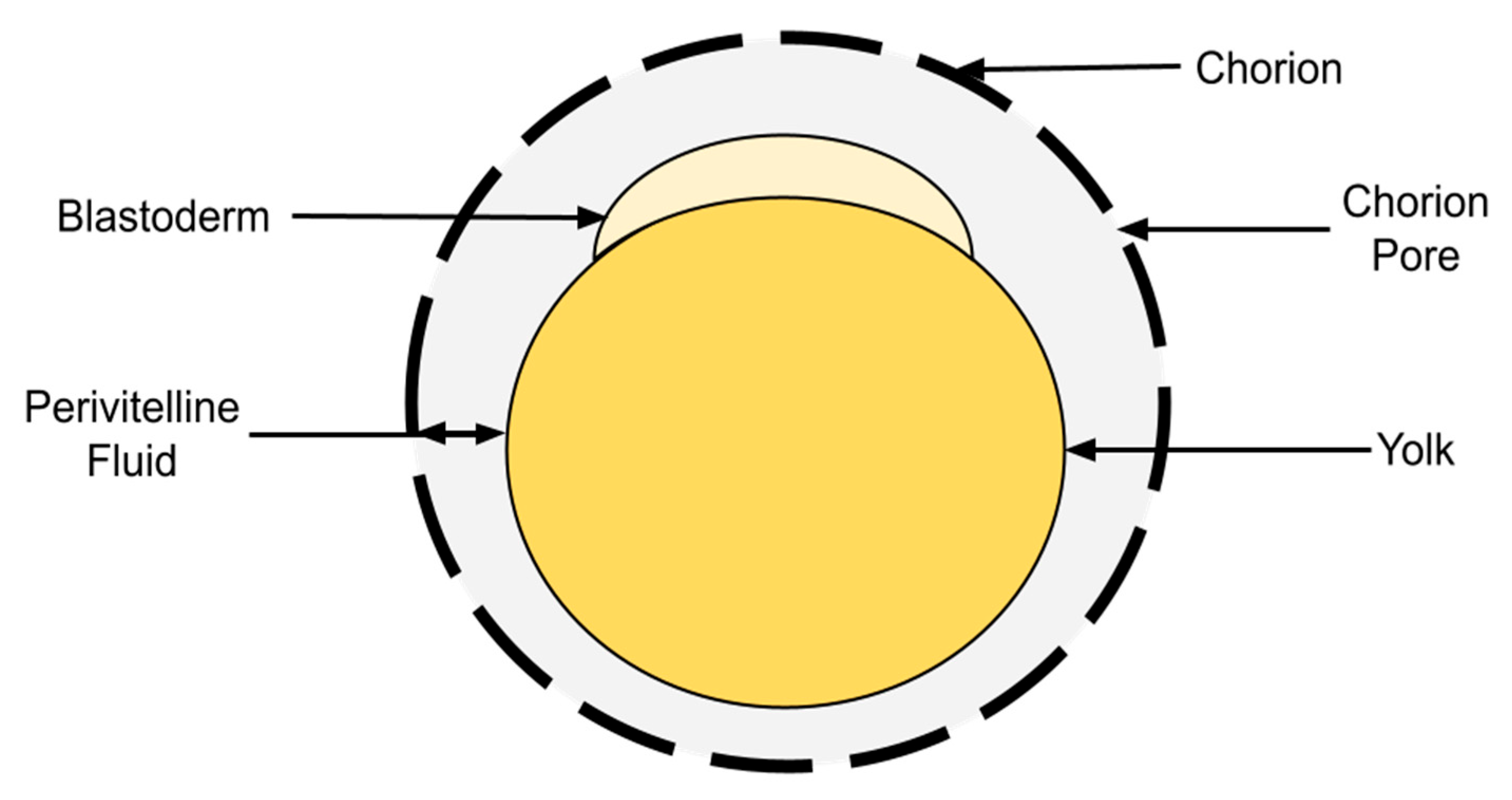
Figure 1. The major structural components of the early developing zebrafish embryo.
Fishes 10 00369 g001
Figure 2. The configuration of the pulled needles which were used for the micro-aspiration of zebrafish perivitelline fluid (PVF). (A) The length and shape of the needle prior to breaking the tip. (B) The preferred cut site and angle of the needle tip for efficient PVF extraction, with uncut depicted above and cut depicted below. Needles were broken at an approximately 45° angle to provide the sharp edge used to push through the chorion.
Fishes 10 00369 g002
Figure 3. Zebrafish eggs undergoing perivitelline fluid (PVF) extraction. Individual ovulated eggs were placed into separate wells of a 72-microwell plate containing fresh Milli-Q water. Approximately 60% of the PVF was extracted from the egg in the top-left well.
Fishes 10 00369 g003
Figure 4. Histological observations of zebrafish eggs prior to activation (A), and 6 min post-activation (B) and post-perivitelline fluid extraction (C). Black boxes represent the approximate portion of the image (AC), portrayed at increased magnification (A2C2). All histological sections were embedded in Technovit 7100, cut at 2 μm, and stained with periodic acid–Schiff stain (s2). Y = yolk, C = chorion, PVS = perivitelline space, CA = cortical alveoli, PPC = periodic acid–Schiff-positive core of cortical alveoli post-exocytosis.
Fishes 10 00369 g004
Figure 5. Protein samples of perivitelline fluid (PVF), salmon ovarian fluid (SOF), and whole zebrafish eggs pre-activation and 8 min post-activation (T0, T8, respectively) were run on a 1D SDS-PAGE gradient gel (8–15%) and stained with Coomassie Brilliant Blue R-250. Novex™ Sharp pre-stained protein standard was run as a reference protein ladder (L).
Fishes 10 00369 g005
Disclaimer/Publisher’s Note: The statements, opinions and data contained in all publications are solely those of the individual author(s) and contributor(s) and not of MDPI and/or the editor(s). MDPI and/or the editor(s) disclaim responsibility for any injury to people or property resulting from any ideas, methods, instructions or products referred to in the content.