Article

Transcriptome Analysis Reveals Hyperglycemic Hormone and Excitatory Amino Acid Transporter 3 Are Involved in the Thermal Adaptation of Eriocheir sinensis

Tianjin Key Laboratory of Animal and Plant Resistance/College of Life Sciences, Tianjin Normal University, Tianjin 300387, China
*
Authors to whom correspondence should be addressed.
These authors contributed equally to this work.
Fishes 2025, 10(7), 361; https://doi.org/10.3390/fishes10070361
Submission received: 11 June 2025 / Revised: 14 July 2025 / Accepted: 18 July 2025 / Published: 21 July 2025
(This article belongs to the Section Aquatic Invertebrates)

Abstract

Temperature is one of the critical factors influencing the survival, growth, and reproduction of organisms. The molting and developmental mechanisms of crustaceans are highly sensitive to temperature, yet the regulatory mechanisms underlying their thermal adaptation remain unclear. In this work, transcriptome sequencing was performed to analyze the gene expression profiles of Eriocheir sinensis under normal temperature (22 °C) and high-temperature (27 °C and 32 °C) conditions. A total of 377 differentially expressed genes (DEGs) were identified, including 149 up-regulated and 227 down-regulated genes. Through Gene Ontology (GO) enrichment analysis of these DEGs, 11 significantly temperature-regulated signaling pathways were identified, including the estrogen and androgen receptor signaling pathways, and two neurotransmission signaling pathways. These findings suggest that temperature may influence sex regulation in E. sinensis, while the dopamine receptor and neuropeptide signaling pathways may play a role in its thermal adaptation. Further validation via RT-qPCR of DEGs involved in neurotransmission signaling pathways revealed that crustacean hyperglycemic hormone (CHH) and excitatory amino acid transporter 3 (EAA3) genes are likely involved in the thermal adaptation of E. sinensis. In addition, the hemolymph glucose levels associated with the elevated temperatures were detected and consistent variations between glucose levels and CHH expressions were found. This indicates that the eyestalk CHH is strongly correlated with the hemolymph glucose levels and likely mediates the response to temperature changes by regulating blood glucose in E. sinensis. The results of this study not only provide key molecular targets for elucidating the mechanisms by which temperature affects molting and development in E. sinensis, but also establish a theoretical foundation for further research into thermal adaptation strategies in crustaceans.
Key Contribution: The estrogen and androgen receptor signaling pathways and two neurotransmission signaling pathways have been found to be involved in the thermal adaptation of E. sinensis. Furthermore, crustacean hyperglycemic hormone (CHH) and excitatory amino acid transporter 3 (EAA3) genes are found to participate in the thermal adaptation.

1. Introduction

Temperature is one of the critical factors influencing the survival, development, and reproduction of organisms. Organisms have evolved diverse mechanisms to detect temperature changes and mount adaptive responses [1,2]. Thermosensation, a vital physiological function universally present in organisms, enables them to perceive high-temperature stimuli and execute adaptive reactions for self-protection [3]. The impacts of high temperatures and the corresponding response mechanisms vary among different organisms [4,5]. As poikilothermic organisms, arthropods are fundamentally dependent on ambient thermal conditions for thermoregulation. While these species demonstrate considerable plasticity in physiological adjustments and behavioral adaptations to optimize their thermal environment within species-specific thermal limits, exposure to temperatures exceeding their upper critical thresholds can induce physiological stress and threaten their survival capacity [6]. Although arthropods lack complex endocrine organs like those found in mammals, they have evolved unique neuroendocrine regulatory strategies to cope with temperature variations. For instance, in Drosophila melanogaster, environmental temperature is initially detected by primary thermosensory neurons in the peripheral nervous system. These neurons project signals to specific regions of the central nervous system, where the temperature information undergoes integration and processing. The signals are then transmitted to the brain, ultimately involving histamine in the regulation of temperature preference behavior [7]. In addition, studies have shown that high-temperature exposure can inhibit the sperm generation of D. melanogaster and affect its reproductive ability [8].
As an important group within the phylum Arthropoda, crustaceans are highly sensitive to temperature throughout their life cycle due to their unique molting-based growth and development strategies. Temperature plays a particularly crucial role in influencing disease resistance, feeding, molting, development, and reproduction in crustaceans [9]. Studies have shown that moderately high temperatures enhance embryonic development in the southern crab Ovalipes trimaculatus, but when exceeding the thermal viability threshold, embryos may develop abnormally or even die [10]. In addition, the high temperature of 33 °C will weaken the immune response of blood cells of neohelice granulolata to lipopolysaccharide and have a certain inhibitory effect on its immune system [11].
The eyestalks of crustaceans possess dual functions of sensing the external environment and neuroendocrine regulation, capable of secreting various neuropeptide hormones [12] and biogenic amines [13,14]. The X-organ–sinus gland complex (XO-SG) in the eyestalk is structurally and functionally analogous to the hypothalamus–pituitary system in vertebrates, and serves as the endocrine control center of crustaceans [15]. Additionally, the expression and release of neuropeptide hormones in eyestalks are regulated by neurons or factors secreted by peripheral cells and tissues, which transmit signals encoding internal and external environmental information [13]. Existing studies demonstrate that light, darkness, and circadian rhythms influence the expression and release of neuropeptide hormones and biogenic amines [16]. Therefore, crustacean eyestalks likely receive external environmental signals (e.g., temperature) and regulate molting and growth through neuroendocrine adaptations. As typical aquatic poikilotherms, crustaceans utilize the specialized XO-SG neuroendocrine system to convert temperature fluctuations into endocrine signals that govern critical physiological processes such as growth, molting, and reproduction. For instance, elevated temperatures increase the synthesis of crustacean hyperglycemic hormone (CHH) in Metacarcinus magister to potentially regulate energy metabolism under heat stress, while also accelerating molting, but extreme temperatures may cause mortality [17]. High temperatures further alter the protein expression profile in XO-SG, affecting neuropeptide synthesis and secretion. Notably, heat shock proteins and antioxidant enzymes significantly increase under thermal stress to protect neuroendocrine cells from heat damage [18].
The Chinese mitten crab (Eriocheir sinensis), belonging to the phylum Arthropoda, class Malacostraca, order Decapoda, family Varunidae, and genus Eriocheir, is an important aquatic economic animal. E. sinensis undergoes discontinuous growth and development through molting. It requires over twenty times of molting throughout its lifetime to reach maturity. Temperature is one of the most critical environmental factors influencing the growth and development of E. sinensis, exerting its effects throughout the entire lifecycle by directly impacting metabolic rate, feeding, molting, gonad development, and survival rate. Temperature can affect the inter-molt period and molt increment in E. sinensis, thereby influencing its growth and development [19]. When temperature exceeds 30 °C, the mortality rate of E. sinensis increases rapidly [20]. Studies indicate that high temperature and hypoxia can act synergistically, further exacerbating the toxic effects of hypoxia on the intestine of E. sinensis, which may contribute to the increased mortality [21]. Following exposure to high-temperature air, even upon re-immersion in water, the changes in gut microbiota composition and metabolic levels induced by the air exposure cannot be effectively reversed [22]. Furthermore, temperature stress is associated with immune responses, oxidative stress pathways, and hormone synthesis [23,24]. It is found that E. sinensis exhibits thermal tolerance during increases in water temperature in the summer, but the specific mechanisms remain unclear [23].
The eyestalk optic ganglia of E. sinensis are located beneath the compound eye layer and consist of three primary structures: the medulla externa, medulla interna, and medulla terminalis. The X-organ, situated at the outer margin of the medulla terminalis, is formed by clusters of neurosecretory cells that synthesize and secrete various hormones [25]. The sinus gland, positioned laterally between the medulla interna and medulla terminalis, acts as a storage and release site for hormones and is composed of axonal terminals from neurosecretory cells. Multiple hormones synthesized and secreted by the X-organ are transported via axon bundles from neurosecretory cells to the sinus gland, where they are released to regulate critical physiological processes such as molting, growth, and reproduction in the crab [26]. Current research on E. sinensis eyestalks has primarily focused on photoperiod and circadian rhythms [27,28,29], while studies on temperature effects have been limited to gut microbial communities [21,22,30]. The specific molecular mechanisms by which temperature regulates molting and developmental processes in E. sinensis through the eyestalk neuroendocrine center remain unclear. This research gap hinders a comprehensive understanding of environmental adaptation mechanisms in crustaceans.
In this work, transcriptome sequencing was performed to analyze the gene expression levels in E. sinensis under normal (22 °C) and high-temperature conditions (27 °C and 32 °C). Differentially expressed genes (DEGs) were analyzed to identify thermal tolerance genes, while Gene Ontology (GO) enrichment analysis was employed to determine temperature-regulated signal transduction pathways. A further validation of DEGs involved in neural signaling pathways was conducted using real-time quantitative PCR (RT-qPCR). The influence of temperature on the hemolymph glucose levels was also detected. These approaches aimed to investigate the impact of high temperature on neural signal transmission, providing critical insights for elucidating the molecular mechanisms by which temperature regulates molting and developmental processes in E. sinensis.

2. Materials and Methods

2.1. Culture Conditions of E. sinensis

The E. sinensis were sourced from a crab aquaculture base in Ninghe District, Tianjin, China. Healthy individuals with robust physique, vigorous activity, and intact appendages (body weight: 13.8 ± 1.5 g) were selected as experimental subjects. Prior to the experiment, the crabs were acclimated for one week in fully aerated tap water tanks. During acclimation, water temperature was maintained at 25 ± 0.1 °C, with half of the tank volume replaced every two days. Daily feeding of high-quality formulated crab feed (Tianjin Modern Tianjiao Aquatic Feed Co., Ltd., Tianjin, China) was conducted at scheduled times with measured amounts, corresponding to approximately 5% of the crabs’ total body weight. After one week of acclimation, healthy E. sinensis individuals were transferred to plastic tanks (19 cm × 35 cm × 20 cm) for culture. Following a three-day adaptation period, crabs in the inter-molt stage were selected for subsequent experiments.

2.2. Sample Collection and Storage

The experiment included six temperature groups (7 °C, 12 °C, 17 °C, 22 °C, 27 °C, and 32 °C), each with three parallel replicates. For each replicate, 16 uniformly sized crabs were randomly assigned. Temperature control was achieved using chromatography refrigerators and illuminated incubators, maintaining water temperatures at designated levels. Throughout the experiment, all groups were gradually transferred from the acclimation temperature of 25 °C to target temperatures with an increase rate of 0.6 °C/min and a decrease rate of 0.2 °C/min. The time required to reach the target temperature varied among groups. Therefore, the time required for the water temperature (25 °C) to reach each target temperature was firstly calculated, and then the thermal stimulation was initiated according to this calculated time to ensure that samples could be collected at either 10:00 a.m. or 10:00 p.m. after reaching the target temperature and 24 h of exposure to the target temperature. A 12 h dark/12 h light photoperiod was consistently maintained.
At each temperature, 48 crabs (with three replicates of 16 crabs each) were used for sampling, resulting in a total of 288 crabs at six temperatures used for reference genome assembly. In each replicate, the 16 crabs maintained a 1:1 sex ratio, and all crabs were non-reproductive (not sexually mature). Eyestalks were dissected by severing the connective membrane between the eyestalk and body using a sterile scalpel. The entire eyestalk was immersed in pre-cooled crab-specific saline solution. Under a dissecting microscope, the eyestalk exoskeleton was opened with sterile forceps and scissors, followed by the removal of the pigment layer from the compound eye to isolate the internal X-organ–sinus gland complex. A total of 18 samples were collected (three replicates per temperature). Samples were flash-frozen in liquid nitrogen and stored at −80 °C.

2.3. Reference Genome Assembly

Total RNA was first extracted from E. sinensis eyestalk samples across all temperature groups using the Trizol method (TRIzol® Reagent, Invitrogen, Carlsbad, CA, USA), with extracted RNA stored in a −80 °C ultra-low temperature freezer. A portion of RNA from each temperature group was pooled and subjected to third-generation transcriptome sequencing on the PacBio Sequel platform to generate a reference genome. Total RNA was first reverse transcribed into first-strand cDNA using the UMI base PCR cDNA Synthesis Kit (BGI, Shenzhen, China). The first-strand cDNA was then amplified via PCR to generate second-strand cDNA, producing double-stranded DNA (dsDNA). This dsDNA underwent a second round of PCR amplification. The resulting product was subsequently used for SMRTbell library preparation and sequencing. Subread data obtained post-sequencing were processed via the SMRT Analysis Suite (BGI, Sehenzhen, China) for circular consensus sequence (CCS) identification. CCSs were categorized into full-length non-chimeric reads and non-full-length reads based on the presence of complete 5′ primers, polyA tails, and 3′ primers. Only full-length non-chimeric reads were retained for downstream analysis. High-quality full-length sequences from all libraries were merged, clustered, and de-duplicated to obtain isoforms. Finally, the assembled transcripts were evaluated using the Benchmarking Universal Single-Copy Orthologs (BUSCO) database [31], a single-copy ortholog database, to assess transcriptome completeness by comparing conserved genes. The reference genome of E. sinensis has been submitted to the GEO database with the accession number GSE298339.

2.4. Transcriptome Sequencing of Samples from Normal and High-Temperature Conditions

Existing studies indicate that from 25 °C to 30 °C, the survival time of E. sinensis gradually decreases and mortality gradually increases with rising temperature. Above 30 °C, their survival time is significantly reduced and mortality increases rapidly [20]. Given that 22 °C is the optimal temperature for their growth, two high-temperature exposure groups were established at 27 °C and 32 °C using 5 °C intervals to analyze the effects of elevated temperature on the eyestalks of E. sinensis. Here, 27 °C and 32 °C can serve as a sub-lethal high temperature and a lethal high temperature, respectively. Therefore, transcriptome sequencing was conducted at three temperatures: 22 °C (optimal growth temperature), 27 °C (sub-lethal high temperature), and 32 °C (lethal high temperature). Crabs were sampled after 24 h of exposure to each temperature. No crab died at 22 °C, while 2 crabs died at 27 °C and 5 crabs died at 32 °C. Second-generation transcriptome sequencing was performed on nine samples (three replicates per temperature) using the DNBSEQ platform.
The cDNA library was constructed with the BGI optimal series dual module mRNA Library Construction Kit (BGI, Sehenzhen, China). Total RNA is processed using either the mRNA enrichment method or the rRNA removal method. The obtained RNA is fragmented using a fragmentation buffer. First-strand cDNA is synthesized using random N6 primers, followed by second-strand synthesis to form double-stranded DNA (dsDNA). The ends of the synthesized dsDNA are blunted and phosphorylated at the 5′ end, while a single ‘A’ overhang is added to the 3′ end. A Y-shaped adapter with a single ‘T’ overhang at the 3′ end is then ligated to the dsDNA. The ligation products are amplified by PCR using specific primers. The PCR products are heat-denatured into single strands. A bridging primer is used to circularize the single-stranded DNA, resulting in a single-stranded circular DNA library, which is then ready for sequencing on the machine.
The raw sequencing data, termed “raw reads,” included low-quality sequences, adapter-contaminated reads, and reads with excessive unknown bases (N content). To ensure data reliability, SOAPnuke software (version v2.3; Dundee, UK) [32] was employed for preprocessing to remove adapter-containing reads, reads with >1% unknown N bases, and low-quality reads (defined as sequences where bases with quality scores <20 accounted for >40% of total bases). The filtered data, referred to as “clean reads”, were subjected to quality validation using Q20, Q30, and base composition distribution as quality control parameters to confirm suitability for downstream analysis. After quality control, clean reads were aligned to the reference genome using Bowtie2 (version v2.4.5; UMD, College Park, MD, USA) [33]. Alignment results were further evaluated through mapping rates and read distribution across transcripts to determine compliance with secondary quality control criteria. Gene expression levels for each sample were then quantified using RSEM (v1.3.1; Madison, WI, USA) [34]. Gene expression levels were quantified using FPKM (Fragments Per Kilobase of exon model per Million mapped fragments). Genes with FPKM values ≥ 1 were considered expressed. The transcriptome sequencing data of E. sinensis have been submitted to the GEO database with the accession number GSE299373.

2.5. Screening and GO Enrichment Analysis of Thermal-Responsive Genes

Differential gene expression analysis was performed using the criteria of |log2FoldChange| ≥ 1 and q-value ≤ 0.05 to identify DEGs in the 22 °C vs. 27 °C and 22 °C vs. 32 °C comparison groups. To more precisely pinpoint thermal tolerance related genes, the intersection of DEGs from both comparison groups was analyzed. This approach identified thermal-responsive DEGs consistently differentially expressed across both temperature gradients. The thermal-responsive DEGs were then subjected to GO annotation and enrichment analysis using Blast2GO (version: v2.5.0, Moncada, Valencia, Spain) [35]. Genes whose GO annotations included “signaling pathway” were subsequently screened out. The bubble plot for GO enrichment analysis results was drawn with an online chiplot tool (https://www.chiplot.online/, accessed on 5 May 2025).

2.6. RT-qPCR Validation

RT-qPCR was performed to validate the genes involved in neurotransmission signaling pathways identified through GO enrichment analysis. The reverse transcription of RNA was performed using the HiScript III RT SuperMix for qPCR (+gDNA wiper) kit (Vazyme, Nanjing, China). First, genomic DNA was removed from the eyestalk samples using a reaction system containing 1 μL of RNA template and 4 μL of 4 × gDNA wiper mix, brought up to 16 μL with RNase-free H2O. The mixture was thoroughly mixed and incubated at 42 °C for 2 min. Subsequently, 4 μL of 5 × HiScriptIII qRT SuperMix was added to prepare the reverse transcription reaction system. The PCR program was set as follows: 37 °C for 15 min, followed by 85 °C for 5 s.
β-actin was used as the internal reference gene, and each sample was analyzed in triplicate to detect the expression levels of the target genes. RT-qPCR primers for the genes to be validated were designed using Primer Premier (version 5.00, Premier Biosoft International, Palo Alto, CA, USA) [36], with primer sequences listed in Table 1. Following the instructions of the ChamQ Universal SYBR qPCR Master Mix kit (Vazyme, Nanjing, China), the reaction mixture was prepared in quantitative eight-tube strips as follows: 10 μL of 2× ChamQ Universal SYBR qPCR Master Mix, 0.4 μL each of forward and reverse primers, 2 μL of 10× diluted cDNA, and ddH2O to a final volume of 20 μL.
The PCR program included an initial denaturation at 95 °C for 5 min, followed by 40 cycles of 95 °C for 10 s and 60 °C for 30 s, and a final melt curve analysis step (95 °C for 15 s, 60 °C for 1 min, and 95 °C for 15 s). Gene expression levels were calculated using the comparative Ct method (2−ΔΔCt method).

2.7. Detection of Hemolymph Glucose at Different Temperatures

Healthy E. sinensis in the inter-molt stage were selected. Following a 24 h acclimation period at 22 °C, 27 °C, and 32 °C, hemolymph samples were collected. Hemolymph was drawn from the soft membrane at the base of the third or fourth pereiopod into 1.5 mL centrifuge tubes using a 1 mL sterile syringe. Three replicate samples from different individuals were collected for each temperature. The samples were kept at 4 °C overnight, and then centrifuged at 10,000 rpm for 20 min at 4 °C. The resulting supernatant (serum) was carefully aspirated into new centrifuge tubes and stored at 4 °C for subsequent analysis. Serum glucose levels were quantified using a Glucose Assay Kit (Nanjing Jiancheng Bioengineering Institute, Nanjing, China) with the glucose oxidase method.

3. Results

3.1. Reference Genome Assembly

To obtain more comprehensive results due to the incomplete genome coverage of the current E. sinensis genome sequencing, third-generation transcriptome sequencing was performed using a pooled sample, named ‘Es-7-32’, from 18 individuals across six temperature conditions (7 °C, 12 °C, 17 °C, 22 °C, 27 °C, and 32 °C) to construct a reference genome for the subsequent transcriptomic analysis. The mixed samples from the 18 individuals yielded a total of 14,086,755 subreads (70.91 GB of data) with an N50 length of 5448 bp. SMRT cell analysis generated 506,250 circular consensus sequences (CCSs). After splitting, 1,450,467 full-length non-chimeric sequences were obtained. These sequences were clustered into consensus transcripts and de-duplicated, resulting in 390,023 isoforms.
The quality of the assembled transcripts was evaluated using the BUSCO database, as shown in Figure 1. Most isoforms were fully aligned to sequences in the BUSCO database, indicating high completeness of the transcriptome assembly. These isoforms were subsequently used for downstream transcriptomic analyses.

3.2. Transcriptome Sequencing

To investigate the effects of high temperature on gene expression in E. sinensis, transcriptome sequencing was conducted on nine samples collected at three temperatures: 22 °C, 27 °C, and 32 °C. The sequencing results for these nine samples are summarized in Table 2. All samples exhibited Q20 scores above 96% and Q30 scores above 89%. The alignment rates of clean reads to the reference genome are also presented in Table 2, ranging from 79% to 84% across all samples. The base composition distribution of clean reads across the samples is shown in Supplementary File S1. In each sample, the contents of ‘A’ and ‘T’ are basically the same, and the contents of ‘C’ and ‘G’ are basically the same, which conforms to the principle of base complementary pairing. The content of ‘N’ (green line) is extremely low and almost coincides with the X axis, indicating that there is almost no sequencing failure and sample degradation problem. For paired-end sequencing, 0 bp represents the starting position of read 1, and 150 bp represents the starting position of read 2. The fluctuations observed at 0 bp and 150 bp in Supplementary File S1 are due to the use of 6 bp random primers during reverse transcription to synthesize cDNA in the transcriptome library construction process. These primers introduce a certain degree of nucleotide composition bias at the initial positions of the sequencing data, and this fluctuation is a normal phenomenon. The distribution of reads across transcripts is illustrated in Supplementary File S2. The central region of the distribution curve was relatively flat, while a slight elevation was observed at the 3′ end, likely due to mRNA degradation. These data collectively demonstrate the high quality of the samples.

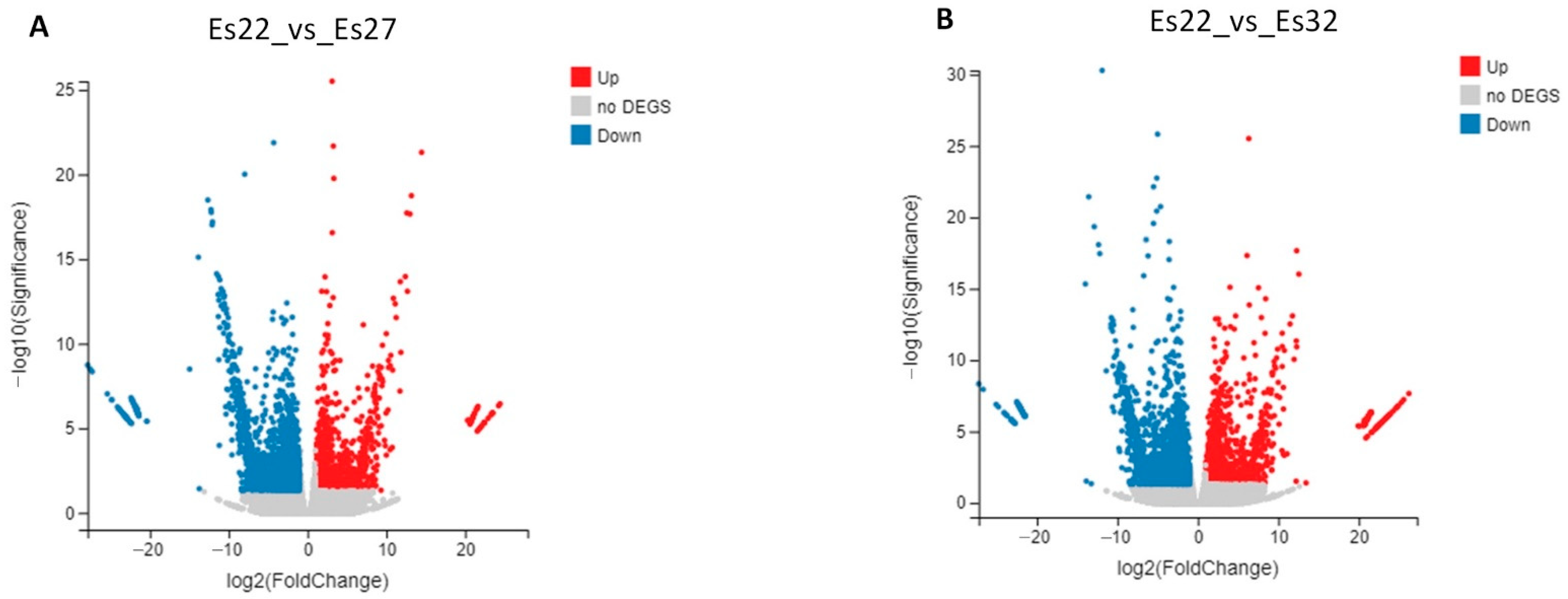
3.3. Analysis of DEGs

Volcano plots of DEGs for the comparisons Es22_vs_Es27 and Es22_vs_Es32 are shown in Figure 2. To identify genes more accurately associated with thermal regulation, the intersection of DEGs from both comparisons was analyzed (Table 3). A total of 377 common genes were obtained, including 149 up-regulated and 227 down-regulated genes (Supplementary File S3). Notably, only one gene, isoform_62262 (splicing factor 1-like), was down-regulated in the Es22_vs_Es27 comparison but up-regulated in the Es22_vs_Es32 comparison.

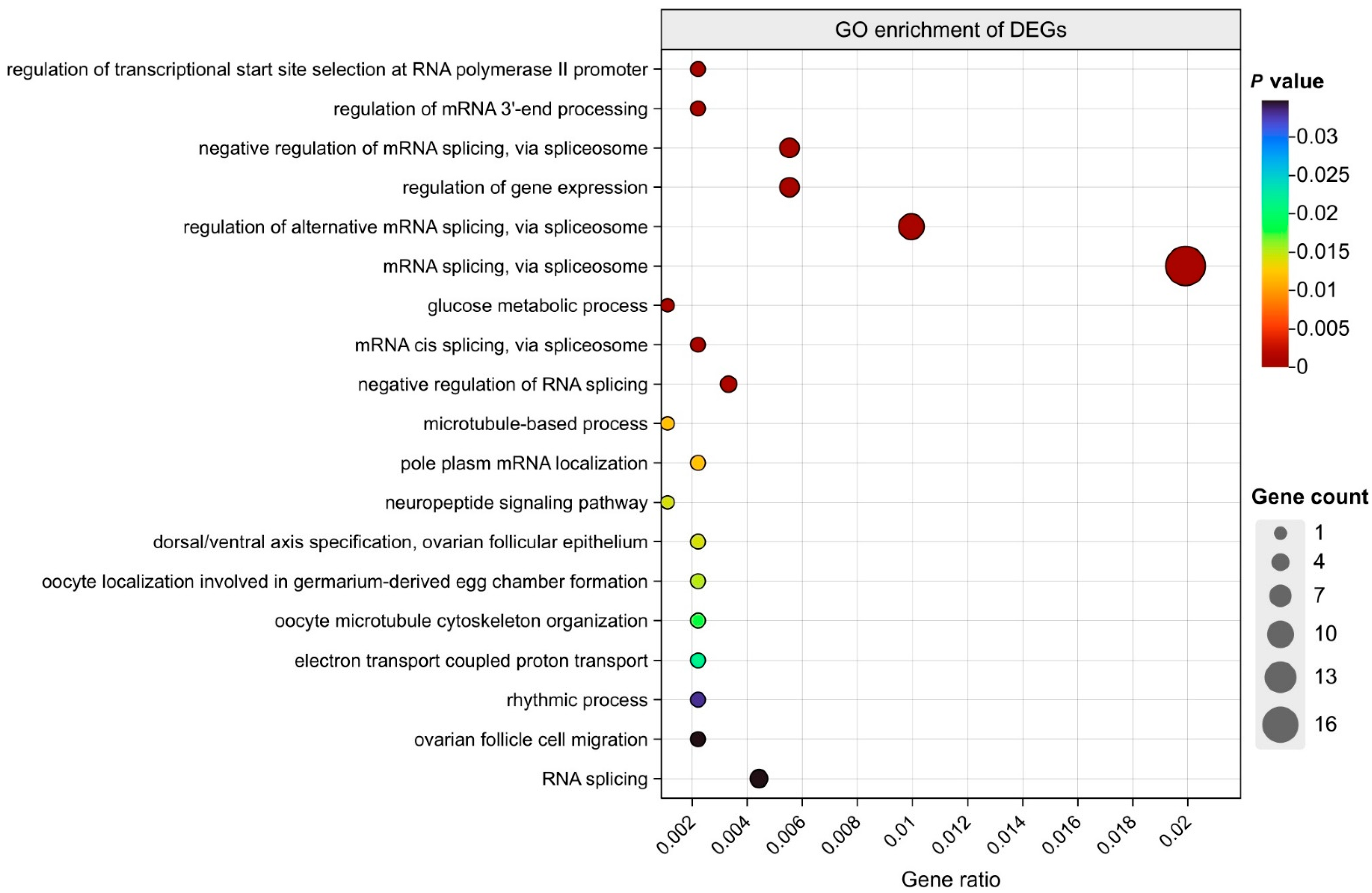
3.4. GO Enrichment Analysis of Common DEGs

GO annotation and enrichment analysis of biological processes were performed on the common DEGs between the Es22_vs_Es27 and Es22_vs_Es32 groups, with results shown in Figure 3 (Supplementary File S4). The analysis revealed that these common DEGs were predominantly enriched in RNA-level regulatory processes, glucose metabolic processes, and the neuropeptide signaling pathway. Notably, the same set of genes was enriched in both the glucose metabolic process and neuropeptide signaling pathway, suggesting a role of the neuropeptide signaling pathway in temperature regulation. To further explore whether other signaling pathways contribute to thermal adaptation, an enrichment analysis of these DEGs across all signaling pathways was conducted (Figure 4). The results demonstrated significant enrichment not only in the neuropeptide signaling pathway but also in 10 additional signaling pathways. Among these, the androgen receptor signaling pathway and intracellular estrogen receptor signaling pathway each contained one enriched gene, implying a potential link between sex hormone regulation and temperature response.
Moreover, the glutamate receptor, dopamine receptor, cytokine-mediated, and fibroblast growth factor receptor signaling pathways exhibited the highest number of enriched genes, with the first three pathways sharing identical sets of genes. Both the dopamine receptor and neuropeptide signaling pathways are associated with neurotransmission. Previous studies in mammals and D. melanogaster have shown that temperature adaptation in organisms correlates with neural regulation [7,37]. Based on these findings, the genes enriched in these two neurotransmission-related signaling pathways were further validated using RT-qPCR.

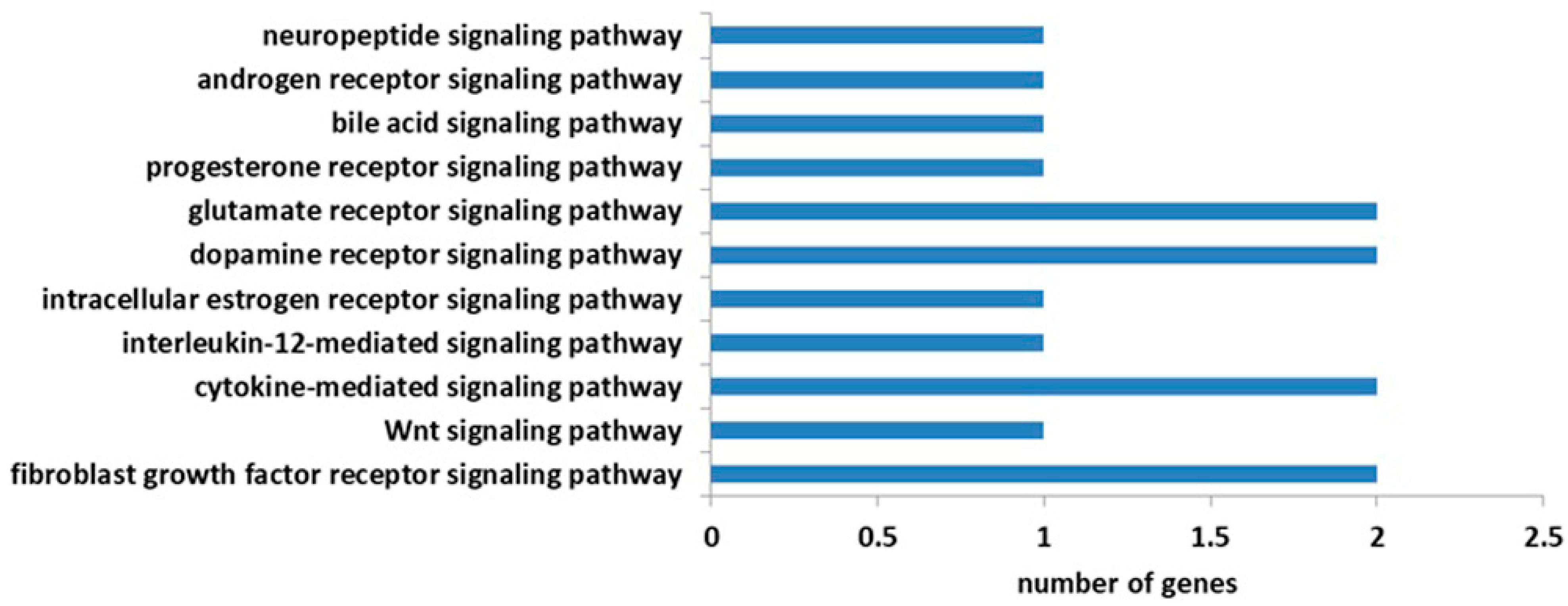
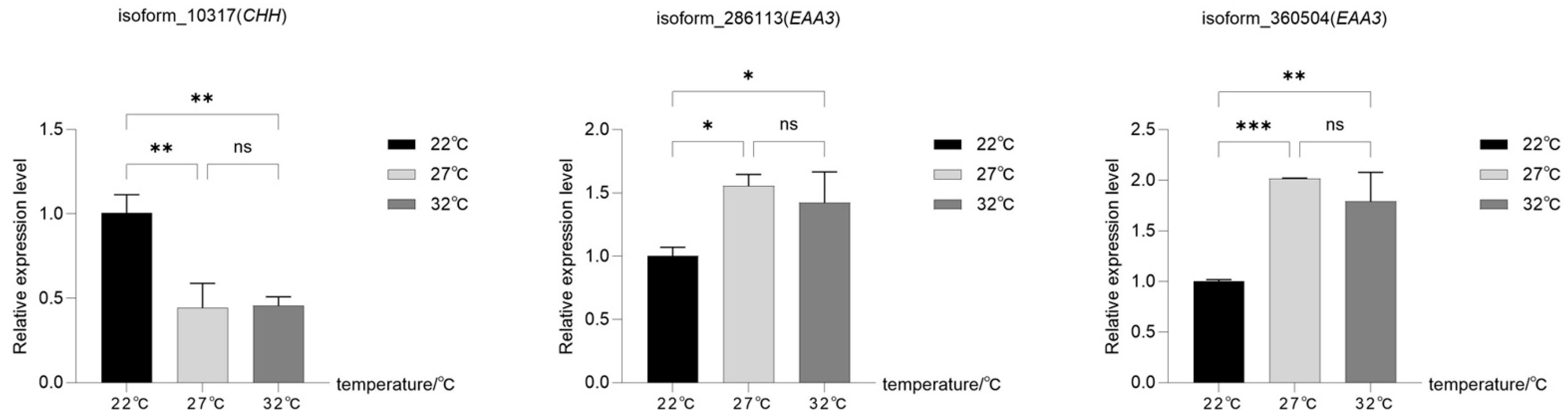
3.5. RT-qPCR Validation of Genes in Neurotransmission Signaling Pathways

The dopamine receptor and neuropeptide signaling pathways were totally enriched with three genes: isoform_10317 (CHH) in the neuropeptide signaling pathway, and isoform_286113 (excitatory amino acid transporter 3, EAA3) and isoform_360504 (EAA3) in the dopamine receptor signaling pathway. Transcriptomic analysis revealed that isoform_10317 was down-regulated under high-temperature conditions (27 °C and 32 °C) compared to the normal temperature (22 °C), while isoform_286113 and isoform_360504 exhibited up-regulated expression at high temperatures.
The expression patterns of these three genes at 22 °C, 27 °C, and 32 °C were further validated using RT-qPCR. The results are shown in Figure 5. The relative expression level of isoform_10317 was highest at 22 °C, and shows significantly down-regulated expression in both 22 °C_vs_27 °C and 22 °C_vs_32 °C groups, whereas isoform_286113 and isoform_360504 showed significantly up-regulated expression for both 22 °C_vs_27 °C and 22 °C_vs_32 °C groups. The RT-qPCR results were consistent with the transcriptomic analysis.

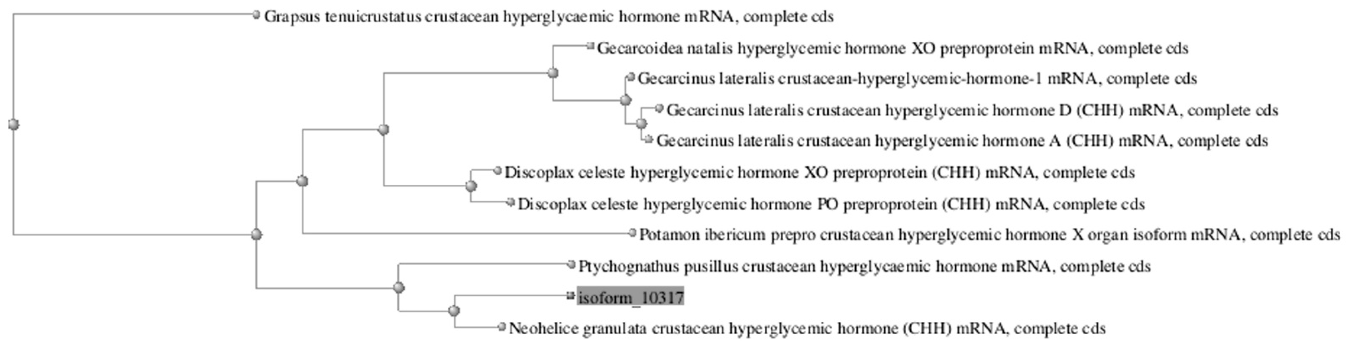
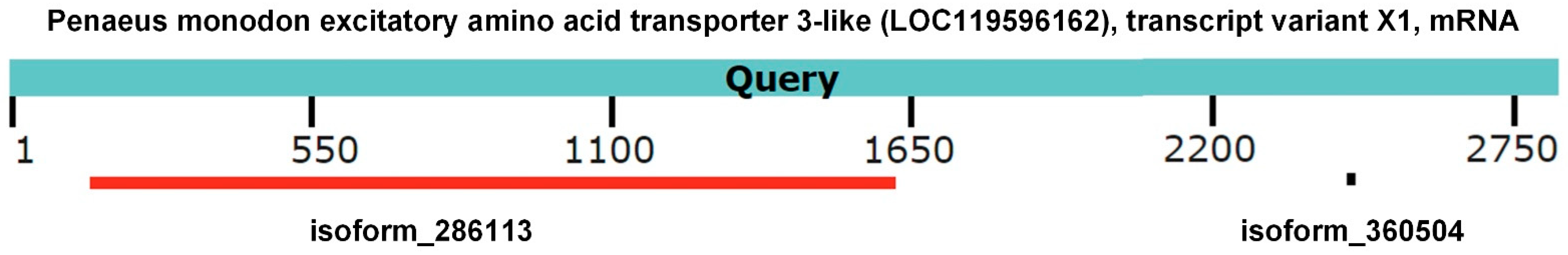
3.6. Sequence Similarity Analysis

In order to find the closest gene homolog of isoform_10317, a BLAST alignment of isoform_10317 was performed against the NCBI database (https://blast.ncbi.nlm.nih.gov/Blast.cgi, accessed on 9 July 2025) to reveal its similarity to CHH genes from other species, as shown in Table 4. Using NCBI’s phylogenetic tree analysis function, an evolutionary analysis was conducted on the top ten CHH sequences with high similarity, resulting in the phylogenetic tree presented in Figure 6. The homologous gene most closely related to isoform_10317 and exhibiting the highest identity is Neohelice granulata CHH, while the homologous gene with the highest alignment score is Ptychognathus pusillus CHH. However, currently no studies exist on the relationship between CHH and heat stress in these two species. Additionally, isoform_10317 shows homology to Gecarcinus lateralis CHH-1, CHH D, and CHH A, with almost the same identity. Its identity with Discoplax celeste XO-CHH is higher than with pericardial organ PO-CHH. Research in Callinectes sapidus suggests that XO-CHH and PO-CHH may be subject to differential regulation of transcription: XO-CHH expression is closely associated with hemolymph glucose levels, while PO-CHH does not induce hyperglycemia in the hemolymph [38]. It is evident that isoform_10317 is more similar to XO-CHH.
Isoform_286113 (5411 bp length) and isoform_360504 (4148 bp length) were both annotated as the EAA3 gene. The similar region of the two genes covers 76.47% of isoform_286113 and 99.95% of isoform_360504. The identity of the two genes is 99.83%. Isoform_286113 exhibits significant similarity (79.49% identity) to the 148–1621 nucleotides region of the “Penaeus monodon excitatory amino acid transporter 3-like (LOC119596162), transcript variant X1, mRNA”. In contrast, isoform_360504 shows significant similarity (84% identity) to the 1098–1621 nucleotides region of the same gene (Figure 7). This suggests that these two genes might be different subtypes of the EAA3 gene in E. sinensis. The sequences of isoform_10317, isoform_286113, and isoform_360504 are provided in Supplementary File S5.

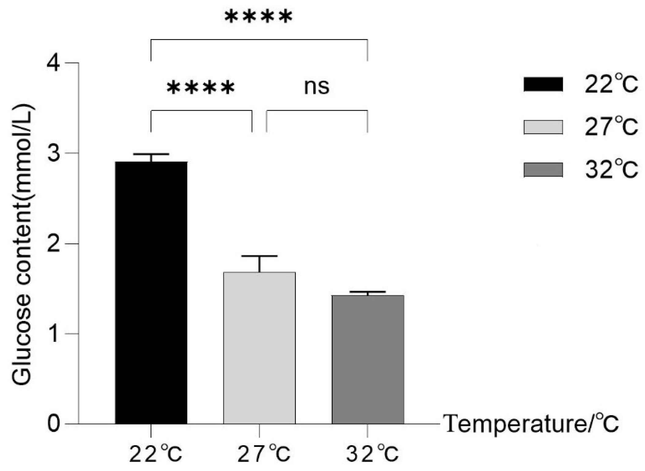
3.7. Influence of Thermal Stress on Hemolymph Glucose

The hemolymph glucose levels detected after 24 h of acclimation at 22 °C, 27 °C, and 32 °C are presented in Figure 8. Under elevated temperatures (27 °C and 32 °C), the hemolymph glucose levels were significantly reduced. Glucose content was lower at 32 °C compared to 27 °C, but this difference was not statistically significant. Furthermore, distinct behavioral changes of crabs were observed. Crabs exhibited a significant decrease in the frequency and intensity of activity, remaining largely sedentary at 32 °C. At 22 °C, crabs displayed greater activity, engaging actively in exploratory behavior and locomotion. At 27 °C, the activity level of the crabs was intermediate between that observed at 22 °C and 32 °C. These results indicate that increasing temperature significantly affects both hemolymph glucose levels and the behavior of E. sinensis.

4. Discussion

4.1. Biological Processes Related to Thermal Regulation

Based on the gene enrichment analysis of common DEGs, multiple biological processes related to RNA splicing were most significantly enriched, indicating that the changes in gene expression might be related to the variable RNA splicing. Furthermore, four biological processes linked to ovarian and oocyte development were enriched: dorsal/ventral axis specification; ovarian follicular epithelium; oocyte localization involved in germarium-derived egg chamber formation; oocyte microtubule cytoskeleton organization; and ovarian follicle cell migration. These findings suggest that temperature significantly impacts ovarian function, consistent with subsequent observations of altered gene expression in the androgen receptor signaling pathway and intracellular estrogen receptor signaling pathway. Notably, previous studies have demonstrated that temperature-dependent sex determination is widespread across species. For example, in the red-eared slider turtle Trachemys scripta, high temperatures (32 °C) during the sex determination period of gonad development induce female differentiation, whereas low temperatures (26 °C) promote male development. Mechanistically, elevated temperatures increase cytosolic calcium levels in somatic cells. Calcium influx into gonadal somatic cells triggers the phosphorylation and activation of STAT3 (pSTAT3), which subsequently relieves the inhibition of the epigenetic regulator Kdm6b. This leads to suppression of the male sex-determining gene Dmrt1, thereby blocking male development [39]. Additionally, pSTAT3 directly binds to the promoter of FoxI2 under high temperatures, initiating female developmental pathways [40]. In the nematode Caenorhabditis elegans, high temperatures (30 °C) promote male development, while low temperatures favor hermaphrodite development. This occurs because elevated temperatures reduce the activity of BiP, an endoplasmic reticulum molecular chaperone that senses thermal stress. Reduced BiP activity increases the degradation of TRA-2, a key driver of hermaphrodite fate, thereby enhancing male gonad development [41].
Temperature fluctuations often coincide with changes in water salinity. The eyestalk may maintain hemolymph osmolality stability by regulating hormones such as molt-inhibiting hormone (MIH) and crustacean CHH, thereby ensuring normal cellular function under thermal stress. Osmoregulation is closely linked to ion channel activity, which may explain why the electron transport-coupled proton transport pathway showed enriched DEGs. Studies in Scylla paramamosain suggest that CHH may regulate carbonic anhydrase expression by activating the PKA signaling pathway, thereby influencing cellular osmoregulation and associated metabolic processes [42]. Additionally, research on Litopenaeus vannamei has demonstrated that the eyestalk is involved in salinity adaptation and stress-related endocrine responses [43].

4.2. Neurotransmission Signaling Pathways and Genes Related to Temperature Regulation

The eyestalk of E. sinensis contains the XO-SG, a critical neuroendocrine regulatory center in crustaceans [15]. This structure secretes various hormones that directly regulate metabolism, osmoregulation, and stress responses, potentially indirectly influencing the crab’s thermal adaptation capacity. Studies have shown that dopamine can regulate the sensitivity to temperature changes in D. melanogaster. Specifically, dopamine-treated flies exhibit an extended heat tolerance duration under high-temperature conditions, accompanied by significant alterations in heat shock protein (HSP) gene expression levels [44]. Therefore, neurosecretory cells in the eyestalk may activate hormone release through temperature-sensitive neural signals (e.g., thermosensitive neurons) triggered by environmental temperature changes, thereby modulating physiological adaptations. However, the precise mechanisms remain unclear.
In this study, the GO enrichment analysis revealed two neurotransmission-related signaling pathways—the dopamine receptor signaling pathway and the neuropeptide signaling pathway—that exhibited significant alterations during high-temperature adaptation. The dopamine receptor signaling pathway and neuropeptide signaling pathway contain three DEGs, isoform_10317, isoform_286113, and isoform_360504. Isoform_10317 is a CHH gene, which is a hormone found in the sinus gland of isopods and decapods which controls the blood sugar level. It has a secret agogue action over the amylase released from the midgut gland. It may act as a stress hormone and may be involved in the control of molting and reproduction [45]. In addition, the fact that CHH genes are enriched in both the glucose metabolic process and neuropeptide signaling pathway indicates that glucose metabolism is associated with the neuropeptide. This is consistent with the function of CHH in the regulation of glucose metabolism in crustaceans as a response to environmental stress [46]. CHH may respond to temperature changes by regulating glucose metabolism under high-temperature conditions.
Many studies have demonstrated that CHH is associated with temperature response in crustaceans. However, the response to temperature varies among different species and under different conditions. In the freshwater crayfish Cherax quadricarinatus, the CHH level was the highest under conditions of high salinity (10 g/L) and low temperature (20 °C) [47]. In L. vannamei, the expression of CHH-A and CHH-B2 in the eyestalk was lowest at a low temperature (20 °C) and highest at a high temperature (32 °C). Salinity had little effect on CHH expression at low and normal temperatures, whereas under high-temperature conditions, the levels of both CHH genes increased significantly under hyperosmotic conditions compared to low salinity conditions [48]. Conversely, Rajendiran et al. [49] found that both excessively high and low temperatures could reduce CHH levels in the blue swimmer crab Portunus pelagicus. They measured CHH and glucose levels in the hemolymph of P. pelagicus after 3 h of heat stress (at 24 °C, 26 °C, 30 °C, and 32 °C) followed by 3 h of recovery at the normal temperature (28 °C). Results showed that when the temperature decreased to 26 °C, hemolymph CHH levels increased significantly. However, CHH levels decreased significantly when the temperature dropped to 24 °C or rose to 30 °C. At 32 °C, CHH levels were also lower than at the normal temperature. Correspondingly, at 26 °C, the induced heat stress significantly affected hemolymph glucose levels, causing hyperglycemia, while higher or lower temperatures alleviated this hyperglycemia. In contrast, another study by Vasudevan et al. [50] reported that in P. pelagicus, after heat stress ended, the hemolymph CHH level was highest at 24 °C compared to the normal temperature (28 °C), while CHH levels at 26 °C and 30 °C were significantly lower than at 28 °C. CHH contents at 30 °C were even lower than at 26 °C, whereas at 32 °C, CHH contents showed a marked increase compared to 28 °C. Correspondingly, blood glucose was higher when exposed to 24 °C and 32 °C, consuming more glucose to meet the elevated metabolic demands. Glucose levels were significantly lower at 26 °C and 30 °C.
These results indicate that the response of CHH varies significantly across different species and under different combinations of temperature and salinity conditions. It does not universally increase or decrease significantly due to heat stress in all species. The findings of this study show that in E. sinensis, CHH expression in the eyestalk significantly decreased when the temperature rose by 5 °C and 10 °C compared to the optimal growth temperature (22 °C), while no significant change occurred between 27 °C and 32 °C.
A similar finding across these studies is that all of them show that CHH has hyperglycemic effects. In fact, the CHH gene family is large and multifunctional. Certain members possess hyperglycemic effects, while others are non-responsive. For example, in the mud crab Scylla olivacea, the Sco-CHH from eyestalk ganglia exerted hyperglycemic and molt-inhibiting activity, whereas Sco-CHH-L (CHH-like peptide) from extra-eyestalk tissues did not exhibit these functions [51]. According to the similarity analysis, the CHH genes obtained in this study had higher similarity with the eyestalk CHH genes, which possess hyperglycemic effects. In this work, the variation in hemolymph glucose levels across different temperatures further indicates that the CHH expressions associated with thermal response are correlated with glucose levels in E. sinensis. At 22 °C, where CHH expression increased, the hemolymph glucose concentration was the highest. In contrast, under elevated temperatures (27 °C and 32 °C), CHH expression was significantly reduced, accompanied by a significant decrease in glucose concentration. Between 27 °C and 32 °C, CHH expression levels showed little change, remaining similarly low, and the glucose concentration also exhibited minimal variation between these two temperatures. These findings demonstrate that CHH expression in the eyestalk is strongly correlated with hemolymph glucose levels and likely mediates the response to temperature changes by regulating blood glucose in E. sinensis. Combined with the observation of crab behavior at different temperatures, it is hypothesized that under high-temperature conditions, due to the significantly reduced activity frequency and intensity of crabs, the metabolism in crabs becomes less active than at normal temperatures, requiring less energy consumption. Consequently, there is no need to induce CHH release to synthesize more glucose for providing energy.
Isoform_286113 and isoform_360504 are both EAA3 gene, which mediates the uptake of L-glutamate and also L-aspartate and D-aspartate [52,53]. Current research indicates that EAA3 plays significant roles in glutamatergic synapses within the midbrain, GABAergic synapses in the hippocampus, and in supporting cellular survival through its cysteine transport capacity in midbrain and hippocampal neurons [54]. It critically maintains low local glutamate concentrations, and its predominant postsynaptic localization enables the buffering of nearby glutamate receptors, thereby modulating excitatory neurotransmission and synaptic plasticity. Furthermore, as the primary neuronal cysteine uptake system, EAA3 acts as the rate-limiting factor for glutathione synthesis—a potent antioxidant—in neurons expressing this transporter. On GABAergic neurons, EAA3 additionally supplies glutamate as a precursor for GABA synthesis [55]. In this study, EAA3 exhibited up-regulated expression under high-temperature conditions (27 °C and 32 °C). Studies have shown that the intake of L-glutamate is associated with an increase in body temperature in rats [56]. Temperature-dependent L-aspartate transporters have also been identified in rats [57]. Additionally, in chicks, the intake of D-aspartate has been found to reduce body temperature by decreasing the expression of avUCP mRNA in the pectoral muscle [58]. In crustaceans, the uptake of glutamate has been found in the large muscle fibers of Balanus nubilus [59] and the synaptic vesicle of crayfish [60]. The temperature-dependent L-aspartate transporters were also found to influence the axon–glia signaling in crayfish [61]. However, the relationship between these amino acids and the thermal adaptation of crustaceans has not yet been studied. The findings of this study provide valuable clues to this area of research. The results of this study suggest that CHH and EAA3 may play roles in temperature adaptation in E. sinensis. These findings provide valuable insights for further investigations into the mechanistic role of the eyestalk—a neuroendocrine hub—in regulating molting and growth/development processes in E. sinensis.

5. Conclusions

The molting and growth development mechanisms of crustaceans are highly sensitive to temperature, yet the regulatory mechanisms underlying their thermal adaptation remain unclear. This study conducted transcriptome sequencing on E. sinensis under normal (22 °C) and elevated temperature conditions (27 °C and 32 °C) to analyze the changes in gene expression levels. Through a GO enrichment analysis of DEGs, temperature-affected signaling pathways were identified, including estrogen and androgen receptor signaling pathways along with two neurotransmission signaling pathways. These findings suggest that temperature might influence gender regulation in E. sinensis, while simultaneously indicating the potential involvement of dopamine receptor and neuropeptide signaling pathways in thermal adaptation. Subsequent RT-qPCR validation of DEGs within neurotransmission pathways revealed that EAA3 and CHH genes might participate in high-temperature adaptation regulation. The parallel changes in hemolymph glucose levels and CHH expression under elevated temperatures confirm that eyestalk CHH strongly correlates with glucose regulation in E. sinensis and likely mediates thermal responses by controlling blood glucose. This research provides crucial insights into the molecular mechanisms through which temperature affects molting and developmental processes in E. sinensis, while also offering valuable references for studies on thermal adaptation regulation in crustaceans.

Supplementary Materials

The following supporting information can be downloaded at https://www.mdpi.com/article/10.3390/fishes10070361/s1, Supplementary File S1. Base content distribution of clean reads. A: adenine; G: guanine; C: cytosine; T: thymine; and N: uncertain. Supplementary File S2. Distribution of reads on transcripts. The x-axis represents the position of transcripts (200 sliding windows are set), and the y-axis represents the number of reads (calculated by each sliding window). Supplementary File S3. The common DEGs between Es22_vs_Es27 and Es22_vs_Es32 groups. Supplementary File S4. The result of the GO enrichment analysis of common DEGs. Supplementary File S5. The sequences of isoform_10317, isoform_286113, and isoform_360504.

Author Contributions

Conceptualization, T.H. and J.S.; Data curation, X.L., R.Z. (Runlin Zhou), and R.Z. (Ruiqi Zhang); Formal analysis, Z.H. and M.Z.; Funding acquisition, T.H. and J.S.; Investigation, Z.H. and M.Z.; Methodology, X.L., R.Z. (Runlin Zhou), R.Z. (Ruiqi Zhang), Z.H., and T.H.; Project administration, T.H. and J.S.; Resources, X.L. and R.L.; Software, R.Z. (Runlin Zhou); Supervision, R.L., T.H., and J.S.; Validation, X.L., R.Z. (Runlin Zhou), and R.Z. (Ruiqi Zhang); Writing—original draft, X.L., R.L., and T.H.; Writing—review and editing, T.H. and J.S. All authors have read and agreed to the published version of the manuscript.

Funding

This work was supported by the Tianjin Aquatic Seed Industry Innovation Project (SCZYGG202402), Tianjin Science and Technology Planning Project (S25ZY2404005), and Tianjin Development Program for Innovation and Entrepreneurship team (ITTFRS2017007).

Institutional Review Board Statement

All experimental procedures were approved by the College of Life Sciences, Tianjin Normal University, with approval code No. 2025-04 and an approval date of 6 April 2025.

Informed Consent Statement

Not applicable.

Data Availability Statement

All data generated or analyzed during this study are included in the Supplementary Materials.

Conflicts of Interest

The authors declare that the research was conducted in the absence of any commercial or financial relationships that could be construed as a potential conflict of interest.

References

  1. Tansey, E.A.; Johnson, C.D. Recent advances in thermoregulation. Adv. Physiol. Educ. 2015, 39, 139–148. [Google Scholar] [CrossRef] [PubMed]
  2. Tan, C.L.; Knight, Z.A. Regulation of Body Temperature by the Nervous System. Neuron 2018, 98, 31–48. [Google Scholar] [CrossRef] [PubMed]
  3. Lim, C.L. Fundamental Concepts of Human Thermoregulation and Adaptation to Heat: A Review in the Context of Global Warming. Int. J. Environ. Res. Public Health 2020, 17, 7795. [Google Scholar] [CrossRef] [PubMed]
  4. Mota-Rojas, D.; Titto, C.G.; Orihuela, A.; Martínez-Burnes, J.; Gómez-Prado, J.; Torres-Bernal, F.; Flores-Padilla, K.; Carvajal-de la Fuente, V.; Wang, D. Physiological and Behavioral Mechanisms of Thermoregulation in Mammals. Animals 2021, 11, 1733. [Google Scholar] [CrossRef] [PubMed]
  5. Johnson, J.M.; Kellogg, D.L., Jr. Thermoregulatory and thermal control in the human cutaneous circulation. Front. Biosci. (Schol. Ed.) 2010, 2, 825–853. [Google Scholar] [PubMed]
  6. Abram, P.K.; Boivin, G.; Moiroux, J.; Brodeur, J. Behavioural effects of temperature on ectothermic animals: Unifying thermal physiology and behavioural plasticity. Biol. Rev. Camb. Philos. Soc. 2017, 92, 1859–1876. [Google Scholar] [CrossRef] [PubMed]
  7. Volonté, C.; Liguori, F.; Amadio, S. A Closer Look at Histamine in Drosophila. Int. J. Mol. Sci. 2024, 25, 4449. [Google Scholar] [CrossRef] [PubMed]
  8. Gandara, A.C.P.; Drummond-Barbosa, D. Chronic exposure to warm temperature causes low sperm abundance and quality in Drosophila melanogaster. Sci. Rep. 2023, 13, 12331. [Google Scholar] [CrossRef] [PubMed]
  9. Johnson, K.H.; Dobkowski, K.A.; Seroy, S.K.; Fox, S.; Meenan, N. Feeding preferences and the effect of temperature on feeding rates of the graceful kelp crab, Pugettia gracilis. PeerJ 2023, 11, e15223. [Google Scholar] [CrossRef] [PubMed]
  10. Martelli, A.; Barón, P.J. Effects of temperature and salinity on the development and survival of the embryos and zoeae I from the southern surf crab Ovalipes trimaculatus (Brachyura: Portunidae). An. Acad. Bras. Cienc. 2021, 93, e20190999. [Google Scholar] [CrossRef] [PubMed]
  11. Ayres, B.S.; Varela Junior, A.S.; Corcini, C.D.; Lopes, E.M.; Nery, L.E.M.; Maciel, F.E. Effects of high temperature and LPS injections on the hemocytes of the crab Neohelice granulata. J. Invertebr. Pathol. 2024, 205, 108144. [Google Scholar] [CrossRef] [PubMed]
  12. Chung, J.S.; Zmora, N.; Katayama, H.; Tsutsui, N. Crustacean hyperglycemic hormone (CHH) neuropeptidesfamily: Functions, titer, and binding to target tissues. Gen. Comp. Endocrinol. 2010, 166, 447–454. [Google Scholar] [CrossRef] [PubMed]
  13. Kotsyuba, E.; Dyachuk, V. Immunocytochemical Localization of Enzymes Involved in Dopamine, Serotonin, and Acetylcholine Synthesis in the Optic Neuropils and Neuroendocrine System of Eyestalks of Paralithodes camtschaticus. Front. Neuroanat. 2022, 16, 844654. [Google Scholar] [CrossRef] [PubMed]
  14. Christie, A.E.; Stanhope, M.E.; Gandler, H.I.; Lameyer, T.J.; Pascual, M.G.; Shea, D.N.; Yu, A.; Dickinson, P.S.; Hull, J.J. Molecular characterization of putative neuropeptide, amine, diffusible gas and small molecule transmitter biosynthetic enzymes in the eyestalk ganglia of the American lobster, Homarus americanus. Invert. Neurosci. 2018, 18, 12. [Google Scholar] [CrossRef] [PubMed]
  15. Christie, A.E. Crustacean neuroendocrine systems and their signaling agents. Cell Tissue Res. 2011, 345, 41–67. [Google Scholar] [CrossRef] [PubMed]
  16. Strauss, J.; Dircksen, H. Circadian clocks in crustaceans: Identified neuronal and cellular systems. Front. Biosci. 2010, 15, 1040–1074. [Google Scholar] [CrossRef] [PubMed]
  17. Wittmann, A.C.; Benrabaa, S.A.M.; López-Cerón, D.A.; Chang, E.S.; Mykles, D.L. Effects of temperature on survival, moulting, and expression of neuropeptide and mTOR signalling genes in juvenile Dungeness crab (Metacarcinus magister). J. Exp. Biol. 2018, 221, jeb187492. [Google Scholar]
  18. Lacerda, J.T.; David, D.D.; Castrucci, A.M.L. The effect of thermal stress on the X-organ/sinus gland proteome of the estuarine blue crab Callinectes sapidus during the intermolt and premolt stages. J. Proteom. 2025, 313, 105382. [Google Scholar] [CrossRef] [PubMed]
  19. Yuan, Q.; Wang, Q.D.; Zhang, T.L.; Li, Z.J.; Liu, J.S. Effects of water temperature on growth, feeding and molting of juvenile Chinese mitten crab (Eriocheir sinensis). Aquaculture 2017, 468, 169–174. [Google Scholar] [CrossRef]
  20. Peng, J.; Zhao, Y.L.; Xu, Z.G.; Liu, B.Q.; Duan, C.C.; Tang, Y.C. Effect of temperature stress on the survival of juvenile Chinese mitten crab (Eriocheir sinensis). Iran. J. Fish. Sci. 2019, 18, 763–774. [Google Scholar]
  21. Deng, D.; Hu, S.; Lin, Z.; Geng, J.; Qian, Z.; Zhang, K.; Ning, X.; Cheng, Y.; Zhang, C.; Yin, S. High temperature aggravated hypoxia-induced intestine toxicity on juvenile Chinese mitten crab (Eriocheir sinensis). Comp. Biochem. Physiol. Part D Genom. Proteom. 2024, 52, 101288. [Google Scholar] [CrossRef] [PubMed]
  22. Deng, X.; Yang, Y.; Li, Z.; Luo, L.; Wang, S.; Zhang, R.; Guo, K.; Zhao, Z. Effects of re-enter water on antioxidant, immune, intestinal flora and metabolome of Chinese mitten crab (Eriocheir sinensis) after high temperature air exposure. Comp. Biochem. Physiol. C Toxicol. Pharmacol. 2025, 296, 110232. [Google Scholar] [CrossRef] [PubMed]
  23. Pan, T.; Li, T.; Yang, M.; Jiang, H.; Ling, J. Integrative Analysis of Hepatopancreas Transcriptome and Proteome in Female Eriocheir sinensis under Thermal Stress. Int. J. Mol. Sci. 2024, 25, 7249. [Google Scholar] [CrossRef] [PubMed]
  24. Yin, C.; Pang, A.; Liu, R.; Yang, W.; Wu, H.; Yang, J.; Xuan, J.; Sun, X.; Ding, G.; Zhang, H.; et al. Regulation of gene expression under temperature stress and genome-wide analysis of heat shock protein family in Eriocheir sinensis. Int. J. Biol. Macromol. 2025, 308, 142503. [Google Scholar] [CrossRef] [PubMed]
  25. Keller, R. Crustacean neuropeptides: Structures, functions and comparative aspects. Experientia 1992, 48, 439–448. [Google Scholar] [CrossRef] [PubMed]
  26. Simões, L.A.R.; Normann, R.S.; Chung, J.S.; Vinagre, A.S. A brief and updated introduction to the neuroendocrine system of crustaceans. Mol. Cell. Endocrinol. 2024, 590, 112265. [Google Scholar] [CrossRef] [PubMed]
  27. Li, Y.; Han, Z.; She, Q.; Zhao, Y.; Wei, H.; Dong, J.; Xu, W.; Li, X.; Liang, S. Comparative transcriptome analysis provides insights into the molecular basis of circadian cycle regulation in Eriocheir sinensis. Gene 2019, 694, 42–49. [Google Scholar] [CrossRef] [PubMed]
  28. Pang, Y.Y.; Zhang, C.; Xu, M.J.; Huang, G.Y.; Cheng, Y.X.; Yang, X.Z. The transcriptome sequencing and functional analysis of eyestalk ganglions in Chinese mitten crab (Eriocheir sinensis) treated with different photoperiods. PLoS ONE 2019, 14, e0210414. [Google Scholar] [CrossRef] [PubMed]
  29. Yu, C.; Huang, Z.; Xu, Y.; Zhang, B.; Li, Y. Deep sequencing of microRNAs reveals circadian-dependent microRNA expression in the eyestalks of the Chinese mitten crab Eriocheir sinensis. Sci. Rep. 2023, 13, 5253. [Google Scholar] [CrossRef] [PubMed]
  30. Liang, M.; Feng, W.; Chen, X.; Tang, Y.; Li, J.; Li, W. Effects of different temperatures on growth and intestinal microbial composition of juvenile Eriocheir sinensis. Front. Physiol. 2023, 14, 1163055. [Google Scholar] [CrossRef] [PubMed]
  31. Seppey, M.; Manni, M.; Zdobnov, E.M. BUSCO: Assessing Genome Assembly and Annotation Completeness. Methods Mol. Biol. 2019, 1962, 227–245. [Google Scholar] [PubMed]
  32. Chen, Y.; Shi, C.; Huang, Z.; Zhang, Y.; Li, S.; Li, Y.; Ye, J.; Yu, C.; Li, Z.; Zhang, X.; et al. SOAPnuke: A MapReduce acceleration-supported software for integrated quality control and preprocessing of high-throughput sequencing data. GigaScience 2018, 7, gix120. [Google Scholar] [CrossRef] [PubMed]
  33. Langmead, B.; Salzberg, S.L. Fast gapped-read alignment with Bowtie 2. Nat. Methods 2012, 9, 357–359. [Google Scholar] [CrossRef] [PubMed]
  34. Li, B.; Dewey, C.N. RSEM: Accurate transcript quantification from RNA-Seq data with or without a reference genome. BMC Bioinform. 2011, 12, 323. [Google Scholar] [CrossRef] [PubMed]
  35. Conesa, A.; Gotz, S.; Garcia-Gomez, J.M.; Terol, J.; Talon, M.; Robles, M. Blast2GO: A universal tool for annotation, visualization and analysis in functional genomics research. Bioinformatics 2005, 21, 3674–3676. [Google Scholar] [CrossRef] [PubMed]
  36. Singh, V.K.; Mangalam, A.K.; Dwivedi, S.; Naik, S. Primer premier: Program for design of degenerate primers from a protein sequence. Biotechniques 1998, 24, 318–319. [Google Scholar] [CrossRef] [PubMed]
  37. Kichko, T.I.; Reeh, P.W. TRPV1 controls acid- and heat-induced calcitonin gene-related peptide release and sensitization by bradykinin in the isolated mouse trachea. Eur. J. Neurosci. 2009, 29, 1896–1904. [Google Scholar] [CrossRef] [PubMed]
  38. Chung, J.S.; Zmora, N. Functional studies of crustacean hyperglycemic hormones (CHHs) of the blue crab, Callinectes sapidus—The expression and release of CHH in eyestalk and pericardial organ in response to environmental stress. FEBS J. 2008, 275, 693–704. [Google Scholar] [CrossRef] [PubMed]
  39. Weber, C.; Zhou, Y.; Lee, J.G.; Looger, L.L.; Qian, G.; Ge, C.; Capel, B. Temperature-dependent sex determination is mediated by pSTAT3 repression of Kdm6b. Science 2020, 368, 303–306. [Google Scholar] [CrossRef] [PubMed]
  40. Wu, P.; Wang, X.; Ge, C.; Jin, L.; Ding, Z.; Liu, F.; Zhang, J.; Gao, F.; Du, W. pSTAT3 activation of Foxl2 initiates the female pathway underlying temperature-dependent sex determination. Proc. Natl. Acad. Sci. USA 2024, 121, e2401752121. [Google Scholar] [CrossRef] [PubMed]
  41. Shi, J.; Sheng, D.; Guo, J.; Zhou, F.; Wu, S.; Tang, H. Identification of BiP as a temperature sensor mediating temperature-induced germline sex reversal in C. elegans. EMBO J. 2024, 43, 4020–4048. [Google Scholar] [CrossRef] [PubMed]
  42. Mo, N.; Shao, S.; Cui, Z.; Bao, C. Roles of eyestalk in salinity acclimatization of mud crab (Scylla paramamosain) by transcriptomic analysis. Comp. Biochem. Physiol. Part D Genom. Proteom. 2024, 52, 101276. [Google Scholar] [CrossRef] [PubMed]
  43. Farhadi, A.; Liu, Y.; Xu, C.; Han, T.; Wang, X.; Li, E. Evidence from transcriptome analysis unravelled the roles of eyestalk in salinity adaptation in Pacific white shrimp (Litopenaeus vannamei). Gen. Comp. Endocrinol. 2022, 329, 114120. [Google Scholar] [CrossRef] [PubMed]
  44. Ueno, T.; Tomita, J.; Kume, S.; Kume, K. Dopamine modulates metabolic rate and temperature sensitivity in Drosophila melanogaster. PLoS ONE 2012, 7, e31513. [Google Scholar] [CrossRef] [PubMed]
  45. Kegel, G.; Reichwein, B.; Weese, S.; Gaus, G.; Peter-Katalinić, J.; Keller, R. Amino acid sequence of the crustacean hyperglycemic hormone (CHH) from the shore crab, Carcinus maenas. FEBS Lett. 1989, 255, 10–14. [Google Scholar] [CrossRef] [PubMed]
  46. Zhang, X.; Pan, L.; Tong, R.; Li, Y.; Si, L.; Chen, Y.; Wu, M.; Wang, Q. Effects of crustacean hyperglycaemic hormone RNA interference on regulation of glucose metabolism in Litopenaeus vannamei after ammonia-nitrogen exposure. Br. J. Nutr. 2022, 127, 823–836. [Google Scholar] [CrossRef] [PubMed]
  47. Prymaczok, N.C.; Pasqualino, V.M.; Viau, V.E.; Rodriguez, E.M.; Medesani, D.A. Involvement of the crustacean hyperglycemic hormone (CHH) in the physiological compensation of the freshwater crayfish Cherax quadricarinatus to low temperature and high salinity stress. J. Comp. Physiol. B 2016, 186, 181–191. [Google Scholar] [CrossRef] [PubMed]
  48. Giffard-Mena, I.; Ponce-Rivas, E.; Sigala-Andrade, H.M.; Uranga-Solis, C.; Re, A.D.; Diaz, F.; Camacho-Jimenez, L. Evaluation of the osmoregulatory capacity and three stress biomarkers in white shrimp Penaeus vannamei exposed to different temperature and salinity conditions: Na+/K+ ATPase, Heat Shock Proteins (HSP), and Crustacean Hyperglycemic Hormones (CHHs). Comp. Biochem. Physiol. B Biochem. Mol. Biol. 2024, 271, 110942. [Google Scholar] [CrossRef] [PubMed]
  49. Rajendiran, S.; Iqbal, B.M.M.; Vasudevan, S. Induced thermal stress on serotonin levels in the blue swimmer crab, Portunus pelagicus. Biochem. Biophys. Rep. 2016, 5, 425–429. [Google Scholar] [CrossRef] [PubMed]
  50. Vasudevan, S.; Rajendran, S. Thermal stress induced hyperglycemia in the blue swimmer crab, Portunus pelagicus. J. Therm. Biol. 2021, 100, 103076. [Google Scholar] [CrossRef] [PubMed]
  51. Chang, C.C.; Tsai, K.W.; Hsiao, N.W.; Chang, C.Y.; Lin, C.L.; Watson, R.D.; Lee, C.Y. Structural and functional comparisons and production of recombinant crustacean hyperglycemic hormone (CHH) and CHH-like peptides from the mud crab Scylla olivacea. Gen. Comp. Endocrinol. 2010, 167, 68–76. [Google Scholar] [CrossRef] [PubMed]
  52. Lin, C.I.; Orlov, I.; Ruggiero, A.M.; Dykes-Hoberg, M.; Lee, A.; Jackson, M.; Rothstein, J.D. Modulation of the neuronal glutamate transporter EAAC1 by the interacting protein GTRAP3-18. Nature 2001, 410, 84–88. [Google Scholar] [CrossRef] [PubMed]
  53. Bjoras, M.; Gjesdal, O.; Erickson, J.D.; Torp, R.; Levy, L.M.; Ottersen, O.P.; Degree, M.; Storm-Mathisen, J.; Seeberg, E.; Danbolt, N.C. Cloning and expression of a neuronal rat brain glutamate transporter. Brain Res. Mol. Brain Res. 1996, 36, 163–168. [Google Scholar] [CrossRef] [PubMed]
  54. Underhill, S.M.; Ingram, S.L.; Ahmari, S.E.; Veenstra-VanderWeele, J.; Amara, S.G. Neuronal excitatory amino acid transporter EAAT3: Emerging functions in health and disease. Neurochem. Int. 2019, 123, 69–76. [Google Scholar] [CrossRef] [PubMed]
  55. Bjorn-Yoshimoto, W.E.; Underhill, S.M. The importance of the excitatory amino acid transporter 3 (EAAT3). Neurochem. Int. 2016, 98, 4–18. [Google Scholar] [CrossRef] [PubMed]
  56. Sengupta, T.; Jaryal, A.K.; Kumar, V.M.; Mallick, H.N. L-glutamate microinjection in the preoptic area increases brain and body temperature in freely moving rats. NeuroReport 2014, 25, 28–33. [Google Scholar] [CrossRef] [PubMed]
  57. Lao, L.; Franklin, R.B.; Costello, L.C. High-affinity L-aspartate transporter in prostate epithelial cells that is regulated by testosterone. Prostate 1993, 22, 53–63. [Google Scholar] [CrossRef] [PubMed]
  58. Do, P.H.; Tran, P.V.; Bahry, M.A.; Yang, H.; Han, G.; Tsuchiya, A.; Asami, Y.; Furuse, M.; Chowdhury, V.S. Oral administration of a medium containing both D-aspartate-producing live bacteria and D-aspartate reduces rectal temperature in chicks. Br. Poult. Sci. 2017, 58, 569–577. [Google Scholar] [CrossRef] [PubMed]
  59. Revest, P.A.; Baker, P.F. Glutamate transport in large muscle fibres of Balanus nubilus. J. Neurochem. 1988, 50, 94–102. [Google Scholar] [CrossRef] [PubMed]
  60. Tabb, J.S.; Ueda, T. Phylogenetic studies on the synaptic vesicle glutamate transport system. J. Neurosci. 1991, 11, 1822–1828. [Google Scholar] [CrossRef] [PubMed]
  61. Kane, L.S.; Buttram, J.G.; Urazaev, A.K.; Lieberman, E.M.; Grossfeld, R.M. Uptake and metabolism of glutamate at non-synaptic regions of crayfish central nerve fibers: Implications for axon-glia signaling. Neuroscience 2000, 97, 601–609. [Google Scholar] [CrossRef] [PubMed]
Figure 1. BUSCO assessment result. C (Complete): Sequences fully aligned to the BUSCO database. F (Fragmented): Sequences partially aligned to the BUSCO database. D (Duplicate): Multiple genes aligned to the same BUSCO entry. M (Missing): Sequences filtered out or undetected.
Fishes 10 00361 g001
Figure 2. Volcano map of DEGs in Es22_vs_Es27 (A) and Es22_vs_Es32 (B) groups.
Fishes 10 00361 g002
Figure 3. GO enrichment analysis of common DEGs.
Fishes 10 00361 g003
Figure 4. Enrichment of common DEGs in signaling pathways.
Fishes 10 00361 g004
Figure 5. Results of RT-qPCR validation. Statistical analysis was performed via one-way ANOVA (multiple comparison—Tukey’s post hoc test). All results are represented as the means ± SD of three independent experiments. ‘*’ at the 0.1 level, ‘**’ at the 0.01 level, ‘***’ at the 0.001 level, and ‘ns’ represents no significance.
Fishes 10 00361 g005
Figure 6. Phylogenetic analysis of isoform_10317 and its homologous genes. Isoform_10317 was shown in grey color.
Fishes 10 00361 g006
Figure 7. Similar regions of isoform_286113 and isoform_360504 to the P. monodon EAA3-like gene.
Fishes 10 00361 g007
Figure 8. Hemolymph glucose content at different temperatures after 24 h of acclimation. Statistical analysis was performed via one-way ANOVA (multiple comparison—Tukey’s post hoc test). All results are represented as the means ± SD of three independent experiments. ‘****’ at the 0.0001 level, and ‘ns’ represents no significance.
Fishes 10 00361 g008
Table 1. RT-qPCR primers.
GeneLength of Primers (bp)Sequence of Primers (5′–3′)
β-actin-qF20CCCATCTACGAGGGCTACGC
β-actin-qR23CCTTGATGTCTCGCACGATTTCT
isoform_10317-qF19GCCACCCTCTGGATAAACG
isoform_10317-qR20CGGAAGACGAGGTTGCTGTA
isoform_286113-qF19CCCCTCATAGTCTCCTCCC
isoform_286113-qR25GTGCGTCTCGAAGCAGGCCTGGATC
isoform_360504-qF21TCATAGTCTCCTCCCTGGTGT
isoform_360504-qR17CCGTCTTGTGCGTCTCG
Table 2. Results of transcriptome sequencing.
Sample NameRaw Reads/MClean Reads/MQ20/%Q30/%Clean Reads Ratio/%Total Mapping/%
Es220122.8822.0497.6692.6496.3383.94
Es220222.8822.0397.6992.7296.2883.24
Es220322.8822.1197.6692.6596.6382.43
Es270123.222.0496.5289.899579.39
Es270223.0422.0496.5990.1495.6680.21
Es270323.222.0796.690.1995.1380.13
Es320122.8822.0496.5589.9896.3380.86
Es320223.3622.0997.9793.5894.5679.78
Es320322.8822.1297.8393.1196.6881.37
Table 3. DEGs in Es22_vs_Es27 and Es22_vs_Es32 groups.
GroupNumber of DEGsUp-RegulatedDown-Regulated
Es22_vs_Es271243447796
Es22_vs_Es321486621865
Common377149227
Table 4. Results of similarity analysis of isoform_10317.
GenesScoreCoverageE ValueIdentity
Neohelice granulata crustacean hyperglycemic hormone (CHH) mRNA, complete cds86118%093.37%
Ptychognathus pusillus crustacean hyperglycaemic hormone mRNA, complete cds110328%086.2%
Gecarcoidea natalis hyperglycemic hormone XO preproprotein mRNA, complete cds44024%1.00 × 10−11773.28%
Discoplax celeste hyperglycemic hormone XO preproprotein (CHH) mRNA, complete cds42519%3.00 × 10−11376.7%
Gecarcinus lateralis crustacean-hyperglycemic-hormone-1 mRNA, complete cds40816%2.00× 10−10877.76%
Gecarcinus lateralis crustacean hyperglycemic hormone D (CHH) mRNA, complete cds40817%2.00× 10−10877.76%
Gecarcinus lateralis crustacean hyperglycemic hormone A (CHH) mRNA, complete cds40417%1.00× 10−10677.57%
Potamon ibericum prepro crustacean hyperglycemic hormone X organ isoform mRNA, complete cds33515%4.00× 10−8676.92%
Discoplax celeste hyperglycemic hormone PO preproprotein (CHH) mRNA, complete cds41919%8.00× 10−6474.22%
Grapsus tenuicrustatus crustacean hyperglycaemic hormone mRNA, complete cds25315%1.00× 10−6172.49%
Disclaimer/Publisher’s Note: The statements, opinions and data contained in all publications are solely those of the individual author(s) and contributor(s) and not of MDPI and/or the editor(s). MDPI and/or the editor(s) disclaim responsibility for any injury to people or property resulting from any ideas, methods, instructions or products referred to in the content.

