Article

Isolation, Characterization, and Assessment of Probiotic Lactococcus lactis from the Intestinal Tract of Largemouth Bass (Micropterus salmoides)

1
Fisheries College, Jimei University, Xiamen 361021, China
2
Key Laboratory of Special Aquatic Feed for Fujian, Fujian Tianma Technology Company Limited, Fuzhou 350308, China
*
Authors to whom correspondence should be addressed.
Fishes 2025, 10(6), 291; https://doi.org/10.3390/fishes10060291
Submission received: 27 April 2025 / Revised: 8 June 2025 / Accepted: 13 June 2025 / Published: 16 June 2025
(This article belongs to the Section Welfare, Health and Disease)

Abstract

:
The health benefits associated with microbial species inhabiting aquatic animals have garnered increasing attention, as it is expected that the colonization and efficacy of native probiotic bacteria adapted to the internal environment of the target species will be more active than non-native bacteria. In this study, six isolates were obtained from the intestinal tract of largemouth bass. Three of these isolates demonstrated higher growth ability compared to the others and were further characterized using in vitro assays. Lactococcus lactis LBM15 was found to exhibit antibacterial activity against common pathogens affecting largemouth bass; the adhesion inhibition capabilities of the isolates were systematically evaluated through competitive, repulsive, and substitutive adhesion assays. The strain inhibited adhesion to all six tested pathogen strains, with competitive adhesion inhibition rates ranging from 42% to 54%, the highest of which was observed against V. anguillarum. Repulsive adhesion inhibition rates ranged from 27% to 55%, with the highest rate noted for Edwardsiella tarda. Additionally, substitutive adhesion inhibition rates were found to range from 48% to 76%, with the highest inhibition observed against Aeromonas hydrophila. Furthermore, LBM15 exhibited favorable antimicrobial susceptibility profiles, showing sensitivity to 21 antibiotics tested. Notably, safety assessment trials were performed exposing fish to LBM15 at a concentration of 1 × 10⁹ CFU/mL by injection and at a concentration of 1 × 10⁸ CFU/mL by feed administration. No clinical abnormalities, behavioral alterations, or mortality were documented in either exposure group, confirming the safety of LBM15 for application in aquaculture. The results suggested that LBM15 isolates from largemouth bass have potential for further investigation and possible application as probiotic candidates.
Key Contribution: A probiotic Lactococcus lactis LBM15 was isolated from the intestinal tract of largemouth bass. LBM15 exhibited strong potential probiotic properties that make it an ideal probiotic with potential for application in aquaculture.

1. Introduction

In recent years, the continuously growing demand for aquatic products and the promotion of intensive farming patterns have led to the rapid growth of the aquaculture industry [1]. Contemporary aquaculture systems face emerging challenges characterized by disease outbreaks exacerbated by high-density cultivation, environmental degradation from nutrient loading, and evolution of antimicrobial in pathogenic strains [2,3,4]. Initially, antibiotics were employed in aquaculture to prevent and control diseases as well as to improve the yield of aquaculture production [5]. However, studies have shown that the excessive use of antibiotics results in residues that pose safety risks to aquatic products, surrounding aquatic environments, and even human health [6]. To address this challenge, there is a need to explore alternatives to antibiotics in order to reduce the industry’s reliance on them and mitigate environmental impacts. At the same time, research has shown that the health, growth, and immune function of aquatic species are closely linked to their gut microbiome [7]. Meanwhile, numerous studies have demonstrated the advantages of probiotics as a friendly alternative to antibiotics. Strategic probiotic supplementation represents a promising therapeutic approach to enhance growth performance and immune competence while mitigating the ecological risks associated with indiscriminate antimicrobial use [3,8], indicating that probiotics have significant potential for application in the aquaculture industry [9,10,11].
In the past, most of the probiotics used in aquaculture were derived from terrestrial mammals or livestock, ignoring fundamental differences in the animals [12]. Accumulating evidence underscores that endogenous probiotics are safer and more effective in expressing beneficial effects than exogenous probiotics [13,14,15]. Probiotic bacteria isolated from the respective fish host are perceived to be more practical compared with those derived from terrestrial animals, milk or cheese [16,17]. This phenomenon may be attributed to the dynamic microecological interface between aquatic organisms and their environments, where probiotic efficacy is intrinsically linked to microbial origin, as indigenous probiotics isolated from host intestinal ecosystems might demonstrate enhanced ecological fitness, exhibiting preferential colonization and proliferation within conspecifics [18] that are considered more effective, exerting longer lasting beneficial effects once applied [19]. Screening for native isolates should consequently be prioritized in the future.
Of particular interest are lactic acid bacteria (LAB), which constitute a critical symbiotic component of piscine gastrointestinal microbiota [15]. LAB are a group of Gram-positive bacteria that produce lactic acid as a primary fermentation product from sugars. They typically exhibit spherical or rod-shaped morphology and thrive in acidic environments with an optimal pH range of 5.0 to 7.0. Due to their diverse species, LAB are commonly employed as probiotics in aquaculture [15,20]. LAB have multiple beneficial effects in aquaculture, such as improving feed quality and value, enhancing gastrointestinal function, promoting growth in aquatic animals, modulating immune responses and improving environmental conditions [14,21,22], their functional versatility has positioned LAB are recognized as excellent probiotics in the aquaculture industry [23]. During fermentation, they produce various amino acids and vitamins that are not present in raw feed ingredients, thereby enhancing productivity in aquatic animal farming [24]. Their metabolic processes also generate antibacterial substances and components that regulate immunity, thereby boosting aquatic animals’ resistance to bacterial diseases [14]. Additionally, lactic acid bacteria fermentation improves the palatability of aquatic feeds and enhances feeding efficiency [25]. As heterotrophic microorganisms, lactic acid bacteria can degrade and utilize a variety of organic carbon and nitrogen compounds under anaerobic or facultative anaerobic conditions, making them crucial in biological wastewater treatment [26,27].
The largemouth bass (Micropterus salmoides) has emerged as a globally significant aquaculture species, exhibiting distinct advantages including rapid growth rates, wide temperature tolerance, and superior flesh quality that underpin its economic value [28]. The study on the isolation of native probiotics from aquatic animals remains insufficiently explored, as there is currently limited research on host-associated LAB, particularly Lactococcus lactis in largemouth bass.
In this study, we isolated a strain of Lactococcus lactis LBM15 from the intestinal tract of largemouth bass. Comprehensive in vitro tests were conducted to investigate its probiotic characteristics, with a particular focus on their potential antibacterial activity and ability to prevent and control bacterial diseases of largemouth bass. Our findings will establish a fundamental basis for the potential application of host-derived Lactococcus lactis as an alternative to antibiotics in largemouth bass aquaculture.

2. Materials and Methods

All the animal experiments conducted in this paper were in accordance with the laboratory animal care committee and all animal experiments were approved by the Jimei University Animal Ethics Committee (Licence No. JMULAC2011-58).

2.1. Isolation of Strains from Intestine

Healthy largemouth bass (300–400 g) were anesthetized with MS-222 (100 ppm, West Gene, Chengdu, China) prior to experimental procedures; the external surfaces were aseptically prepared and the intestinal tracts were removed. After three rinses with sterile PBS, the intestinal tissue was homogenized with PBS at the ratio of weight (g):volume (mL) = 1:2 under ice-water bath conditions. After serial diluting, the homogenates were spread on MRS agar plates that were incubated at 37 °C for 24 h. Single colonies were purified thrice by using the plate streaking method.

2.2. Characterization of the Bacterial Isolates

2.2.1. Determination of Acid Production Capacity

MRS agar plates supplemented with 1% bromocresol purple were prepared. The wells were punched in the agar, and 100 μL of bacterial culture (OD600 nm = 0.6) was added to each well. After 24 h of incubation at 37 °C, the diameter of the color change zone was measured.

2.2.2. Growth Curve Determination

Growth curve was determined following previous procedures [24] with minor modifications. The isolates were grown in MRS broth at 37 °C until the optical density (OD600 nm) reached 0.6. The cultures were diluted 105-fold in sterile MRS broth. Aliquots of 200 μL from each diluted culture were inoculated to a 96-well microplate, with an equal volume of sterile MRS broth serving as a blank control. The OD600 nm of the cultures was measured hourly at 37 °C using a Synergy H1 Microplate Reader (Winooski, VT, USA, Biotek) for 24 h, with nine replicates per each isolate.

2.2.3. Determination of Enzymatic Activities

Agar plates for amylase, lipase, and protease were prepared according to previous methods [29]. Aliquots of 100 μL of bacterial cultures (OD600 nm = 0.6) of each isolate were inoculated into each well in the test plates and then incubated at 37 °C for 24 h. Lugol’s iodine solution was added to observe clear zones for amylases. White precipitate zones were observed for lipase and clear zones were observed for protease. The diameter of zones of clearance and precipitate were measured. Three replicates were performed for each isolate.

2.3. Determination of Antagonistic Activity to Aquatic Pathogens

The antagonistic activities of the LAB isolates to aquatic pathogens were assayed according to Vinderola’s method [30] with minor modifications. Overnight cultures (OD600 nm = 0.6) of Vibrio alginolyticus, Vibrio harveyi, Pseudomonas plecoglossicida, Edwardsiella tarda, Aeromonas hydrophila, and Vibrio anguillarum were evenly spread on LB agar plates. Three holes per plate were punched, each filled with 100 μL of overnight culture (OD600 nm = 0.6) of each isolate. The plates were then incubated at 37 °C for 24 h, and the diameter of inhibition zones was measured. Three replicates were performed for each isolate.

2.4. Identification of Isolate

Morphological observations and Gram staining were carried out for preliminary identification of isolate, which were cultured in LB broth at 37 °C for 24 h. DNA was extracted by using a FastPure Cell DNA Isolation Mini Kit (Vazyme, Nanjing, China), following the manufacturer’s instructions. Amplification of 16S rDNA was conducted by using polymerase chain reaction (PCR) with the primers 27F: AGAGTTTGATCMTGGCTCAG and 1492R: TACGGYTACCTTGTTACGACT. The PCR products were detected by electrophoresis on a 1% agarose gel containing ethidium bromide. The sequencing of the PCR products was carried out by Shanghai Shengong Bioengineering Co., Ltd. (Shanghai, China). Sequence analysis was performed using ElasticBLAST 1.4.0 (National Institutes of Health, Bethesda, MD, USA; https://blast.ncbi.nlm.nih.gov/Blast.cgi, accessed on 23 May 2024). Multisequence alignments were obtained using CLUSTALW implemented in MEGA 7.0. Phylogenetic reconstructions of sequencing data were performed using the neighbor-joining (NJ) method, with branching reliability tested using bootstrap resampling with 1000 pseudo-replicates.

2.5. Tolerance Assay

pH tolerance determination [31]: MRS broth pH was adjusted using 1 mol/L HCl or 1 mol/L NaOH (Scilux, Shantou, China) to pH 2, 3, 4, 5, 6, 8, 10, and 12. Bacterial cultures with an initial OD600 nm of 0.6 were diluted 1:50 into each medium. Cultures were incubated on a shaker at 37 °C and 220 rpm for 4 h, followed by measurement of absorbance at 600 nm.
NaCl tolerance determination: MRS broth was adjusted to NaCl (Scilux, Shantou, China) concentrations of 0.5%, 1.5%, 2.5%, 3.5%, 4.5%, 5.5%, and 6.5%. Bacterial cultures with an initial OD600 nm of 0.6 were diluted 1:50 into each medium and incubated for 24 h at 37 °C. Absorbance at 600 nm was measured.
Simulated gastric and intestinal fluid tolerance determination [32]: Artificial gastric fluid was prepared by adding pepsin (Solarbio, Beijing, China) to MRS broth (1 g/100 mL) and adjusting pH to 2. Simulated intestinal fluid was prepared by adding KH2PO4 (0.68 g/100 mL) (Macklin, Shanghai, China) and trypsin (1 g/100 mL) (Solarbio, Beijing, China) to MRS medium, adjusting the pH to 6.80, followed by sterile filtration through a 0.22 μm membrane filter. Bacterial cultures with an initial OD600 nm of 0.6 were diluted 1:50 into each medium and incubated for 4 h at 37 °C. Absorbance at 600 nm was measured every hour.
The ability of the isolates to grow in the bile salt was determined according to the method of Jose [13]. MRS broth was adjusted to bile salt (Solarbio, Beijing, China) concentrations of 0.15%, 0.30%, and 0.45%, with a control of 0% bile salts. Bacterial cultures with an initial OD600 nm of 0.6 were diluted 1:50 into each medium and incubated for 24 h at 37 °C. Absorbance at 600 nm was measured.

2.6. Adhesion Capability Determination

2.6.1. Preparation of Fish Mucus

The experiment was modeled on Quwehand [33]. Largemouth bass intestinal tract was rinsed with sterile PBS, and mucus was gently scraped off with clean slides. The collected mucus was mixed in PBS, centrifuged twice at 4000× g and 4 °C for 30 min, and filtered sequentially through 0.45 μm and 0.22 μm filters. Protein concentration was determined using a protein assay kit (Solarbio, Beijing, China), adjusted with PBS to a concentration of 1 mg/mL, and stored at −20 °C.

2.6.2. Glass Slide-Based Comprehensive Adhesion Measurement

A total of 20 μL of largemouth bass mucus (1 mg protein/mL) was evenly spread on 22 × 22 mm2 glass slides and allowed to air-dry overnight in a laminar flow hood. After drying, 200 μL of 4% methanol solution (Liuqiao, Beijing, China) was applied to fix the mucus for 2 h. The overnight cultures of the isolates and Vibrio alginolyticus, Vibrio harveyi, Pseudomonas plecoglossicida, Edwardsiella tarda, Aeromonas hydrophila, and Aeromonas salmonicida were each adjusted to an OD600 of 0.6 ± 0.01 using sterile PBS.
In vitro adhesion capability determination [34]: A total of 200 μL of bacterial suspension was added to the mucus-coated area and incubated at 37 °C for 2 h in a humid environment. The slides were gently washed five times with sterile PBS.
Competitive inhibition adhesion assay: A total of 100 μL of probiotic suspension and 100 μL of each pathogenic bacterial suspension were separately added to the mucus-coated area and incubated at 37 °C for 1 h in a humid environment. The slides were washed gently five times with sterile PBS.
Replacement inhibition adhesion assay: A total of 100 μL of each pathogenic bacterial suspension was separately added to the mucus-coated area and incubated at 37 °C for 1 h in a humid environment. The slides were washed gently five times with sterile PBS, then 100 μL of probiotic suspension was added and incubated at 37 °C for another 1 h. The slides were washed gently five times with sterile PBS.
Exclusion inhibition adhesion assay: A total of 100 μL of probiotic suspension was added to the mucus-coated area and incubated at 37 °C for 1 h in a humid environment. The slides were washed gently five times with sterile PBS, then 100 μL of each pathogenic bacterial suspension was added and incubated at 37 °C for another 1 h. The slides were washed gently five times with sterile PBS.
The slides were fixed with 200 μL of 4% methanol for 30 min and then subjected to Gram staining. After air-drying, the slides were observed under a light microscope at 400× magnification (Leica DM4000 B LED, Leica, Wetzlar, Germany). For each group, 20 clear images were randomly captured and photographed, and each experiment was performed in triplicate.

2.6.3. Fluorescence-Based Comprehensive Adhesion Measurement

The experiment was modeled on Wang [35], with some modifications. Hoechst 33258 staining solution (2 μg/mL; Beyotime, Shanghai, China) was prepared according to the protocol described by Yao et al. [10], and bacterial suspensions were stained following the same method.
Inhibition of adhesion assay: Mucus proteins (1 mg protein/mL) from California sea bass were thawed at 4 °C, and 100 μL was added to each well of a 96-well black polystyrene culture plate. The plates were incubated at 4 °C for 24 h, then washed twice with 200 μL Hepes–Hanks solution to remove unbound mucus proteins.
Competitive inhibition adhesion assay: Each well was added with 100 μL of stained pathogenic bacteria and unstained probiotics. The plates were incubated at 37 °C for 1 h, then washed twice with Hepes–Hanks solution. Finally, 200 μL of 1% SDS-0.1 mol/L NaOH lysis solution was added to each well and incubated at 60 °C for 1 h to elute adhered bacteria. The positive controls were treated similarly with stained pathogenic bacteria alone, while the negative controls contained unstained pathogenic bacteria and LBM15. Fluorescence intensity was measured using a fluorescence microplate reader with excitation at 340 nm and emission at 460 nm.
Replacement inhibition adhesion assay: Each well was filled with 100 μL of unstained probiotics and incubated at 37 °C for 1 h. After washing twice with Hepes–Hanks solution, each well was filled with 100 μL of stained pathogenic bacteria and incubated at 37 °C for another 1 h. The plates were then washed twice with Hepes–Hanks solution, followed by elution with 200 μL of 1% SDS-0.1 mol/L NaOH lysis solution at 60 °C for 1 h. The positive controls were treated similarly with stained pathogenic bacteria alone, while the negative controls contained unstained pathogenic bacteria and probiotics. Fluorescence intensity was measured using a fluorescence microplate reader with excitation at 340 nm and emission at 460 nm.
Exclusion inhibition adhesion assay: Each well was filled with 100 μL of unstained probiotics and incubated at 37 °C for 1 h. After washing twice with Hepes–Hanks solution, each well was added with 100 μL of stained pathogenic bacteria and incubated at 37 °C for another 1 h. The plates were then washed twice with Hepes–Hanks solution, followed by elution with 200 μL of 1% SDS-0.1 mol/L NaOH lysis solution at 60 °C for 1 h. The positive controls were treated similarly with stained pathogenic bacteria alone, while the negative controls contained unstained pathogenic bacteria and probiotics. Fluorescence intensity was measured using a fluorescence microplate reader with excitation at 340 nm and emission at 460 nm.
Adhesion inhibition rate (%) = (Fluorescence intensity of positive control − Fluorescence intensity of experimental group)/(Fluorescence intensity of positive control − Fluorescence intensity of negative control) × 100.

2.7. Safety Tests

2.7.1. Hemolytic Assay

Overnight cultures of bacterial strains were adjusted to OD600 nm = 0.6. Subsequently, 10 μL was spotted onto agar plates containing 1% defibrinated sheep blood (Guangdong Huankai Microbial Technology Co., Ltd., Zhuhai, China) for hemolysis testing. A 1% Triton X-100 solution served as the positive control and culture medium as the negative control. After 24 h of incubation at 37 °C, hemolysis zones were observed. Strains with a grass-green zone around colonies were classified as α-hemolytic; those with well-defined, completely transparent zones were classified as β-hemolytic; and strains showing no zones around colonies were classified as non-hemolytic.

2.7.2. Antibiotic Sensitivity Test

The antibiotic sensitivity of probiotic strains was determined using the paper disk diffusion method. A commercial antibiotic susceptibility test kit (Hangzhou Microbial Reagent Co., Ltd., Hangzhou, China), containing 32 commonly used antibiotic disks, was employed. Overnight cultures of probiotics adjusted to OD600 nm = 0.6 were evenly spread on MRS agar plates. Standard antibiotic disks were aseptically placed onto the inoculated plate using forceps and incubated at 37 °C for 24 h. The diameter of inhibition zones was measured.

2.7.3. In Vivo Experiment

The biosafety of potential probiotics was evaluated through both feeding and injection trials. Healthy largemouth bass (26.28 ± 3.40 g) sourced from Minxi Aquaculture Farm (Guangdong, Zhuhai, China) were acclimatized for two weeks under laboratory conditions. Throughout the acclimation period, continuous oxygenation was maintained, with water temperature regulated at 27 ± 1 °C and pH at 7.3 ± 0.2. The fish were fed a formulated largemouth bass diet (Fujian Tianma Technology Group Co., Ltd., Fuqing, China) twice daily at 07:30 and 17:30. Residual feed and feces were siphoned 30 min post-feeding, and one-third of the water volume was replaced daily. Following acclimatization, 120 largemouth bass (26.28 ± 3.40 g) were randomly assigned to four experimental groups (n = 30 per group). For the injection groups, fish received either an intraperitoneal injection of 0.1 mL bacterial suspension (1 × 10⁹ CFU/mL) and the other group was injected with an equal volume of PBS buffer as a control. For the feeding groups, the fish were fed 15 g of basal diet containing LBM15 (1 × 10⁸ CFU/g) in one group, and the same amount of basal diet with PBS buffer as control in the other group for two consecutive weeks. The fish were monitored daily for 15 days to assess feeding behavior, swimming patterns, and mortality rates. All clinical changes were documented in detail, and post-mortem examinations were performed on deceased fish to determine the cause of death.

3. Results

3.1. Isolation and Physiological Properties of Isolates

Six bacterial strains were isolated from the intestinal tract of largemouth bass and designated as LBM4, LBM5, LBM10, LBM12, LBM13, and LBM15. The GenBank accession numbers for their 16S rRNA gene sequences are PV652828.1, PV652828.2, PV652828.3, PV652828.4, PV652828.5, and PV652828.6, respectively. The range of discoloration diameters was measured from 24.17 mm to 30.43 mm after 24 h of incubation in bromothymol blue medium, as shown in Table 1. Based on statistical analysis of growth curve parameters including latency time and maximum OD600 nm value, it was concluded that the top three growth rates were LBM12, LBM15, and LBM10, as shown in Figure 1. Among the six isolates, only LBM4 and LBM15 had hydrolysis halos on protease medium, measuring 25.17 mm and 26.36 mm, respectively.

3.2. Antagonistic Activity

Antimicrobial activity has a very important role in the process of selecting strains as potential probiotics. The results of the present study (Table 2) showed LBM15’s inhibition of V. alginolyticus, V. anguillarum, V. harveyi, A. hydrophila, and E. tarda growth, with the strongest inhibition observed against V. alginolyticus.
After combining the above test results, LBM15 was selected to carry out the following tests.

3.3. Identification of LBM15

Strain LBM15 was isolated from the intestinal tract of largemouth black bass. The colony morphology showed round, milky-white colonies with elevation, smooth surfaces, and regular edges, as depicted in Figure 2A. Gram staining revealed violet cocci under high-power microscopy, confirming it as a Gram-positive bacterium (Figure 2B). BLAST analysis of the 16S rRNA sequence indicated 99.74% similarity to known L. lactis strains, confirming its classification.

3.4. Probiotic Potential of LAB Isolates

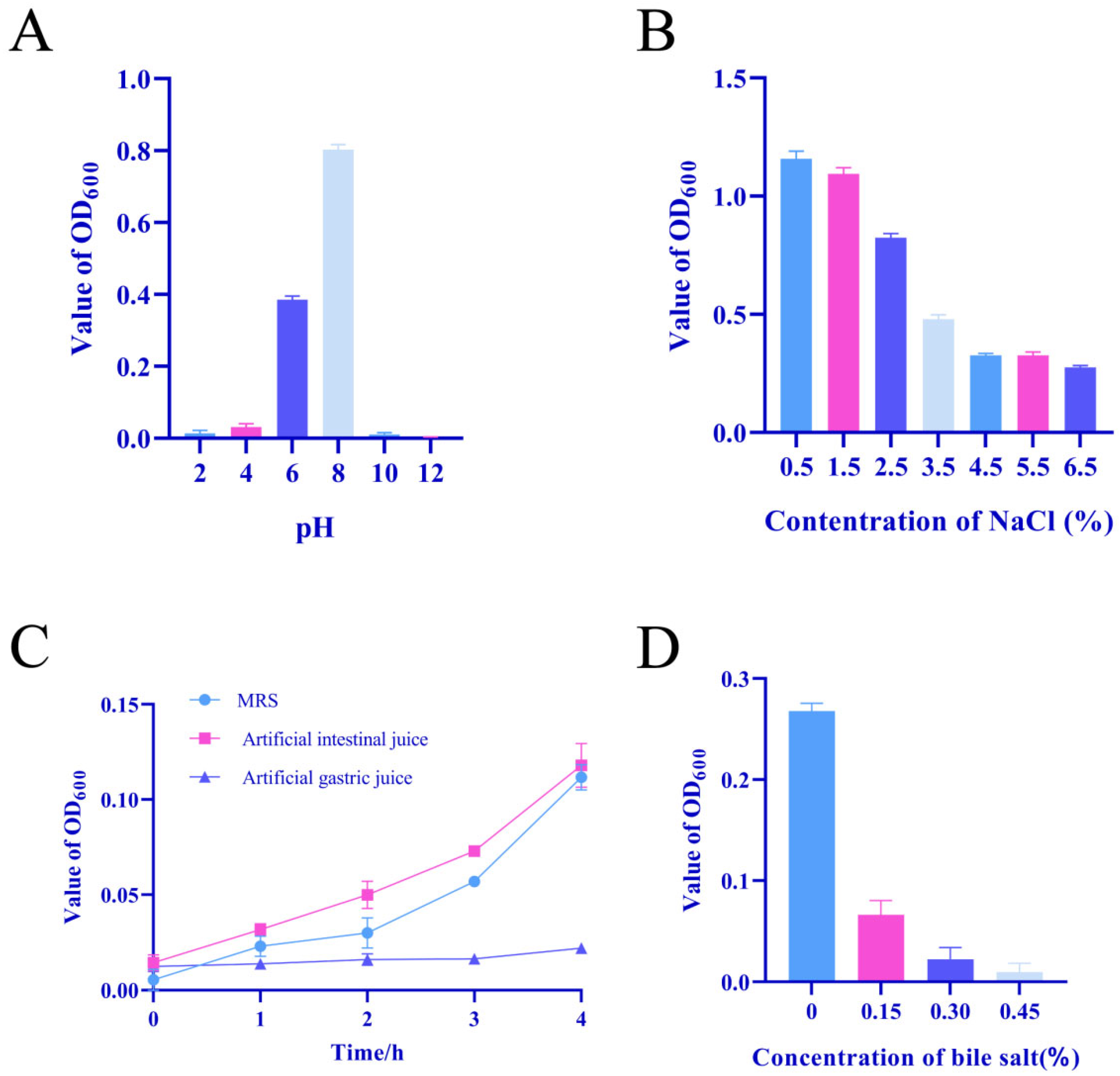
LBM15 showed differential growth in MRS broth at varying pH levels, as depicted in Figure 3A. Within 4 h, the OD600 values gradually increased with rising pH, reaching maximum growth at pH 8, indicating optimal conditions. Growth was inhibited at pH 4 and nearly absent at pH 2, 10, and 12. LBM15 exhibited wide salt tolerance, as illustrated in Figure 3B. It grew in MRS broth containing NaCl concentrations ranging from 0% to 6.5% and showed good growth at 0.5%, 1.5%, and 2.5% of NaCl concentration. With decreasing OD600 values, NaCl concentration increased.
LBM15 demonstrated robust tolerance in artificial intestinal fluid, with no growth inhibition observed within 4 h, growth was severely restricted in simulated gastric fluid, as shown in Figure 3C.
LBM15’s tolerance to different bile salt concentrations is depicted in Figure 3D. Compared to the control group, growth inhibition was observed in all concentration groups. At low bile salt concentrations (0.15%), OD600 values of LBM15 cultures continued to increase, while growth was significantly hindered at 0.30% and 0.45% bile salt concentrations.

3.5. Adherence Properties of LAB Strains

Microscopic observations using the slide method demonstrated L. lactis LBM15’s effective ex vivo adhesion, exerting varying degrees of inhibitory effects against six pathogenic strains (Figure 4), with the results of fluorescence method as a supplementary illustration.
The results of fluorescence measurement showed that LBM15 demonstrated competitive inhibition rates exceeding 40% against all six pathogenic bacteria, with the highest inhibition rate of 54.96% observed against V. anguillarum. In exclusion assays, LBM15 exhibited varying degrees of inhibitory effects, showing inhibition rates of 55.52% against V. anguillarum and 45.62% against Edwardsiella tarda. Among the three adhesion inhibition mechanisms (competition, exclusion, and displacement), displacement showed the strongest effect, with inhibition rates ranging from 48.81% to 76.07%. Notably, LBM15 exhibited the most potent displacement inhibition against Aeromonas hydrophila, followed by V. anguillarum.

3.6. Safety Aspects of LBM15

LBM15 exhibited no hemolytic activity on agar plates containing 1% defibrinated sheep blood. Sensitivity testing against 18 common antibiotics showed susceptibility, with four displaying moderate sensitivity, the isolates displayed marked resistance against aminoglycosides (AK, GM, K, N) (Table 3). Biosafety studies revealed no significant differences in feeding behavior between the experimental and control groups. Post-mortem examination showed normal visceral organs in both groups (Figure 5), and no mortality was observed throughout the study period.

4. Discussion

Although numerous studies have documented the isolation of LAB from aquatic animals and environments as potential probiotics [36], such as Lactococcus lactis from grouper [22] and Lactobacillus fermentum from C. mriala [37], there are few reports on the isolation of Lactococcus lactis from largemouth bass. This study presents the first systematic screening of L. lactis probiotics derived from largemouth bass. In this study, we employed a targeted approach to screen LAB strains with therapeutic potential, with a particular focus on their ability to inhibit pathogen adhesion, distinguishing our method from conventional approaches. Through systematic isolation and functional characterization, Lactococcus lactis strain LBM15 emerged as a promising probiotic candidate.
Our experimental strategy prioritized in vitro screening as a cost-effective and time-efficient precursor to subsequent in vivo validation [38,39]. This tiered validation approach—progressing from controlled laboratory conditions to biological systems—not only optimizes resource allocation but also aligns with global initiatives to develop non-antibiotic therapeutics [40,41,42,43]. This methodological selection proves particularly pertinent given the urgent need to identify probiotic alternatives capable of countering prevalent pathogens in largemouth bass aquaculture, including A. hydrophila, E. tarda, and V. anguillarum—species that are associated with economic losses and zoonotic transmission risks.
Notably, the demonstrated the capacity of LAB to synthesize growth-inhibitory compounds such as organic acids, bacteriocins, and hydrogen peroxide, while LAB prevent pathogen proliferation [27,44,45], as well as the attachment and colonization of pathogens in the gastro-intestinal tract.
The antimicrobial efficacy of lactic acid bacteria against Gram-negative pathogens has been extensively documented, facilitated by the permeability of low molecular weight and water-soluble metabolites such as lactic acid through bacterial membranes [46,47,48,49]. Early investigations dating back to 1997 documented Lactobacillus-mediated suppression of E. tarda [50], followed by reports of L. lactis exhibiting antagonism against V. anguillarum in 1998 [51]. A growing body of evidence further corroborates the inhibitory effects of Lactobacillus spp. on A. hydrophila [26,52,53,54,55,56,57]. Our experimental findings align with these observations that LBM15 demonstrated robust antagonistic activity against several pathogens, particularly A. hydrophila, suggesting its probiotic formulations might enhance disease resistance in aquatic hosts like Micropterus salmoides [58].
Probiotic attachment to the gastrointestinal tract represents a fundamental requirement for probiotics to establish residency and mediate their beneficial effects [15,21,33]. On the other hand, beyond mere intestinal attachment, the capacity of probiotics to competitively exclude or suppress pathogenic colonization is critical for maintaining host-microbe homeostasis and mitigating infection risks [28,35]. This dual functionality not only reinforces intestinal barrier integrity but also serves as a defense against pathogen invasion, thereby preserving gut ecological balance [4].
The adhesion mechanisms of lactic acid bacteria involve intricate surface molecular interactions during their engagement with host enterocytes [59,60,61]. Key surface-associated components, including S-layer proteins, lipoteichoic acids, and exopolysaccharides, facilitate ligand-receptor binding to intestinal mucins and epithelial cells [62,63]. These molecular adaptations enable LAB to occupy ecological niches, thereby limiting pathogen access to adhesion sites and nutrients. For instance [64], L. plantarum CLFP 238 reduces the adhesion of A. hydrophila and A. salmonicida in the fish intestinal tract [15], corroborating the role of adhesion competition in pathogen suppression.
Our research evaluated the adhesion capability of LBM15 using a mucus model to the fish’s gut. The results showed that LBM15 exhibited significant adhesion ability, effectively displacing pathogens through competitive exclusion. Although L. lactis isolation from M. salmoides was preliminarily noted [28], LBM15’s 76.07% displacement inhibition against A. hydrophila represents a breakthrough for combating hemorrhagic septicemia. This pathogen-specific efficacy, combined with cross-protection against V. anguillarum (54.96%) and E. tarda (55.52%), addresses the polymicrobial infection crisis in intensive aquaculture. These findings underscore the potential of LBM15 as a probiotic for enhancing gut health and pathogen resistance in aquaculture and position LBM15 as a promising candidate for developing antibiotic alternatives in sustainable aquaculture. While intestinal mucus was used to characterize adhesion mechanisms, conducting cellular adhesion assays would enable a more comprehensive assessment of LBM15’s adhesion capacity.
The tolerance of candidate probiotics to various conditions is crucial for their growth and survival in the gastrointestinal tract of fish, making it a key requirement for probiotic selection [65]. Probiotics need to withstand the acidic environment of the gastrointestinal tract to colonize the host’s intestines, survive and grow, and exert their beneficial properties. This study focused on screening the Lactococcus lactis strain LBM15, which exhibited good acid production and protease activity. In media with pH values ranging from 4 to 8, the OD600 increased, suggesting that LBM15 might grow in the digestive tract, which aligns with Roslina Jawan’s viewpoint [66]. After 4 h of incubation in media at pH 2 and pH 4, as well as in simulated gastric juice, there was no significant change in OD600, but showed a slow growth trend at pH 4. This is consistent with previous findings that low acidity, especially pH 2, significantly affects the growth of lactic acid bacteria [67,68]. However, the OD600 value measured in this study only reflects the culture’s growth after 4 h, and further research is needed to determine the survival rate of LBM15 in gastric juice at pH 2, such as by calculating its viability using the viable cell count method. In media with pH values ranging from 4 to 8, the OD600 increased, suggesting that LBM15 might grow in the digestive tract, which aligns with Roslina Jawan’s viewpoint [66]. The average level of 0.3% bile salt has been considered in many studies for bile salt tolerance of potential probiotic LAB. LBM15 was able to grow within bile salt concentrations ranging from 0.1% to 0.45%. This result is similar to the research by Mathara [69], which showed that LAB strains exhibit bile salt tolerance between 0.1% and 0.5%. Probiotics are expected to survive in the gastrointestinal tract while withstanding bile salts and acidic gastric fluids, thereby exerting their beneficial effects. The tolerance of potential LAB probiotic strains is critical not only for overcoming gastrointestinal stress but also for ensuring prolonged survival in acidic environments, which provides guidance for preparing probiotic formulations.
While the functionality of candidate probiotics is essential, safety evaluations are equally necessary, which generally involve investigating the intrinsic characteristics of probiotics and their interactions with the host [70]. Hemolytic activity is an important criterion for assessing probiotic safety [38]. The L. lactis LBM15 strain exhibited γ-hemolysis on sheep blood agar plates. Furthermore, antimicrobial sensitivity testing is one of the standard methods for evaluating potential probiotics [38]. The sensitivity of candidate probiotic strains to commonly used antibiotics must be assessed to avoid serious issues in treating microbial infections, and this process is crucial for detecting the potential transfer of antibiotic-resistant genes [39]. The examined LBM15 strains showed varying degrees of resistance toward Amikacin, Gentamcin, Kanamycin, Neomycin, Erythromycin, Norfloxacin, Polymy xin B, and compound Sulfamethoxazole. In the literature, aminoglycoside (gentamycin and streptomycin) resistance have been reported in LAB, which is associated in most cases with their innate resistance resulting from the impermeability of their membrane [71]. Resistance to broad-spectrum antibiotics such as kanamycin and streptomycin has also been observed in Lactobacillus and Bifidobacterium species [43]. However, in most cases, antibiotic resistance is non-transferable, Lactic acid bacteria strains with non-infectious antibiotic resistance typically do not pose safety concerns [70].
In animal safety evaluations, no signs of illness or death were observed in the experimental group of L. lactis LBM15, indicating it is harmless to fish and can be formulated as a probiotic to replace antibiotics for preventing and treating diseases in aquaculture. While zootechnical parameters are valuable for commercial probiotics, their assessment requires large-scale farming trials with optimized doses—a logical next step following safety validation, this tiered approach aligns with global probiotic development frameworks [72]. Future work will evaluate growth and immune parameters in farm-scale trials and advance the development of LBM15 as a probiotic formulation for aquaculture applications.

5. Conclusions

L. lactis LBM15 exhibited strong potential probiotic properties, including tolerance to acid, salt, and bile salts, high acid production capacity, good extracellular enzyme activity, significant inhibition of pathogens, and excellent antibiotic susceptibility. These properties make LBM15 an ideal probiotic with potential for application in aquaculture to improve fish health and performance. Future studies should further explore the effects and mechanisms of its application in real aquaculture environments.

Author Contributions

Investigation, X.C. and W.Z.; Resources, J.Z. (Jiaolin Zhang); Data curation, J.Z. (Jiaonan Zhang); Writing—original draft, X.C.; Writing—review & editing, Q.Y. All authors have read and agreed to the published version of the manuscript.

Funding

This work was supported by the National Key Research and Development Program (2023YFD2400700) and the Open Fund of Fujian Province Key Laboratory of Special Aquatic Formula Feed under contract No. TMKJZ2402.

Institutional Review Board Statement

This study was approved by Jimei University Animal Ethics Committee No. JMULAC2011-58, approval date: 2021-01-22.

Informed Consent Statement

Not applicable.

Data Availability Statement

All data generated or analysed during this study are included in this published article.

Conflicts of Interest

Jiaonan Zhang and Jiaolin Zhang were employed by the company Fujian Tianma Technology Company Limited. The remaining authors declare that the research was conducted in the absence of any commercial or financial relationships that could be construed as a potential conflict of interest.

References

  1. Guillen, J.; Natale, F.; Carvalho, N.; Casey, J.; Hofherr, J.; Druon, J.-N.; Fiore, G.; Gibin, M.; Zanzi, A.; Martinsohn, J.T. Global seafood consumption footprint. Ambio 2019, 48, 111–122. [Google Scholar] [CrossRef] [PubMed]
  2. Sagada, G.; Chen, J.M.; Shen, B.Q.; Huang, A.X.; Sun, L.H.; Jiang, J.H.; Jin, C.H. Optimizing protein and lipid levels in practical diet for juvenile northern snakehead fish (Channa argus). Anim. Nutr. 2017, 3, 156–163. [Google Scholar] [CrossRef] [PubMed]
  3. Banerjee, G.; Ray, A.K. The advancement of probiotics research and its application in fish farming industries. Res. Vet. Sci. 2017, 115, 66–77. [Google Scholar] [CrossRef] [PubMed]
  4. Etyemez, M.; Balcazar, J.L. Isolation and characterization of bacteria with antibacterial properties from Nile tilapia (Oreochromis niloticus). Res. Vet. Sci. 2016, 105, 62–64. [Google Scholar] [CrossRef]
  5. Quiñones, R.A.; Fuentes, M.; Montes, R.M.; Soto, D.; León-Muñoz, J. Environmental issues in Chilean salmon farming: A review. Rev. Aquac. 2019, 11, 375–402. [Google Scholar] [CrossRef]
  6. Ahmad, A.; Abdullah, S.R.S.; Abu Hasan, H.; Othman, A.R.; Ismail, N.I. Aquaculture industry: Supply and demand, best practices, effluent and its current issues and treatment technology. J. Environ. Manag. 2021, 287, 112271. [Google Scholar] [CrossRef]
  7. Ray, A.K.; Ghosh, K.; Ringo, E. Enzyme-producing bacteria isolated from fish gut: A review. Aquac. Nutr. 2012, 18, 465–492. [Google Scholar] [CrossRef]
  8. Martinez Cruz, P.; Ibanez, A.L.; Monroy Hermosillo, O.A.; Ramirez Saad, H.C. Use of probiotics in aquaculture. ISRN Microbiol. 2012, 2012, 916845. [Google Scholar] [CrossRef]
  9. Wang, X.; Yao, Y.X.; Ge, H.; Zhang, J.N.; Zhang, J.L.; Yan, Q.P. Isolation and identification of probiotic Bacillus subtilis AJQ03 from the intestinal tract of Anguilla japonica (Japanese eel). Front. Microbiol. 2024, 15, 1446299. [Google Scholar] [CrossRef]
  10. Yao, Y.; Wang, X.; Lin, X.; Wu, J.; Wang, P.; Zhu, C.; Yan, Q. Isolation and characterization of probiotic Lysinibacillus species from the gastrointestinal tract of large yellow croaker (Larimichthys crocea). Front. Mar. Sci. 2024, 11, 1408979. [Google Scholar] [CrossRef]
  11. Wang, P.; Wang, S.; Zhu, C.; Sun, Y.; Yan, Q.; Yi, G. Monascus purpureus M-32 fermented soybean meal improves the growth, immunity parameters, intestinal morphology, disease resistance, intestinal microbiota and metabolome in Pacific white shrimp (Litopenaeus vannamei). Anim. Nutr. 2024, 17, 283–296. [Google Scholar] [CrossRef] [PubMed]
  12. Wanka, K.M.; Damerau, T.; Costas, B.; Krueger, A.; Schulz, C.; Wuertz, S. Isolation and characterization of native probiotics for fish farming. BMC Microbiol. 2018, 18, 119. [Google Scholar] [CrossRef] [PubMed]
  13. Jose, N.M.; Bunt, C.R.; Hussain, M.A. Comparison of Microbiological and Probiotic Characteristics of Lactobacilli Isolates from Dairy Food Products and Animal Rumen Contents. Microorganisms 2015, 3, 198–212. [Google Scholar] [CrossRef] [PubMed]
  14. Ringo, E.; Gatesoupe, F.J. Lactic acid bacteria in fish: A review. Aquaculture 1998, 160, 177–203. [Google Scholar] [CrossRef]
  15. Balcazar, J.L.; Vendrell, D.; de Blas, I.; Ruiz-Zarzuela, I.; Muzquiz, J.L.; Girones, O. Characterization of probiotic properties of lactic acid bacteria isolated from intestinal microbiota of fish. Aquaculture 2008, 278, 188–191. [Google Scholar] [CrossRef]
  16. Llewellyn, M.S.; Boutin, S.; Hoseinifar, S.H.; Derome, N. Teleost microbiomes: The state of the art in their characterization, manipulation and importance in aquaculture and fisheries. Front. Microbiol. 2014, 5, 207. [Google Scholar] [CrossRef]
  17. Ibrahem, M.D. Evolution of probiotics in aquatic world: Potential effects, the current status in Egypt and recent prospectives. J. Adv. Res. 2015, 6, 765–791. [Google Scholar] [CrossRef]
  18. Butt, R.L.; Volkoff, H. Gut Microbiota and Energy Homeostasis in Fish. Front. Endocrinol. 2019, 10, 9. [Google Scholar] [CrossRef]
  19. Lazado, C.C.; Caipang, C.M.A.; Rajan, B.; Brinchmann, M.F.; Kiron, V. Characterization of GP21 and GP12: Two Potential Probiotic Bacteria Isolated from the Gastrointestinal Tract of Atlantic Cod. Probiotics Antimicrob. Proteins 2010, 2, 126. [Google Scholar] [CrossRef]
  20. Hua, X.J.; Li, C.; Xiao, Y.C.; Lu, Y.N.; Liu, X.Q. Oral administration of recombinant Lactococcus lactis expressing largemouth bass (Micropterus salmoides) IFNa3 protein enhances immune response against largemouth bass virus (LMBV) infection. Fish Shellfish Immunol. 2024, 154, 109875. [Google Scholar] [CrossRef]
  21. Todorov, S.D.; Lima, J.M.S.; Bucheli, J.E.V.; Popov, I.V.; Tiwari, S.K.; Chikindas, M.L. Probiotics for Aquaculture: Hope, Truth, and Reality. Probiotics Antimicrob. Proteins 2024, 16, 2007–2020. [Google Scholar] [CrossRef] [PubMed]
  22. Sun, Y.; He, M.W.; Cao, Z.J.; Xie, Z.Y.; Liu, C.S.; Wang, S.F.; Guo, W.L.; Zhang, X.; Zhou, Y.C. Effects of dietary administration of Lactococcus lactis HNL12 on growth, innate immune response, and disease resistance of humpback grouper (Cromileptes altivelis). Fish Shellfish Immunol. 2018, 82, 296–303. [Google Scholar] [CrossRef] [PubMed]
  23. Ringo, E.; Hoseinifar, S.H.; Ghosh, K.; Van Doan, H.; Becks, B.R.; Song, S.K. Lactic Acid Bacteria in Finfish-An Update. Front. Microbiol. 2018, 9, 1818. [Google Scholar] [CrossRef] [PubMed]
  24. He, L.; Zhao, L.; Li, Q.; Huang, L.; Qin, Y.; Zhuang, Z.; Wang, X.; Huang, H.; Zhang, J.; Zhang, J.; et al. Pseudomonas plecoglossicida fliP gene affects the immune response of Epinephelus fuscoguttatus ♀ × Epinephelus lanceolatus ♂ to infection. Fish Shellfish Immunol. 2023, 140, 108971. [Google Scholar] [CrossRef]
  25. Li, Q.; Jiang, B.; Zhang, Z.; Huang, Y.; Xu, Z.; Chen, X.; Huang, Y.; Jian, J.; Yan, Q. Involvement and characterization of NLRCs and pyroptosis-related genes in Nile tilapia (Oreochromis niloticus) immune response. Fish Shellfish Immunol. 2022, 130, 602–611. [Google Scholar] [CrossRef]
  26. Ayyash, M.M.; Abdalla, A.K.; AlKalbani, N.S.; Baig, M.A.; Turner, M.S.; Liu, S.Q.; Shah, N.P. Invited review: Characterization of new probiotics from dairy and nondairy products-Insights into acid tolerance, bile metabolism and tolerance, and adhesion capability. J. Dairy Sci. 2021, 104, 8363–8379. [Google Scholar] [CrossRef]
  27. Alakomi, H.L.; Skyttä, E.; Saarela, M.; Mattila-Sandholm, T.; Latva-Kala, K.; Helander, I.M. Lactic acid permeabilizes gram-negative bacteria by disrupting the outer membrane. Appl. Environ. Microbiol. 2000, 66, 2001–2005. [Google Scholar] [CrossRef]
  28. Xia, L.; Cheng, G.; Wang, P.; Wang, X.; Dong, Z.; Mu, Q.; Yu, J.; Jiang, Z.; Xiao, J.; Feng, H.; et al. Screening and identification of probiotics from the intestinal tract of largemouth bass (Micropterus salmoides) for use as a feed additive and bacterial infection control. Aquaculture 2024, 584, 740661. [Google Scholar] [CrossRef]
  29. León, J.; Pellón, F.J.; Unda, V.; David, J.; Anaya, C.A.M.; Mendoza, V.Í. Producción de enzimas extracelulares por bacterias aisladas de invertebrados marinos. Rev. Peru. Biol. 2014, 7, 202–210. [Google Scholar] [CrossRef]
  30. Vinderola, G.; Capellini, B.; Villarreal, F.; Suárez, V.; Quiberoni, A.; Reinheimer, J. Usefulness of a set of simple in vitro tests for the screening and identification of probiotic candidate strains for dairy use. LWT-Food Sci. Technol. 2008, 41, 1678–1688. [Google Scholar] [CrossRef]
  31. Aarti, C.; Khusro, A.; Varghese, R.; Arasu, M.V.; Agastian, P.; Al-Dhabi, N.A.; Ilavenil, S.; Choi, K.C. In vitro investigation on probiotic, anti-Candida, and antibiofilm properties of Lactobacillus pentosus strain LAP1. Arch. Oral Biol. 2018, 89, 99–106. [Google Scholar] [CrossRef] [PubMed]
  32. Charteris, W.P.; Kelly, P.M.; Morelli, L.; Collins, J.K. Development and application of an in vitro methodology to determine the transit tolerance of potentially probiotic Lactobacillus and Bifidobacterium species in the upper human gastrointestinal tract. J. Appl. Microbiol. 1998, 84, 759–768. [Google Scholar] [CrossRef] [PubMed]
  33. Ouwehand, A.C.; Kirjavainen, P.V.; Grönlund, M.M.; Isolauri, E.; Salminen, S.J. Adhesion of probiotic micro-organisms to intestinal mucus. Int. Dairy J. 1999, 9, 623–630. [Google Scholar] [CrossRef]
  34. He, L.; Mao, M.; Zhao, L.; Li, Q.; Zhuang, Z.; Wang, X.; Huang, H.; Wang, Q.; Yan, Q. A novel small non-coding RNA 562 mediates the virulence of Pseudomonas plecoglossicida by regulating the expression of fliP, a key component of flagella T3SS. Fish Shellfish Immunol. 2024, 151, 109752. [Google Scholar] [CrossRef]
  35. Wang, J.; Wu, Z.; Wang, S.; Wang, X.; Zhang, D.; Wang, Q.; Lin, L.; Wang, G.; Guo, Z.; Chen, Y. Inhibitory effect of probiotic Bacillus spp. isolated from the digestive tract of Rhynchocypris Lagowskii on the adhesion of common pathogenic bacteria in the intestinal model. Microb. Pathog. 2022, 169, 105623. [Google Scholar] [CrossRef]
  36. Rahayu, S.; Amoah, K.; Huang, Y.; Cai, J.; Wang, B.; Shija, V.M.; Jin, X.; Anokyewaa, M.A.; Jiang, M.Y. Probiotics application in aquaculture: Its potential effects, current status in China and future prospects. Front. Mar. Sci. 2024, 11, 1455905. [Google Scholar] [CrossRef]
  37. Govindaraj, K.; Samayanpaulraj, V.; Narayanadoss, V.; Uthandakalaipandian, R. Isolation of Lactic Acid Bacteria from Intestine of Freshwater Fishes and Elucidation of Probiotic Potential for Aquaculture Application. Probiotics Antimicrob. Proteins 2021, 13, 1598–1610. [Google Scholar] [CrossRef]
  38. Nemcova, R. Selection criteria of lactobacilli for probiotic use. Vet. Med. 1997, 42, 19–27. [Google Scholar]
  39. Reuben, R.C.; Roy, P.C.; Sarkar, S.L.; Alam, R.-U.; Jahid, I.K. Isolation, characterization, and assessment of lactic acid bacteria toward their selection as poultry probiotics. BMC Microbiol. 2019, 19, 253. [Google Scholar] [CrossRef]
  40. Carvalho, I.T.; Santos, L. Antibiotics in the aquatic environments: A review of the European scenario. Environ. Int. 2016, 94, 736–757. [Google Scholar] [CrossRef]
  41. O’Flaherty, E.; Cummins, E. Antibiotic resistance in surface water ecosystems: Presence in the aquatic environment, prevention strategies, and risk assessment. Hum. Ecol. Risk Assess. 2017, 23, 299–322. [Google Scholar] [CrossRef]
  42. Cabello, F.C.; Godfrey, H.P.; Tomova, A.; Ivanova, L.; Doelz, H.; Millanao, A.; Buschmann, A.H. Antimicrobial use in aquaculture re-examined: Its relevance to antimicrobial resistance and to animal and human health. Environ. Microbiol. 2013, 15, 1917–1942. [Google Scholar] [CrossRef] [PubMed]
  43. Smith, P. Antimicrobial resistance in aquaculture. Rev. Sci. Tech.-Off. Int. Epizoot. 2008, 27, 243–264. [Google Scholar] [CrossRef]
  44. De Vuyst, L.; Leroy, F. Bacteriocins from lactic acid bacteria: Production, purification, and food applications. J. Mol. Microbiol. Biotechnol. 2007, 13, 194–199. [Google Scholar] [CrossRef]
  45. Li, H.; Meiqin, M.; Lingmin, Z.; Qi, L.; Hui, G.; Jiao-nan, Z.; Jiaolin, Z.; Qingpi, Y. sRNA113 regulates the motility of Pseudomonas plecoglossicida to affect the immune response against infection in pearl gentian grouper. Zool. Res. 2025, 46, 152. [Google Scholar]
  46. Luis Balcazar, J.; De Blas, I.; Ruiz-Zarzuela, I.; Vendrell, D.; Calvo, A.C.; Marquez, I.; Girones, O.; Luis Muzquiz, J. Changes in intestinal microbiota and humoral immune response following probiotic administration in brown trout (Salmo trutta). Br. J. Nutr. 2007, 97, 522–552. [Google Scholar] [CrossRef]
  47. Yoshiyama, M.; Wu, M.; Sugimura, Y.; Takaya, N.; Kimoto-Nira, H.; Suzuki, C. Inhibition of Paenibacillus larvae by lactic acid bacteria isolated from fermented materials. J. Invertebr. Pathol. 2013, 112, 62–67. [Google Scholar] [CrossRef]
  48. Hwanhlem, N.; Buradaleng, S.; Wattanachant, S.; Benjakul, S.; Tani, A.; Maneerat, S. Isolation and screening of lactic acid bacteria from Thai traditional fermented fish (Plasom) and production of Plasom from selected strains. Food Control. 2011, 22, 401–407. [Google Scholar] [CrossRef]
  49. Tang, Y.; Jiao, J.; Zhao, L.; Zhuang, Z.; Wang, X.; Fu, Q.; Huang, H.; Huang, L.; Qin, Y.; Zhang, J.; et al. The contribution of exbB gene to pathogenicity of Pseudomonas plecoglossicida and its interactions with Epinephelus coioides. Fish Shellfish Immunol. 2022, 120, 610–619. [Google Scholar] [CrossRef]
  50. Byun, J.W.; Park, S.C.; Benno, Y.; Oh, T.K. Probiotic effect of Lactobacillus sp. DS-12 in flounder (Paralichthys olivaceus). J. Gen. Appl. Microbiol. 1997, 43, 305–308. [Google Scholar] [CrossRef]
  51. Harzevili, A.R.S.; Van Duffel, H.; Dhert, P.; Swings, J.; Sorgeloos, P. Use of a potential probiotic Lactococcus lactis AR21 strain for the enhancement of growth in the rotifer Brachionus plicatilis (Muller). Aquac. Res. 1998, 29, 411–417. [Google Scholar] [CrossRef]
  52. Gatesoupe, F.J. The use of probiotics in aquaculture. Aquaculture 1999, 180, 147–165. [Google Scholar] [CrossRef]
  53. García-Hernández, Y.; Pérez-Sánchez, T.; Boucourt, R.; Balcázar, J.L.; Nicoli, J.R.; Moreira-Silva, J.; Rodríguez, Z.; Fuertes, H.; Nuñez, O.; Albelo, N.; et al. Isolation, characterization and evaluation of probiotic lactic acid bacteria for potential use in animal production. Res. Vet. Sci. 2016, 108, 125–132. [Google Scholar] [CrossRef] [PubMed]
  54. Pimentel, T.C.; Costa, W.K.A.d.; Barão, C.E.; Rosset, M.; Magnani, M. Vegan probiotic products: A modern tendency or the newest challenge in functional foods. Food Res. Int. 2021, 140, 110033. [Google Scholar] [CrossRef]
  55. Wang, W.; Gänzle, M. Chapter Three—Toward rational selection criteria for selection of probiotics in pigs. In Advances in Applied Microbiology; Gadd, G.M., Sariaslani, S., Eds.; Academic Press: Cambridge, MA, USA, 2019; Volume 107, pp. 83–112. [Google Scholar]
  56. Verschuere, L.; Rombaut, G.; Sorgeloos, P.; Verstraete, W. Probiotic Bacteria as Biological Control Agents in Aquaculture. Microbiol. Mol. Biol. Rev. 2000, 64, 655–671. [Google Scholar] [CrossRef]
  57. Vaughan, E.E.; Heilig, H.; Ben-Amor, K.; de Vos, W.M. Diversity, vitality and activities of intestinal lactic acid bacteria and bifidobacteria assessed by molecular approaches. Fems Microbiol. Rev. 2005, 29, 477–490. [Google Scholar] [CrossRef]
  58. Li, X.; Ringo, E.; Hoseinifar, S.H.; Lauzon, H.L.; Birkbeck, H.; Yang, D. The adherence and colonization of microorganisms in fish gastrointestinal tract. Rev. Aquac. 2019, 11, 603–618. [Google Scholar] [CrossRef]
  59. Kos, B.; Suskovic, J.; Vukovic, S.; Simpraga, M.; Frece, J.; Matosic, S. Adhesion and aggregation ability of probiotic strain Lactobacillus acidophilus M92. J. Appl. Microbiol. 2003, 94, 981–987. [Google Scholar] [CrossRef]
  60. Chantanawilas, P.; Pahumunto, N.; Teanpaisan, R. Aggregation and adhesion ability of various probiotic strains and Candida species: An in vitro study. J. Dent. Sci. 2024, 19, 2163–2171. [Google Scholar] [CrossRef]
  61. Xin, G.; Zhao, L.; Zhuang, Z.; Wang, X.; Fu, Q.; Huang, H.; Huang, L.; Qin, Y.; Zhang, J.; Zhang, J.; et al. Function of the rpoD gene in Pseudomonas plecoglossicida pathogenicity and Epinephelus coioides immune response. Fish Shellfish Immunol. 2022, 127, 427–436. [Google Scholar] [CrossRef]
  62. Mack, D.R.; Michail, S.; Wei, S.; McDougall, L.; Hollingsworth, M.A. Probiotics inhibit enteropathogenic E. coli adherence in vitro by inducing intestinal mucin gene expression. Am. J. Physiol.-Gastrointest. Liver Physiol. 1999, 276, G941–G950. [Google Scholar] [CrossRef] [PubMed]
  63. Yang, D.; Zhao, L.; Li, Q.; Huang, L.; Qin, Y.; Wang, P.; Zhu, C.; Yan, Q. flgC gene is involved in the virulence regulation of Pseudomonas plecoglossicida and affects the immune response of Epinephelus coioides. Fish Shellfish Immunol. 2023, 132, 108512. [Google Scholar] [CrossRef] [PubMed]
  64. Ren, P.; Xu, L.; Yang, Y.; He, S.; Liu, W.; Ringø, E.; Zhou, Z. Lactobacillus planarum subsp. plantarum JCM 1149 vs. Aeromonas hydrophila NJ-1 in the anterior intestine and posterior intestine of hybrid tilapia Oreochromis niloticus ♀ × Oreochromis aureus ♂: An ex vivo study. Fish Shellfish Immunol. 2013, 35, 146–153. [Google Scholar] [CrossRef] [PubMed]
  65. Ehrmann, M.A.; Kurzak, P.; Bauer, J.; Vogel, R.F. Characterization of lactobacilli towards their use as probiotic adjuncts in poultry. J. Appl. Microbiol. 2002, 92, 966–975. [Google Scholar] [CrossRef]
  66. Jawan, R.; Abbasiliasi, S.; Tan, J.S.; Mustafa, S.; Halim, M.; Ariff, A.B. Influence of Culture Conditions and Medium Compositions on the Production of Bacteriocin-Like Inhibitory Substances by Lactococcus lactis Gh1. Microorganisms 2020, 8, 1454. [Google Scholar] [CrossRef]
  67. Guo, X.-H.; Kim, J.-M.; Nam, H.-M.; Park, S.-Y.; Kim, J.-M. Screening lactic acid bacteria from swine origins for multistrain probiotics based on in vitro functional properties. Anaerobe 2010, 16, 321–326. [Google Scholar] [CrossRef]
  68. Angmo, K.; Kumari, A.; Savitri; Bhalla, T.C. Probiotic characterization of lactic acid bacteria isolated from fermented foods and beverage of Ladakh. Lwt-Food Sci. Technol. 2016, 66, 428–443. [Google Scholar] [CrossRef]
  69. Mathara, J.M.; Schillinger, U.; Kutima, P.M.; Mbugua, S.K.; Guigas, C.; Franz, C.; Holzapfel, W.H. Functional properties of Lactobacillus plantarum strains isolated from Maasai traditional fermented milk products in Kenya. Curr. Microbiol. 2008, 56, 315–321. [Google Scholar] [CrossRef]
  70. Salminen, S.; von Wright, A.; Morelli, L.; Marteau, P.; Brassart, D.; de Vos, W.M.; Fonden, R.; Saxelin, M.; Collins, K.; Mogensen, G.; et al. Demonstration of safety of probiotics—A review. Int. J. Food Microbiol. 1998, 44, 93–106. [Google Scholar] [CrossRef]
  71. Yanina Bustos, A.; Font de Valdez, G.; Raya, R.; de Almeida, A.M.; Fadda, S.; Pia Taranto, M. Proteomic analysis of the probiotic Lactobacillus reuteri CRL1098 reveals novel tolerance biomarkers to bile acid-induced stress. Food Res. Int. 2015, 77, 599–607. [Google Scholar] [CrossRef]
  72. Arya, M.; Shahi, N.; Bisht, I.; Pandey, N.; Mallik, S.K. Probiotic potential of Bacillus velezensis STPB10 sourced from the gut microbiota of a hillstream fish Schizothorax richardsonii (Gray, 1832) for aquaculture applications. Sci. Rep. 2025, 15, 17580. [Google Scholar] [CrossRef]
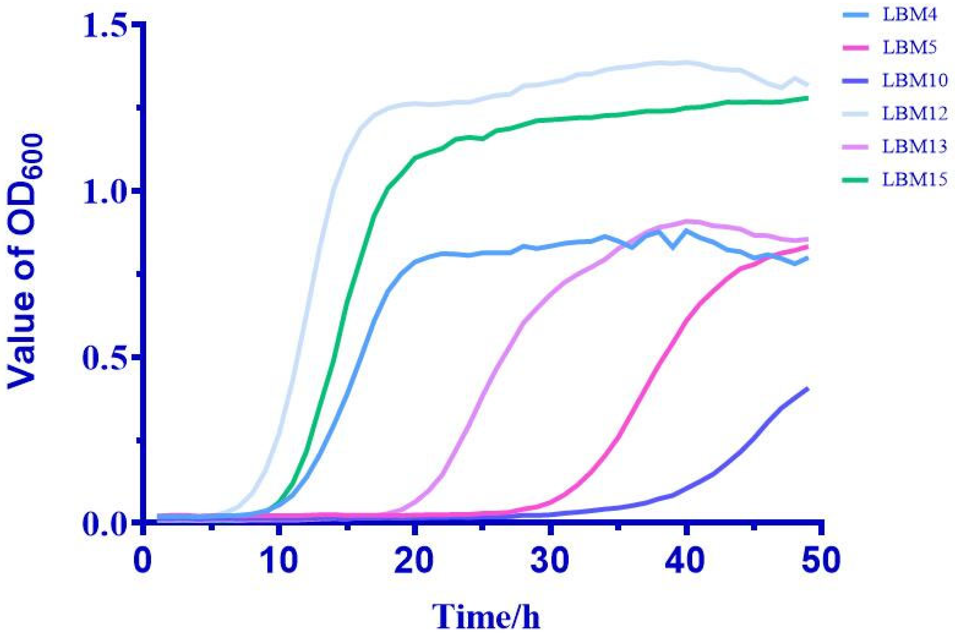
Figure 1. Growth curve of isolates.
Fishes 10 00291 g001
Figure 2. Morphology and phylogenetic tree of LBM15. (A) The colony morphology of LBM15 on MRS medium; (B) microscopic view of showed Gram-positive spherical and spherical shaped; (C) phylogenetic tree of LBM15 and related genera based on partial 16S rRNA sequence.
Fishes 10 00291 g002
Figure 3. (A) Growth of LBM15 in different pH values; (B) growth of LBM15 under different NaCl concentrations; (C) growth of LBM15 under simulated gastric and simulated intestinal fluids; (D) growth of LBM15 under different levels of bile salts.
Fishes 10 00291 g003
Figure 4. LBM15’s inhibitory adhesion against pathogens, assessed using glass slides.
Fishes 10 00291 g004
Figure 5. Viscera after feeding and injection of LBM15. (A): Intestine, liver, and spleen of largemouth bass after being fed LBM15; (B): Intestine, liver, and spleen of largemouth bass after being injected with LBM15.
Fishes 10 00291 g005
Table 1. Diameter of discoloration of isolates on bromothymol blue medium.
StrianZone Diameter/mm
LBM427.56 ± 0.49
LBM530.43 ± 0.51
LBM1027.88 ± 0.47
LBM1224.17 ± 0.71
LBM1327.63 ± 0.58
LBM1526.99 ± 0.44
Table 2. Antagonistic effects of LBM15 against pathogens.
PathogensInhibition Zone Diameter/mm
V. alginolyticus18.22 ± 0.32
V. anguillarum11.97 ± 0.60
V. harveyi11.71 ± 0.32
A. hydrophila12.35 ± 0.50
E. tarda10.15 ± 0.33
P. plecoglossicida/
“/” indicates no inhibition zone.
Table 3. Antibiotic sensitivity of LBM15.
AntibioticDrug Doses (μg)Inhibition Zone Diameters (mm)Sensitivity
Penicillin G1032.61 ± 0.40S
Oxacillin117.33 ± 0.52S
Ampicillin1032.71 ± 0.44S
Carbenicillin10033.63 ± 0.46S
Piperacillin10031.02 ± 0.54S
Cefalexin3023.10 ± 0.20S
Cefazolin3026.16 ± 0.36S
Cephradine3025.36 ± 0.25S
Cefuroxime3034.70 ± 0.53S
Ceftazidime3023.96 ± 0.35S
Ceftriaxone3032.72 ± 0.38S
Cefoperazone7529.61 ± 0.50S
Amikacin3010.52 ± 0.17R
Gentamicin107.61 ± 0.33R
Kanamycin30/R
Neomycin30/R
Tetracycline3015.17 ± 0.69I
Doxycycline3017.54 ± 0.43S
Minocycline3015.65 ± 0.35I
Erythromycin159.93 ± 0.43R
Midecamycin3019.53 ± 0.54S
Norfloxacin309.92 ± 0.44R
Ofloxacin518.14 ± 0.53S
Ciprofloxacin516.21 ± 0.50I
Vancomycin3017.74 ± 0.12S
Polymyxin B300/R
Sulfamethoxazole23.7512.95 ± 0.52R
Furazolidone30017.64 ± 0.51S
Chloramphenicol3025.92 ± 0.68S
Clindamycin217.41 ± 0.29I
Antibiotic susceptibility was classified as resistant (R), intermediate susceptible (I), and susceptible (S), depending on microbial responses. “/” indicates no inhibition zone.
Disclaimer/Publisher’s Note: The statements, opinions and data contained in all publications are solely those of the individual author(s) and contributor(s) and not of MDPI and/or the editor(s). MDPI and/or the editor(s) disclaim responsibility for any injury to people or property resulting from any ideas, methods, instructions or products referred to in the content.