Article

Cryopreservation of Ovarian Tissue at the Stage of Vitellogenesis from Yellow Drum (Nibea albiflora) and Its Effects on Cell Viability and Germ Cell-Specific Gene Expression

1
Engineering Research Center of Mindong Aquatic Product Deep-Processing, Fujian Province University, College of Ocean Studies, Ningde Normal University, Ningde 352100, China
2
State Key Laboratory of Mariculture Breeding, Fufa Fisheries Company Limited, Ningde 352103, China
3
Ningde Yiye Marine Industry Development Co., Ltd., Ningde 352103, China
*
Authors to whom correspondence should be addressed.
Fishes 2025, 10(6), 288; https://doi.org/10.3390/fishes10060288
Submission received: 30 April 2025 / Revised: 5 June 2025 / Accepted: 10 June 2025 / Published: 12 June 2025

Abstract

The cryopreservation of ovarian tissues from fish has recently been carried out for several endangered and commercially valuable species. However, previous studies in this context have focused on the cryopreservation of immature ovaries—mainly through slow freezing and vitrification—which requires specialized freezing equipment or higher cryoprotectant concentrations to keep cell viability. Therefore, the aim of this study was to explore a convenient, rapid, efficient and less toxic method for the cryopreservation of ovaries at the stage of vitellogenesis from yellow drum (Nibea albiflora), an economically important marine fish. The ovaries at the stage of vitellogenesis were isolated and cut into blocks of approximately 1 cm3, then cryopreserved with 15% propylene glycol (PG), fetal bovine serum (FBS) and 0.2 M trehalose as cryoprotectants. Finally, the samples were treated using three different freezing procedures, including a −80 °C refrigerator, liquid nitrogen, and their combination. After 7 days, the tissues were thawed and digested, and the cell survival rates and gene expression levels were detected using cell viability assay kits and qRT-PCR, respectively. The results of the viability assay showed that the procedure of ovarian tissue storage at −80 °C in a refrigerator for 1 h, followed by transfer to liquid nitrogen, resulted in the highest cell survival rate (>90%). Furthermore, the germ cells at various phases were of normal size; presented a full, smooth surface and regular shape; and did not show any signs of cell rupture, atrophy, depression, granulation or cavitation. Furthermore, the qRT-PCR results revealed that genes related to reproductive development, such as vasa, foxl2, zp3 and gsdf, were all down-regulated under the optimal protocol, while the expression of the nanos2 gene (which is specifically distributed in oogonia) maintained a higher level, similar to that in the control group. This indicated that the viability of germ stem cells (oogonia) was not weakened after freezing and that oogonia could be isolated from the cryopreserved ovaries for germ cell transplantation. The present study successfully establishes an optimal cryopreservation protocol for ovarian tissues from Nibea albiflora, providing reference for the preservation of ovaries at the stage of vitellogenesis from other species.
Key Contribution: The present study successfully establishes an optimal cryopreservation protocol for ovarian tissues at the stage of vitellogenesis from Nibea albiflora. This is a convenient; rapid; efficient and less-toxic method that can be applied for the cryopreservation of germplasm resources obtained from economically important marine fish.

1. Introduction

Fish maternal germplasm resources carry abundant genetic information, which helps to maintain the genetic diversity of species and enhance their ability to adapt to environmental changes. The individual females usually pass on good traits such as disease resistance and high yield to their offspring, which are crucial in breeding practices aimed at assisting in the formation of new lines. Therefore, as an important part of biobanking, the cryopreservation of maternal germplasm resources is of great significance, especially for endangered species. The eggs, oocytes, embryos and ovarian tissue are important maternal germplasm resources [1,2]. However, compared with sperm, it is still difficult to cryopreserve eggs, oocytes and embryos due to the large volume of cells, high intracellular water and yolk compartment [1].
In recent years, ovarian tissue from several teleosts, including the rainbow trout (Oncorhynchus mykiss) [3], brown trout (Salmo trutta) [4], cyprinid honmoroko (Gnathopogon caerulescens) [5], common carp (Cyprinus carpio) [6], Piracanjunba (Brycon orbignyanus) [7], Murray River Rainbowfish (Melanotaenia fluviatilis) [8], sturgeon [9,10,11] and zebrafish [12], has been successfully cryopreserved. In previous research, the ovarian tissues were mostly isolated from immature individuals for cryopreservation, when the tissue mainly contained a large number of oogonia (i.e., germ stem cells), which could be used as donor cells for transplantation after thawing. This method provides a new approach for the conservation and utilization of precious, endangered and elite germplasm resources. In rainbow trout, common carp and Siberian sturgeon (Acipenser baerii), the ovaries were cooled at −1 °C/min for 90 min using specific equipment (e.g., a Bicell plastic freezing container) before immersion in liquid nitrogen, and the oogonia after cryopreservation were successfully used for transplantation [3,6,9]. Different from the slow freezing method, in brown trout, Piracanjunba and cyprinid honmoroko, ovaries with high concentrations of cryoprotectants have been rapidly cryopreserved via vitrification, resulting in high cell integrity and viability [4,5,7]. Whether slow freezing or vitrification was performed, the cryoprotectants played a vital role in the freezing process. Cryoprotectants can be categorized into two types: permeating and non-permeating [13]. Permeating cryoprotectants permeate cell membranes, which include dimethyl sulfoxide (DMSO), methanol (MeOH), propylene glycol (PG), ethylene glycol (EG) and glycerol (Gly). Meanwhile, non-permeating cryoprotectants include trehalose, fetal bovine serum (FBS), bovine serum albumin (BSA), sucrose, egg yolk powder and skim milk powder. A combination of cryoprotectants at appropriate concentrations can minimize the damage caused by the formation of ice crystals to cells or tissues during freezing. The cryoprotectants DMSO, PG, EG, MeOH, trehalose and FBS have been widely applied for the cryopreservation of ovarian tissues. It has been shown that DMSO and PG have a good preservation effect when used in the vitrification of salmon and sturgeon ovaries [4,5,10]. Meanwhile, trehalose and FBS have mostly been used for slow freezing in some species [3,11].
Although a small number of endangered or commercially valuable fish have been cryopreserved with relatively high cell survival rates, there remain some problems to be solved. Slow freezing and vitrification, as two common cooling procedures, have certain limitations [14]. Slow freezing methods require complex and expensive equipment to control the temperature precisely and are not suitable for the timely preservation of ovary materials in the sea, field, laboratory or farm without relevant equipment [15,16]. Meanwhile, vitrification—unlike slow cooling—only requires high concentrations of cryoprotectants (especially permeating agents such as DMSO) [17]. However, at high concentrations, although effectively reducing the formation of ice crystals, these cryoprotectants may be highly toxic to cells [18]. In addition, ovarian cryopreservation approaches are mainly applied to immature ovaries, which are limited by the developmental period and the small size of the tissue. Compared to immature ovaries, the ovaries at the stage of vitellogenesis are generally larger and contain various types of germ cells. If they can be cryopreserved successfully, not only can the oogonia be separated for transplantation and other operations, but the vitellogenic oocytes can also be induced for artificial insemination. This is expected to expand the scope of preservation and application of frozen germplasm.
The yellow drum (Nibea albiflora) is an important fish species in the Sciaenidae family. It displays asynchronous oocyte development, with ovaries containing oocytes at multiple developmental stages simultaneously. This allows for batch spawning during the reproductive season (May–July). As a marine culture fish, it is of great economic value in China. In recent years, with the rapid development of marine aquaculture, yellow drums have suffered from higher rates of disease, leading to significant economic losses [19]. Furthermore, overwinter mortalities limit the aquaculture of this species [20]. Therefore, it is very important to cultivate new strains that are resistant to disease and stress and keep them frozen in time for the healthy and sustainable development of yellow drum culture. However, to date, the cryopreservation of yellow drum ovaries has not been reported. In this study, we aimed to establish a simple and efficient preservation system for ovaries at the stage of vitellogenesis from yellow drum. The ovaries were isolated from female individuals in May and cut into tissue blocks of approximately 1 cm3, then immersed in 15% PG, 15% FBS and 2M trehalose as cryoprotectants for 1 h on ice. Subsequently, the tissues were removed and cryopreserved using three different freezing procedures. After 7 days, the tissues were thawed and digested, and the morphology, structure and survival rate of cells were detected using the AO-EB Double Fluorescence Staining Kit. Furthermore, the expression levels of genes related to female reproductive development were analyzed via RT-qPCR in order to estimate cell viability. This study provides a new method for the preservation of yellow drum germplasm, thus promoting the development of artificial culture while providing a reference for similar preservation in other fish.

2. Materials and Methods

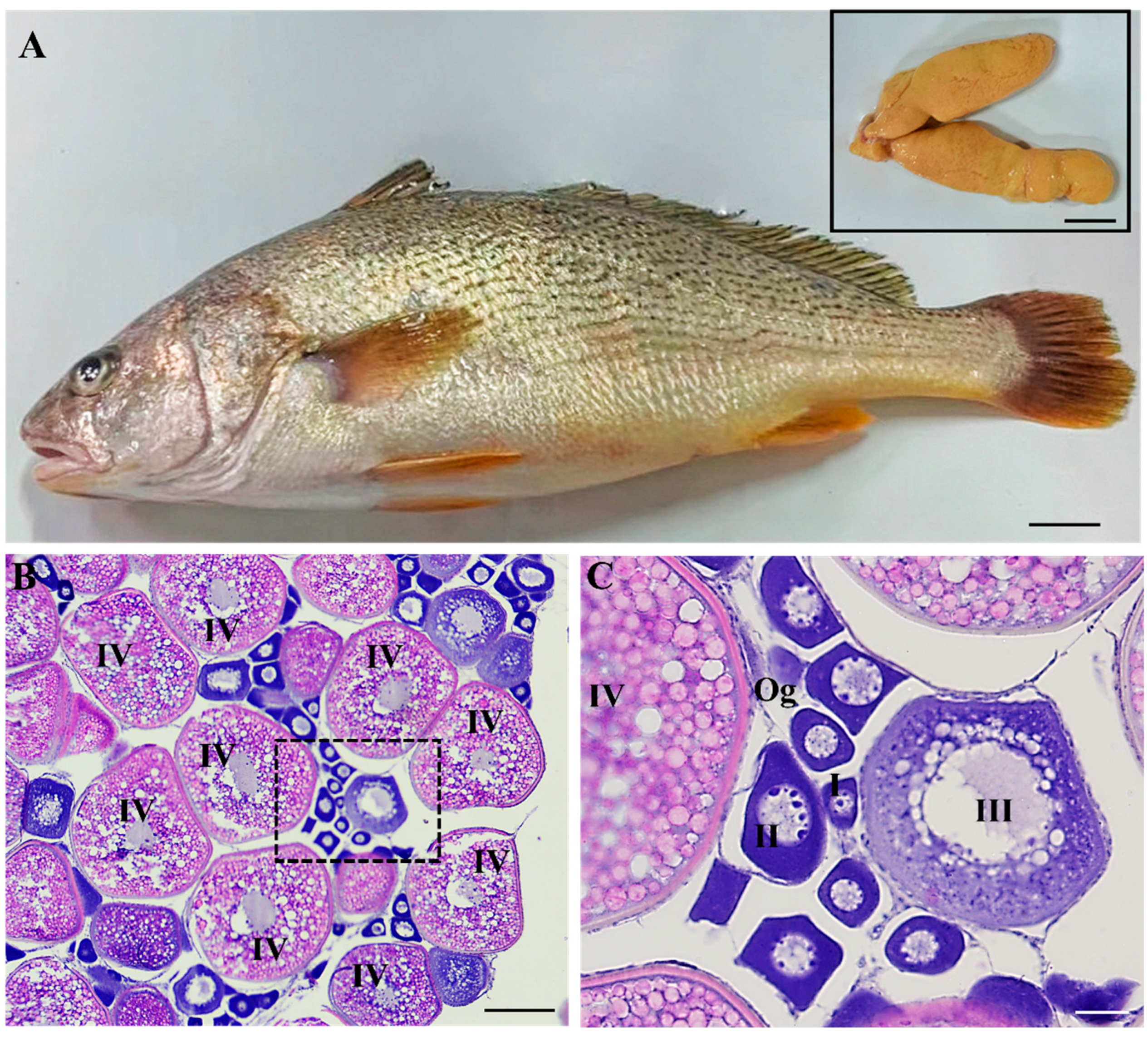
2.1. Collection of Fish and Ovaries

Five female yellow drum in May were obtained from Ningde Yiye Marine Industry Development Co., Ltd. The basic indicators of body weight, total length and body length were measured after anesthetization by MS-222 (Sigma-Aldrich, St. Louis, MI, USA). The total length was approximately 31 ± 2.0 cm, body length was 28 ± 1.5 cm, and body weight was 450 ± 35 g. Subsequently, the ovaries of the fish were removed and weighed, of which one portion was fixed in Bouin’s reagent for histology and the other was placed in L-15 medium on ice for cryopreservation. Based on the weight of the ovaries and body, the gonadosomatic index (GSI) was obtained as 8.72 ± 1.2, suggesting that these female individuals were at the stage of vitellogenesis.
The cryopreservation of ovarian tissue from yellow drum was approved by the Ethics Committee of Ningde Normal University (NDNU-LL-202508).

2.2. Histology

The tissue sections and H&E staining of the ovary were performed according to Zhao et al. [21]. Briefly, samples were fixed, dehydrated, clarified and embedded in paraffin, and then 5 μm thick sections were cut by a manual microtome (Leica RM2125, Wetzlar, Germany). Finally, the images were photographed using a fluorescence microscope (Leica DM7500, Wetzlar, Germany).

2.3. Experimental Design

Referring to previous studies [3,10,11], PG (SINOPHARM), FBS (AusgeneX, Shanghai, China) and trehalose (Bomei, Maanshan, China) were selected for use as cryoprotectants in this study, which were diluted into a cryoprotectant comprising 15% (v/v) PG, 15% (v/v) FBS and 0.2 M trehalose with 50% extender. The 100% extender was based on the study of Lee et al. [3], which contained 55.27 mM Hepes, 375.48 mM NaCl, 7.28 mM KCl, 23.10 mM KH2PO4, 3.82 mM Na2HPO4, 3.64 mM sodium pyruvate, 2.6 mM CaCl2·2H2O and 1.4 mM MgCl2·6H
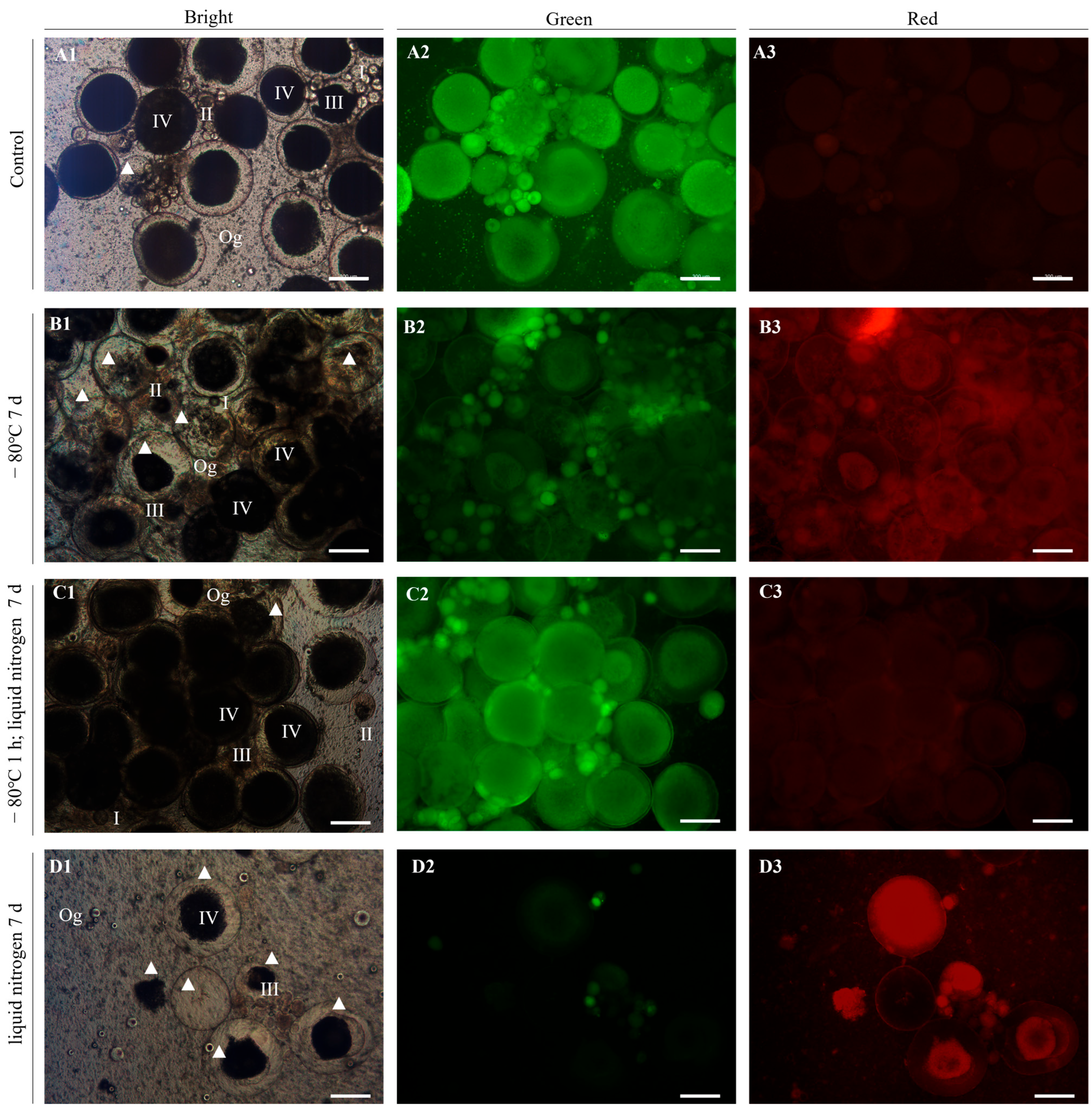
2O. In order to explore the cryopreservation effect of yellow drum ovaries under different freezing procedures. According to preliminary laboratory research, three procedures were adopted with −80 °C refrigerator, liquid nitrogen, and their combination (−80 °C refrigerator for 1 h; liquid nitrogen).

2.4. Cryopreservation Protocols

The isolated ovaries were cut into tissue blocks of approximately 1 cm3, then covered with about 1 mL of cryoprotectant mixture in a 2 mL cryopreservation tube. Three parallel samples were used. Subsequently, the ovary tissues were equilibrated on ice for 60 min, and the cryoprotectants were removed from the tissues after equilibrium had been reached. Then, the three parallel samples were cryopreserved via one of the three freezing procedures, respectively. Upon cryopreservation for 7 days, the tissues were thawed by a water bath (10 °C; 8 min), according to the previous studies of Lee et al. [3] and Lujić et al. [10] with minor modifications. Finally, the ovaries were rehydrated and restored to room temperature in L-15 for about 10 min. Untreated fresh ovarian tissue served as a control.

2.5. Detection of Cell Survival Rate

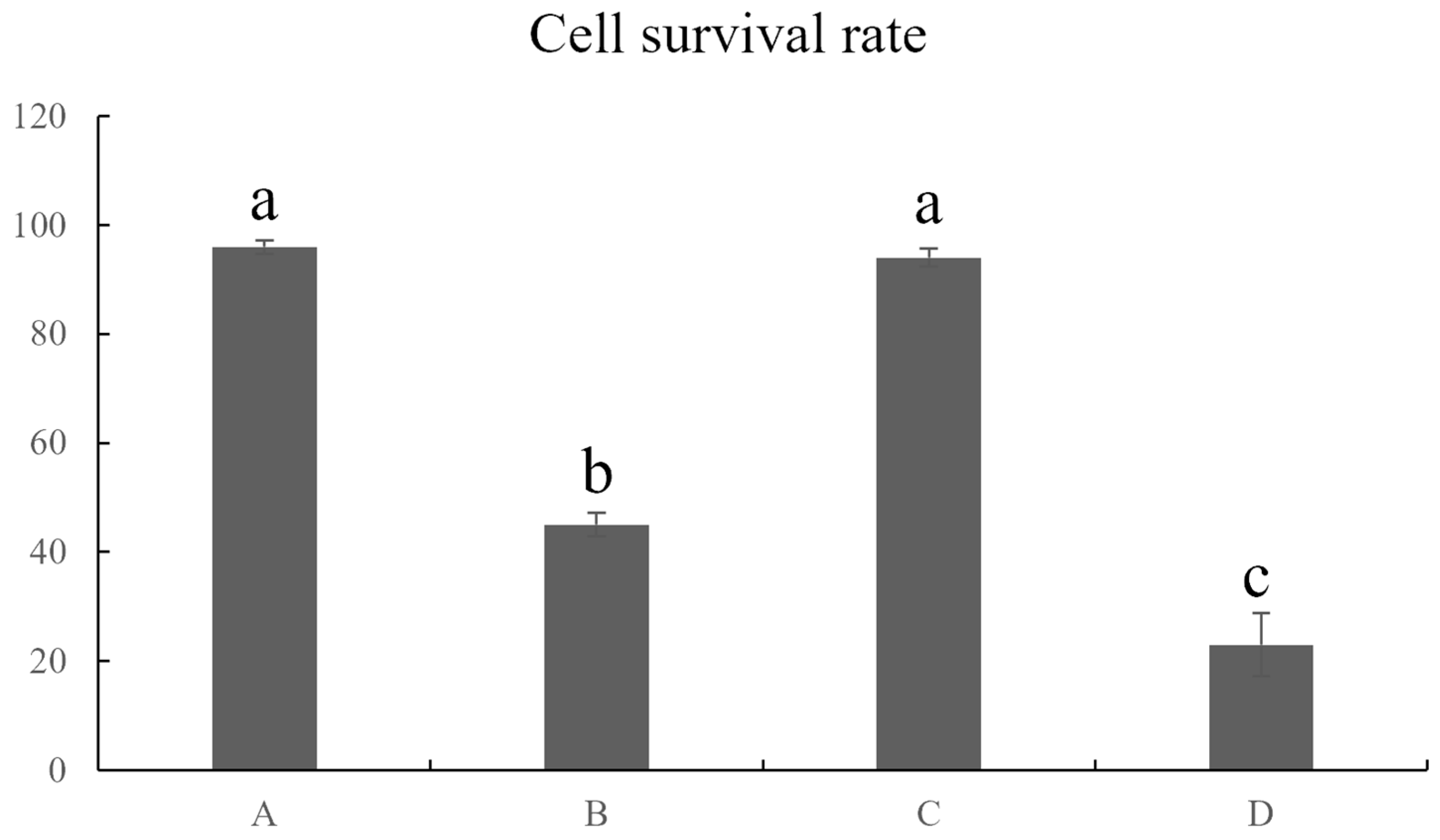
The AO-EB Double Fluorescence Staining Kit (Phygene, Fuzhou, China) was used to detect the survival rate of germ cells. The thawed tissue blocks were processed into dispersed cells with 0.25% Trypsin–EDTA solution (Pricella, Wuhan, China) for 2 h at 28 °C after being washed with L-15. Then, according to the manuals of the kit, the working solution of AO/EB was configured for cell staining. Based on the kit’s instructions, under a fluorescence microscope (Leica DMI8, Wetzlar, Germany), the live and dead cells could show green and red fluorescence after treatment with the working solution, respectively. Therefore, the cell survival rate can be calculated by the following formula. Cell viability = green cells/total cells × 100%.

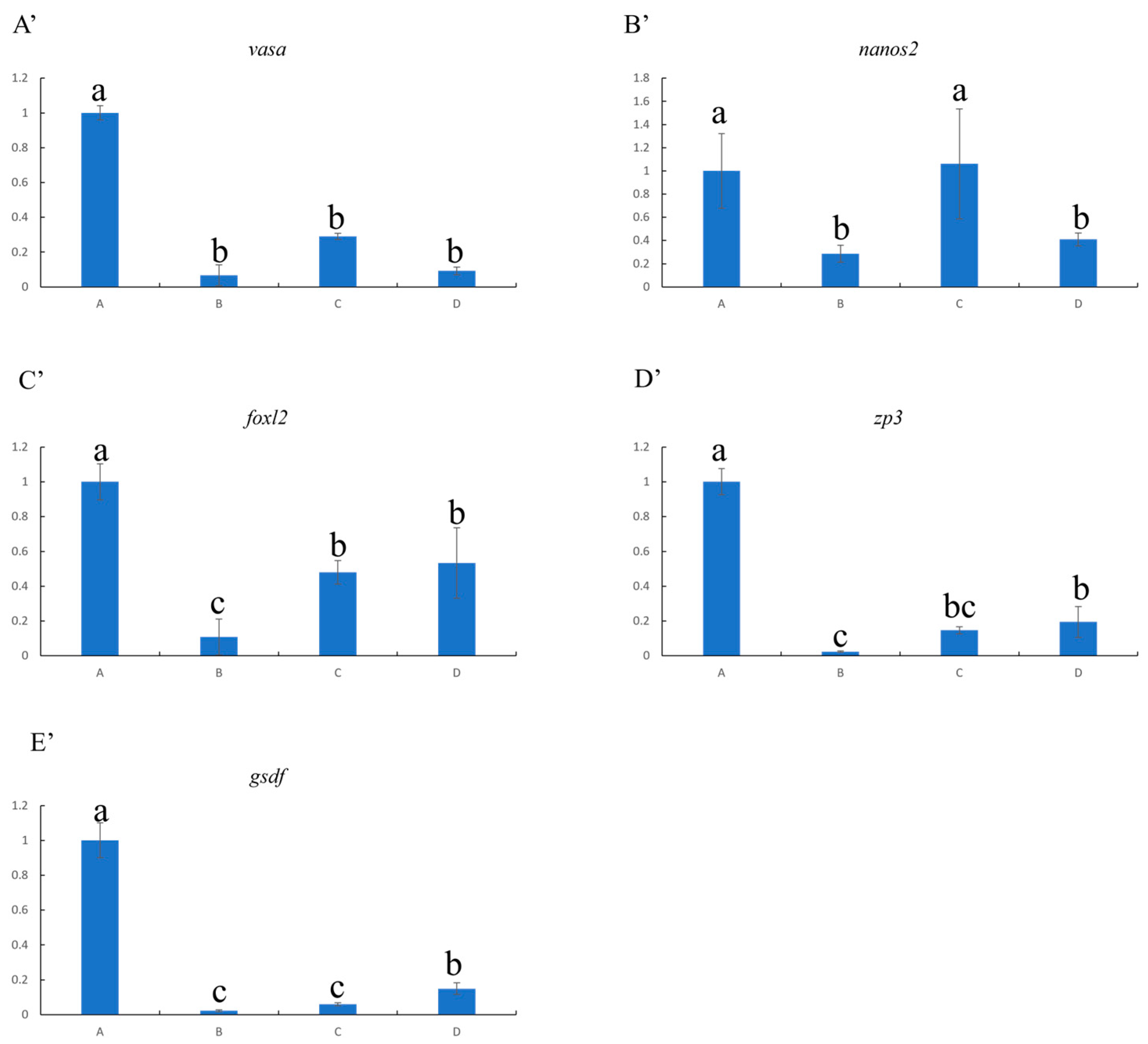
2.6. Analysis of Gene Expression Related to Reproductive Development

After thawing and digestion, some of the isolated cells were also subjected to quantitative real-time polymerase chain reaction (qRT-PCR). β-actin and 18s were used as internal controls, and genes related to reproductive development, such as vasa, nanos2, foxl2, zp3 and gsdf, were detected to assess the effect of cryopreservation. The qRT-PCR primers for these genes were designed using the Primer 5 software and are shown in Table 1. According to the manufacturer’s instructions, total RNA of isolated cells from the control and experiment groups was extracted using the SPARK easy Tissue/Cell RNA kit (Sparkjade, Jinan, China), and cDNA synthesis was performed using the PrimeScriptTM RT reagent kit with gDNA Eraser (Takara, Osaka, Japan). qRT-PCR was performed with TB Green® Premix Ex TaqTM II (Tli RNaseH Plus) (Takara, Osaka, Japan) using a QuantStudio 3 system (Applied Biosystems, Foster City, CA, USA). Briefly, a total volume of 20 μL, including 4 μL of 2× TB Green® Premix Ex Taq II, 0.4 μL of 50 × ROX Reference Dye II, 0.8 μL of forward and reverse primers (10 μM), 6 μL of RNase-free water and 2 μL (<100 ng) of template DNA, was used, and the program was set as follows: 95 °C for 30 s, 35 cycles of 95 °C for 5 s, and 60 °C for 34 s. The results were analyzed according to the comparative Ct method. In this method, vasa, nanos2, foxl2, zp3 and gsdf expression levels were normalized against β-actin and 18s expression, generating a ΔCt. Relative expression was calculated according to the 2−ΔΔCt equation.

2.7. Statistical Analysis

The data of cell survival rate and gene expression was analyzed by software SPSS 26. Each group was presented as mean ± standard error (SE). And the different groups were carried out to one-way ANOVA, then performed Tukey’s test. The p < 0.05 presented significant differences between the different groups.

3. Results

3.1. Appearance of Yellow Drum and Ovary Morphological Histology

The obtained female yellow drum had a slender and flattened body, appearing grayish yellow, with many slanting gray–black stripes on the sides and a silver–white belly (Figure 1A). The ovaries were orange rods filled with eggs, located in the abdominal cavity above the swim bladder and below the spine, which gathered at the genital pore, forming a “V” shape (Figure 1A). The histology results indicated that the ovary contained germ cells at various stages, such as oogonia (Og, 10–20 μm), early previtellogenic phase (I, 20–50 μm), late previtellogenic phase (II, 50–100 μm), early vitellogenic phase (III, 100–300 μm) and late vitellogenic phase (IV, 300–700 μm) (Figure 1B,C). The oogonia and I-II oocytes were small, with large nuclei, prominent nucleoli, little cytoplasm, strong basophilia and dark staining under the microscope (Figure 1C). With the development of oocytes, the volume of oocytes increased significantly, and a few small and scattered yolk granules appeared in III oocytes (Figure 1C). The oocytes were enlarged further, and the IV oocytes were filled with yolk granules, resulting in uniform eosinophilic staining of the cytoplasm (Figure 1B). It is worth noting that the ovary contained a large number of IV oocytes. The histology results further proved that the ovaries were at the stage of vitellogenesis, conforming to the characteristics of asynchronous oocyte development during the reproductive season.

3.2. Cell Survival Rate After Cryopreservation

The ovarian germ cells from the control group showed normal morphology and structure under microscopic observation, containing germ cells at various stages, such as Og and I-IV oocytes (Figure 2(A1)). The viability of control group cells was further verified using the AO-EB Double Fluorescence Staining Kit. The results demonstrated that almost all the cells showed green fluorescence instead of red fluorescence, indicating that they were all viable (Figure 2(A2,A3)).
Compared to the control group, the ovaries treated with cryoprotectants and the three freezing procedures showed obviously different preservation effects. Among them, in the tissues that were stored in a −80 °C refrigerator for 1 h and then transferred to liquid nitrogen for 7 d, the germ cells were of normal size; presented a full, regular shape and smooth surface; and did not show signs of cell rupture, atrophy, depression, granulation or cavitation (Figure 2(C1)). The detected results regarding this treatment indicated the best cryopreservation effect, with most cells presenting green fluorescence, similar to the control group (Figure 2(C2,C3)). To the contrary, when the tissues were stored directly in liquid nitrogen for 7 d, the germ cells presented signs of yolk granulation, shrinkage and even rupture of the egg membrane, resulting in their contents flowing out (Figure 2(D1)). Only a few oogonia showed normal morphology (Figure 2(D1)). The cell viability results further showed that the germ cells almost all presented red fluorescence (except for the very small cells), suggesting that almost all oocytes except a few oogonia had died (Figure 2(D2,D3)). In addition, when the tissues were stored directly in a −80 °C refrigerator for 7 d, the phase IV oocytes presented significant yolk shrinkage, membrane rupture and/or outflow of content (Figure 2(B1)). The cell viability detection results were consistent with the observed morphology and structure, with the oogonia and primary oocytes presenting obvious green fluorescence, the phase II and III oocytes showing weak green fluorescence, and no green fluorescence observed for phase IV oocytes (Figure 2(B1–B3)).

3.3. Cell Survival Rate Statistics After Cryopreservation

The cell survival rates were determined according to the results obtained using the AO−EB Double Fluorescence Staining kit (Figure 3). The results showed that the ovaries stored at −80 °C in a refrigerator for 1 h and then transferred to liquid nitrogen for 7 d presented the best preservation effect, demonstrating a significant difference when compared with other experiment groups (p < 0.05). The cell survival rate in this group was higher than 90%, similar to that in the control group, with which there was no significant difference (p > 0.05). The remaining two experimental groups had poor preservation effects, and the cell survival rate was less than 50%, especially for the ovaries stored directly in liquid nitrogen for 7 d. In conclusion, the results obtained for the ovaries from yellow drum with the used cryoprotectant combination—namely, 15% (v/v) PG, 15% (v/v) FBS and 0.2 M trehalose—when cryopreserved at −80 °C in a refrigerator for 1 h and then transferred to liquid nitrogen indicate this protocol as the most suitable of those tested.

3.4. Gene Expression Analysis After Cryopreservation

After cryopreservation, the results of qRT-PCR indicated that genes related to reproductive development were all down-regulated (except for nanos2) when compared to the control group (Figure 4). In fish, the germ gene vasa is widely distributed throughout various germ cells, while nanos2 is mainly expressed in germline stem cells (e.g., oogonia). Although the ovaries refrigerated at −80 °C for 1 h and then transferred to liquid nitrogen for 7 d demonstrated relatively high expression of the vasa gene, there were significant differences in vasa between the control and all treatment groups (Figure 4A’). However, this experimental group presented similar expression of the nanos2 gene as the control group (no significant difference), indicating that the viability of germ stem cells was not weakened after freezing (Figure 4B’).
In addition, genes associated with ovarian differentiation and development—including foxl2, zp3 and gsdf—were also influenced by cryopreservation (Figure 4C’–E’). Among them, the ovaries stored directly in a −80 °C refrigerator for 7 d showed the lowest expression levels of the three experimental groups (Figure 4C’–E’). In terms of these genes, there was no obvious advantage of refrigerating the ovaries at −80 °C for 1 h before transferring them to liquid nitrogen, compared with their direct placement in liquid nitrogen (Figure 4C’–E’). Taken together, the procedure of refrigerating ovaries at −80 °C for 1 h before their transfer to liquid nitrogen was determined as the optimal protocol, in terms of the expression of relevant genes, consistent with the results of the cell viability assay.

4. Discussion

The present study performed the optimal cryopreservation of ovaries at the stage of vitellogenesis from yellow drum for the first time (Figure 5). The ovaries were isolated from female individuals during the reproduction period and cryopreserved with a cryoprotectant combination using three different freezing procedures. After thawing, the ovaries refrigerated at −80 °C for 1 h and then transferred to liquid nitrogen for 7 d showed the highest cell survival rate (>90%), as determined using the AO-EB Double Fluorescence Staining kit. Furthermore, the qRT-PCR results revealed that genes related to reproductive development, including vasa, foxl2, zp3 and gsdf, were all down-regulated, while the expression of the nanos2 gene—which is specifically distributed in oogonia—was maintained at a higher level, similar to that in the control group. These results indicate that the viability of germ stem cells (oogonia) was not weakened after freezing, and the oogonia could be isolated from the cryopreserved ovaries for germ cell transplantation. These results are similar to those relating to the cryopreservation of ovarian tissues at early stages from Siberian sturgeon [9] and Murray River Rainbowfish [8]. As such, this study established a simple, rapid, efficient and less toxic cryopreservation system for yellow drum, which is of great significance for the preservation of the germplasm resources of Sciaenidae family members.
Regarding the cryopreservation of fish ovaries, previous studies have mainly focused on the preservation of immature ovaries [7,9,11]. In this study, we attempted to cryopreserve ovaries at the stage of vitellogenesis obtained from yellow drum. According to the results of the cell morphology and structure, cell viability and gene expression analyses after freezing, it can be concluded that the effect of the optimal cryopreservation protocol proposed in this study after freezing was good for germ cells, especially oogonia. It can be speculated that, in addition to cryoprotectant agents and procedures, the ovarian support structure may also promote the efficiency of this cryopreservation protocol. In contrast to previous studies, the cryopreservation of ovaries at the stage of vitellogenesis allows for not only the preservation of isolated oogonia (as is the case for cryopreserved immature ovaries) but also vitellogenic oocytes, enabling the possibility of subsequent artificial insemination. This could be considered as an alternative route for eggs and embryos that are traditionally hard to preserve. Moreover, considering the large volume of ovaries at the stage of vitellogenesis, they cannot be frozen whole, as is generally the case for immature ovary cryopreservation. Therefore, in this study, the ovaries were first cut into tissue blocks of approximately 1 cm3 before preservation.
In previous studies, slow cooling and vitrification methods have been mainly used for the cryopreservation of fish ovaries [3,4,5,6]. In rainbow trout, Siberian sturgeon and Murray River Rainbowfish, slow cooling methods showed a good preservation effect, yielding cell viabilities above 70% [3,8,9]. However, slow cooling requires special temperature control equipment (e.g., freezer containers) to cool the ovaries at a controlled rate of −1 °C/minute over the course of more than 1 h before their transfer into liquid nitrogen, which greatly increases the cost associated with preservation. Furthermore, this approach is not suitable in cases where the timely preservation of ovaries is necessary, such as sea, field, laboratory or farm conditions without the required equipment. In addition, vitrification usually involves the use of high concentrations of cryoprotectants—especially permeating agents—to rapidly cool the ovaries. For example, in cyprinid honmoroko, the cryopreservation of whole ovaries required a vitrification medium including 5M PG and 5M DMSO [5]. It should be noted that high concentrations of permeating cryoprotectants, such as DMSO, exhibit good preservation ability but increase germ cell toxicity. Compared to the previous studies, the present study achieved the cryopreservation of yellow drum ovaries using a low-toxicity cryoprotectant without requiring any special temperature control equipment.

5. Conclusions

This study is the first to explore the cryopreservation of ovaries at the stage of vitellogenesis from an economically important marine fish species. The cryoprotectant, comprising 15% PG, 15% FBS and 0.2 M trehalose, demonstrated a high cell survival rate (>90%) when used for the cryopreservation of yellow drum ovaries (especially for oogonia), with good cell viability confirmed through morphological and molecular identification. In the process of cryopreservation, the large volumes of ovaries were cut into tissue blocks, then refrigerated at −80 °C for 1 h before being transferred to liquid nitrogen for 7 d. Compared with the commonly used methods of slow cooling and vitrification, the preservation of ovarian tissues in this study did not require special equipment and the smaller tissue blocks facilitated the penetration of cryoprotectants, thus reducing the cytotoxicity associated with high concentrations of cryoprotectants. The present study provided a convenient, rapid, efficient and less toxic method for the cryopreservation of ovaries at the stage of vitellogenesis, which is important for germplasm resource preservation and fish breeding.

Author Contributions

L.Z., writing—original draft, investigation, formal analysis, data curation, conceptualization, funding acquisition; F.L., investigation, methodology, conceptualization; Z.S., investigation; J.C., investigation; K.H., writing—review and editing, supervision. All authors have read and agreed to the published version of the manuscript.

Funding

This research was funded by the Scientific Research Program of Ningde Normal University (2022ZDK02 and 2022Y06), the Natural Science Foundation of Fujian (2022J05270 and 2020J02011), the Open Project of the State Key Laboratory of Mariculture Breeding (2023KF006), and the Central Government Guiding Local Science and Technology Development Program of Fujian (2021L063).

Institutional Review Board Statement

The animal study protocol was approved by Ethics Committee of Ningde Normal University (Approval Code: NDNU-LL-202508, approval date: 25 February 2025).

Informed Consent Statement

Not applicable.

Data Availability Statement

Data will be made available on request.

Conflicts of Interest

The authors Jia Chen and Zhaohan Sun were employed by the Ningde Yiye Marine Industry Development Co. Ltd. The remaining authors declare that the research was conducted in the absence of any commercial or financial relationships that could be construed as a potential conflict of interest.

References

  1. Seki, S.; Yano, M.; Higashiya, M.; Oikawa, T.; Yamazaki, W.; Yoshizaki, G. Vitrification of medaka whole testis with a trehalose-containing solution and production of medaka individuals derived from the vitrified cells. Cryobiology 2024, 116, 104936. [Google Scholar] [CrossRef]
  2. Jaiswal, A.N.; Vagga, A. Cryopreservation: A Review Article. Cureus 2022, 14, e31564. [Google Scholar] [CrossRef]
  3. Lee, S.; Katayama, N.; Yoshizaki, G. Generation of juvenile rainbow trout derived from cryopreserved whole ovaries by intraperitoneal transplantation of ovarian germ cells. Biochem. Biophys. Res. Commun. 2016, 478, 1478–1483. [Google Scholar] [CrossRef]
  4. Lujić, J.; Marinović, Z.; Sušnik Bajec, S.; Djurdjevič, I.; Kása, E.; Urbányi, B.; Horváth, Á. First successful vitrification of salmonid ovarian tissue. Cryobiology 2017, 76, 154–157. [Google Scholar] [CrossRef]
  5. Higaki, S.; Todo, T.; Teshima, R.; Tooyama, I.; Fujioka, Y.; Sakai, N.; Takada, T. Cryopreservation of male and female gonial cells by vitrification in the critically endangered cyprinid honmoroko Gnathopogon caerulescens. Fish Physiol. Biochem. 2018, 44, 503–513. [Google Scholar] [CrossRef]
  6. Franek, R.; Tichopad, T.; Steinbach, C.; Xie, X.; Lujic, J.; Marinovic, Z.; Horváth, Á.; Kašpar, V.; Pšenička, M. Preservation of female genetic resources of common carp through oogonial stem cell manipulation. Cryobiology 2019, 87, 78–85. [Google Scholar] [CrossRef]
  7. Santos Marques, L.; Rodrigues de Freitas, T.; Batista Rodrigues, R.; Dos Santos Teixeira, N.; Pérez-Atehortúa, M.; Rosa-Silva, H.T.; Fonseca Moreira, J.C.; Streit, D.P., Jr. Vitrification protocol for immature Brycon orbignyanus ovarian tissue as an extinction escape strategy. Cryobiology 2021, 103, 116–122. [Google Scholar] [CrossRef]
  8. Rivers, N.; Daly, J.; Jones, R.; Currie, P.D.; Temple-Smith, P. Cryopreservation and Flow Cytometric Analysis of Ovarian Tissue in Murray River Rainbowfish, Melanotaenia fluviatilis. Animals 2022, 12, 794. [Google Scholar] [CrossRef]
  9. Pšenička, M.; Saito, T.; Rodina, M.; Dzyuba, B. Cryopreservation of early stage Siberian sturgeon Acipenser baerii germ cells, comparison of whole tissue and dissociated cells. Cryobiology 2016, 72, 119–122. [Google Scholar] [CrossRef]
  10. Lujić, J.; Franěk, R.; Marinović, Z.; Kašpar, V.; Xie, X.; Horváth, Á.; Pšenička, M.; Urbányi, B. Vitrification of the ovarian tissue in sturgeons. Theriogenology 2023, 196, 18–24. [Google Scholar] [CrossRef]
  11. Ye, H.; Zhou, C.; Yue, H.; Wu, M.; Ruan, R.; Du, H.; Li, C.; Wei, Q. Cryopreservation of germline stem cells in American paddlefish (Polyodon spathula). Anim. Reprod. Sci. 2021, 224, 106667. [Google Scholar] [CrossRef]
  12. de Freitas, T.R.; Rodrigues, R.B.; Marques, L.S.; Dantas, R.V.; Torres-Lozano, K.G.; França, T.S.; Lima, L.C.O.; Santos, F.W.; Nicoleti, E.T.; Chaves, T.F.; et al. Biodegradable capsules as a sustainable and accessible container for vitrification of gonadal tissue using the zebrafish animal model. Cryobiology 2024, 116, 104944. [Google Scholar] [CrossRef]
  13. Whaley, D.; Damyar, K.; Witek, R.P.; Mendoza, A.; Alexander, M.; Lakey, J.R. Cryopreservation: An Overview of Principles and Cell-Specific Considerations. Cell Transplant. 2021, 30, 963689721999617. [Google Scholar] [CrossRef]
  14. Zeng, Y.; Li, Y.; Du, H.; Li, C.; Dai, W.; Wu, R. Comparative effectiveness of vitrification and slow freezing after heterotopic transplantation of human ovarian tissues. BMC Women’s Health 2024, 24, 654. [Google Scholar] [CrossRef]
  15. Jang, T.H.; Park, S.C.; Yang, J.H.; Kim, J.Y.; Seok, J.H.; Park, U.S.; Choi, C.W.; Lee, S.R.; Han, J. Cryopreservation and its clinical applications. Integr. Med. Res. 2017, 6, 12–18. [Google Scholar] [CrossRef]
  16. Tsai, S.; Rawson, D.M.; Zhang, T. Development of cryopreservation protocols for early stage zebrafish (Danio rerio) ovarian follicles using controlled slow cooling. Theriogenology 2009, 71, 1226–1233. [Google Scholar] [CrossRef]
  17. El Cury-Silva, T.; Nunes, M.E.G.; Casalechi, M.; Comim, F.V.; Rodrigues, J.K.; Reis, F.M. Cryoprotectant agents for ovarian tissue vitrification: Systematic review. Cryobiology 2021, 103, 7–14. [Google Scholar] [CrossRef]
  18. Bizarro-Silva, C.; Bergamo, L.Z.; Costa, C.B.; González, S.M.; Yokomizo, D.N.; Rossaneis, A.C.; Verri Junior, W.A.; Sudano, M.J.; Andrade, E.R.; Alfieri, A.A.; et al. Evaluation of Cryopreservation of Bovine Ovarian Tissue by Analysis of Reactive Species of Oxygen, Toxicity, Morphometry, and Morphology. Vet. Sci. 2024, 11, 579. [Google Scholar] [CrossRef]
  19. Cui, Y.; Luo, S.; Wu, B.; Li, Q.; Han, F.; Wang, Z. Immunomodulatory Effects of SPHK1 and Its Interaction with TFAP2A in Yellow Drum (Nibea albiflora). Int. J. Mol. Sci. 2024, 25, 13641. [Google Scholar] [CrossRef] [PubMed]
  20. Song, H.; Xu, D.; Tian, L.; Chen, R.; Wang, L.; Tan, P.; You, Q. Overwinter mortality in yellow drum (Nibea albiflora): Insights from growth and immune responses to cold and starvation stress. Fish Shellfish Immunol. 2019, 92, 341–347. [Google Scholar] [CrossRef]
  21. Zhao, C.; Liu, Q.; Xu, S.; Xiao, Y.; Wang, W.; Yang, J.; Yang, Y.; Wang, Y.; Song, Z.; Li, J. Identification of type A spermatogonia in turbot (Scophthalmus maximus) using a new cell-surface marker of Lymphocyte antigen 75 (ly75/CD205). Theriogenology 2018, 113, 137–145. [Google Scholar] [CrossRef] [PubMed]
Figure 1. The appearance and ovary morphological histology of yellow drum. (A) Appearance and ovary morphology; (B,C) paraffin sections and H&E staining of ovary tissue. Panel (C) shows a magnification of the black dashed box in (B). Og, oogonia; I, early previtellogenic phase; II, late previtellogenic phase; III, early vitellogenic phase; IV, late vitellogenic phase. Scale bar, 1 cm (A); 300 μm (B); 50 μm (C).
Fishes 10 00288 g001
Figure 2. Germ cell viability detection in ovaries using the AO-EB Double Fluorescence Staining Kit. Live cells show green fluorescence, while dead cells show red fluorescence. (A1A3) Control; ovaries with the cryoprotectants (15% PG, 15% FBS, and 0.2 M trehalose) stored with three freezing procedures—(B1B3): −80 °C refrigeration for 7 d; (C1C3) −80 °C refrigeration followed by liquid nitrogen for 7 d; (D1D3) immersion in liquid nitrogen for 7 d. The arrows indicate oocyte rupture, shrinkage or granulation of the yolk. Og, oogonia; I–II, early/late previtellogenic phase; III–IV, early/late vitellogenic phase. Scale bar = 300 μm.
Fishes 10 00288 g002
Figure 3. The statistics of germ cell viability in ovaries after cryopreservation. Panel A: Control; panel B: −80 °C refrigeration for 7 d; panel C: −80 °C refrigeration for 1 h followed by liquid nitrogen for 7 d; panel D: liquid nitrogen for 7 d. Differences among groups were evaluated via one-way ANOVA followed by Tukey’s test. Values with different superscripts were significantly different among groups (p < 0.05).
Fishes 10 00288 g003
Figure 4. Analysis of expression of genes related to reproductive development in ovaries after cryopreservation. A: Control; B: −80 °C refrigeration for 7 d; C: −80 °C refrigeration for 1 h followed by liquid nitrogen for 7 d; D: liquid nitrogen for 7 d. The (A’E’) represented the relative expression of vasa, nanos2, foxl2, zp3 and gsdf genes under different control and freezing procedures. Differences among groups were evaluated via one-way ANOVA followed by Tukey’s test. Values with different superscripts were significantly different among groups (p < 0.05).
Fishes 10 00288 g004
Figure 5. Schematic illustration of cryopreservation of ovaries at stage of vitellogenesis from yellow drum.
Fishes 10 00288 g005
Table 1. The qRT-PCR primers used in the study.
PrimerSequence
actinFTTATGAAGGCTATGCCCTGCC
actinRTGAAGGAGTAGCCACGCTCTGT
18sFATTCGTATTGTGCCGCTAGAGGTG
18sRGGAATAACGCCGCCGGATCG
vasaFGCCTGAAGACGAAGACACCA
vasaRGGCTTCACGTAGCCAGACTT
foxl2FGGAGAAAGAGCGACCTAAAGAG
foxl2RTGGCAATGAGAGCGACATAAG
zp3FCTGGGCCTCAAGCTGATAAA
zp3RGCTGTGCAGCTCATATTCAAAG
gsdfFCAACTGGGTGATCCATCCTAAA
gsdfRCGCTGAATTCATCCACAAAGAC
Disclaimer/Publisher’s Note: The statements, opinions and data contained in all publications are solely those of the individual author(s) and contributor(s) and not of MDPI and/or the editor(s). MDPI and/or the editor(s) disclaim responsibility for any injury to people or property resulting from any ideas, methods, instructions or products referred to in the content.

