Article

Influence of Eucommia ulmoides Extract on the Growth, Glucose Metabolism, and Antioxidant Capacity of Largemouth Bass (Micropterus salmoides)

1
College of Fisheries, Tianjin Agricultural University, Tianjin 300392, China
2
Key Laboratory of Integrated Rice-Fish Farming Ecology, Ministry of Agriculture and Rural Affairs, Freshwater Fisheries Research Center, Chinese Academy of Fishery Sciences, Wuxi 214081, China
3
Wuxi Fisheries College, Nanjing Agricultural University, Wuxi 214081, China
*
Authors to whom correspondence should be addressed.
Fishes 2025, 10(6), 269; https://doi.org/10.3390/fishes10060269
Submission received: 5 May 2025 / Revised: 30 May 2025 / Accepted: 1 June 2025 / Published: 3 June 2025
(This article belongs to the Special Issue Largemouth Bass Aquaculture)

Abstract

This study aimed to evaluate the impact of Eucommia ulmoides extract (EE) supplementation on the expression of genes related to glucose metabolism and antioxidant capacity of M. salmoides in response to different starch levels. In order to evaluate the effect of EE on fish metabolism and especially to enhance the metabolism of M. salmoides towards glucose metabolism, especially in high and low starch formulations, we designed six experimental feed groups: PC (high-starch control), NC (low-starch control), and four groups supplemented with EE on the basis of PC, with EE concentrations of 0.05%, 0.10%, 0.15%, and 0.20%, respectively. Each feed was administered to fish with an average weight of 36.98 ± 0.08 g, which were cultured for seven weeks, and the water temperature was 31–33 °C. The results demonstrated that increasing the EE concentration in the feed significantly influenced fish growth without affecting the body composition. Regarding the antioxidant activity, the highest CAT (catalase) enzyme activity in the intestine was recorded in the 0.15% EE group. Additionally, the mRNA expression of the antioxidant gene keap1 (kelch-like ECH-associated protein1) increased with higher EE supplementation, and sod (superoxide dismutase) mRNA expression was significantly elevated in the 0.10% EE group compared to that in the PC group. A plasma biochemical analysis revealed a significant increase in the ALP (alkaline phosphatase) activity in the 0.05% EE group relative to the PC group, while the TG (triglycerides) levels progressively decreased as the EE levels increased. Furthermore, the GLU (glucose) levels were significantly reduced in both the EE-supplemented and NC groups compared to those in the PC group. Among the genes associated with glucose metabolism, both gk (glucokinase) and pepck (phosphoenol pyruvate carboxykinase) exhibited a pattern of initially decreasing, followed by an increase, as the EE levels rose, with the pepck (phosphoenol pyruvate carboxykinase) expression being lowest in the 0.10% EE group. In conclusion, appropriate EE supplementation in the diet may promote growth performance, enhance antioxidant capacity, and support the expression of genes related to glucose metabolism of M.salmoides in response to different starch levels.
Key Contribution: Different concentrations of Eucommia ulmoides extract were added to the feeds in this experiment as a way to improve the glucose metabolism in M. salmoides and alleviate the metabolic liver disease in M. salmoides, to reduce the cost of the formulation, and also to lay the foundation for the healthy and sustainable development of M. salmoides aquaculture industry in China.

Graphical Abstract

1. Introduction

In aquaculture, antibiotics and other chemicals are added to feeds to increase economic returns; however, this leads to the presence of drug residues and accumulation in the cultured organisms [1,2,3]. It is thus necessary to find an effective alternative that can ensure both economic benefits and the health of the farmed animals. In response to the need to reduce drug residues and aquaculture costs, plant-based feed additives have drawn the attention of researchers. The abundance of plant feed additives and their rich array of active compounds make them an ideal alternative for enhancing the growth performance and stress resilience of aquaculture species. These plant-based additives encompass a broad spectrum of bioactive compounds, many of which have demonstrated positive effects on animal health and productivity [4,5]. Recent studies suggest that plant feed additives can enhance fish growth, immunity, and glucose metabolism. For example, dietary inclusion of Ginkgo biloba leaf extract [6], Citrus sinensis peel extract [7], and Jujube (Ziziphus jujube) fruit extract [8] has been shown to promote growth, immune function, and antioxidant capacity. The use of plant extracts not only reduces farming costs but is also environmentally friendly because of higher biodegradability than chemical alternatives. They also confer resistance to a broad range of pathogens, owing to the molecular diversity inherent in plant extracts. This evidence highlights the growing use of plant-based feed additives to enhance fish growth, immune responses, and glucose metabolism in aquaculture.
Eucommia ulmoides Oliv, commonly known as rubber tree wood, belongs to the Eucommiaceae family and is the sole species within the Eucommia genus [9]. This plant contains a diverse range of chemical constituents, such as pinoresinol diglucoside containing not less than 0.10%, chlorogenic acid containing not less than 0.08%, and geniposidic acid containing 1.85%, which possess various pharmacological properties, offering protective effects such as antioxidant and anti-inflammatory activities [10,11]. E. ulmoides has long been utilized in traditional Asian medicine and is listed in the Chinese Pharmacopoeia, with its bark and leaves recognized as the primary medicinal parts. These components have been shown to promote growth and enhance immunity in animals [9]. Studies have shown that the inclusion of EE (Eucommia extract) in feeds enhanced their growth and antioxidant capabilities, including turbot (Scophthalmus maximus L.) [12], juvenile red claw crayfish (Cherax quadricarinatus) [13], genetically improved farmed tilapia (Oreochromis niloticus) [11], and grass carp (Ctenopharyngodon idellus) [14]. In addition, it has also been shown that there is an important role for EE in lipid metabolism regulation [15]. In grass carp, EE supplementation effectively improves lipid metabolism, reduces fat accumulation, and promotes overall fish health [15,16]. However, limited research exists regarding the effects of E. ulmoides on fish glycometabolism.
Micropterus salmoides, commonly referred to as California bass, is native to North America and was introduced to China in the 1970s [17]. As a typical carnivore, M. salmoides requires high levels of dietary protein for optimal growth and has limited ability to utilize glucose [18]. Although previous studies have explored the effects of EE on the immunity and antioxidant capacity of various species, the potential of EE to enhance the antioxidant capacity and improve glucose metabolism in M. salmoides remains unexplored. Therefore, this study aims to investigate the effects of EE as a dietary supplement on the growth, antioxidant capacity, and expression of genes related to glucose metabolism of M. salmoides in response to different starch levels.

2. Materials and Methods

2.1. Preparation of the Diets

The diet formulations and nutritional levels are presented in Table 1. A total of six isonitrogenous and isoenergetic diets were formulated, including two control groups (PC group: high starch level; NC group: low starch level), as well as four experimental groups supplemented with EE at varying concentrations under the glucose level of the PC group: 0.05%EE group (0.05% EE), 0.10%EE group (0.10% EE), 0.15%EE group (0.15% EE), and 0.20%EE group (0.20% EE). The E. ulmoides extract (with the main ingredients of chlorogenic acid, astragalin, and various polysaccharides) was supplied by HANOVE Animal Health Products Co., Ltd., located in Wuxi, China. All raw materials were crushed through 80-mesh sieves, and the ingredients were accurately weighed and mixed with water according to the specified proportions. The mixture was then pelletized using a feed expander (model SJPS56x2 (Jiangsu Muyang Holding Co., Ltd. (Jiangsu, China))) (granule diameter 3 mm). The specific procedure for instrument operation followed that of our previous methodology [19]. Once air-dried, the feed was stored in an airtight container and kept at −20 °C for future use.

2.2. Experimental Fish and Management

The aquaculture experiment was conducted at the Freshwater Fisheries Research Centre (FFRC) of the Chinese Academy of Fisheries Sciences, located in Wuxi, China, where the M. salmoides were sourced from the local breeding farm. The training phase before the start of the experiment used a 3 m × 3 m × 2 m net cage with about 500 fish in the net cage and was fed with an M. salmoides commercial compound feed (48% protein and 12% lipid) purchased from Tongwei Co. in Wuxi, China, fish were fed twice daily (at 7:30 a.m. and 6:30 p.m.) to satiety for a duration of two weeks. Subsequently, 360 healthy fish, each with an average weight of 36.98 ± 0.08 g, were evenly distributed into 18 cages (1 m × 1 m × 1 m), with three replicates per group and 20 fish per cage. All cages were in the same pond, the size of which was 2000 m2 (width: 25 m; length: 80 m). Fish were fed twice daily (at 7:30 a.m. and 6:30 p.m.) to satiety for a duration of seven weeks. Mortality and feeding rates were recorded daily. Water quality parameters were also monitored daily using the YSI ProDSS Multiparameter Water Quality Meter (Columbus, OH, USA), ensuring dissolved oxygen levels remained above 5 mg/L, pH ranged between 7.0 and 8.0, and the water temperature was maintained at 31–33 °C.

2.3. Sample Collection

At the end of the cultivation period, the fish were subjected to a 24-h fasting period. Following this, the total fish population in each net cage was counted, and the fish were weighed to determine growth indices. Five fish were randomly selected from each cage: two were analyzed for whole-body composition, while the remaining three were used for blood sampling. Blood samples were drawn from the tail vein, collected in centrifuge tubes, labeled, and left to stand. The samples were then centrifuged for 10 min at 3000 revolutions per minute (rpm) at 4 °C. The supernatant was carefully transferred into separate centrifuge tubes, which were then stored at −80 °C for subsequent plasma biochemical analysis. After blood collection, the fish were euthanized, and the intestinal and liver tissues were dissected, placed into corresponding sample tubes, and stored at −80 °C for later; the intestine was used for the analysis of antioxidant enzyme activities, and the liver was used for the analysis of glucose metabolism- and antioxidant-related genes.

2.4. Growth Index Test

Upon completion of the cultivation period, the initial and final average fish weights were recorded. Growth performance indicators, such as feed efficiency, were calculated using the following formulas:
Feed conversion ratio (FCR) = W1 (g)/(W2 − W0);
Weight gain rate (WGR, %) = (W2 − W0)/W0 × 100%;
Specific growth rate (SGR, %/d) = (ln W2 − ln W0) × 100/d;
Survival rate (SR, %) = N2/N1 × 100%;
In these formulas, W0 represents the mean weight of the fish at the start of the experiment (g), W1 denotes the weight of the dry feed provided (g), W2 represents the mean weight of the fish at the end of the experiment (g), N1 denotes the initial number of fish at the beginning of the experiment, and N2 denotes the number of fish remaining at the conclusion of the experiment.

2.5. Body Composition and Biochemical Analysis

The composition of the experimental feeds and the entire fish body was analyzed using the AOAC method [20]. This was performed by making three replicates of the crude composition of both whole fish and feed, and before each operation, each sample needed to be dried in an oven at 105 °C, after which it was weighed and calculated to determine the moisture (%) of both whole fish and feed. Crude protein (%) was then determined in a Kjeldahl nitrogen apparatus, crude fat content (%) was determined using an automatic fat analyzer through Soxhlet extraction, and ash (%) was obtained by placing the samples in a muffle furnace at 560 °C for 6 h. Feed crude fiber was determined by the FiberCap method using a fiber analysis system (FiberCap™ 2021, FOSS Analytical Co., Ltd., Hilleroed, Denmark,). Plasma biochemical parameters were measured with an automated biochemical analyzer (Mindray BS-400, Shenzhen, China) according to the recommendations of the International Federation of Clinical Chemistry, following the specific instructions of the kit (TG (Triglycerides)-105-000449-00; ALT(alanine aminotransferase)-105-000442-00; TP (total protein)-105-000451-00; TC (total cholesterol)-105-000448-00; AST (aspartate aminotransferase)-15-00443-000; GLU (glucose)-105-000460-00). Intestinal antioxidant indexes were assessed using a specific assay kit from Nanjing JianCheng Bioengineering Institute (Nanjing, China). The specific protocol was as follows: CAT (catalase) was determined using a molybdenum acid method kit, SOD (superoxide dismutase) was determined using a WST-1 method kit, and GSH (glutathione) was determined using a microplate method kit, all of which were purchased from Jiancheng Bioengineering Research Institute (Nanjing, China); a spectrophotometer (Thermo Fisher Scientific Multiskan GO) was used for the determination of the levels of each. All of the above were purchased from Jiancheng Bioengineering Institute (Nanjing, China); the results were obtained using a spectrophotometer (Thermo Fisher Multiskan GO, Shanghai, China).

2.6. Real-Time PCR Analysis

PCR conditions for gene expression analysis: First, RNA was extracted from the liver tissue of M. salmoides using an RNA extraction reagent (Vazyme, Nanjing, China). Second, the concentration and quality of the RNA were determined using a NanoDrop 2000 spectrophotometer (Shanghai, China). The concentration of the RNA samples was adjusted to 60 ng/μL, ensuring that the A260/A280 ratio was between 1.8 and 2.0. Subsequently, the qPCR assay was performed on a CFX96 Touch Real-Time PCR Detection System (Bio-Rad, Hercules, CA, USA) using the One-Step SYBR PrimeScript™ PLUS RT-PCR Kit (Takara, Dalian, China). The specific reaction procedure for qPCR was as follows: reverse transcription at 42 °C for 5 min, initial denaturation at 95 °C for 10 s, followed by 40 cycles of denaturation at 95 °Cfor 5 s and annealing/extension at 60 °C for 30 s. The temperature was increased from 65 °C to 95 °C, and then the melting curve was analyzed. Gapdh, due to its stable and high expression, was selected as the reference gene. The primer sequences used are listed in Table 2. Gene expression levels were quantified using the standard curve method [21].

2.7. Statistical Analysis

Statistical analysis of the experimental data was conducted with data presented as the mean ± standard error (mean ± S.E.). All analyses were performed using SPSS version 26, and graphical representations were generated using GraphPad Prism software (GraphPad Prism version 9.1.0 for Windows (GraphPad Software, San Diego, CA, USA). A one-way ANOVA was used to determine significant differences, with multiple comparisons conducted using the Duncan test (p < 0.05).

3. Results

3.1. Whole-Fish Growth Index and Body Composition Analysis

The findings, as summarized in Table 3, indicate that the final FBW (body weight), WGR (weight gain rate), and SGR (specific growth rate) were significantly higher in the 0.15% EE and 0.20% EE group compared to those of the PC group (p < 0.05). In addition, the feed conversion ratio (FCR) of the 0.15% EE group was significantly lower than that of the PC group (p < 0.05). The whole-fish body composition data also aligned with these trends, showing no significant differences when compared to the PC group data (Table 4). The 0.15% EE group exhibited slightly more moisture than the 0.20% EE group (p < 0.05).

3.2. Analysis of Antioxidant Indexes in Intestine

As presented in Table 5, the 0.15%EE group demonstrated the highest CAT (catalase) activity, which was statistically significant (p < 0.05). However, no significant differences were observed in the GSH (glutathione) content and SOD (superoxide dismutase) activity among the groups (p > 0.05).

3.3. Plasma Biochemical Analysis

Figure 1 illustrates that the plasma ALP (alkaline phosphatase) activity was highest in the 0.05% EE group, with increasing levels of EE supplementation compared to the PC group (p < 0.05). TG (triglycerides)were significantly lower in the 0.10% EE, 0.15% EE, and 0.20% EE groups compared to those in the PC group (p < 0.05). The NC group and all of the EE-supplemented groups exhibited significantly reduced plasma GLU (glucose) levels compared to those of the PC group (p < 0.05). The plasma levels of ALT (alanine aminotransferase), TP (total protein), AST (aspartate aminotransferase), and TC (total cholesterol) were unaffected by the EE supplementation (p > 0.05).

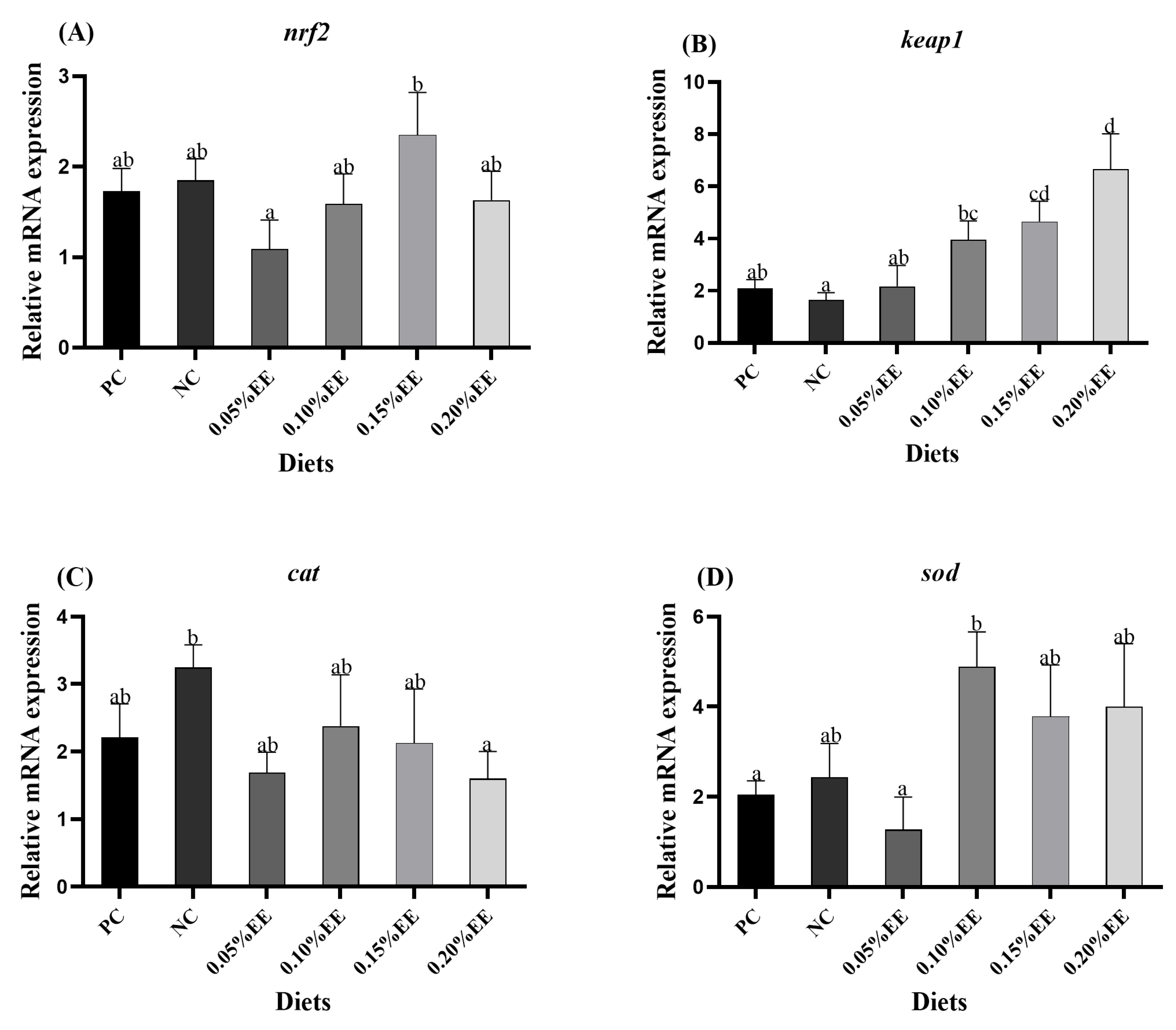
3.4. Analysis of Antioxidant-Related Pathways

Figure 2 shows that the expression of keap1 (kelch-like ECH-associated protein1) mRNA progressively increased with higher levels of EE supplementation, reaching significantly higher levels in the 0.15% EE and 0.20% EE groups compared to those in the PC group (p < 0.05). Additionally, the sod (superoxide dismutase) expression was significantly higher in the 0.10% EE group compared to that in the PC group (p < 0.05). The nrf2 (nuclear factor erythroid 2-related factor 2) expression was significantly higher in the 0.15% EE group compared to that in the 0.05% EE group (p < 0.05), while the cat (catalase) mRNA expression remained unaffected by the EE supplementation (p > 0.05).

3.5. Expression Analysis of Glucose Metabolism-Related Genes

Figure 3 reveals that the mRNA expression of pepck (phosphoenol pyruvate carboxykinase) in the 0.05% EE to 0.20% EE groups was significantly reduced compared to that in the PC group (p < 0.05), with the NC group also showing a significant decrease compared to the PC group (p < 0.05). However, the expression levels of pk (pyruvate kinase) and gk (glucokinase) were not significantly affected by the increasing EE supplementation (p > 0.05).

4. Discussion

The incorporation of EE into aquatic feed has shown growth-promoting effects in various species, including grass carp [22], large yellow croaker (Larimichthys crocea) larvae [23], and Japanese eel (Anguilla japonica) [24]. In this study, supplementing the diet of M. salmoides with EE improved the growth performance, with the 0.15% EE group showing the highest FBW (body weight), WGR (weight gain rate), and SGR (specific growth rate) while also achieving the lowest FCR (feed conversion ratio). These effects may be attributed to the pharmacological properties of Eucommia components like chlorogenic acid, lignans, phenolics, and steroids, which have been shown to enhance growth, reduce inflammation, and promote antioxidant activity in biological systems [10]. For instance, the addition of chlorogenic acid (400 mg/kg) to feed was found to improve both the meat quality and growth performance in grass carp [25]. In this experiment, Eucommia leaf extract (ELE) supplementation had no significant impact on the body composition of M. salmoides except for the moisture content, which is consistent with studies on large yellow croaker larvae [23] and genetically improved farmed tilapia [14]. Interestingly, the 0.15% EE group showed a significantly higher moisture content compared to the 0.20% EE group. Previous studies on large yellow croaker [26], and rice field eel (Monopterus albus) [27] found that adding Eucommia and its extracts to feed generally reduces the body’s moisture content, which aligns with the present findings.
Plasma biochemical indices are important indicators of fish health [28], and the activities of enzymes such as ALP (alkaline phosphatase), ALT (alanine aminotransferase), and AST (aspartate aminotransferase) are particularly crucial in the enzyme leakage associated with liver injury [29,30,31,32]. In this study, the 0.05% EE group exhibited the highest plasma ALP (alkaline phosphatase) activity compared to the PC group. This suggests that supplementation with 0.05% EE in high-starch feed could enhance the ALP (alkaline phosphatase) activity, which may subsequently improve liver immunity. TP (total protein) serves as a major component of hepatic biosynthesis [33], TG (triglycerides) and TC (total cholesterol) are key indicators of lipid metabolism [34]. Previous studies have shown that adding 0.10–0.20% ELE to feed reduced the plasma TP (total protein) and TC (total cholesterol) levels in growing–finishing pigs [35]. However, in our study, no significant differences in the plasma TP (total protein) and TC (total cholesterol) were observed between the groups fed high-starch diets. Interestingly, the plasma triglyceride (TG) levels were significantly lower in the 0.10–0.20% EE groups than in the PC control group, which aligns with findings in obese mice [36,37]. GLU (glucose) levels are an important indicator of intestinal health in M. salmoides. In this experiment, the NC group exhibited markedly lower plasma GLU (glucose) levels compared to those of the PC group, suggesting that the increased glucose levels in the feed elevated the glycogen content in the fish, potentially impacting their health. Additionally, under the same high-starch conditions as in this experiment, the addition of different levels of EE to the feed reduces the levels of GLU (glucose) in the plasma. This supports previous research indicating that EE can lower blood glucose and enhance glucose metabolism [10].
Gene expression levels related to glucose metabolism are critical biomarkers for evaluating liver health in fish [38], with pk, gk (glucokinase), and pepck (phosphoenol pyruvate carboxykinase) playing pivotal roles [39,40]. Pk (pyruvate kinase) and gk (glucokinase) are key rate-limiting enzymes in glycolysis, while pepck (phosphoenol pyruvate carboxykinase) is primarily involved in gluconeogenesis. In previous studies, the gk (glucokinase) mRNA expression in Atlantic salmon (Salmo salar) [41] and Gilthead sea bream (Sparus aurata) juveniles [42] was shown to increase under high-starch feeding conditions. In the present study, while no significant differences in the mRNA expression of pk (pyruvate kinase) and gk (glucokinase) were observed in the EE-supplemented group compared to the PC group, a gradual increase in expression was noted with EE supplementation under the same starch level. This suggests that EE may promote glycolysis in M. salmoides. Additionally, higher pepck (phosphoenol pyruvate carboxykinase) mRNA expression was observed in the PC group compared to the NC group, consistent with findings from glucose-loading studies in rainbow trout [43]. This observation can be attributed to the limited carbohydrate metabolism capacity in fish and particularly carnivorous species [44]. High starch levels are known to induce gluconeogenesis, leading to hyperglycemia [45]. These findings align with the observed plasma GLU (glucose) levels in this study. Notably, the EE supplementation at levels ranging from 0.05% to 0.20% significantly reduced the pepck (phosphoenol pyruvate carboxykinase) activity compared to that in the PC group, indicating that EE supplementation in high-starch diets inhibits gluconeogenesis, thereby lowering plasma GLU (glucose) levels and maintaining glucose homeostasis. Previous studies have shown that Eucommia extract components, such as chlorogenic acid [46] and flavonol glycosides [47], possess blood glucose-lowering abilities. Specifically, flavonol glycosides such as quercetin 3-O-α-l-arabinopyranosyl-(1 → 2)-β-d-glucopyranoside, kaempferol 3-O-β-d-glucopyranoside (known as astragaloside), and quercetin 3-O-β-d-glucopyranoside (isoquercitrin) have demonstrated similar antiglycation effects to aminoguanidine, a well-known antiglycation agent, leading to reduced blood glucose levels. These compounds exhibit a comparable capacity to inhibit glycosylation, thus contributing to decreased blood glucose [47].
Antioxidant capacity can be expressed through the activity of antioxidant enzymes and the expression of antioxidant-related genes [48]. Among these, the nrf2 (nuclear factor erythroid 2-related factor 2) signaling pathway plays a pivotal role in cellular defense mechanisms against oxidative stress [49]. In the current study, although the addition of EE to the feed did not significantly affect the nrf2 (nuclear factor erythroid 2-related factor 2) expression, a trend of an initial increase followed by a decrease was observed in the EE-supplemented groups. Additionally, EE supplementation influenced the expression levels of keap1 (kelch-like ECH-associated protein1) and sod (superoxide dismutase) within the related pathways. Notably, the keap1 (kelch-like ECH-associated protein1) expression was significantly elevated at higher levels of EE supplementation compared to that in the PC group, potentially inhibiting the fish’s antioxidant capacity. On the other hand, the sod (superoxide dismutase) mRNA expression was highest in the 0.10% EE group, suggesting that EE enhances the expression of certain antioxidant genes in M. salmoides, thereby improving its antioxidant properties. A similar enhancement in antioxidant gene activity was observed in channel catfish (Ictalurus punctatus) [50]. This effect may be attributed to the abundance of chemical compounds in E. ulmoides, such as chlorogenic acid, which are known for their ability to neutralize free radicals [11]. However, the cat (catalase) mRNA expression was not influenced by EE supplementation in this study.
Notably, the experimental analysis of the intestinal antioxidant enzyme activity revealed a significant increase in the CAT (catalase) enzyme activity in the 0.15% EE group compared to both the PC and control groups. CAT (catalase) is known to neutralize free radicals [51,52], and in this study, the 0.15% EE group exhibited significantly higher CAT (catalase) activity than the PC group. A similar result was observed in a study on yellow croaker [23], where moderate EE supplementation was found to enhance the antioxidant capacity. However, no significant effects were observed on other antioxidant enzymes, such as SOD (superoxide dismutase) and GSH (glutathione), in the intestinal tissues of the fish. E. ulmoides is known to promote antioxidant enzyme activity [53], and its antioxidants have been shown to reduce free radicals [54,55], thereby alleviating diseases induced by oxidative stress [56,57]. Additionally, EE has been reported to improve erythrocyte viability and increase SOD (superoxide dismutase) and CAT (catalase) activity, among other benefits [58]. Taken together with the keap1 (kelch-like ECH-associated protein1) and sod (superoxide dismutase) mRNA expression levels, it can be hypothesized that moderate EE supplementation promotes the intestinal antioxidant capacity of M. salmoides fed a high-starch diet.

5. Conclusions

In summary, high starch feed levels lead to increased blood glucose in M. salmoides. However, the addition of Eucommia extract at 0.15 g/kg and 0.20 g/kg promotes glycolysis, inhibits gluconeogenesis, and enhances growth performance to some extent. Moreover, the inclusion of Eucommia extract at 0.1–0.15 g/kg effectively improves the antioxidant capacity of M. salmoides in response to different starch levels.

Author Contributions

Conceptualization, J.G. and D.H.; methodology, D.H.; formal analysis, S.Z.; data curation, M.R. and H.L.; writing—original draft preparation, S.Z.; writing—review and editing, M.R. and H.L.; project administration, H.L. All authors have read and agreed to the published version of the manuscript.

Funding

This study was financially supported by the earmarked fund for CARS (CARS-46), National Key R & D Program of China (2023YFD2400601), the National Natural Science Foundation of China (32102806).

Institutional Review Board Statement

The study was approved by the Laboratory Animal Ethics Committee of the Freshwater Fisheries Research Center (LAECFFRC-2022-09-19), Approval Date: 19 September 2022.

Informed Consent Statement

Not applicable.

Data Availability Statement

Data are contained within the article.

Conflicts of Interest

The authors declare no conflicts of interest.

References

  1. Liu, X.; Steele, J.C.; Meng, X.Z. Usage, residue, and human health risk of antibiotics in Chinese aquaculture: A review. Environ. Pollut. 2017, 223, 161–169. [Google Scholar] [CrossRef] [PubMed]
  2. Li, W.; Shi, Y.; Gao, L.; Liu, J.; Cai, Y. Occurrence of antibiotics in water, sediments, aquatic plants, and animals from Baiyangdian Lake in North China. Chemosphere 2012, 89, 1307–1315. [Google Scholar] [CrossRef] [PubMed]
  3. Hossain, A.; Nakamichi, S.; Habibullah-Al-Mamun, M.; Tani, K.; Masunaga, S.; Matsuda, H. Occurrence, distribution, ecological and resistance risks of antibiotics in surface water of finfish and shellfish aquaculture in Bangladesh. Chemosphere 2017, 188, 329–336. [Google Scholar] [CrossRef] [PubMed]
  4. Hashemi, S.R.; Davoodi, H. Herbal plants and their derivatives as growth and health promoters in animal nutrition. Vet. Res. Commun. 2011, 35, 169–180. [Google Scholar] [CrossRef]
  5. Gadde, U.; Kim, W.H.; Oh, S.T.; Lillehoj, H.S. Alternatives to antibiotics for maximizing growth performance and feed efficiency in poultry: A review. Anim. Health Res. Rev. 2017, 18, 26–45. [Google Scholar] [CrossRef]
  6. Tan, X.; Sun, Z.; Liu, Q.; Ye, H.; Zou, C.; Ye, C.; Lin, H.; Wang, A. Effects of dietary ginkgo biloba leaf extract on growth performance, plasma biochemical parameters, fish composition, immune responses, liver histology, and immune and apoptosis-related genes expression of hybrid grouper (Epinephelus lanceolatus♂× Epinephelus fuscoguttatus♀) fed high lipid diets. Fish Shellfish Immunol. 2018, 72, 399–409. [Google Scholar] [PubMed]
  7. Shabana, M.S.; Karthika, M.; Ramasubramanian, V. Effect of dietary Citrus sinensis peel extract on growth performance, digestive enzyme activity, muscle biochemical composition, and metabolic enzyme status of the freshwater fish, Catla catla. J. Basic Appl. Zool. 2019, 80, 51. [Google Scholar] [CrossRef]
  8. Hoseinifar, S.H.; Khodadadian Zou, H.; Paknejad, H.; Ahmadifar, E.; Van Doan, H. Non-specific immune responses and intestinal immunity of common carp (Cyprinus carpio) fed Jujube (Ziziphus jujube) fruit extract. Aquac. Res. 2018, 49, 2995–3003. [Google Scholar] [CrossRef]
  9. He, X.; Wang, J.; Li, M.; Hao, D.; Yang, Y.; Zhang, C.; He, R.; Tao, R. Eucommia ulmoides Oliv.: Ethnopharmacology, phytochemistry and pharmacology of an important traditional Chinese medicine. J. Ethnopharmacol. 2014, 151, 78–92. [Google Scholar] [CrossRef]
  10. Hussain, T.; Tan, B.E.; Liu, G.; Oladele, O.A.; Rahu, N.; Tossou, M.C.; Yin, Y. Health-promoting properties of Eucommia ulmoides: A review. Evid.-Based Complement. Altern. Med. 2016, 2016, 5202908. [Google Scholar] [CrossRef]
  11. Huang, D.; Zhu, J.; Zhang, L.; Ge, X.; Ren, M.; Liang, H. Dietary supplementation with Eucommia ulmoides leaf extract improved the intestinal antioxidant capacity, immune response, and disease resistance against Streptococcus agalactiae in genetically improved farmed tilapia (GIFT; Oreochromis niloticus). Antioxidants 2022, 11, 1800. [Google Scholar] [CrossRef] [PubMed]
  12. Zhang, B.; Li, C.; Wang, X.; Zhou, H.; Mai, K.; He, G. The effects of dietary Eucommia ulmoides Oliver on growth, feed utilization, antioxidant activity and immune responses of turbot (Scophthalmus maximus L.). Aquac. Nutr. 2019, 25, 367–376. [Google Scholar] [CrossRef]
  13. Lu, Y.-P.; Zheng, P.-H.; Xu, J.-R.; Cao, Y.-L.; Li, J.-T.; Hao, C.-G.; Zhang, Z.-L.; Xian, J.-A.; Zhang, X.-X.; Wang, A.-L. Effects of Dietary Eucommia ulmoides Leaf Extract on Growth, Muscle Composition, Hepatopancreas Histology, Immune Responses and Microcystin-LR Resistance of Juvenile Red Claw Crayfish (Cherax quadricarinatus). Fishes 2023, 8, 20. [Google Scholar] [CrossRef]
  14. Yang, H.; Xu, Z.; Li, X.Q.; Tan, S.M.; Cheng, Z.; Leng, X.J. Influences of dietary Eucommia ulmoides extract on growth, flesh quality, antioxidant capacity and collagen-related genes expression in grass carp (Ctenopharyngodon idellus). Anim. Feed Sci. Technol. 2021, 277, 114965. [Google Scholar] [CrossRef]
  15. Yang, H.; Xu, Z.; Tan, S.; Zhang, C.; Li, X.; Leng, X. In vitro effects of Eucommia ulmoides and its active components on the growth, lipid metabolism and collagen metabolism of grass carp (Ctenopharyngodon idellus) hepatocyte and intramuscular fibroblast. J. Fish Biol. 2022, 101, 597–612. [Google Scholar] [CrossRef]
  16. Yang, H.; Xu, Z.; Xu, X.; Rahman, M.M.; Li, X.; Leng, X. Transcriptomic and biochemical analyses revealed the improved growth, lipid metabolism, and flesh quality of grass carp (Ctenopharyngodon idellus) by dietary Eucommia ulmoides bark and leaf supplementation. J. Anim. Sci. 2022, 100, skac250. [Google Scholar] [CrossRef]
  17. Bai, J.; Li, S. Development of largemouth bass (Micropterus salmoides) culture. In Aquaculture in China: Success Stories and Modern Trends; John Wiley & Sons Ltd.: Hoboken, NJ, USA, 2018; pp. 421–429. [Google Scholar]
  18. Zhang, B.Y.; Yang, H.L.; Yuan, Z.H.; Sun, X.O.; Yin, J.H.; Xu, J.J.; Cai, G.H.; Sun, Y.Z. Commensal microorganisms ameliorate the adverse effects of high wheat starch diet on the growth performance, glucose and lipid metabolisms in juvenile largemouth bass, Micropterus salmoides. Aquac. Rep. 2024, 39, 102452. [Google Scholar] [CrossRef]
  19. Ren, M.; Liao, Y.; Xie, J.; Liu, B.; Zhou, Q.; Ge, X.; Cui, H.; Pan, L.; Chen, R. Dietary arginine requirement of juvenile blunt snout bream, Megalobrama amblycephala. Aquaculture 2013, 414, 229–234. [Google Scholar] [CrossRef]
  20. AOAC. Official Methods of Analysis of the Association of Official Analytical Chemists, 15th ed.; Association of Official Analytical Chemists, Inc.: Arlington, VA, USA, 2003. [Google Scholar]
  21. Stone, D.A.; Gaylord, T.G.; Johansen, K.A.; Overturf, K.; Sealey, W.M.; Hardy, R.W. Evaluation of the effects of repeated fecal collection by manual stripping on the plasma cortisol levels, TNF-α gene expression, and digestibility and availability of nutrients from hydrolyzed poultry and egg meal by rainbow trout, Oncorhynchus mykiss (Walbaum). Aquaculture 2008, 275, 250–259. [Google Scholar]
  22. Sun, W.T.; Li, X.Q.; Xu, H.B.; Chen, J.N.; Xu, X.Y.; Leng, X.J. Effects of dietary chlorogenic acid on growth, flesh quality and serum biochemical indices of grass carp (Ctenopharyngodon idella). Aquac. Nutr. 2017, 23, 1254–1263. [Google Scholar] [CrossRef]
  23. Huang, W.; Yao, C.; Liu, Y.; Xu, N.; Yin, Z.; Xu, W.; Miao, Y.; Mai, K.; Ai, Q. Effects of dietary Eucommia ulmoides leaf extract on growth performance, expression of feeding-related genes, activities of digestive enzymes, antioxidant capacity, immunity and cytokines expression of large yellow croaker (Larimichthys crocea) larvae. Br. J. Nutr. 2022, 128, 1711–1719. [Google Scholar] [CrossRef] [PubMed]
  24. Lim, J.W.; Mendis, W.R.H.; Jeong, B.C.; Lim, T.J.; Ahn, J.C.; Jung, S.J.; Kang, S.Y. Optimization of Eucommia ulmoides Oliver bark extracts by Box-Behnken design and their effects on growth performance, plasma biochemical and immunological parameters, and antioxidant enzyme-and inflammation-related genes expression in Japanese eel, Anguilla japonica. Aquac. Rep. 2022, 26, 101320. [Google Scholar]
  25. Sun, W.T.; He, M.; Xu, X.Y.; Li, X.Q.; Pan, W.Q.; Leng, X.J. Comparison study of three compounds in Eucommia ulmoides on growth, flesh quality of grass carp (Ctenopharyngodon idella). Aquac. Nutr. 2019, 25, 906–916. [Google Scholar] [CrossRef]
  26. Li, J.; Xu, W.; Lai, W.; Kong, A.; Ai, Q. Effect of dietary methionine on growth performance, lipid metabolism and antioxidant capacity of large yellow croaker (Larimichthys crocea) fed with high lipid diets. Aquaculture 2021, 536, 736388. [Google Scholar] [CrossRef]
  27. Luo, A.; Song, C.; Wu, X.; Li, M.; Shi, C.; Wu, S.; Wei, L.; Peng, F.; Mo, P. Effects of compound plant extracts on growth performance, antioxidant capacity, and histomorphology of liver and intestine of rice field eel (Monopterus albus). J. World Aquac. Soc. 2024, 55, e13065. [Google Scholar] [CrossRef]
  28. Yilmaz, S.; Ergün, S.; Yıgıt, M. Effects of dietary FARMARIN® XP supplement on immunological responses and disease resistance of rainbow trout (Oncorhynchus mykiss). Aquaculture 2018, 496, 211–220. [Google Scholar] [CrossRef]
  29. Mensinger, A.F.; Walsh, P.J.; Hanlon, R.T. Blood biochemistry of the oyster toadfish. J. Aquat. Anim. Health 2005, 17, 170–176. [Google Scholar] [CrossRef]
  30. Wagner, T.; Congleton, J.L. Blood chemistry correlates of nutritional condition, tissue damage, and stress in migrating juvenile chinook salmon (Oncorhynchus tshawytscha). Can. J. Fish. Aquat. Sci. 2004, 61, 1066–1074. [Google Scholar] [CrossRef]
  31. Coz-Rakovac, R.; Strunjak-Perovic1, I.; Hacmanjek, M.; Popovic, N.T.; Lipej, Z.; Sostaric, B. Blood chemistry and histological properties of wild and cultured sea bass (Dicentrarchus labrax) in the North Adriatic Sea. Vet. Res. Commun. 2005, 29, 677–687. [Google Scholar] [CrossRef]
  32. Metón, I.; Mediavilla, D.; Caseras, A.; Cantó, E.; Fernández, F.; Baanante, I.V. Effect of diet composition and ration size on key enzyme activities of glycolysis–gluconeogenesis, the pentose phosphate pathway and amino acid metabolism in liver of gilthead sea bream (Sparus aurata). Br. J. Nutr. 1999, 82, 223–232. [Google Scholar] [CrossRef]
  33. Kumar, S.; Sahu, N.P.; Pal, A.K.; Choudhury, D.; Yengkokpam, S.; Mukherjee, S.C. Effect of dietary carbohydrate on haematology, respiratory burst activity and histological changes in L. rohita juveniles. Fish Shellfish Immunol. 2005, 19, 331–344. [Google Scholar] [CrossRef] [PubMed]
  34. Tang, Y.; Wang, D.; Zhang, H.; Zhang, Y.; Wang, J.; Qi, R.; Yang, J.; Sheng, H.; Xu, Y.; Li, M. Rapid responses of adipocytes to iron overload increase serum TG level by decreasing adiponectin. J. Cell. Physiol. 2021, 236, 7544–7553. [Google Scholar] [CrossRef] [PubMed]
  35. Yang, Y.; Li, F.; Guo, Q.; Wang, W.; Zhang, L.; Yin, Y.; Gong, S.; Han, M.; Yin, Y. Effects of Different Supplemental Levels of Eucommia ulmoides Leaf Extract in the Diet on Carcass Traits and Lipid Metabolism in Growing–Finishing Pigs. Front. Vet. Sci. 2022, 8, 828165. [Google Scholar] [CrossRef]
  36. Hirata, T.; Ikeda, T.; Fujikawa, T.; Nishibe, S. The chemistry and bioactivity of Eucommia ulmoides Oliver leaves. Stud. Nat. Prod. Chem. 2014, 41, 225–260. [Google Scholar]
  37. Hirata, T.; Kobayashi, T.; Wada, A.; Ueda, T.; Fujikawa, T.; Miyashita, H.; Ikeda, T.; Tsukamoto, S.; Nohara, T. Anti-obesity compounds in green leaves of Eucommia ulmoides. Bioorganic Med. Chem. Lett. 2011, 21, 1786–1791. [Google Scholar] [CrossRef]
  38. Gharaei, A.; Ghaffari, M.; Keyvanshokooh, S.; Akrami, R. Changes in metabolic enzymes, cortisol and glucose concentrations of Beluga (Huso huso) exposed to dietary methylmercury. Fish Physiol. Biochem. 2011, 37, 485–493. [Google Scholar] [CrossRef] [PubMed]
  39. Peng, K.; Wang, G.; Zhao, H.; Wang, Y.; Mo, W.; Wu, H.; Huang, Y. Effect of high level of carbohydrate and supplementation of condensed tannins on growth performance, serum metabolites, antioxidant and immune response, and hepatic glycometabolism gene expression of Lateolabrax japonicus. Aquac. Rep. 2020, 18, 100515. [Google Scholar] [CrossRef]
  40. Enes, P.; Sanchez-Gurmaches, J.; Navarro, I.; Gutiérrez, J.; Oliva-Teles, A. Role of insulin and IGF-I on the regulation of glucose metabolism in European sea bass (Dicentrarchus labrax) fed with different dietary carbohydrate levels. Comp. Biochem. Physiol. Part A Mol. Integr. Physiol. 2010, 157, 346–353. [Google Scholar] [CrossRef]
  41. Borrebaek, B.; Waagbø, R.; Christophersen, B.; Tranulis, M.A.; Hemre, G.I. Adaptable hexokinase with low affinity for glucose in the liver of Atlantic salmon (Salmo salar). Comp. Biochem. Physiol. Part B Comp. Biochem. 1993, 106, 833–836. [Google Scholar] [CrossRef]
  42. Enes, P.; Peres, H.; Couto, A.; Oliva-Teles, A. Growth performance and metabolic utilization of diets including starch, dextrin, maltose or glucose as carbohydrate source by gilthead sea bream (Sparus aurata) juveniles. Fish Physiol. Biochem. 2010, 36, 903–910. [Google Scholar] [CrossRef]
  43. Jin, J.; Médale, F.; Kamalam, B.S.; Aguirre, P.; Véron, V.; Panserat, S. Comparison of glucose and lipid metabolic gene expressions between fat and lean lines of rainbow trout after a glucose load. PLoS ONE 2014, 9, e105548. [Google Scholar] [CrossRef] [PubMed]
  44. Hemre, G.I.; Mommsen, T.P.; Krogdahl, Å. Carbohydrates in fish nutrition: Effects on growth, glucose metabolism and hepatic enzymes. Aquac. Nutr. 2002, 8, 175–194. [Google Scholar] [CrossRef]
  45. Zhao, L.; Liao, L.; Tang, X.; Liang, J.; Liu, Q.; Luo, W.; Adan, A.; Luo, J.; Li, Z.; Yang, S.; et al. High-carbohydrate diet altered conversion of metabolites, and deteriorated health in juvenile largemouth bass. Aquaculture 2022, 549, 737816. [Google Scholar] [CrossRef]
  46. de Sotillo, D.V.R.; Hadley, M. Chlorogenic acid modifies plasma and liver concentrations of: Cholesterol, triacylglycerol, and minerals in (fa/fa) Zucker rats. J. Nutr. Biochem. 2002, 13, 717–726. [Google Scholar] [CrossRef]
  47. Kim, H.Y.; Moon, B.H.; Lee, H.J.; Choi, D.H. Flavonol glycosides from the leaves of Eucommia ulmoides O. with glycation inhibitory activity. J. Ethnopharmacol. 2004, 93, 227–230. [Google Scholar] [CrossRef] [PubMed]
  48. Feng, L.; Xiao, W.W.; Liu, Y.; Jiang, J.; Hu, K.; Jiang, W.D.; Li, S.H.; Zhao, X.Q. Methionine hydroxy analogue prevents oxidative damage and improves antioxidant status of intestine and hepatopancreas for juvenile Jian carp (Cyprinus carpio var. Jian). Aquac. Nutr. 2011, 17, 595–604. [Google Scholar] [CrossRef]
  49. Ma, Q. Role of nrf2 in oxidative stress and toxicity. Annu. Rev. Pharmacol. Toxicol. 2013, 53, 401–426. [Google Scholar] [CrossRef]
  50. Zhang, F.L.; Hao, Q.; Zhang, Q.S.; Lv, H.Y.; Yang, Y.L.; Zhang, Z.; Zhou, Z.G. Influences of dietary Eucommia ulmoides leaf extract on the hepatic lipid metabolism, inflammation response, intestinal antioxidant capacity, intestinal microbiota, and disease resistance of the channel catfish (Ictalurus punctatus). Fish Shellfish Immunol. 2022, 123, 75–84. [Google Scholar] [CrossRef]
  51. Graça-Souza, A.V.; Maya-Monteiro, C.; Paiva-Silva, G.O.; Braz, G.R.; Paes, M.C.; Sorgine, M.H.; Oliveira, M.F.; Oliveira, P.L. Adaptations against heme toxicity in blood-feeding arthropods. Insect Biochem. Mol. Biol. 2006, 36, 322–335. [Google Scholar] [CrossRef]
  52. Parihar, M.S.; Javeri, T.; Hemnani, T.; Dubey, A.K.; Prakash, P. Responses of superoxide dismutase, glutathione peroxidase and reduced glutathione antioxidant defenses in gills of the freshwater catfish (Heteropneustes fossilis) to short-term elevated temperature. J. Therm. Biol. 1997, 22, 151–156. [Google Scholar] [CrossRef]
  53. Takamura, C.; Hirata, T.; Ueda, T.; Ono, M.; Miyashita, H.; Ikeda, T.; Nohara, T. Iridoids from the green leaves of Eucommia ulmoides. J. Nat. Prod. 2007, 70, 1312–1316. [Google Scholar] [CrossRef] [PubMed]
  54. Cai, Y.; Luo, Q.; Sun, M.; Corke, H. Antioxidant activity and phenolic compounds of 112 traditional Chinese medicinal plants associated with anticancer. Life Sci. 2004, 74, 2157–2184. [Google Scholar] [CrossRef] [PubMed]
  55. Dragland, S.; Senoo, H.; Wake, K.; Holte, K.; Blomhoff, R. Several culinary and medicinal herbs are important sources of dietary antioxidants. J. Nutr. 2003, 133, 1286–1290. [Google Scholar] [CrossRef] [PubMed]
  56. Akinmoladun, A.C.; Obuotor, E.M.; Farombi, E.O. Evaluation of antioxidant and free radical scavenging capacities of some Nigerian indigenous medicinal plants. J. Med. Food 2010, 13, 444–451. [Google Scholar] [CrossRef]
  57. Hendra, R.; Ahmad, S.; Oskoueian, E.; Sukari, A.; Shukor, M.Y. Antioxidant, anti-inflammatory and cytotoxicity of Phaleria macrocarpa (Boerl.) Scheff fruit. BMC Complement. Altern. Med. 2011, 11, 110. [Google Scholar] [CrossRef]
  58. Park, S.A.; Choi, M.S.; Jung, U.J.; Kim, M.J.; Kim, D.J.; Park, H.M.; Park, Y.B.; Lee, M.K. Eucommia ulmoides Oliver leaf extract increases endogenous antioxidant activity in type 2 diabetic mice. J. Med. Food 2006, 9, 474–479. [Google Scholar] [CrossRef]
Figure 1. Effect of different EE levels on plasma biochemical indices of M. salmoides. ALP (alkaline phosphatase) (A); TG (triglycerides) (B); ALT (alanine aminotransferase) (C); TP (total protein) (D); TC (total cholesterol) (E); AST (aspartate aminotransferase) (F); GLU (glucose) (G). The data are displayed as mean ± S.E.M, with statistically significant differences indicated by varying letters (a, b, c) (p < 0.05).
Fishes 10 00269 g001
Figure 2. Impacts of varying EE concentrations on antioxidant genes in the partial liver of M. salmoides. nrf2: nuclear factor erythroid 2-related factor 2 (A); keap1: kelch-like ECH-associated protein1 (B); cat: catalase (C); sod: superoxide dismutase (D); Data are expressed as mean ± S.E.M. Different letters indicate statistical significance (p < 0.05).
Fishes 10 00269 g002
Figure 3. Different levels of EE supplementation on liver glucose metabolism-related gene expression in M. salmoides. pk: pyruvate kinase (A); gk: glucokinase (B), and pepck: phosphoenolpyruvate carboxykinase (C). Data are expressed as mean ± S.E.M. Different letters indicate statistical significance (p < 0.05).
Fishes 10 00269 g003
Table 1. M. salmoides test feed composition and nutrition level (dry matter, %).
IngredientsPCNC0.05%EE0.10%EE0.15%EE0.20%EE
Fish meal 147.0047.0047.0047.0047.0047.00
Blood meal 16.006.006.006.006.006.00
Soybean meal 114.0014.0014.0014.0014.0014.00
Wheat flour 16.006.006.006.006.006.00
Tapioca starch6.004.006.006.006.006.00
Rice bran7.007.007.007.007.007.00
Microcrystalline cellulose3.485.483.433.383.333.28
Shrimp paste2.002.002.002.002.002.00
Fish oil0.500.500.500.500.500.50
Vitamin premix 21.001.001.001.001.001.00
Mineral premix 21.001.001.001.001.001.00
Calcium dihydrogen phosphate1.001.001.001.001.001.00
Soybean oil4.524.524.524.524.524.52
Choline chloride0.500.500.500.500.500.50
EE(%) 30.000.000.050.100.150.20
Total100.00100.00100.00100.00100.00100.00
Analyzed proximate composition
Dry matter (%)90.4090.4490.0290.2090.2490.15
Crude protein (%)47.1047.0047.0847.1547.0947.12
Crude lipid (%)12.1012.1312.1512.1812.0912.05
Crude ash (%)10.0310.1110.0610.1410.1010.05
Crude fibre (%)
NFE (%) 4
6.53
14.64
8.42
12.78
6.48
14.25
6.42
14.31
6.38
14.58
6.33
14.60
1 The materials were sourced from Wuxi Tongwei feedstuffs Co., Ltd., based in Wuxi, China. 2 The vitamin and mineral supplements were procured from Wuxi Hanove Animal Health Products Co., Ltd. (Wuxi, China). Vitamin premix (IU or mg/kg of premix): vitamin A, 800,000 IU; vitamin D3, 150,000–250,000 IU; vitamin E, 4500 IU; vitamin K3, 600 mg; thiamin, 800 mg; riboflavin, 800 mg; calcium pantothenate, 2000 mg; pyridoxine HCl, 2500 mg; cyanocobalamin, 8 mg; biotin, 16 mg; folic acid, 400 mg; niacin, 2800 mg; inositol, 10,000 mg; vitamin C, 10,000 mg. Mineral premix (g/kg of premix): magnesium sulphate, 1.0–1.5%; ferrous sulphate, 15–30 g; zinc sulphate, 8–13.5 g; cupric sulphate, 0.35–0.8 g; manganese sulphate, 2–6 g; rice chaff and zeolite were used as a carrier. 4 g. 3 The EE (Eucommia ulmoides extract) was sourced from HANOVE Animal Health Products Co., Ltd. in Wuxi. 4 NFE (nitrogen free extract, %) = dry matter (%) − (crude protein (%)  +  crude lipid (%)  +  crude ash (%)  +  crude fiber (%)).
Table 2. Real-time PCR primer sequences.
GenesForward Primer (5′-3′)Reverse Primer (5′-3′)Accession No.
nrf2AGAGACATTCGCCGTAGATCGCAGTAGAGCAATCCTNM_212855.2
catCTATGGCTCTCACACCTTCTCCTCTACTGGCAGATTCTMK614708.1
sodGGTGTTTAAAGCCGTTTGTGTTCCTCTGATTTCTCCTGTCACCTXM_038708943.1
pkCACGCAACACTGGCATCATCTCGAAGCTCTCACATGCCTCMT431526.1
gkCCCTTGTGGGCAGGAGAAAAACAACTGAGTCCTCCTTGCGXP_023260296.1
pepckGGCAAAACCTGGAAGCAAGGATAATGGCGTCGATGGGGACMT431525.1
keap1CGTACGTCCAGGCCTTACTCTGACGGAAATAACCCCCTGCXP_018520553.1
gapdhACTGTCACTCCTCCATCTTCACGGTTGCTGTATCCAAAZA04761.1
Note: nrf2 (nuclear factor erythroid 2-related factor 2); cat (catalase); sod (superoxide dismutase); pk (pyruvate kinase); gk (glucokinase); pepck (phosphoenol pyruvate carboxykinase); keap1 (recombinant kelch like ech associated protein 1).
Table 3. Impact of various EE concentrations on the growth metrics of Micropterus salmoides.
GroupsIBW (g)FBW (g)FCRWGR (%)SGR (%/day)SR (%)
PC36.95 ± 0.06131.30 ± 2.79 ab1.52 ± 0.02 d255.4 ± 7.99 ab2.11 ± 0.04 ab96.7 ± 1.67
NC37.07 ± 0.07129.23 ± 1.24 a1.50 ± 0.03 cd248.7 ± 4.00 a2.08 ± 0.02 a98.3 ± 1.67
0.05%EE37.05 ± 0.10128.66 ± 0.68 a1.53 ± 0.03 d247.3 ± 2.38 a2.07 ± 0.01 a96.7 ± 1.67
0.10%EE36.92 ± 0.09135.26 ± 0.56 b1.45 ± 0.01 bc266.4 ± 1.48 b2.16 ± 0.01 b95.0 ± 0.00
0.15%EE37.02 ± 0.10146.89 ± 1.45 c1.37 ± 0.01 a296.8 ± 3.05 c2.30 ± 0.01 c98.3 ± 1.67
0.20%EE37.02 ± 0.04145.10 ± 1.18 c1.43 ± 0.01 ab292.0 ± 2.71 c2.28 ± 0.01 c96.7 ± 1.67
Note: The absence of a shoulder note in the table signifies that the difference is not statistically significant (p > 0.05), whereas a statistically significant difference is denoted when the data points do not have a common superscript letter (p < 0.05). IBW: initial body weight; FBW: body weight; FCR: feed conversion ratio; WGR: weight gain rate; SGR: specific growth rate); SR: survival rate.
Table 4. Influence of varying EE concentrations on the body composition of Micropterus salmoides.
GroupsMoisture (%)Lipid (%)Ash (%)Protein (%)
PC69.73 ± 0.16 ab7.80 ± 0.724.46 ± 0.1419.00 ± 0.18
NC70.06 ± 0.49 ab6.97 ± 0.704.63 ± 0.1617.69 ± 0.09
0.05%EE69.27 ± 0.47 ab7.39 ± 0.514.89 ± 0.1618.14 ± 0.32
0.10%EE69.33 ± 0.11 ab7.24 ± 0.864.59 ± 0.1418.57 ± 0.08
0.15%EE70.76 ± 0.11 a6.35 ± 0.084.70 ± 0.2218.85 ± 0.36
0.20%EE68.46 ± 0.31 b6.78 ± 0.994.65 ± 0.1518.26 ± 0.11
Note: A lack of a shoulder note in the table means that the difference is not statistically significant (p > 0.05), whereas a difference is considered statistically significant if the corresponding data lack a shared superscript letter (p < 0.05).
Table 5. Influence of varying EE concentrations on the intestinal antioxidant indexes in Micropterus salmoides.
GroupsCAT (U/mgprot)SOD (U/mgprot)GSH (umol/mL)
PC328.87 ± 77.64 a4.27 ± 0.2530.74 ± 17.44
NC292.64 ± 55.02 a4.66 ± 0.2225.98 ± 7.58
0.05%EE424.24 ± 65.03 a4.84 ± 0.3120.43 ± 10.79
0.10%EE239.95 ± 29.40 a4.64 ± 0.2827.60 ± 13.10
0.15%EE1347.94 ± 72.10 b5.00 ± 0.1223.22 ± 17.72
0.20%EE380.69 ± 78.55 a4.89 ± 0.3425.25 ± 11.49
Note: CAT (catalase); SOD (superoxide dismutase); GSH (glutathione). The absence of a shoulder note in the table signifies that the differences are not statistically significant (p > 0.05), whereas differences are statistically significant if the data points do not have a common superscript letter (p < 0.05).
Disclaimer/Publisher’s Note: The statements, opinions and data contained in all publications are solely those of the individual author(s) and contributor(s) and not of MDPI and/or the editor(s). MDPI and/or the editor(s) disclaim responsibility for any injury to people or property resulting from any ideas, methods, instructions or products referred to in the content.

