Article

Characterization of Immune Response Against Mycobacterium marinum Infection in Coho Salmon (Oncorhynchus kisutch)

Marine Science Research Institute of Shandong Province, Qingdao 266104, China
*
Author to whom correspondence should be addressed.
Fishes 2025, 10(6), 268; https://doi.org/10.3390/fishes10060268
Submission received: 23 April 2025 / Revised: 19 May 2025 / Accepted: 22 May 2025 / Published: 3 June 2025

Abstract

:
Mycobacterium marinum is an opportunistic pathogen prevalent in aquatic environments, causing significant morbidity in fish, including Coho salmon (Oncorhynchus kisutch), a species increasingly cultured in Chinese salmonid aquaculture. This study investigated the immune response of Coho salmon to M. marinum infection and the bacterial proliferation dynamics in the liver and kidney. Transcriptome analysis revealed 5028 differentially expressed genes (DEGs) in the kidney and 3419 DEGs in the liver at 6 weeks post-infection. Gene Ontology and KEGG enrichment analysis highlighted pathways such as cytokine–cytokine receptor interaction, metabolic pathways, and Toll-like receptor signaling in the kidney, while the DEGs in the liver were enriched in metabolic pathways, immune system processes, and stress and defense responses. The temporal expression profiling of 15 immune-related genes, including acute-phase proteins (serum amyloid A-5 and hepcidin), cytokines (TNF-α, IL-1β, IL-17A), chemokines (CXCL13 and CCL19), pattern recognition receptors (Toll-like receptor 13), and other immune-related genes, showed significant upregulation against M. marinum infection, with stronger responses in the liver. Furthermore, it was found that there was a progressive proliferation of M. marinum in the infected liver and kidney from approximately 2.5 log10 cfu/g at week 2 to about 6 log10 cfu/g by 6 weeks, with a significantly higher load in the liver. These findings provide critical insights into the immune mechanisms of Coho salmon against M. marinum and the pathogen’s tissue-specific proliferation, offering a foundation for developing targeted control strategies against M. marinum in aquaculture.
Key Contribution: This study elucidated the immune response of Coho salmon (Oncorhynchus kisutch) to M. marinum infection and characterized the pathogen’s proliferation dynamics in the liver and kidney. We identified 5028 DEGs in the kidney and 3419 DEGs in the liver 6 weeks post-infection, revealing the activation of key immune pathways, including cytokine signaling and stress responses. Notably, 15 immune-related genes demonstrated significant upregulation, particularly in the liver, highlighting the organ-specific immune response. Moreover, we established a qPCR standard curve for M. marinum quantification, revealing a marked increase in bacterial load over time, with higher levels in the liver compared to the kidney. These findings enhance our understanding of Coho salmon’s immune mechanisms against M. marinum and provide a vital basis for developing targeted control strategies in aquaculture.

1. Introduction

Mycobacterium marinum is a slow-growing, opportunistic, nontuberculous mycobacterium that is commonly found in aquatic environments [1]. This bacterium is known to cause a potentially serious skin infection called aquarium granuloma, particularly in individuals with compromised immune systems or those with open wounds [2]. In fish, M. marinum can cause a systemic granulomatous disease characterized by skin lesions, a loss of scales, and ulcerations, leading to significant morbidity and mortality in affected aquatic species [3,4]. These infections may present as either acute or chronic conditions, and they can also remain asymptomatic for extended periods before advancing to clinically apparent disease [5]. M. marinum infections have been reported in a wide range of fish species, both in wild and captive populations [6]. The prevalence of infection can be as high as 40–50% in some fish species, particularly in aquaculture settings [7], which leads to significant production and economic losses in the fish farming industry. Also, amphibians and reptiles could be infected by M. marinum, with reports of infections in frogs, salamanders, and turtles [8], posing a potential threat to the health of aquatic animals.
Salmonid aquaculture, particularly rainbow trout (Oncorhynchus mykiss), has significantly expanded in China in recent years, driven by high market demand [9,10]. However, the intensification of aquaculture, including the use of recirculating aquaculture systems (RASs), creates environments favorable for disease outbreaks and opportunistic pathogen proliferation [11,12,13]. While rainbow trout farming dominates, other salmonids like Coho salmon (Oncorhynchus kisutch) and Atlantic salmon (Salmo salar) are also increasingly cultured [14]. With the rapid development of salmonid farming in China, disease issues have become increasingly prominent, and several diseases have affected the salmonid aquaculture industry, causing heavy economic losses, including bacterial infections such as Vibriosis and Aeromonas, viral diseases like infectious pancreatic necrosis, viral hemorrhagic septicemia, and infectious hematopoietic necrosis, and parasitic infections such as Gyrodactylus and Ichthyopthirius [15,16]. M. marinum, as one of the opportunistic pathogens, was found to be prevalent in Chinese salmonid aquaculture, including Coho salmon cultured in RASs. Despite extensive research, effective treatment for mycobacteriosis in fish remains elusive due to the lack of potent and efficacious interventions [17]. To date, no systematic transcriptomic analysis of salmonid immune responses to M. marinum has been reported. Understanding the immune mechanisms in Coho salmon (a species now increasingly farmed) is therefore of great value, as it could guide management practices and the development of potential vaccine strategies against mycobacteriosis in salmonids.
Understanding the immune response of Coho salmon to M. marinum is crucial for developing effective vaccines and therapies. This study utilizes transcriptome profiling to analyze the immune response of Coho salmon to M. marinum infection. The resulting transcriptome profiles will provide valuable information on Coho salmon gene sequences and differentially expressed genes (DEGs) following infection, offering insights into disease mechanisms and guiding future prevention and control strategies.

2. Materials and Methods

2.1. Bacterial Challenge and Sample Collection

A total of 100 healthy Coho salmon (50 ± 10 g) were purchased from a marine farm in Shandong province, China. The fish were maintained in tanks containing aerated sand-filtered seawater at 17 ± 0.5 °C for one week prior to processing. The M. marinum strain used in this study was isolated from the liver of Coho salmon naturally infected with mycobacteriosis, and its virulence was validated by a challenge experiment in healthy Coho salmon. The M. marinum was cultured at 28 °C on solid-phase Middlebrook 7H10 Agar for 10 days. For infection, the concentration of M. marinum was determined by colony-forming unit (CFU) counting on Middlebrook 7H10 agar and then adjusted to 5 × 10⁶ CFU/mL, and each fish was intraperitoneally injected with 100 μL of bacteria suspension, the livers and kidneys were separately sampled from three infected fish at 0, 2, 4, and 6 weeks after infection for gene expression analysis and bacterial load examination, and the tissues sampled at 0 and 6 weeks were used for RNA-seq analysis. For RNA-seq, tissues from 15 fish were sampled at each time point, and every 5 individual samples were pooled to create one composite sample. The samples were quickly frozen with liquid nitrogen and stored at −80 °C. Before injection, the fish were anesthetized with 100 mg/mL of MS-222 (Sigma-Aldrich, City of Saint Louis, MO, USA). For sampling, the fish were over-anesthetized with 300 mg/mL of MS-222.

2.2. RNA Extraction, Library Construction, and Sequencing

Total RNA was extracted using the Trizol reagent kit (Invitrogen, Carlsbad, CA, USA), according to the manufacturer’s protocol. RNA quality was assessed on an Agilent 2100 Bioanalyzer (Agilent Technologies, Palo Alto, CA, USA) and checked using RNase-free agarose gel electrophoresis. After total RNA was extracted, eukaryotic mRNA was enriched by Oligo (dT) beads, while prokaryotic mRNA was enriched by removing rRNA by Ribo-ZeroTM Magnetic Kit (Epicentre, Madison, WI, USA). Then, the enriched mRNA was fragmented into short fragments using fragmentation buffer and reverse transcripted into cDNA with random primers. Second-strand cDNA was synthesized by DNA polymerase I, RNase H, dNTP, and buffer. Then, the cDNA fragments were purified with the QiaQuick PCR extraction kit (Qiagen, Venlo, The Netherlands), and end-repaired poly(A) was added and ligated to Illumina sequencing adapters. The ligation products were size selected by agarose gel electrophoresis, PCR amplified, and sequenced using Illumina HiSeq2500 by Gene Denovo Biotechnology Co. (Guangzhou, China).

2.3. Basic Bioinformatics Analysis

Reads obtained from the sequencing machines include raw reads containing adapters or low-quality bases, which will affect the following assembly and analysis. Thus, to obtain high-quality clean reads, reads were further filtered by fastp (version 0.18.0). The parameters were set and performed as follows: (1) removing reads containing adapters; (2) removing reads containing more than 10% of unknown nucleotides (N); (3) removing low-quality reads containing more than 50% of low-quality (Q-value ≤ 20) bases. The reference genome was obtained from the NCBI database (GCA_002021735.2). An index was built using hisat2-build from HISAT2 (v2.2.4) to facilitate read alignment. Paired-end clean reads were mapped to the indexed genome using hisat2, generating SAM files. The mapped reads of each sample were assembled by using StringTie v1.3.1 in a reference-based approach. For each transcription region, an FPKM value was calculated to quantify its expression abundance and variations, using StringTie software. RNA differential expression analysis was performed by DESeq2 software between two different groups (healthy and infected fish at week 6). The healthy fish sampled before infection was used as the control. The genes/transcripts with the parameter of a false discovery rate (FDR) below 0.05 and absolute fold change ≥2 were considered differentially expressed genes/transcripts.

2.4. GO and Pathway Enrichment and Analysis

All DEGs were mapped to Gene Ontology (GO) terms in the Gene Ontology database (http://www.geneontology.org/ (accessed on 30 May 2024)), gene numbers were calculated for every term, and significantly enriched GO terms in DEGs compared to the genome background were defined by a hypergeometric test. GO annotations of transcripts were obtained by searching against the non-redundant database using the Blast2GO program [18], and the GO functional classifications were carried out using WEGO software [19]. Kyoto Encyclopedia of Genes and Genomes (KEGG) pathways were assigned to the assembled transcripts using the online KEGG Automatic Annotation Server (http://www.genome.jp/kegg/kaas/ (accessed on 30 May 2024)). The bi-directional best-hit (BBH) method was used to obtain KEGG orthology assignments.

2.5. qPCR Analysis of Immune-Related Genes

Based on the results of transcriptome analysis, 15 genes related to Mycobacterium marinum infection (Serum amyloid A-5, hepcidin, N-acetylmuramoyl-L-alanine amidase (NAMLAA), mannose-binding protein C, Toll-like receptor 13, pentraxin-3 (PTX3), tumor necrosis factor α (TNF-α), Hemicentin-2, interleukin-1β (IL-1β), interleukin-17A (IL-17A), C-X-C motif chemokine 13, C-C motif chemokine 19, cis-aconitate decarboxylase, and claudin-1) were selected for time-course expression analysis using qRT-PCR. SYBR GreenIMaster (Roche, Basel, Switzerland) was used as a reagent and real-time PCR was carried out in the LightCycler® 480 II System (Roche, Basel, Switzerland). To normalize expression data, β-actin was used as an internal control gene. The expression levels of these genes were analyzed by the 2−△△Ct method, and the primers used are listed in Table 1.

2.6. Quantitative PCR for Determining Mycobacterial Loads

The infected Coho salmon were euthanized at designated time points (2, 4, and 6 weeks post-infection) using MS-222 ( Sigma-Aldrich, City of Saint Louis, MO, USA). The liver and kidney were aseptically excised, rinsed in sterile phosphate-buffered saline (PBS) to remove blood, and placed into pre-weighed 1.5 mL tubes. Tissue weights were recorded for normalization. Then, the samples were homogenized and processed using the QIAamp DNA Microbiome kit (Qiagen, Venlo, The Netherlands), according to the manufacturer’s instructions. Extracted DNA was measured with a NanoDrop 8000 spectrophotometer (Thermo Scientific, Waltham, MA, USA). For Quantitative qPCR, the primers targeting the M. marinum 16S-23S rRNA ITS region (forward: 5′-CACCACGAGAAACACTCCAA-3′, reverse: 5′-ACATCCCGAAAC CAACAGAG-3′) were used in 20 µL reactions. The qPCR amplification system and conditions were performed as previously described [21]. All the samples were run in triplicate on an Applied Biosystems 7500 system. A standard curve (101–107 CFU) was prepared from M. marinum DNA, and the amplification program was followed by a melting curve. The bacterial load was calculated as the CFU per gram of tissue.

3. Results

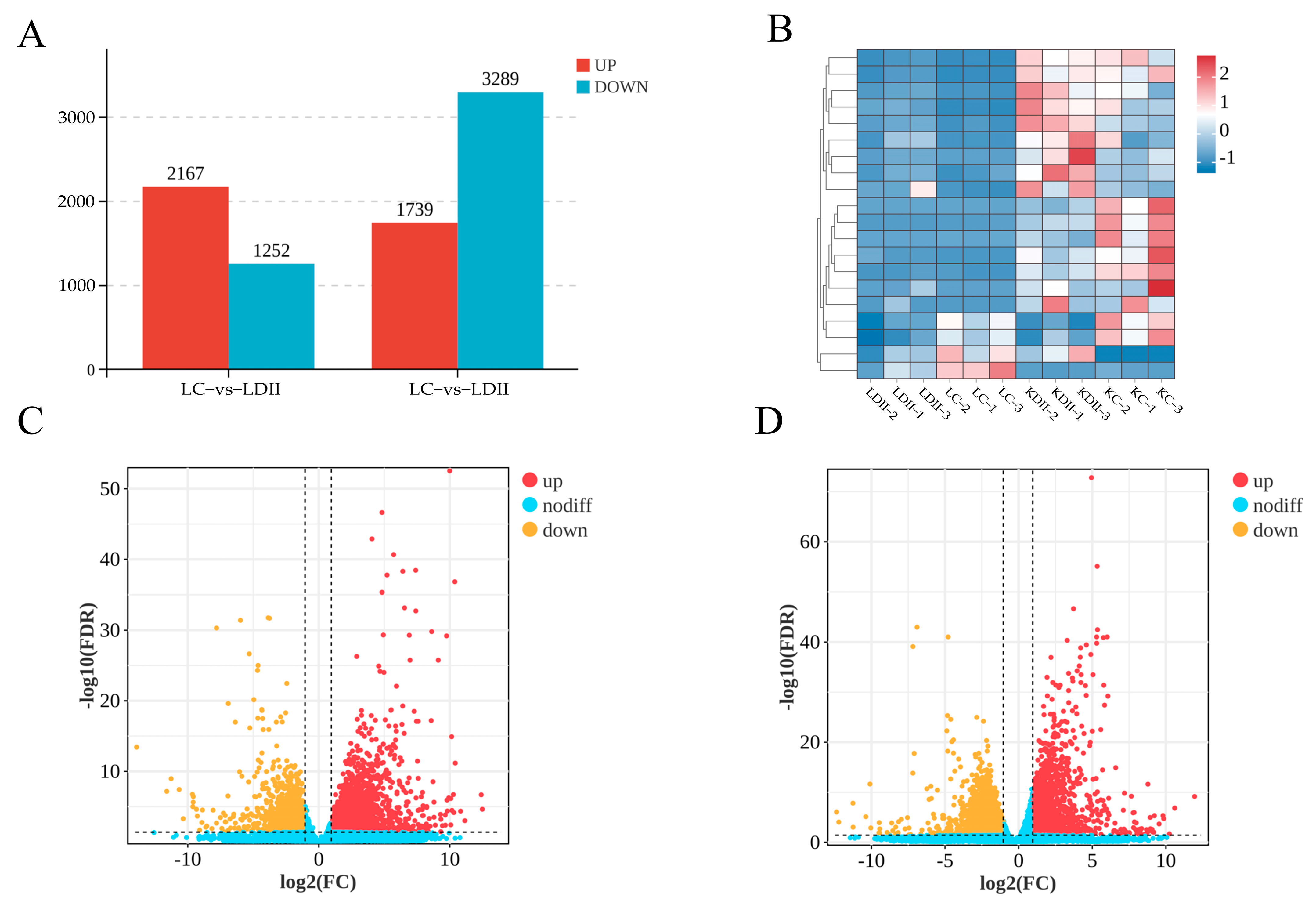
3.1. Analysis of Sample Relationships and Differentially Expressed Genes

To ensure the greater reliability of the results of differential gene analysis between samples, the correlation analysis of sample tissues was carried out. The PCA results demonstrated that the positions of three parallel sample points were similar within the groups, and there was an obvious grouping trend among the four distinct groups (Figure 1A). Additionally, the results of the correlation heat map indicated that the correlation coefficient of parallel samples in each group was greater than 0.99 (Figure 1B), suggesting that the experimental treatment had excellent repeatability, and there were significant differences in gene expression between kidney and liver tissues post-infection.
The DEGs in the kidney and liver tissues of the infected group and the control group were both analyzed, and genes with log2 > 2 and FDR < 0.05 were selected as DEGs. As depicted in Figure 2, compared with the control group, 5028 differential genes were identified in the kidney tissues after infection with Mycobacterium marinum, including 1739 upregulated genes and 3289 downregulated genes. A total of 3419 differential genes were identified in the liver tissue, including 2167 upregulated genes and 1252 downregulated genes.

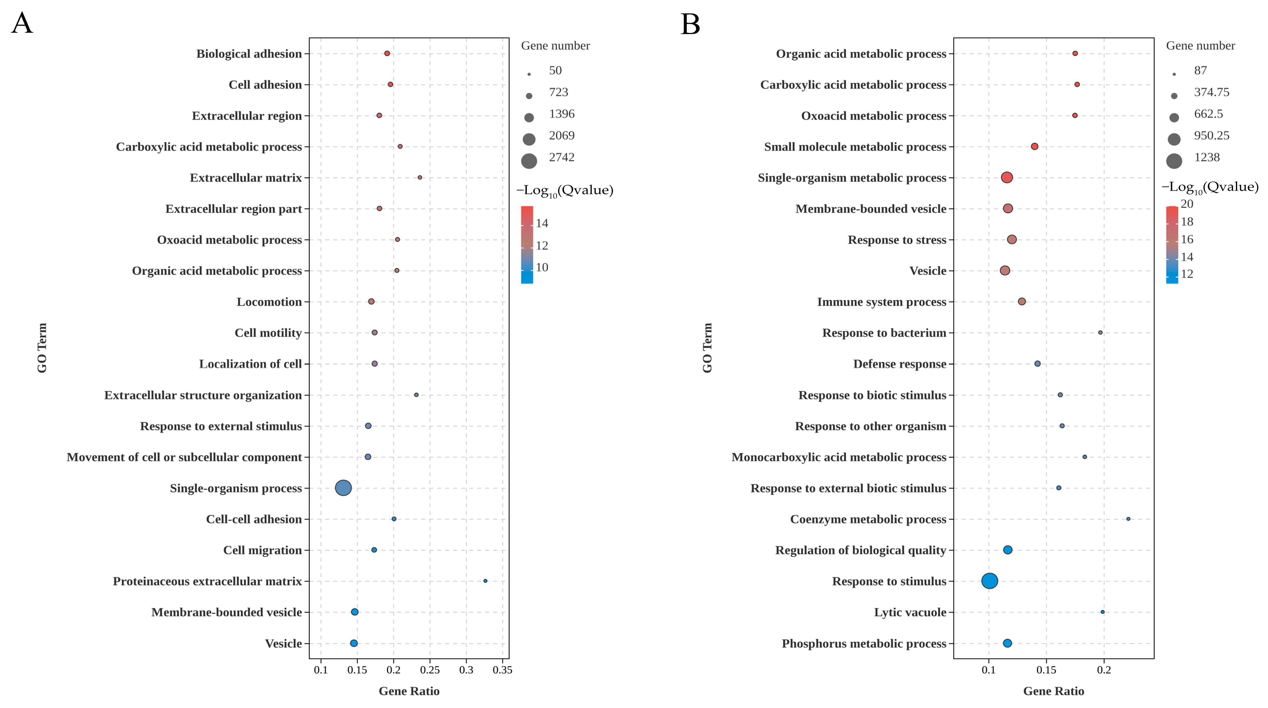
3.2. GO Enrichment of the DEGs in Kidney and Liver

The DEGs were annotated to Gene Ontology (GO) terms and subjected to enrichment analysis across three categories: Biological Process (BP), Molecular Function (MF), and Cellular Component (CC). The top 20 GO terms were ranked based on their FDR values. As seen in Figure 3A, the DEGs in the kidney were predominantly associated with processes such as bioadhesion, cell adhesion, carboxylic acid metabolism, oxoacid metabolism, organic acid metabolism, motility, cell motility, cell localization, extracellular structure organization, response to external stimuli, subcellular component motility, monobiotic processes, cell–cell adhesion, and cell migration. Enriched cellular components included the extracellular region, extracellular matrix, extracellular matrix structural components, proteinaceous extracellular matrix, membrane-bounded vesicles, and vesicles. As seen in Figure 3B, the DEGs in liver were mainly enriched in biological processes such as organic acid metabolism, carboxylic acid metabolism, oxoacid metabolism, small-molecule metabolic processes, single-organism metabolic processes, stress responses, immune system processes, bacterial response, defense response, response to biological stimuli, interactions with other organisms, monocarboxylic acid metabolism, response to external biological stimuli, coenzyme metabolism, and biological quality control. Enriched cellular components included membrane-bounded vesicles, vesicles, and lysosomal vacuoles.

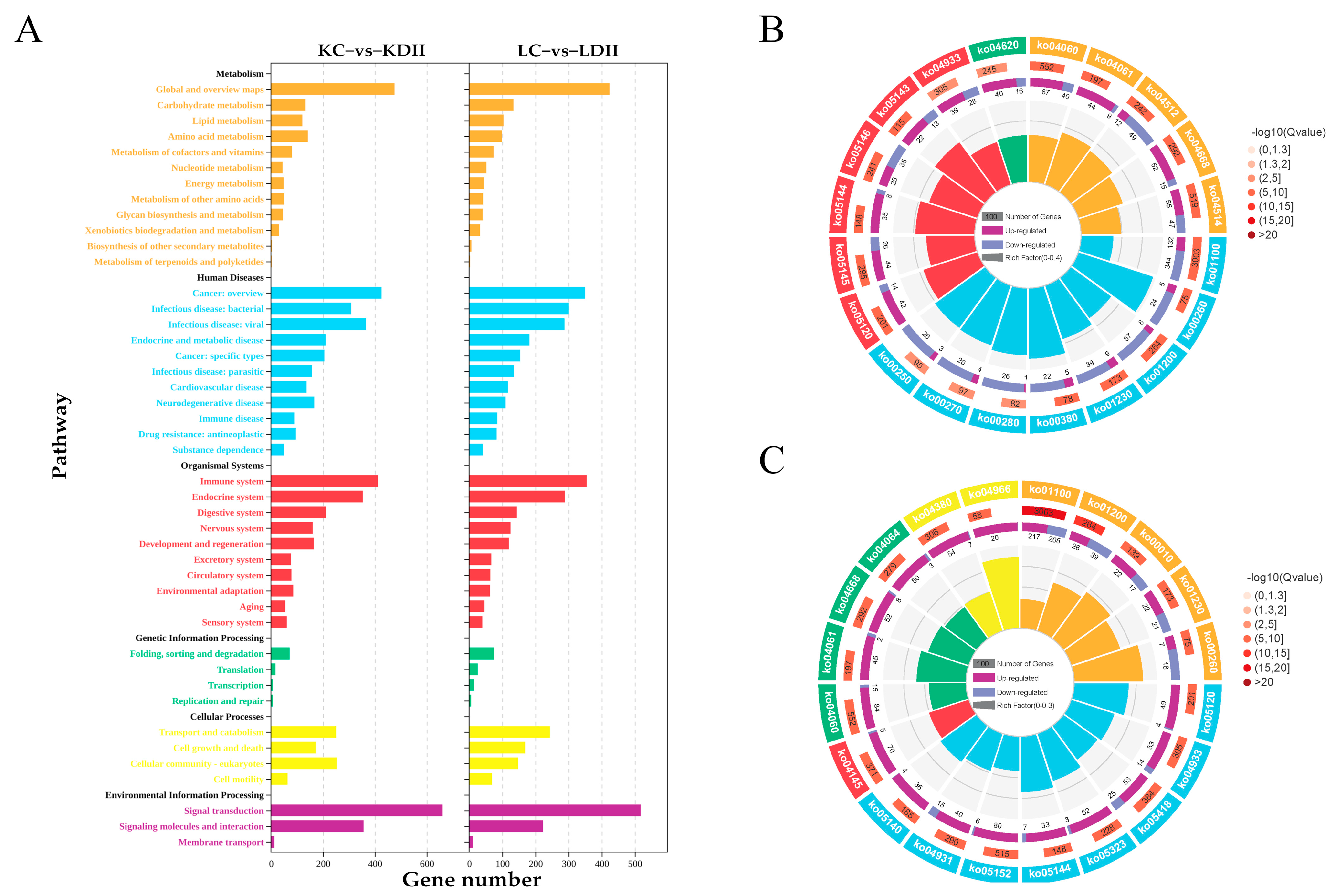
3.3. KEGG Enrichment of the DEGs in the Kidney and Liver

The KEGG enrichment analysis identified the top 20 signaling pathways based on FDR values. As shown in Figure 4A, in response to M. marinum infection, DEGs were classified into six major categories: metabolism, human diseases, organismal systems, cellular processes, genetic information processing, and environmental information processing. In kidney tissues (Figure 4B), the most significantly enriched pathways included cytokine–cytokine receptor interaction, metabolic pathways, epithelial cell signaling in Helicobacter pylori infection, amino metabolisms, viral protein–cytokine and cytokine receptor interaction, carbon metabolism, extracellular matrix (ECM)–receptor interaction, toxoplasmosis, amino acid biosynthesis, malaria, amebiasis, the TNF signaling pathway, cell adhesion molecules, Toll-like receptor signaling, and so on. In the liver (Figure 4C), the most enriched pathways included metabolic pathways, carbon metabolism, epithelial cell signaling in Helicobacter pylori infection, the role of AGE-RAGE signaling in diabetic complications, fluid shear stress and atherosclerosis, rheumatoid arthritis, phagosomes, cytokine–cytokine receptor interaction, glycolysis/gluconeogenesis, malaria, viral protein–cytokine and cytokine receptor interaction, amino acid biosynthesis, the TNF signaling pathway, osteoclast differentiation, glycine, serine, threonine metabolism, tuberculosis, bile acid secretion, insulin resistance, leishmaniasis, and the NF-κB signaling pathway.

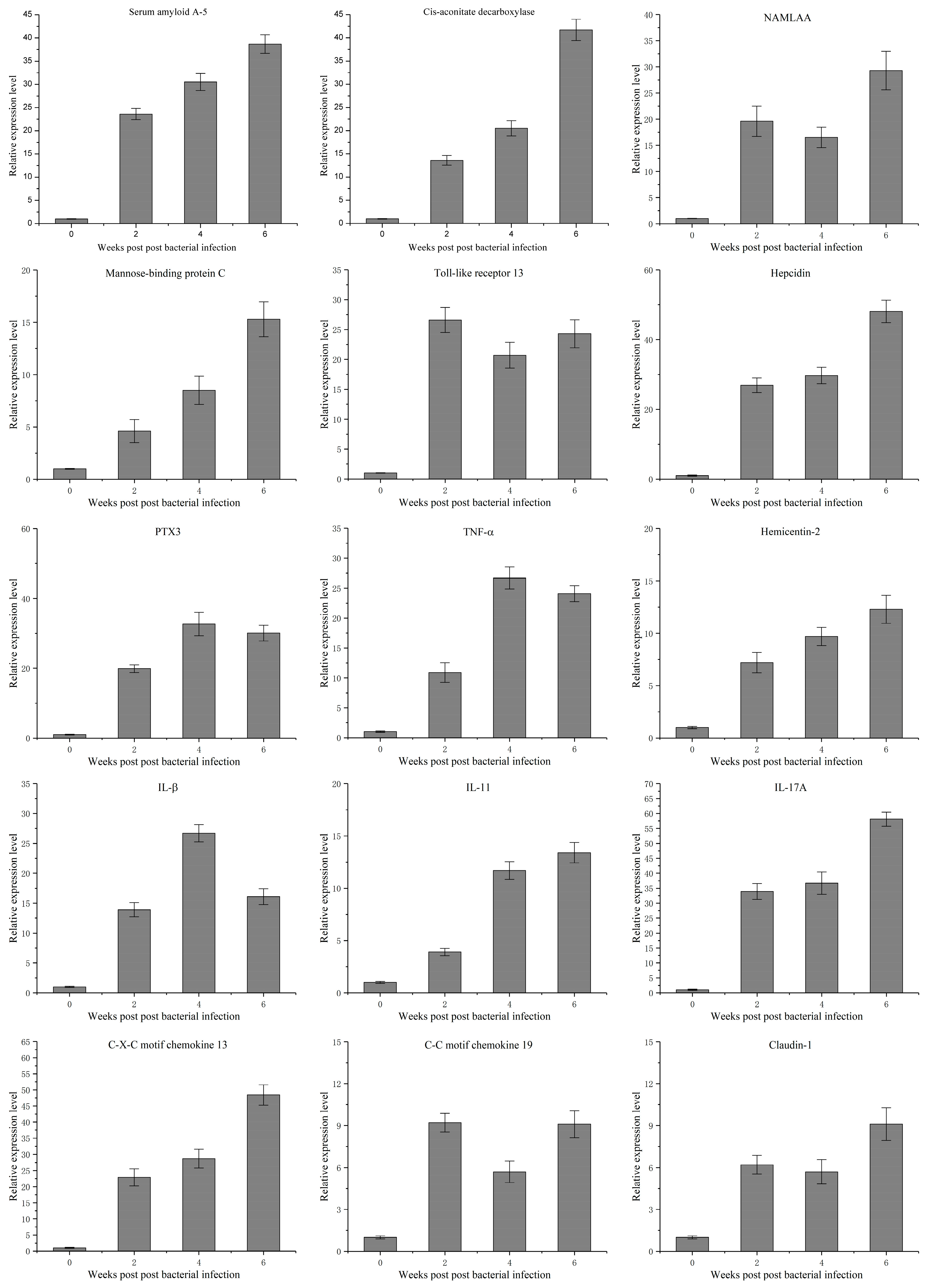
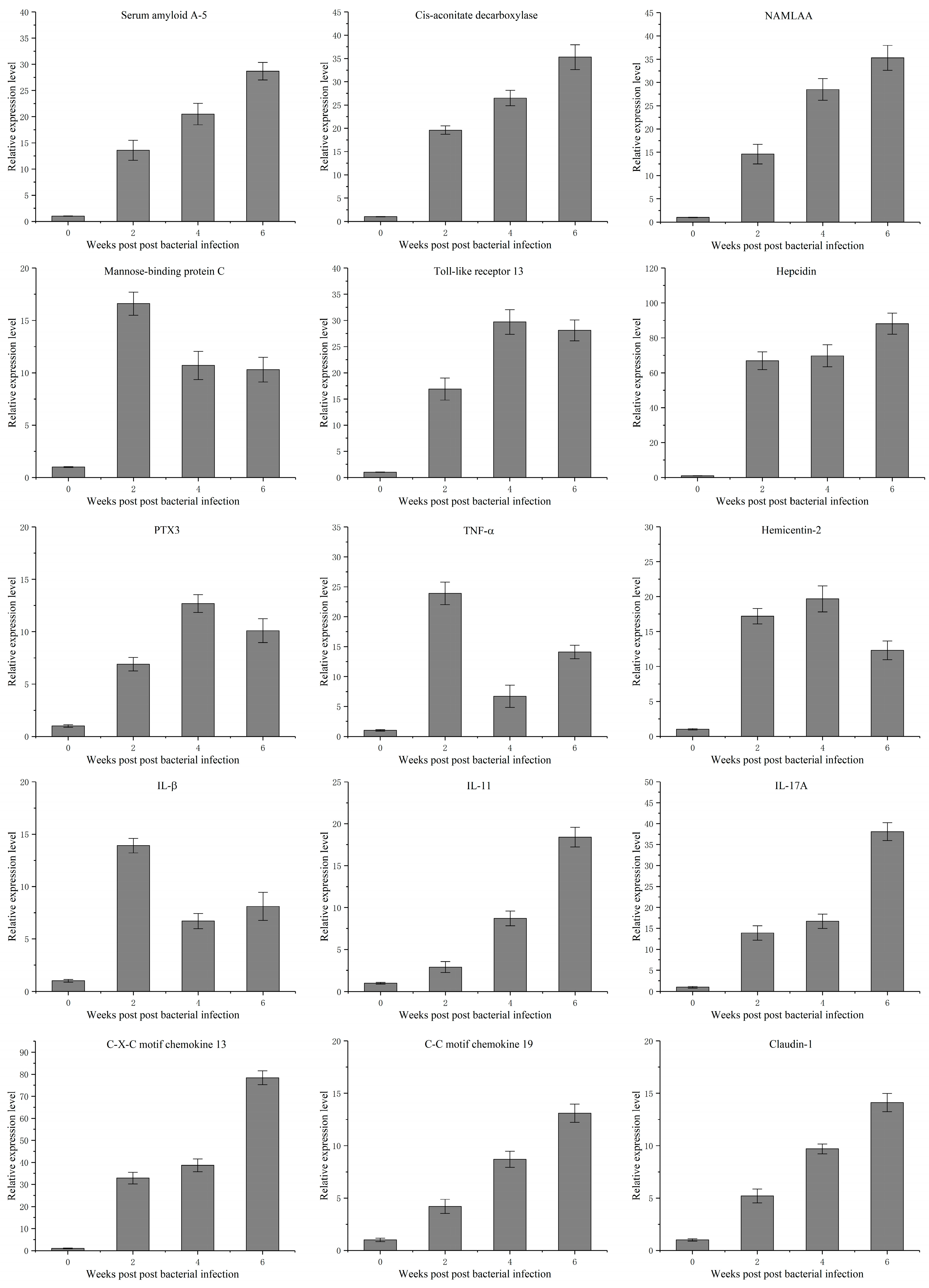
3.4. Temporal Expression Characteristics of 15 Genes Related to Mycobacterium marinum Infection

Based on the results of transcriptomic analysis, we selected 15 significantly differentially expressed genes closely related to mycobacterial infection and immune responses to further study their dynamic expression following Mycobacterium marinum infection across six weeks. The results demonstrated that the 15 selected genes involved in bacterial infection, including acute-phase proteins, pattern recognition receptors, inflammatory cytokines, and other immune-related genes, were all upregulated in the liver and kidney, which was generally consistent with the results obtained from the transcriptome analysis (Figure 5 and Figure 6). The expression levels of acute-phase proteins, including serum amyloid A-5 and hepcidin, exhibited quicker and stronger responses in the liver than in the kidney following infection. Innate immunity-related proteins, including mannose-binding protein C, Toll-like receptor 13, and NAMLAA, exhibited a significant increase and reached peak levels at 2 or 4 weeks post-infection, maintaining elevated levels for 6 weeks. Cytokines, including TNF-α, IL-1β, IL-11, and IL-17A, displayed dynamic regulation in response to M. marinum infection. In the liver, TNF-α and IL-1β showed significant upregulation at 2 weeks post-infection, peaking at 4 weeks. IL-17A expression progressively increased, reaching its highest level at 6 weeks post-infection. IL-11 displayed a modest upregulation at 4 weeks post-infection, with relatively minor changes overall. In the kidney, the expression trends of these cytokines were generally consistent with those in the liver but with reduced magnitudes. Two chemokines, C-X-C motif chemokine 13 and C-C motif chemokine 19, also exhibited a significant increase and peaked at 4 or 6 weeks post-infection, both in the liver and kidney. The expression of other immune-related proteins, including PTX3, Hemectin-2, and claudin-1, displayed diverse patterns following infection. Cis-aconitate decarboxylase, a metabolic enzyme, showed a gradual increase within 6 weeks after infection and reached the peak level at the sixth week.

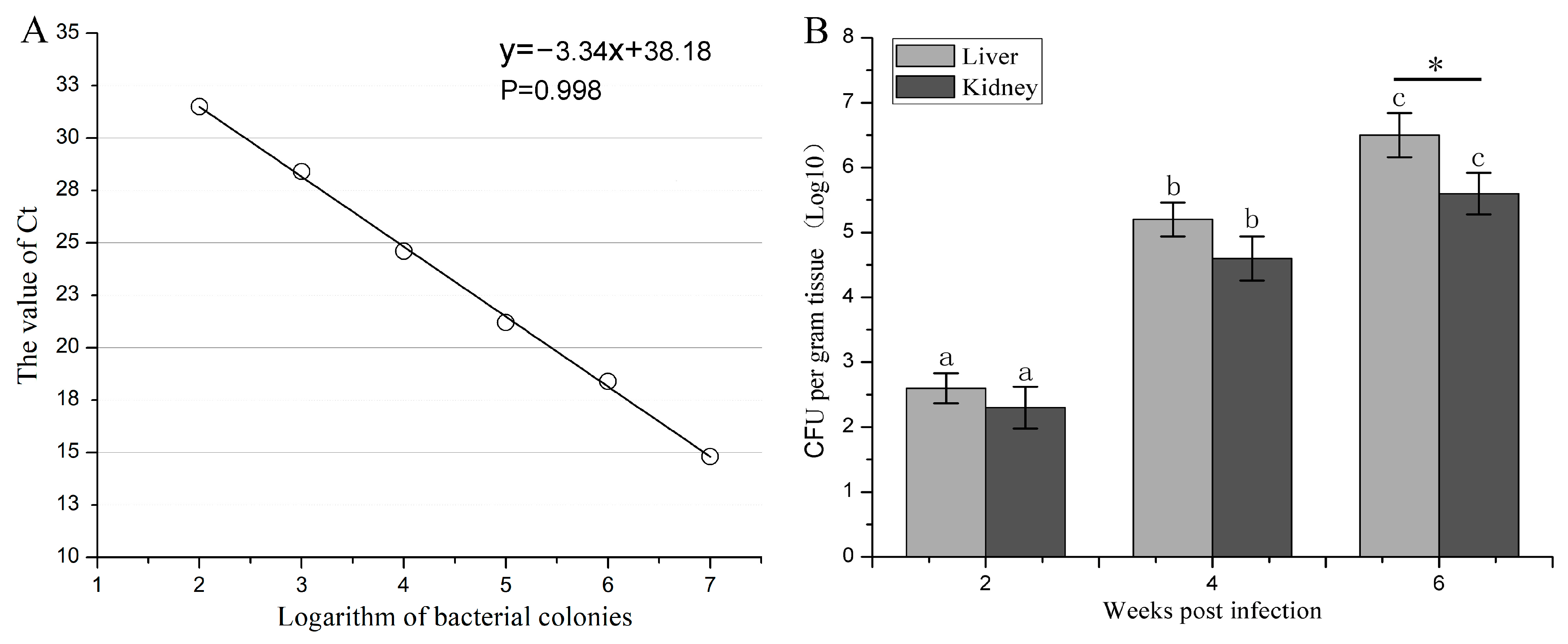
3.5. The Proliferation Dynamic of M. marinum in Liver and Kidney

A linear standard curve was successfully developed using quantitative PCR (qPCR) technology to enable the precise detection and quantification of M. marinum. The relationship between the logarithm of bacterial colonies and the cycle threshold (Ct) value was modeled by the linear equation y = −3.34x + 38.18. This curve exhibited a strong negative correlation, with a p-value of 0.998, confirming excellent linearity and reliability (Figure 7A). The bacterial load of M. marinum in the liver and kidney of Coho salmon was quantified at 2, 4, and 6 weeks post-infection based on the established standard curve, revealing a progressive increase in bacterial proliferation over time in both organs (Figure 1B). At 2 and 4 weeks post-infection, the bacterial loads were around 2.5 and 5 log10 CFU/g in the two tissues, respectively, and no significant difference was found between the liver and kidney. At week 6, the bacterial load peaked significantly, with the liver at approximately 6.5 log10 cfu/g, showing a significantly higher load compared to that in the kidney with 5.6 log10 cfu/g.

4. Discussion

Mycobacterium species are resilient bacteria found in aquatic environments that cause chronic infections and pose a zoonotic risk to aquaculture practitioners [22,23,24,25]. M. marinum threatens aquatic animals, leading to disease outbreaks, growth impairment, and mortality, especially in intensive farming, causing significant economic losses [26,27]. This study provided a comprehensive analysis of the immune response of Coho salmon to M. marinum infection, shedding light on the complex interplay between the host and pathogen. By employing transcriptome profiling and qPCR-based quantification, we also elucidated key aspects of the Coho salmon’s defense mechanisms and the pathogen’s proliferation dynamics in vivo. These findings have the potential to inform the development of future vaccines or immunotherapies for mycobacteriosis in fish.
This study investigated the transcriptional response of Coho salmon to the infection with M. marinum, focusing on gene expression changes in kidney and liver tissues. Principal component analysis (PCA) and correlation analysis demonstrated excellent reproducibility within experimental groups, validating the experimental design and sample consistency [28]. The substantial number of DEGs identified in both kidney and liver tissues highlights the profound impact of M. marinum infection on multiple physiological systems in Coho salmon. The predominance of upregulated genes in both tissues suggests a robust activation of immune responses, alongside significant alterations in metabolic processes and other physiological functions. For instance, the enrichment of Gene Ontology terms related to immune system processes, such as cytokine production and pathogen recognition, indicates a strong immune activation, while terms associated with carboxylic acid metabolism and energy metabolism reflect a metabolic reprogramming to support the host’s defense efforts. Similarly, pathways related to tissue repair and stress responses suggest a coordinated systemic response to mitigate infection-induced damage. These findings suggested the complex, multifaceted response of Coho salmon to M. marinum infection, involving an interplay of immune, metabolic, and physiological adaptations to counter the pathogen and maintain homeostasis. The disparity in the number of up- and downregulated genes between the tissues implies distinct roles for these organs in the overall immune response. The larger number of downregulated immune genes in the kidney suggests a potential pathogen-driven immunosuppression or regulatory response [29]. The significant differences observed in gene expression between kidney and liver tissues suggest the tissue-specific nature of the host response to M. marinum infection.
Gene Ontology enrichment analysis revealed distinct functional categories enriched in each tissue. In the kidney, GO terms related to cell adhesion, extracellular matrix (ECM), and carboxylic acid metabolism were significantly enriched. Cell adhesion and ECM are crucial for tissue repair and remodeling. During infection, damaged tissues need to be repaired, and the ECM often needs to be restructured to facilitate immune cell infiltration and tissue healing. The enrichment of these terms suggests that the organism is actively engaging in tissue repair mechanisms [30,31]. During infection, cells often need to adapt their metabolic processes to meet the increased energy demands of the immune response and to produce necessary molecules for fighting the infection. Therefore, carboxylic acid metabolism enrichment indicates a shift in cellular metabolism [32]. The enrichment of GO terms related to immune system processes, stress response, and metabolic processes in the liver suggests the liver’s critical function as both a metabolic and immune organ. It was documented that the liver plays a crucial role in the systemic immune response and energy metabolism during a pathogen’s infection [33]. The liver also plays an important role in immune functions and homeostasis [34]. These results collectively point to a coordinated, systemic response orchestrated by the liver to address the challenges posed by infection. KEGG pathway analysis further elucidated the complex interplay of signaling pathways involved in the response to M. marinum. The significant enrichment of pathways involved in cytokine–cytokine receptor interaction, various metabolic pathways, and Toll-like receptor signaling in both tissues confirms the activation of both innate and adaptive immune responses [35,36]. In the kidney, the enrichment of pathways related to cytokine–cytokine receptor interaction, metabolic processes, and Toll-like receptor signaling suggests a coordinated effort to activate immune cells, modulate metabolism to support the immune response, and recognize pathogen-associated molecular patterns [37,38]. On the other hand, the liver exhibited enrichment in immune system processes, stress responses, and NF-κB signaling, indicating a more pronounced inflammatory response and the activation of cellular stress pathways [39,40].
The temporal expression profiling of 15 selected immune-related genes provided valuable insights into the kinetics of the immune response. The upregulation of acute-phase proteins, cytokines, chemokines, and pattern recognition receptors confirms the activation of both innate and adaptive immune mechanisms. The quicker and stronger responses observed in the liver for acute-phase proteins suggest a more rapid mobilization of systemic defenses in this tissue. The upregulation of acute-phase proteins (APPs) such as serum amyloid A-5 and hepcidin, particularly prominent in the liver, highlights the pivotal role of APPs in the early stages of the host’s immune response. This early hepatic response aligns with the liver’s function as a primary site for APP production [41,42,43]. The quicker and stronger response of these proteins in the liver compared to the kidney suggests a coordinated, organ-specific prioritization of defense mechanisms, necessary for mitigating pathogen spread [44]. The significant increase in the expression of pattern recognition receptors, such as mannose-binding protein C and Toll-like receptor 13, reflects their integral role in pathogen recognition and the activation of downstream immune pathways. PRRs are the first line of defense in the innate immune system. Their upregulation suggests the active surveillance and detection of pathogens, initiating the immune response [45,46,47]. The sustained elevation of these receptors up to six weeks post-infection highlights their importance not just in immediate pathogen detection but also in prolonged defense, facilitating the recruitment and activation of other immune components, and maintaining a prolonged state of alertness against the pathogen. The dynamic regulation of cytokines, with TNF-α and IL-1β showing early upregulation and IL-17A increasing progressively, suggests a sequential activation of different inflammatory pathways. The timing of these peaks suggests an orchestrated effort to mediate inflammation and direct adaptive immune responses as seen in other aquatic species infected with mycobacteria [48]. Interestingly, IL-11 showed only modest upticks, which could indicate a more nuanced role, possibly in regulating excessive inflammation to prevent tissue damage, a balancing act essential during prolonged infections [49].
The upregulation of chemokines, notably C-X-C motif chemokine 13 and C-C motif chemokine 19, plays a crucial role in the host’s immune response against Mycobacterium marinum infection in fish. These chemotactic cytokines are instrumental in orchestrating leukocyte recruitment, a hallmark of the immune response to mycobacterial infections [50,51]. The observed peak expression of these chemokines at later stages of infection suggests a sustained immune response, potentially indicative of ongoing leukocyte recruitment necessary for pathogen containment and clearance. The varied expression patterns of proteins like PTX3 and claudin-1 further reflected the multifaceted nature of the host immune response. As a pattern recognition molecule, PTX3 plays a crucial role in the identification and opsonization of pathogens, facilitating their clearance by phagocytes [52]. In fish, PTX3 has also been shown to be involved in the acute-phase response and possesses antimicrobial properties [53]. The increased expression of PTX3 in M. marinum infection suggests its involvement in pathogen recognition and the orchestration of innate immune responses, potentially contributing to the containment of mycobacterial spread. The upregulation of claudin-1, a tight junction protein, suggests significant alterations in epithelial barriers as part of the host defense strategy. Claudins play a crucial role in maintaining the integrity of epithelial and endothelial barriers, which are critical first lines of defense against pathogen invasion [54]. In fish, the modulation of claudin expression has been observed in response to various pathogens [55,56]. The increased expression of claudin in M. marinum infection may represent an attempt to enhance barrier function, potentially limiting mycobacterial dissemination and protecting underlying tissues.
The qPCR-based quantification of M. marinum loads revealed a progressive proliferation of the pathogen in both the liver and kidney over time. This finding is consistent with previous studies investigating mycobacterial infections in other fish species, such as zebrafish (Danio rerio), where M. marinum infection typically shows a gradual increase in bacterial burden over time [57]. Interestingly, the bacterial loads in the liver and kidney were similar at 2 and 4 weeks post-infection, but diverged significantly by week 6, with a higher bacterial load in the liver. This tissue-specific difference contrasts with some studies in other fish species. For instance, in goldfish (Carassius auratus), M. marinum infection often results in higher bacterial loads in the kidney compared to the liver [58]. The higher liver burden in Coho salmon at later stages of infection could indicate a species-specific tropism of M. marinum or differences in tissue-specific immune responses. Several factors may contribute to the higher bacterial load observed in the liver of Coho salmon at 6 weeks post-infection. Firstly, the rich vascularization and extensive blood supply in the liver might provide an ideal microenvironment for M. marinum proliferation, as the bacterium is known to infect macrophages and persist intracellularly [59]. Secondly, the route of infection, likely intraperitoneal injection in this study, might also facilitate the bacterial dissemination to the liver. In addition, differences in experimental conditions, such as bacterial strain and infection dose, could also influence tissue tropism. The difference between Coho salmon and other fish species also highlights the need for species-specific research in aquaculture. While zebrafish and goldfish provide valuable models for studying mycobacterial infections, their tissue tropism patterns may not fully represent those in commercially important salmonids. These findings highlight the importance of tailored diagnostic and therapeutic approaches based on the unique physiological and immunological characteristics of Coho salmon.

5. Conclusions

In summary, this study provides a comprehensive characterization of the immune response of Coho salmon to Mycobacterium marinum infection. By integrating transcriptome profiling and qPCR-based quantification, we have identified key immune genes and pathways, elucidated the tissue-specific dynamics of the immune response, and revealed the pathogen’s proliferation dynamics in vivo. These findings contribute to our understanding of the host–pathogen interaction and provide a foundation for developing targeted disease control strategies against mycobacterial infections.

Author Contributions

Conceptualization, J.D.; methodology, L.L., D.X., and C.G.; software, X.Y.; writing—original draft preparation, L.L.; writing—review and editing, J.D.; funding acquisition, J.D. and H.Y. All authors have read and agreed to the published version of the manuscript.

Funding

This study was supported by Shandong Provincial Natural Science Foundation (ZR2021MC089); Marine Science and Technology Innovation Demonstration Project of Qingdao (23-1-3-hysf-2-hy); Modern Agriculture Fish Industry Technology System of Shandong Province (SDAIT-12-01).

Institutional Review Board Statement

This work followed a standard working methodology approved by the Ethics Committee of marine science research institute of Shandong province (Protocol No. HKYLLSC2025009) on 23 April 2025.

Data Availability Statement

Data are available upon reasonable request.

Conflicts of Interest

The authors declare no conflicts of interest.

References

  1. Canetti, D.; Riccardi, N.; Antonello, R.M.; Di Bella, S.; Farinazzo, A.; Matteelli, A.; Codecasa, L.R.; Tacconelli, E.; Alagna, R. Mycobacterium marinum: A brief update for clinical purposes. Eur. J. Intern. Med. 2022, 105, 15–19. [Google Scholar] [CrossRef] [PubMed]
  2. Johnson, M.G.; Stout, J.E. Twenty-eight cases of Mycobacterium marinum infection: Retrospective case series and literature review. Infection 2015, 43, 655–662. [Google Scholar] [CrossRef] [PubMed]
  3. Aubry, A.; Chosidow, O.; Caumes, E.; Robert, J.; Cambau, E. Sixty-three cases of Mycobacterium marinum infection: Clinical features, treatment, and antibiotic susceptibility of causative isolates. Arch. Intern. Med. 2002, 162, 1746–1752. [Google Scholar] [CrossRef] [PubMed]
  4. Wu, T.S.; Chiu, C.H.; Yang, C.H.; Leu, H.S.; Huang, C.T.; Chen, Y.C.; Wu, T.L.; Chang, P.Y.; Su, L.H.; Kuo, A.J.; et al. Fish tank granuloma caused by Mycobacterium marinum. PLoS ONE 2012, 7, e41296. [Google Scholar] [CrossRef]
  5. Cosma, C.L.; Sherman, D.R.; Ramakrishnan, L. The secret lives of the pathogenic mycobacteria. Annu. Rev. Microbiol. 2003, 57, 641–676. [Google Scholar] [CrossRef]
  6. Hashish, E.; Merwad, A.; Elgaml, S.; Amer, A.; Kamal, H.; Elsadek, A.; Marei, A.; Sitohy, M. Mycobacterium marinum infection in fish and man: Epidemiology, pathophysiology and management; a review. Vet. Quart. 2018, 38, 35–46. [Google Scholar] [CrossRef]
  7. Decostere, A.; Hermans, K.; Haesebrouck, F. Piscine mycobacteriosis: A literature review covering the agent and the disease it causes in fish and humans. Vet. Microbiol. 2004, 99, 159–166. [Google Scholar] [CrossRef]
  8. Ramsay, J.M.; Watral, V.; Schreck, C.B.; Kent, M.L. Husbandry stress exacerbates mycobacterial infections in adult zebrafish, Danio rerio (Hamilton). J. Fish Dis. 2009, 32, 931–941. [Google Scholar] [CrossRef]
  9. Chen, W.; Gao, S. Current status of industrialized aquaculture in China: A review. Environ. Sci. Pollut. Res. 2023, 30, 32278–32287. [Google Scholar] [CrossRef]
  10. Duan, Z.; Zhou, Y.; Liu, W.; Zhang, J.; Ren, H.; Li, H.; Zhu, X.; Zhao, Y.; Chen, P.; Li, C. Variations in flavor according to fish size in rainbow trout (Oncorhynchus mykiss). Aquaculture 2020, 526, 735398. [Google Scholar] [CrossRef]
  11. Noble, A.C.; Summerfelt, S.T. Diseases encountered in rainbow trout cultured in recirculating systems. Annu. Rev. Fish Dis. 1996, 6, 65–92. [Google Scholar] [CrossRef]
  12. Rurangwa, E.; Verdegem, M.C.J. Microorganisms in recirculating aquaculture systems and their management. Rev. Aquac. 2015, 7, 117–130. [Google Scholar] [CrossRef]
  13. Aich, N.; Nama, S.; Biswal, A.; Paul, T.; Biswas, P. A review on recirculating aquaculture systems: Challenges and opportunities for sustainable aquaculture. Innov. Farm. 2020, 5, 17–24. [Google Scholar]
  14. Ford, J.S.; Myers, R.A. A global assessment of salmon aquaculture impacts on wild salmonids. PLoS Biol. 2008, 6, e33. [Google Scholar] [CrossRef]
  15. Zhu, L.; Wang, X.; Wang, K.; Su, F.; Chen, J.; Shi, S.; Zhao, L.; Yu, X.; Zeng, L. Outbreak of infectious pancreatic necrosis virus (IPNV) in farmed rainbow trout in China. Acta Trop. 2017, 170, 63–69. [Google Scholar] [CrossRef]
  16. Jia, P.; Breyta, R.B.; Li, Q.; Qian, X.; Wu, B.; Zheng, W.; Wen, Z.; Liu, Y.; Kurath, G.; Liu, H.; et al. Insight into infectious hematopoietic necrosis virus (IHNV) in Chinese rainbow trout aquaculture from virus isolated from 7 provinces in 2010–2014. Aquaculture 2018, 496, 239–246. [Google Scholar] [CrossRef]
  17. Huang, J.; Zhang, S.H.; Di, J.; Dong, X.; Wang, Y.; Yao, Y.; Liu, X.; Liu, Y.; Qin, Q.; Wang, X.; et al. Inactivated Mycobacterium vaccine induces innate immunity against M. ulcerans ecovar Liflandii (MuLiflandii ASM001) infection in Dabry’s sturgeon (Acipenser dabryanus). Aquaculture 2019, 512, 734312. [Google Scholar] [CrossRef]
  18. Conesa, A.; Götz, S.; García-Gómez, J.M.; Terol, J.; Talón, M.; Robles, M. Blast2GO: A universal tool for annotation, visualization and analysis in functional genomics research. Bioinformatics 2005, 21, 3674–3676. [Google Scholar] [CrossRef]
  19. Ye, J.; Fang, L.; Zheng, H.; Zhang, Y.; Chen, J.; Zhang, Z.; Wang, J.; Li, S.; Li, R.; Bolund, L.; et al. WEGO: A web tool for plotting GO annotations. Nucleic Acids Res. 2006, 34, W293–W297. [Google Scholar] [CrossRef]
  20. Zhang, Q.; Guo, M.; Li, F.; Qin, M.; Yang, Q.; Yu, H.; Xu, J.; Liu, Y.; Tong, T. Evaluation of fermented soybean meal to replace a portion fish meal on growth performance, antioxidant capacity, immunity, and mTOR signaling pathway of Coho Salmon (Oncorhynchus kisutch). Aquac. Nutr. 2023, 2558173. [Google Scholar] [CrossRef]
  21. Luukinen, H.; Hammarén, M.M.; Vanha-Aho, L.M.; Parikka, M. Modeling tuberculosis in Mycobacterium marinum infected adult zebrafish. J. Vis. Exp. 2018, 140, e58299. [Google Scholar]
  22. Slany, M.; Makovcova, J.; Jezek, P.; Bodnarova, M.; Pavlik, I. Relative prevalence of Mycobacterium marinum in fish collected from aquaria and natural freshwaters in central Europe. J. Fish Dis. 2014, 37, 527–533. [Google Scholar] [CrossRef] [PubMed]
  23. Gauthier, D.T.; Rhodes, M.W. Mycobacteriosis in fishes: A review. Vet. J. 2009, 180, 33–47. [Google Scholar] [CrossRef] [PubMed]
  24. Dinh-Hung, N.; Dong, H.T.; Senapin, S.; Pimsannil, K.; Thompson, K.D.; Shinn, A.P.; Soontara, C.; Sirimanapong, W.; Chatchaiphan, S.; Rodkhum, C. Insight into characteristics and pathogenicity of five rapidly growing non-tuberculous Mycobacterium species isolated from the Siamese fighting fish, Betta splendens. Aquaculture 2023, 575, 739822. [Google Scholar] [CrossRef]
  25. Dinh-Hung, N.; Dong, H.T.; Senapin, S.; Linh, N.V.; Shinn, A.P.; Pirarat, N.; Hirono, I.; Chatchaiphan, S.; Rodkhum, C. Infection and histopathological consequences in Siamese fighting fish (Betta splendens) due to exposure to a pathogenic Mycobacterium chelonae via different routes. Aquaculture 2024, 579, 740191. [Google Scholar] [CrossRef]
  26. Davidovich, N.; Pretto, T.; Sharon, G.; Zilberg, D.; Blum, S.E.; Baider, Z.; Edery, N.; Klement, E.; Kaidar-Shwartz, I.; Dveyrin, Z.; et al. Cutaneous appearance of mycobacteriosis caused by Mycobacterium marinum, affecting gilthead seabream (Sparus aurata) cultured in recirculating aquaculture systems. Aquaculture 2020, 528, 735507. [Google Scholar] [CrossRef]
  27. Yanong, R.P.E.; Pouder, D.B.; Falkinham, J.O., III. Association of mycobacteria in recirculating aquaculture systems and mycobacterial disease in fish. J. Aquat. Anim. Health 2010, 22, 219–223. [Google Scholar] [CrossRef]
  28. Chen, X.; Zhang, B.; Wang, T.; Bonni, A.; Zhao, G. Robust principal component analysis for accurate outlier sample detection in RNA-Seq data. BMC Bioinform. 2020, 21, 269. [Google Scholar] [CrossRef]
  29. Eslamloo, K.; Caballero-Solares, A.; Inkpen, S.M.; Emam, M.; Kumar, S.; Bouniot, C.; Avendaño-Herrera, R.; Jakob, E.; Rise, M.L. Transcriptomic profiling of the adaptive and innate immune responses of Atlantic salmon to Renibacterium salmoninarum infection. Front. Immunol. 2020, 11, 567838. [Google Scholar] [CrossRef]
  30. Bonnans, C.; Chou, J.; Werb, Z. Remodelling the extracellular matrix in development and disease. Nat. Rev. Mol. Cell Biol. 2014, 15, 786–801. [Google Scholar] [CrossRef]
  31. Parsons, J.T.; Horwitz, A.R.; Schwartz, M.A. Cell adhesion: Integrating cytoskeletal dynamics and cellular tension. Nat. Rev. Mol. Cell Biol. 2010, 11, 633–643. [Google Scholar] [CrossRef] [PubMed]
  32. Kelly, B.; O’Neill, L.A. Metabolic reprogramming in macrophages and dendritic cells in innate immunity. Cell Res. 2015, 25, 771–784. [Google Scholar] [CrossRef] [PubMed]
  33. Xu, L.; Wang, Y.; Niu, W.; Wang, X.; He, H.; Zeng, Q.; Lin, Y.; Jia, Y.; Zhou, J. Liver transcriptome analysis reveals important factors involved in the response to Newcastle disease virus infection. Funct. Integr. Genom. 2015, 15, 671–682. [Google Scholar]
  34. Robinson, M.W.; Harmon, C.; O’Farrelly, C. Liver immunology and its role in inflammation and homeostasis. Cell. Mol. Immunol. 2016, 13, 267–276. [Google Scholar] [CrossRef]
  35. Zhu, L.Y.; Nie, L.; Zhu, G.; Xiang, L.X.; Shao, J.Z. Advances in research of fish immune-relevant genes: A comparative overview of innate and adaptive immunity in teleosts. Dev. Comp. Immunol. 2013, 39, 39–62. [Google Scholar] [CrossRef]
  36. Tort, L.; Balasch, J.C.; Mackenzie, S. Fish immune system. A crossroads between innate and adaptive responses. Immunologia 2003, 22, 277–286. [Google Scholar]
  37. Mokhtar, D.M.; Zaccone, G.; Alesci, A.; Cavestro, T.; Marino, F. Main components of fish immunity: An overview of the fish immune system. Fishes 2023, 8, 93. [Google Scholar] [CrossRef]
  38. Magnadóttir, B. Innate immunity of fish (overview). Fish Shellfish Immunol. 2006, 20, 137–151. [Google Scholar] [CrossRef]
  39. Zou, J.; Secombes, C.J. The function of fish cytokines. Biology 2016, 5, 23. [Google Scholar] [CrossRef]
  40. Doherty, D.G. Immunity, tolerance and autoimmunity in the liver: A comprehensive review. J. Autoimmun. 2016, 66, 60–75. [Google Scholar] [CrossRef]
  41. Bayne, C.J.; Gerwick, L. The acute phase response and innate immunity of fish. Dev. Comp. Immunol. 2001, 25, 725–743. [Google Scholar] [CrossRef] [PubMed]
  42. Rodrigues, P.N.; Vázquez-Dorado, S.; Neves, J.V.; Wilson, J.M. Dual function of fish hepcidin: Response to experimental iron overload and bacterial infection in sea bass (Dicentrarchus labrax). Dev. Comp. Immunol. 2006, 30, 1156–1167. [Google Scholar] [CrossRef] [PubMed]
  43. Villarroel, F.; Bastías, A.; Casado, A.; Amthauer, R.; Concha, M.I. Serum amyloid A: A typical acute-phase reactant in rainbow trout? Dev. Comp. Immunol. 2008, 32, 1160–1169. [Google Scholar] [CrossRef] [PubMed]
  44. Cray, C.; Zaias, J.; Altman, N.H. Acute phase response in animals: A review. Comp. Med. 2009, 59, 517–526. [Google Scholar]
  45. Dang, Y.; Meng, X.; Wang, S.; Li, H.; Zhang, X.; Xu, X.; Xiong, Y.; Wang, X. Mannose-binding lectin and its roles in immune responses in grass carp (Ctenopharyngodon idella) against Aeromonas hydrophila. Fish Shellfish Immunol. 2018, 72, 367–376. [Google Scholar] [CrossRef] [PubMed]
  46. Bergman, I.M. Toll-like receptors (TLRs) and mannan-binding lectin (MBL): On constant alert in a hostile environment. Ups. J. Med. Sci. 2011, 116, 90–99. [Google Scholar] [CrossRef]
  47. Boltaña, S.; Reyes-Lopez, F.; Morera, D.; Goetz, F.; MacKenzie, S.A. Divergent responses to peptidoglycans derived from different E. coli serotypes influence inflammatory outcome in trout, Oncorhynchus mykiss, macrophages. BMC Genom. 2011, 12, 34. [Google Scholar] [CrossRef]
  48. Rajme-Manzur, D.; Gollas-Galván, T.; Vargas-Albores, F.; Martínez-Porchas, M.; Hernández-Oñate, M.Á.; Hernández-López, J. Granulomatous bacterial diseases in fish: An overview of the host’s immune response. Comp. Biochem. Physiol. Part A Mol. Integr. Physiol. 2021, 261, 111058. [Google Scholar] [CrossRef] [PubMed]
  49. Huising, M.O.; Kruiswijk, C.P.; van Schijndel, J.E.; Savelkoul, H.F.; Flik, G.; Verburg-van Kemenade, B.M. Multiple and highly divergent IL-11 genes in teleost fish. Immunogenetics 2005, 57, 432–443. [Google Scholar] [CrossRef]
  50. Cronan, M.R.; Tobin, D.M. Fit for consumption: Zebrafish as a model for tuberculosis. Dis. Model. Mech. 2014, 7, 777–784. [Google Scholar] [CrossRef]
  51. Torraca, V.; Masud, S.; Spaink, H.P.; Meijer, A.H. Macrophage-pathogen interactions in infectious diseases: New therapeutic insights from the zebrafish host model. Dis. Model. Mech. 2014, 7, 785–797. [Google Scholar] [CrossRef] [PubMed]
  52. Bottazzi, B.; Doni, A.; Garlanda, C.; Mantovani, A. An integrated view of humoral innate immunity: Pentraxins as a paradigm. Annu. Rev. Immunol. 2009, 28, 157–183. [Google Scholar] [CrossRef] [PubMed]
  53. Lund, V.; Olafsen, J.A. A comparative study of pentraxin-like proteins in different fish species. Dev. Comp. Immunol. 1998, 22, 185–194. [Google Scholar] [CrossRef]
  54. Günzel, D.; Yu, A.S. Claudins and the modulation of tight junction permeability. Physiol. Rev. 2013, 93, 525–569. [Google Scholar] [CrossRef]
  55. Sun, L.; Liu, S.; Bao, L.; Li, Y.; Feng, J.; Liu, Z. Claudin multigene family in channel catfish and their expression profiles in response to bacterial infection and hypoxia as revealed by meta-analysis of RNA-Seq datasets. Comp. Biochem. Physiol. Part D Genom. Proteom. 2015, 13, 60–69. [Google Scholar] [CrossRef]
  56. Deng, F.; Wang, D.; Chen, F.; Lu, T.; Li, S. Molecular characterization and expression analysis of claudin-4-like in rainbow trout involved in Flavobacterium psychrophilum infection. Fish Shellfish Immunol. 2022, 130, 244–251. [Google Scholar] [CrossRef] [PubMed]
  57. Swaim, L.E.; Connolly, L.E.; Volkman, H.E.; Humbert, O.; Born, D.E.; Ramakrishnan, L. Mycobacterium marinum infection of adult zebrafish causes caseating granulomatous tuberculosis and is moderated by adaptive immunity. Infec. Immun. 2006, 74, 6108–6117. [Google Scholar] [CrossRef]
  58. Talaat, A.M.; Reimschuessel, R.; Wasserman, S.S.; Trucksis, M. Goldfish, Carassius auratus, a novel animal model for the study of Mycobacterium marinum pathogenesis. Infec. Immun. 1998, 66, 2938–2942. [Google Scholar] [CrossRef]
  59. Flynn, J.L.; Chan, J. Tuberculosis: Latency and reactivation. Infec. Immun. 2001, 69, 4195–4201. [Google Scholar] [CrossRef]
Figure 1. The analysis results of principal components and the correlation between samples of Coho salmon infected with M. marinum. (A) Inter-sample principal component analysis diagram. (B) Heat map of correlation between samples. KC: control group of kidney; LC: control group of liver; KDII: kidney from infected fish; LDII: liver from infected fish.
Fishes 10 00268 g001
Figure 2. Statistical results of differentially expressed genes in the Coho salmon model infected by M. marinum. (A) Histogram of differentially expressed genes before and after infection. (B) Heat maps of differentially expressed genes before and after infection with M. marinum. (C) Volcano map of differentially expressed genes in kidney tissue. (D) Volcano map of differentially expressed genes in liver tissue. KC: control group of kidney; LC: control group of liver; KDII: kidney from infected fish; LDII: liver from infected fish.
Fishes 10 00268 g002
Figure 3. GO enrichment analysis of differentially expressed genes (DEGs) in Coho salmon infected by M. marinum. (A) The top 20 GO terms from the enrichment analysis of DEGs in kidney samples of Coho salmon infected with M. marinum. (B) The top 20 GO terms from the enrichment analysis of DEGs in liver samples of Coho salmon infected with M. marinum.
Fishes 10 00268 g003
Figure 4. KEGG enrichment analysis of differentially expressed genes (DEGs) in Coho salmon Infected by M. marinum. (A) Classification and statistical overview of DEGs in Coho salmon infected with M. marinum. (B) Top 20 KEGG-enriched pathways in the kidney of infected Coho salmon. (C) Top 20 KEGG-enriched pathways in the liver of infected Coho salmon.
Fishes 10 00268 g004
Figure 5. The time-course expression profiles of 15 genes related to Mycobacterium marinum infection in the kidney of Coho salmon. Data were expressed as mean ± SD (n = 3).
Fishes 10 00268 g005
Figure 6. The time-course expression profiles of 15 genes related to Mycobacterium marinum infection in the liver of Coho salmon. Data were expressed as mean ± SD (n = 3).
Fishes 10 00268 g006
Figure 7. The changes in mycobacterial loads in the liver and kidney post-Mycobacterium marinum infection by quantitative qPCR. (A) The standard curves constructed for the quantification of M. marinum by the SYBR Green-based qPCR method; three replicates were used for each concentration sample. (B) The mycobacterial loads in the liver and kidney post-infection. The different letters above the columns indicate a significant difference among different time points, and the asterisk (*) indicates a significant difference between the kidney and liver, p < 0.05.
Fishes 10 00268 g007
Table 1. Primers used for the qPCR analysis of selected genes.
Gene NameSequences (5′-3′)Primer Source (Genbank No.)
Serum amyloid A-5 F:GCGACATGAAGGATGCCAACXM_020478634
R:TCCTCATGTCCTCGACCACT
Cis-aconitate decarboxylaseF:CTCCAACGAGGCTCAGAATATC XM_031789418
R: TGAGAGCGTTCCAGAGAGA
N-acetylmuramoyl-L-alanine amidaseF:CTGTGGTGAGGGAAGGAATAAAXM_020486665
R:CACTGACACCGTGGGATAATAG
Mannose-binding protein CF:CTTTGACGATGCTGTTCAGTTCXM_020469364
R:CCTTCAAAGACCTGAGTGAGAG
Toll-like receptor 13F:AGGGACTGTGTCATCTACCTATCXM_031792536
R:GTTTGGGTGCAGGTCCTTAATA
HepcidinF:TTCAGGTTCAAGCGTCAGAGXM_031790947
R:CAGGCAGGTCCTCAGAATTT
Pentraxin-3F:GGACACAGGCTATCCAACTTATCXM_020471418
R:CATCTCTGCTGATGCCACTT
Tumor necrosis factor αF:GGCGAGCATACCACTCCTCTZhang et al., 2023 [20]
R:TCGGACTCAGCATCACCGTA
Hemicentin-2F:GTCAGTCCGTCTGGTGAAATAGXM_020469630
R:CCCGTCCGTTCTTCATTCTT
Interleukin-1βF:GCGACATGGTGCGTTTCCTTTTZhang et al., 2023 [20]
R:TGTCTACCGGTTTGGTGTAGTCCT
Interleukin-17AF:GGTCTTTGGACGGAGAGTAATGXM_020477819
R:GAGAGACCTGGTGGCTTTATG
C-X-C motif chemokine 13F:ACACTACTTGTTGGTCGTACTGXM_020477664
R:GAAACCTGGTGGGAGAGATAAA
C-C motif chemokine 19F:CCACAGAGTGGTGAAGTCATACXM_020458446
R:GCACACAGTCTAAGGTTCTTCT
Claudin-1F:CTTGCTTTGATCGGTCTTGTTGXM_020480182
R:GTTACCGTGCTCTCTGTGTAAG
β-actinF:CCAAAGCCAACAGGGAGAAZhang et al., 2023 [20]
R:AGGGACAACACCGCCTGGAT
Disclaimer/Publisher’s Note: The statements, opinions and data contained in all publications are solely those of the individual author(s) and contributor(s) and not of MDPI and/or the editor(s). MDPI and/or the editor(s) disclaim responsibility for any injury to people or property resulting from any ideas, methods, instructions or products referred to in the content.

