Article

Model Selection Applied to Growth of the Stingray Urotrygon chilensis (Günther, 1872) in the Southeastern Mexican Pacific

by
Ana Bricia Guzmán-Castellanos
1,
Enrique Morales-Bojórquez
2,*,
Hugo Aguirre-Villaseñor
3 and
Javier Tovar-Ávila
4
1
Biology and Conservation of Chondrichthyans, Chondrolab, Department of Marine Sciences and Natural Resources, Universidad de Valparaíso, Av. Borgoño #16344, Viña del Mar 2520000, Chile
2
Dirección de Investigación Pesquera en el Pacífico, Instituto Mexicano de Investigación en Pesca y Acuacultura Sustentables, Avenida México 190 del Carmen, Coyoacán, Ciudad de Mexico 04100, Mexico
3
Centro Regional de Investigación Acuícola y Pesquera-Mazatlán, Instituto Mexicano de Investigación en Pesca y Acuacultura Sustentable, Ave. Camarón Sábalo S/N, Estero del Yugo, Mazatlán 82000, Mexico
4
Centro Regional de Investigación Acuícola y Pesquera-Bahía Banderas, Instituto Mexicano de Investigación en Pesca y Acuacultura Sustentables, Calle Tortuga 1, La Cruz de Huanacaxtle 63732, Mexico
*
Author to whom correspondence should be addressed.
Fishes 2025, 10(5), 232; https://doi.org/10.3390/fishes10050232
Submission received: 21 October 2024 / Revised: 22 April 2025 / Accepted: 29 April 2025 / Published: 16 May 2025
(This article belongs to the Section Biology and Ecology)

Abstract

The present study analyzed the growth pattern of the stingray Urotrygon chilensis caught as bycatch by the shrimp fishery in the southeastern Mexican Pacific. From January to December 2012, the thoracic vertebrae of 491 females and 205 males were collected. Female ages ranged from 0 to 14 years, whereas male ages ranged from 0 to 12 years. The marginal increment and edge analyses suggested the annual formation of growth bands in the vertebrae. The size-at-age data were analyzed using the multimodel inference approach; six candidate growth models were compared, including models with a theoretical age-at-zero total length, mean size-at-birth, and generalized models. Based on Akaike’s information criterion, the best statistical fit to the size-at-age data was the two-phase Gompertz growth model (k = −0.13, G = 1.59, L 0 = 10.40) for males and the two-parameter Gompertz growth model (k = 1.42, α = 0.15, L 0 = 10.90) for females. In this study, we compare the growth parameters among batoid species, finding that U. chilensis has a relatively short lifespan, slower growth, and that females are larger than males.
Key Contribution: (1) Age and growth estimates for U. chilensis in the Mexican Pacific reveal that it is a slow-growing species, although it does not live as long as other elasmobranchs. (2) We recommend using multiple growth models to evaluate the growth characteristics of round stingrays like U. chilensis. (3) The implication of estimating the “best model” to describe size–age data needs to consider the statistical description and the biological information of size-at-age data, assuming that each sex has a different growth pattern.

Graphical Abstract

1. Introduction

The stingray Urotrygon chilensis (Günther, 1872) is widely distributed in the Pacific Ocean, and it can be found just south of the Baja California Peninsula, Mexico, to the north of Chile and lives mainly in shallow coastal sandy bottoms at depths between 10 and 60 m [1]. Urotrygon chilensis is not a target commercial fisheries species in the Mexican Ocean Pacific. However, there are reports from the Mexican trawl shrimp fishery that stingrays are an abundant bycatch [2,3]. Small-scale surveys of landing data from the Gulf of California have identified specific species of batoids, particularly rays, as important fishery targets in this region; for example, Rhinoptera steindachneri is a commercially significant species landed in the artisanal elasmobranch fisheries in the Gulf of California and Bahía Almejas on the Pacific coast of Baja California Sur [2,3], the composition of batoid species in commercial catches is frequently unknown.
Consequently, the species are unmonitored and unmanaged, which can seriously misrepresent fishing mortality and its impact on the population and the ecosystem [4]. The Chilean stingray was last assessed in the IUCN Red List of Threatened Species in 2024 and its conservation status is considered as Near Threatened (NT) with population trend (declining) [5]. Some cases of local extinctions of other dorsoventrally flattened fish have been reported recently (e.g., the disappearance of the common skate Dipturus batis by the trawling fishery operating in the Irish Sea [6] and the close-to-extinction status of the barndoor skate Dipturus laevis in the northwest Atlantic [7]). Impacts on the ecosystem have been reported for the North Sea, where fishing mortality has modified the species composition of the local skate assemblage [8]. Overfishing and habitat degradation in Italian waters have diminished the biomass of several species of sharks and skates that once were widespread and abundant [9]. Around the Islas Malvinas, Bathyraja albomaculata is one of the most valuable bycatch species caught by finfish trawlers [10]. In the Gulf of Mexico, there was a significant decline in several large elasmobranch species during the 20th century, likely due to the impact of pelagic longline fisheries [11]. Many of these species are now listed as at risk by the World Conservation Union (IUCN). Elasmobranchs are particularly vulnerable to overfishing and environmental changes because their biological characteristics limit their ability to sustain fisheries and recover from patterns of overexploitation [12,13,14].
For successful fishery management of U. chilensis, a comprehensive understanding on its population dynamics is required (i.e., recruitment, growth, and mortality). The reproductive biology of U. chilensis has been described in the southern Gulf of California, Mexico. This species displays aplacental viviparity with matrotrophic development, and two reproductive periods have been observed throughout the year; the L T at 50% maturity was estimated as 27.5 cm for females and 25.3 cm for males, and the range birth size was from 11.2 to 15.6 cm L T , with a gestation period of six months [15,16,17]. Although the sizes may be slightly smaller in the southern Mexican Pacific, this species appears to reach a smaller size due to latitudinal variations [16,18]. Understanding age structure and growth rates is essential for comprehending the fundamental biological processes of any population. This knowledge can be utilized to define stock productivity and create strategies for responsible biological management and conservation [19].
The von Bertalanffy growth model is the most commonly used model in fisheries for estimating the life history parameters of elasmobranch species. However, in the last two decades, the use of alternative candidate growth models has increased based on the theoretical criteria of information theory to select the “best candidate model”, and even multimodel inference is commonly applied to incorporate the information from alternative models when there is no “clear winner” [20,21,22]. The resilience of the U. chilensis population in the southeastern Mexican Pacific is currently unknown. Fishing pressure, particularly from bycatch, may also affect the age and length distribution of this species. Therefore, this study aims to provide the first estimates of the age and growth of U. chilensis using multimodel inference. Additionally, it will seek to identify the most appropriate growth model for fitting size-at-age data.

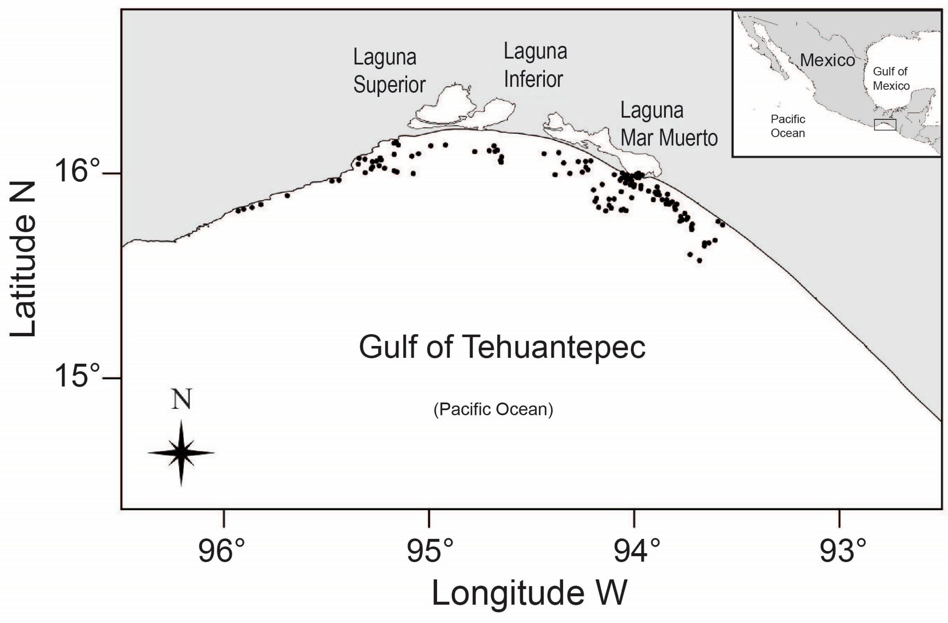
2. Materials and Methods

Biological samples of U. chilensis were collected monthly from January to December 2012, except for June, September, and October, in the Gulf of Tehuantepec (14°10′ N; 92°15′ W–16°13′ N; 95°55′ W), located in the coastal zone of the state of Oaxaca, Mexico (Figure 1).
Biological samples from the shrimp bycatch were collected in accordance with Mexican guidelines and regulations for shrimp fisheries. The organisms were handled ethically and in accordance with the Code of Ethics for Public Servants. No additional permits were needed for this research, which included any endangered or protected species. This emphasizes our commitment to ethical research practices. The samples of U. chilensis were sexed and measured in centimeters from the tip of the snout to the tip of the tail (total body length, L T ), using this as a standard size measure for estimating growth [23,24].

2.1. Vertebrae Preparation

A thoracic vertebral section of each U. chilensis was obtained from the abdominal cavity once the individual was eviscerated. Vertebral samples were manually cleaned by removing the connective tissue and soaking them in a solution of 3% sodium hypochlorite for 5 min to remove the remaining tissue. Once cleaned, the vertebrae were washed under running tap water to remove sodium hypochlorite residuals. Sagittal sections of 0.5 mm in thickness were obtained using a Buehler Isomet low-speed saw. Each section was mounted on a glass slide using clear resin (Cytoseal 60) and examined under a dissecting microscope with transmitted light. Band pairs consisted of one opaque band (wide) and one translucent band (narrow) [25,26]. The relation between L T and vertebral radius R V was fitted by minimizing the sum of squares error to ensure that the growth of each aging structure was proportional to the growth of U. chilensis. Analysis of covariance (ANCOVA) was used to examine differences in the L T R V relationship between sexes.

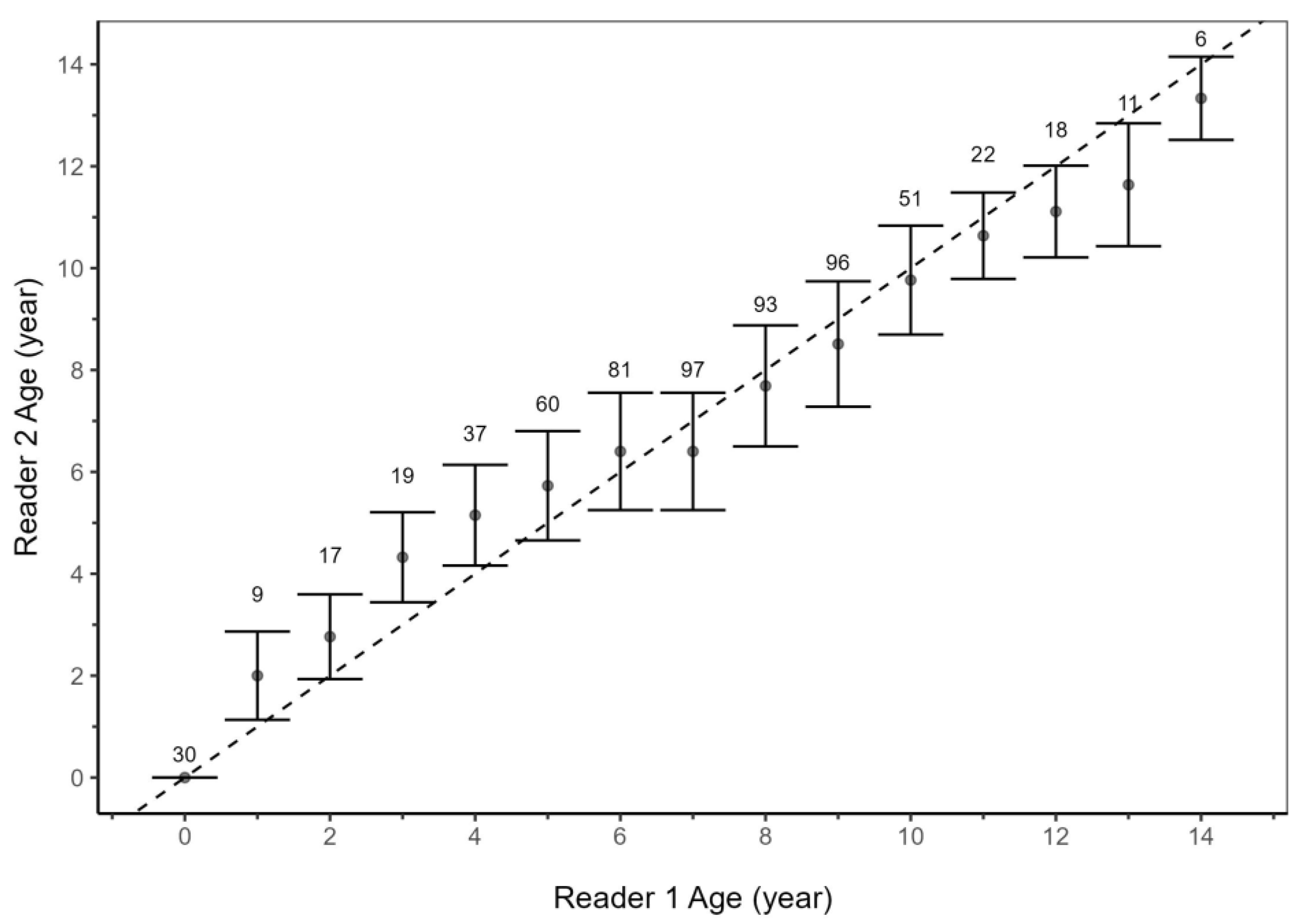
2.2. Precision and Bias

Age estimates were obtained by counting translucent bands, which were considered to represent one year of growth if they were accompanied by an opaque band and if they could be detected on both sides of the corpus calcareum and across the intermedialia. The birthmark (age = 0 years) was identified by a change in the angle of corpus calcareum relative to the intermedialia (Figure 2) and was not included in the age estimate [27]. The changes in the vertebral sections across the total lengths sampled are presented as photocompositions (Figure S1).
The precision of the growth band was estimated from counts performed by two readers; counts and measurements were made without any knowledge of the length or sex of each ray. Two age readers independently counted a reference set of 696 vertebral sections to ensure quality control and precision for the band pair counts. Pairwise inter- and intra-reader comparisons were generated for the reference set and the two counts of the entire sample, made by the primary reader. To ensure the accuracy of our results, we examined the bias and precision of band pair counts using age-biased plots [28] and a paired Student’s t-test. Additionally, the average percent error (APE) and coefficient of variation (CV) were also estimated [29,30].

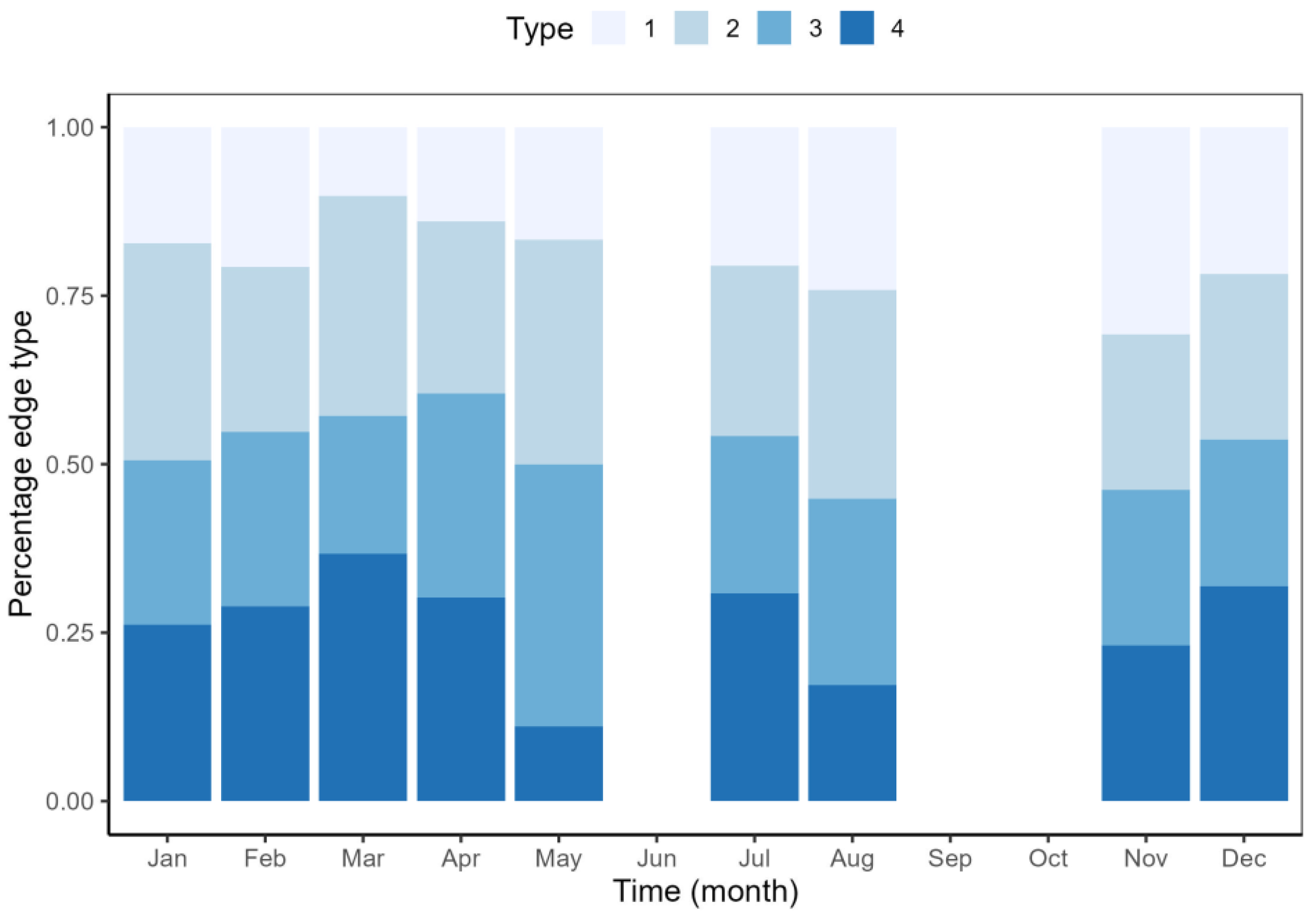
2.3. Age Verification

Opaque and translucent are commonly used to describe growth bands, and they tend to occur in summer and winter, respectively [19]. Alternating opaque and translucent bands representing one band pair were visible in all sections under reflected light (Figure 2). The annual periodicity band pair deposition hypothesis was tested using two indirect techniques: marginal increment ratio analysis (MIR) and edge analysis (EA). Both statistical procedures are very well documented in the literature [31,32,33,34,35]. MIR data were compared against the month of collection, and the Kolmogorov–Smirnov test was applied to determine if the monthly samples were normally distributed. Then, a non-parametric Kruskal–Wallis test was used to determine if there were differences between collection months. If significant statistical differences were estimated in the monthly MIR (p < 0.05), a post hoc Dunn’s test for multiple comparisons was undertaken [36].
Edge analysis was carried out, where the criterion for determining edge type was adapted from Gallagher & Nolan [37] and Yudin & Cailliet [38]. Edge types were defined as the translucent band beginning to form (type 1), a well-formed translucent band (type 2), opaque band beginning to form (type 3), and finally a well-formed opaque band (type 4). Broad and narrow opaque bands were defined based on the relative width of the previously fully formed opaque band. The relative frequencies of translucent and opaque edges were plotted through time. Based on consensus counts and their corresponding maximum differences, it was determined to assess the sources of count variation as an additional measure of precision; this allowed an agreement on the assignation of the final age of each of the organisms. Those readings with a lower percentage of agreement or that were not coincident were discarded.

2.4. Growth Models and Parameter Estimation

Six candidate growth models were fitted for male and female size-at-age data of U. chilensis based on vertebral band counts: the Gompertz model with two (GGM-2) and three (GGM) parameters; the Gompertz model with two phases (GGM-2P); two versions of the Schnute general model (SCHGM-1 and SCHGM-2); and a logistic model with three parameters (LGM) (Table 1). These models involved at least two of the following coefficients: (i) the theoretical age-at-zero total length, which is a position parameter defining the initial condition on the time axis when mean total length-at-age is zero; (ii) the theoretical asymptotic size, representing the average size-at-age that individuals in stock would attain if they grew indefinitely; (iii) the growth coefficient, which is the curvature parameter determining the rate at which the asymptotic size (theoretically the size at an infinite age) is reached; and (iv) the mean size-at-birth (commonly referred as L 0 ), which was estimated by obtaining the mean L T of the largest embryos and the smallest of the neonates sampled [39]. The candidate growth model parameters ( θ i ) (Table 2) were estimated based on a negative log-likelihood objective function proposed by Hilborn & Mangel [40]. The residuals were assumed to have multiplicative error; given that the size-at-age data are more scattered for older individuals, such error allows for stabilizing the variance.

2.5. Confidence Intervals

The confidence intervals of parameters θ i in each candidate growth model were estimated by considering the covariance between parameters [42] using Monte Carlo simulation in a parameterized bootstrap routine. Each candidate growth model was bootstrapped 2000 times. This technique is a consistent estimator by simulation if the simulated data have the same statistical properties as the original data [43,44]. The parametric bootstrap assumes normal probability distribution for the residuals [45]. The confidence intervals were estimated using the bias-corrected percentile method [46].

2.6. Model Selection

Model selection was based on the Akaike information criterion (AIC). Based on fit and parsimony, models with smaller AIC values were favored [20]. The differences were used to rank the models in relation to the best model [20,47,48]. For each model, the likelihood ratio test was performed to compare the growth curves of males and females [49].
The analysis was performed using Visual Basic Application (VBA), available in Microsoft Excel™. Plots were created in ggplot2 (v 3.5.1), available in R package (v 4.4.0).

3. Results

A total of 696 U. chilensis were collected; 205 males ranged in size from 10.5 to 33.8 cm L T (mean 22.5 cm), and 491 females ranged in size from 11.1 to 39.5 cm L T (mean 23.08 cm). The ANCOVA analysis indicated that R V (p < 0.05) significantly affected L T , but sex did not (p > 0.05). Therefore, the results are presented for the entire sample (combined sexes). A linear function of the form described the relationship: L T = (0.1413 × R V ) − 1.0163 (p < 0.0001; R 2   = 0.89; n = 696).

3.1. Age Estimates and Verification

The age estimates generated from vertebral thin sections and used in the growth models ranged from zero to 12 years for males and zero to 14 years for females; the oldest estimated age in females was obtained from two individuals of 39.2 and 39.5 cm L T and two individuals of 33 and 33.8 cm L T in males. The precision between readers was acceptable with values of APE = 6.71% (Student’s t-test = 1.53, p = 0.13) and CV = 9.53% (n = 623). The age bias between readers showed that the precision of age estimates was lower in age groups from 0 to 5 and from 12 to 14 years than that observed for age groups between 6 to 10 years. However, the standard deviation was similar for most of the age comparisons. This pattern indicated an apparent increase in age estimation precision for individuals in middle age (Figure 3).
The results showed that the MIR data were not normally distributed (p < 0.05). Therefore, a Kruskal–Wallis test was applied; the results showed a significant statistical difference in the marginal increment ratio between months (H (8,696) = 12.29; p < 0.001). The Dunn’s post hoc test for multiple comparisons indicated that the average MIR data in January and February were significantly different from those in November and December. Low MIR was observed from January to March with a sharp increase from April to November-December. The low MIR months represented the slow-growing translucent band formation period (mid to late winter and early spring), while the rapid growth of opaque bands occurred during late spring, summer, and fall (Figure 4). According to the vertebral edge analysis, translucent bands (type 1 and type 2) formed mainly during February–April; in these months, the percentage frequency of such bands varied between 62% and 78% (Figure 5).
By contrast, EA showed no clear seasonal pattern. However the opaque edges (type 3 and type 4) were slightly more frequent (over 50%) during February–April, July, and December. According to both methods used (MIR and EA), the formation of translucent bands seemed to occur in late winter to early spring (February–March), followed by the formation of opaque bands in late spring, summer, and fall (from April to December), suggesting that band pairs had annual formation.

3.2. Growth Coefficients and Model Selection

The results of this study indicated that some models used to describe the growth of U. chilensis fit the observed length-at-age data well (Table 3 and Table 4). The estimates of for males were greater than for females according to GGM (♂ = 48.23, ♀ = 40.35), LGM (♂ = 38.59, ♀ = 35.82), SCHGM-1(♂ = 31.13, ♀ = 31.91), and SCHGM-2 (♂ = 46.13, ♀ = 39.66. However, the last model overestimated the size corresponding to the maximum length observed for this species (33.8 cm LT) for males (Figure 6). The likelihood ratio tests indicated statistically significant differences ρ < 0.05 between the growth curves of males and females in all scenarios used (Table 5).
Candidate growth models using L 0 fit the size-at-age data set of U. chilensis. GGM-2, based on the lowest AIC = 1290.77, Δi = 0, and highest wi = 87.5, was the growth function that best fit the size-at-age data for females; however, there was also substantial support for GGM-2P (Δi < 4) (Table 6). By contrast, for males, GGM-2P was the best model that fit the size-at-age data (AIC = 590, Δi = 0, wi = 63.3), followed by GGM-2 (AIC = 591.73, Δi = 1.72, wi = 26.7), GGM (AIC = 594.33, Δi = 4.32, wi = 7.3), and finally SCHGM-2 (AIC = 596.34, Δi = 6.34, wi = 2.7), which had considerably less support (Table 6). LGM and SCHGM-1 had no statistical support to describe the length-at-age data.

4. Discussion

Age and growth studies on elasmobranchs have been fundamental to understanding their life histories and fishery sustainability [50,51,52]. The hypothesis that many species deposit concentric growth band pairs within their vertebral centra on an annual cycle permits age estimation through the enumeration of bands [19,33,53], and the agreement between age estimates generated from vertebral thin sections were acceptable for Urotrygon rogersi [54], U. aspidura [55], and U. chilensis in the present document, suggesting vertebrae as an aging structure for this species. According to Sulikowski et al. [56], samples with an APE of less than 15% are acceptable, although Campana [33] explains that the precision reported in shark age studies is rarely below 10%. The estimates in this study were less than 10% of CV. Thus, our age estimates could be considered consistent.

4.1. Age Verification

Marginal increment analysis (MIR) and edge analysis (EA) are generally more successful when applied to specimens that are still in a rapid state of growth; these methods assume that the width or the density of the outermost increment will exhibit a yearly sinusoidal cycle when plotted against the month of capture if growth bands are formed annually [31,33,57]. In our study, MIR showed two clear periods with different increment ratios; these were defined as (a) slow growth from January to March and (b) rapid growth from April to December. This pattern suggested new opaque band formation starting in spring. Transitions between periods of slow and rapid growth are commonly observed in these organisms, and they have been reported for species like Rhinoptera bonasus, Hypanus dipterurus, Dipturus chilensis, Malacoraja senta, Urotrygon rogersi, and U. aspidura [24,32,54,55,58,59].
On the other hand, Davis et al. [60] faced challenges in validating the annual band formation in Bathyraja trachura using MIR. Their results did not reveal significant statistical differences between MIR data (ANOVA test). The process of estimating cycles becomes intricate when MIR data exhibit outliers [57] or when the data are scattered, both of which are common occurrences in MIR data [33]. Mejía-Falla et al. [54] pointed out that MIR analysis is problematic due to technical difficulties related to resolving the margins of growth bands, which include specific technical difficulties. They also noted that the number of deposited bands and time of deposition may vary with age and proposed that a single narrow band is formed annually on vertebral centra in January, with a peak in November.
Verifying annual band formation by another indirect method, such as EA, has frequently been used in chondrichthyan aging studies, where the percent frequencies of opaque and translucent bands are compared with the month or season. The vertebral edge deposition pattern of U. chilensis in the present document was possible for all age groups; opaque edge types 3 and 4 were present in summer-autumn, which were associated with faster growth, and translucent edge types 1 and 2 were associated with slower growth, which were formed in winter-spring. EA has also been used for verifying the annual band formation for species like H. dipterurus, Platyrhina sinensis, Okamejei acutispina, and Maculobatis astra [23,32,61]. In other batoids, centrum edge analysis was used to demonstrate that round stingrays showed a pattern of seasonal growth in their vertebrae, with faster growth occurring in the summer months and slower growth occurring in the colder winter months. Therefore, the timing of opaque band formation did not necessarily coincide with the time that bands become visible at the edge of the centrum [31].
Natanson [62] suggested that the annual periodicity in growth band deposition is associated with photoperiod for Leucoraja erinaceus. The identification of edge types may be influenced by many factors, e.g., species, lighting methods, inter-annual environmental variation, the width of the forming band, the presence of a circa-annual rhythm, food rations, and hormone secretion [31,63]. For U. chilensis, the false bands observed in the vertebral sections could be explained by changes in environmental conditions in the Gulf of Tehuantepec, such as cold water upwelling events, including the effect of food rations. For Dipturus chilensis, the opaque edges were correlated with the average seasonal sea surface temperature, while translucent edges were more frequent at lower sea surface temperatures [24]. Finally, we consider the use of combined validation techniques, which can provide enhanced details associated with seasonal patterns of band formation, such as MIR and EA. Additionally, it is important to establish a correlation with environmental factors that may influence the periodicity of the bands, such as seasonal variations in sea surface temperature and currents, which are notably pronounced in the study area. In the same way, further research is necessary on the importance of considering the dates of birth and capture to estimate the factional age of individuals for growth estimates, in particular for species like U. chilensis that could have two periods of birth during the year [15].
This study included both juvenile and mature individuals in the age structure of the U. chilensis population in the Gulf of Tehuantepec, Mexico, which tends to be composed of primarily juvenile and mature individuals. The classes between three to seven years were predominant for females, and the longevity estimates were 14 years. For males, the age classes between four and six years were predominant, with a maximum age of 12. These estimates of age indicated that the females were older than the males, as reported by other authors. White & Potter [64] suggested that size, sex, and age segregation is typical for urolophids and that during the reproductive season, mature animals mix indiscriminately.
Rhinoptera bonasus exhibits a difference in the number of age classes by sex; the species showed up to 16 years for males and 18 years for females [58]. Some species showed notable differences in age structure by sex, such as D. dipterura, where males presented 19 age classes and females presented 28 age classes [32]. Similarly, Platyrhina sinensis males presented 5 years, whereas females showed 12 years [23]. Similar results were observed for males and females of Urobatis halleri, with 15 age classes [65]. The predominance of a more significant number of age classes for females was also observed by Kelsey et al. [35], who found that females in this species showed 16 age classes and males only had 12 age classes. Similarly, Maculobatis astra in males presented 19 age classes, whereas females presented 30 age classes [66]. This feature appears common in myliobatoid rays; in general, females attain a more significant size and age while growth is slower in males. This was evidenced in [67], where the difference in growth was associated with the onset of age at first maturity, with females and males of U. rogersi attaining their first maturity in different ages. However, in U. roguersi and U. chilensis, no differences were observed in the length at 50% maturity between sexes [15,54].

4.2. Fitted Growth Models

The procedure for estimating the parameters in the growth models involved assumptions associated with the mathematical function and the information contained in the dataset. A misspecification in the growth model could provide unrealistic parameters and trajectories of fitted curves without biological realism. The datasets provide another source of uncertainty in that they could be biased; for example, the biological samples could exhibit a predominance of age-at-length data for large individuals. Therefore, the parameters estimated for any growth model would provide a high bias in origin. In this study, three mathematical expressions of the Gompertz model were modeled for U. chilensis and two scenarios were considered: (a) the estimation of all parameters in the model, and (b) a mix between fixed parameters (L0) and free parameters estimated for a mathematical iteration algorithm.
Based on this statistical procedure, modifications of the Gompertz models were used for males and females for U. chilensis, where L 0 substituted the parameter t 0 . This variation in the parameter L 0 in different growth models may be solved as follows: (a) L 0 is known, consequently it is a fixed value in the growth model [68]; (b) L 0 is known but there is a range of observed values, consequently it is estimated by the objective function using constraints in this parameter (e.g., smaller size for neonate and upper size for embryonic) [35,69,70]; (c) if L 0 is unknown, then the parameter can be estimated [71]. The advantage of these approaches is that if L 0 is fixed, then one degree of freedom is saved in the model fitting process using any objective function [68,72]. This modification has been used for growth modeling of other elasmobranchs [31,51,58,73].
The growth biology of U. chilensis was described according to two parameters of the Gompertz growth model for females and two phases of the Gompertz growth model for males. Previous studies about the individual growth of batoids have shown that only the von Bertalanffy growth model has fit the data (Tables S1 and S2); in these studies, the best model that fit the data was selected based on the R 2 criterion. Adjusted R 2 and coefficient of variation values are useful as measures of the explained proportion of the variation, but they are not useful in growth model selection [20]. According to Katsanevakis [21], the coefficient of determination commonly used for the selection of growth models needs to be a reliable estimator. The advantage of a fixed size-at-age dataset helps by adding more parameters to the model, thus reducing distance, but it further increases uncertainty in the estimation process. The balance between underfitting and overfitting is clearly shown in the Akaike information criterion (AIC), which penalizes scores based on the number of estimated parameters in the model [74]. Comparatively, for both sexes, LGM using the parameter t 0 was not the best candidate growth model for U. chilensis in females and males. The Schnute growth model describes two curves; the first is S-shaped (an asymptote with two inflection points at different size-at-age growth). However, this model failed to describe the growth pattern of U. chilensis in both sexes. Nevertheless, the second version of this growth model describes the rapid development of a younger organism (Schnute 1981), and this model described the length-at-age data for males (∆i = 6.34), whilst for females, ∆i = 36.35 was much larger. Hence the growth model was omitted.

4.3. Growth Model Selection

Previous studies about the individual growth of batoids have shown that only the von Bertalanffy growth model has fit the data [32,59,64,65,75] (Tables S1 and S2 [23,32,55,59,64,65,66,73,75,76,77,78,79,80,81,82,83,84,85,86,87,88]); in these studies, the best model that fit the data was selected based on the R 2 criterion. However, adjusted R 2 and coefficient of variation values are useful as measures of the explained proportion of the variation, but they are not useful in growth model selection [20,21]. In this study, the general mathematical expression of SCHGM-1 provided a model according to von Bertalanffy; however, the dataset could have been more informative for this model, and the contribution of SCHGM-1 for explaining the datasets for females and males was negligible. The above indicates that U. chilensis does not exhibit growth similar to the von Bertalanffy model; this result contrasts with reports for different batoids species. U. chilensis in the Gulf of Tehuantepec has a growth pattern described by the Gompertz model. This is a particular case of the four parameters of the Richards model and thus belongs to the Richards family of three-parameter sigmoidal growth models, along with similar models, mainly the logistic model and the negative exponential model. The statistical properties of the Gompertz model change the biological interpretation of the parameter k. For this growth model, the parameter is defined as the rate of exponential decrease of the relative growth rate with age, and the average maximum length maintains its biological definition in [41].

5. Conclusions

It has been assumed that all elasmobranchs grow continuously and asymptotically throughout their lives, and their growth is well described by the von Bertalanffy model, especially the growth of sharks. However, we recommend using multiple growth models to evaluate the growth characteristics of round stingrays like U. chilensis that continue to grow in weight but not significantly in length. The implication of using the “best model” to describe size–age data needs to consider the statistical description and the biological information of size-at-age data, assuming that each species has a different growth pattern and may show different growth at various stages of its life cycle. The comparisons of growth parameters in this study for U. chilensis indicate that they have relatively short lives, slower growth, and females have larger sizes than males. These findings are the first contribution for the species in the area (Gulf of Tehuantepec) and the first documentation for the species in the Mexican Pacific.

Supplementary Materials

The following supporting information can be downloaded at: https://www.mdpi.com/article/10.3390/fishes10050232/s1, Table S1. A summary of life history parameters in female bathoids given for the total length ( L T ) or the width of the disk ( W D ). Table S2. A summary of life history parameters in male bathoids given for the total length ( L T )) or the width of the disk ( W D ). Figure S1. Vertebral sections of Urotrygon chilensis are presented for various sizes across the sampled total length range.

Author Contributions

Conceptualization, A.B.G.-C. and E.M.-B.; methodology, A.B.G.-C., E.M.-B., H.A.-V. and J.T.-Á.; validation, E.M.-B. and H.A.-V.; formal analysis, A.B.G.-C., E.M.-B. and H.A.-V.; investigation, A.B.G.-C., E.M.-B., H.A.-V. and J.T.-Á.; data curation, A.B.G.-C.; writing—original draft preparation, A.B.G.-C., E.M.-B., H.A.-V. and J.T.-Á.; writing—review and editing, A.B.G.-C., E.M.-B. and H.A.-V.; visualization, A.B.G.-C. and E.M.-B.; supervision, E.M.-B. and H.A.-V.; project administration, E.M.-B.; funding acquisition, E.M.-B. All authors have read and agreed to the published version of the manuscript.

Funding

This research was funded by Consejo Nacional de Ciencias y Tecnología (CONACYT) under grant no. CIB-2012-01 179322.

Institutional Review Board Statement

The biological samples from shrimp bycatch fauna sampling were taken following Mexican guidelines and regulations. The organisms were handled ethically and in accordance with the Code of Ethics for Public Servants. No other permissions were required to complete this research, and this study was conducted without including any endangered or otherwise protected species. This reaffirms our unwavering commitment to ethical research practices.

Informed Consent Statement

Not applicable.

Data Availability Statement

The data presented in this study can be obtained by request from the corresponding author. Since the data set are reserved for scientific purposes by the Mexican Federal Government; however, the data can be provided whether the purpose is explicitly scientific research.

Acknowledgments

We thank Oswaldo Morales-Pacheco from CRIAP-Salina Cruz for logistical support for this study and the crew members and scientific staff who participated in the sampling cruises. A.B.G.-C. was a recipient of a fellowship (CONACYT 849348).

Conflicts of Interest

The authors declare no conflicts of interest.

References

  1. Beebe, W.; Beebe, W.; Tee-Van, J. Eastern Pacific Expeditions of the New York Zoological Society. XXVIII. Fishes from the Tropical Eastern Pacific. From Cedros Island, Lower California, South to the Galapagos Islands and Northern Peru. Part 3. Rays, Mantas and Chimaeras. Zool. Sci. Contrib. N. Y Zool. Soc. 1941, 26, 245–280. [Google Scholar] [CrossRef]
  2. Villavicencio Garayzar, C.J. Distribución Temporal y Condición Reproductiva de Las Rayas (Pisces: Batoidei), Capturadas Comercialmente En Bahía Almejas, Baja California Sur, México. Rev. Investig. Cient. Rev. Investig. Cient 1995, 6, 1–12. [Google Scholar]
  3. Márquez-Farías, J.F. The Artisanal Ray Fishery in the Gulf of California: Development, Fisheries Research, and Management Issues. Shark News 2002, 14, 12–13. [Google Scholar]
  4. Cisneros-Montemayor, A.M.; Cisneros-Mata, M.A.; Harper, S.; Pauly, D. Extent and Implications of IUU Catch in Mexico’s Marine Fisheries. Mar. Pol. 2013, 39, 283–288. [Google Scholar] [CrossRef]
  5. IUCN The IUCN Red List of Threatened Species. Version 2024-2. Available online: https://www.iucnredlist.org (accessed on 25 February 2025).
  6. Brander, K. Disappearance of Common Skate Raia Batis from Irish Sea. Nature 1981, 290, 48–49. [Google Scholar] [CrossRef]
  7. Casey, J.M.; Myers, R.A. Near Extinction of a Large, Widely Distributed Fish. Science 1998, 281, 690–692. [Google Scholar] [CrossRef]
  8. Bizzarro, J.J.; Smith, W.D.; Márquez-Farías, J.F.; Hueter, R.E. Artisanal Fisheries and Reproductive Biology of the Golden Cownose Ray, Rhinoptera Steindachneri Evermann and Jenkins, 1891, in the Northern Mexican Pacific. Fish. Res. 2007, 84, 137–146. [Google Scholar] [CrossRef]
  9. Dellapa, A.; Kimmel, D.G.; Cl, S. Trends of Fish and Elasmobranch Landings in Italy: Associated Management Implications. ICES J. Mar. Sci. 2012, 69, 1045–1052. [Google Scholar] [CrossRef]
  10. Ruocco, N.L.; Lucifora, L.O.; Díaz De Astarloa, J.M.; Wöhler, O. Reproductive Biology and Abundance of the White-Dotted Skate, Bathyraja albomaculata, in the Southwest Atlantic. ICES J. Mar. Sci. 2006, 63, 105–116. [Google Scholar] [CrossRef]
  11. Baum, J.K.; Myers, R.A. Shifting Baselines and the Decline of Pelagic Sharks in the Gulf of Mexico. Ecol. Lett. 2004, 7, 135–145. [Google Scholar] [CrossRef]
  12. Le Port, A.; Lavery, S.; Montgomery, J.C. Conservation of Coastal Stingrays: Seasonal Abundance and Population Structure of the Short-Tailed Stingray Dasyatis brevicaudata at a Marine Protected Area. ICES J. Mar. Sci. 2012, 69, 1427–1435. [Google Scholar] [CrossRef]
  13. Ainsley, S.M.; Ebert, D.A.; Natanson, L.J.; Cailliet, G.M. A Comparison of Age and Growth of the Bering Skate, Bathyraja interrupta (Gill and Townsend, 1897), from Two Alaskan Large Marine Ecosystems. Fish. Res. 2014, 154, 17–25. [Google Scholar] [CrossRef]
  14. Kadri, H.; Marouani, S.; Saïdi, B.; Bradai, M.N.; Bouaïn, A.; Morize, E. Age, Growth, Sexual Maturity and Reproduction of the Thornback Ray, Raja clavata (L.), of the Gulf of Gabès (South-Central Mediterranean Sea). Mar. Biol. Res. 2014, 10, 416–425. [Google Scholar] [CrossRef]
  15. Alvarez-Fuentes, C.J.; Tovar-Ávila, J.; Payan-Alejo, J.; Chávez-Arrenquín, D.A.; Salgado-Ugarte, I.H.; Amezcua, F. Reproductive Ecology of the Chilean Round Ray (Urotrygon chilensis, Günther, 1872) in the Southern Gulf of California. Fishes 2023, 8, 193. [Google Scholar] [CrossRef]
  16. Ordoñez-López, J. Aspectos Reproductivos y Alimenticios de la Raya Pinta Urotrygon chilensis (Gunther, 1871) (Pisces: Usolophidae) en el Pacifico Central mexicano. Bachelor’s Thesis, Universidad Nacional Autónoma de México, Mexico City, Mexico, 2004. [Google Scholar]
  17. Torres-Huerta, A.M.; López-Pérez, R.A.; Tapia-García, M.; Gracía, A. Distribution of Batoid Demersal Assemblages on the Continental Shelf of the Gulf of Tehuantepec. Mar. Freshw. Res. 2019, 70, 1445–1458. [Google Scholar] [CrossRef]
  18. Guzman-Castellanos, A.B. Historia de Vida de la Raya Chilena Urotrygon chilensis (Günther, 1872) en el Sureste del Pacífico Mexicano. Ph.D. Thesis, Centro de Investigaciones Biológicas del Noroeste S.C., La Paz, Bolivia, 2015. [Google Scholar]
  19. Cailliet, G.M.; Goldman, K.J. Age Determination and Validation in Chondrichthyan Fishes. In Biology of Sharks and Their Relatives; CRC Press: Boca Raton, FL, USA, 2004; pp. 399–448. [Google Scholar] [CrossRef]
  20. Burnham, K.P.; Anderson, D.R. Model Selection and Multimodel Inference: A Practical Information-Theoretic Approach, 2nd ed.; Springer: NewYork, NY, USA, 2002. [Google Scholar]
  21. Katsanevakis, S. Modelling Fish Growth: Model Selection, Multi-Model Inference and Model Selection Uncertainty. Fish. Res. 2006, 81, 229–235. [Google Scholar] [CrossRef]
  22. Flinn, S.A.; Midway, S.R. Trends in Growth Modeling in Fisheries Science. Fishes 2021, 6, 1. [Google Scholar] [CrossRef]
  23. Kume, G.; Furumitsu, K.; Yamaguchi, A. Age, Growth and Age at Sexual Maturity of Fan Ray Platyrhina sinensis (Batoidea: Platyrhinidae) in Ariake Bay, Japan. Fish. Sci. 2008, 74, 736–742. [Google Scholar] [CrossRef]
  24. Aversa, M.I.; Dans, S.L.; García, N.A.; Crespo, E.A. Growth Models Fitted to Dipturus chilensis Length-at-Age Data Support a Two-Phase Growth. Rev. Chil. Hist. Nat. 2011, 84, 33–49. [Google Scholar] [CrossRef]
  25. Cailliet, G.M.; Martin, L.K.; Kushner, D.; Wolf, P.; Welden, B.A. Techniques for Enhancing Vertebral Bands in Age Estimation of California Elasmobranchs. In Proceedings of the International Workshop on Age Determination of Oceanic Pelagic Fishes: Tunas, Billfishes, Sharks, Miami, FL, USA, 15–18 February 1983. NOAA Tech. Rep. NMFS 8. [Google Scholar]
  26. Brown, C.A.; Gruber, S.H. Age Assessment of the Lemon Shark, Negaprion Brevirostris, Using Tetracycline Validated Vertebral Centra. Copeia 1988, 1988, 747–753. [Google Scholar] [CrossRef]
  27. Goldman, K.J. 6. Age and Growth. In Elasmobranch Fisheries Management Techniques; Musick, J.A., Bonfil, R., Eds.; FAO Fisheries Technical Paper No. 474; FAO: Rome, Italy, 2005; pp. 76–102. [Google Scholar]
  28. Graphical and Statistical Methods for Determining the Consistency of Age Determinations—Campana—1995—Transactions of the American Fisheries Society—Wiley Online Library. Available online: https://afspubs.onlinelibrary.wiley.com/doi/abs/10.1577/1548-8659%281995%29124%3C0131%3AGASMFD%3E2.3.CO%3B2 (accessed on 6 September 2024).
  29. Beamish, R.J.; Fournier, D.D.A. A Method for Comparing the Precision of a Set of Age Determinations. Can. J. Fish. Aquat. Sci. 1981, 38, 982–983. [Google Scholar] [CrossRef]
  30. Chang, W.Y.B. A Statistical Method for Evaluating the Reproducibility of Age Determination. Can. J. Fish. Aquat. Sci. 2011, 39, 1208–1210. [Google Scholar] [CrossRef]
  31. Cailliet, G.M.; Smith, W.D.; Mollet, H.F.; Goldman, K.J. Age and Growth Studies of Chondrichthyan Fishes: The Need for Consistency in Terminology, Verification, Validation, and Growth Function Fitting. Environ. Biol. Fishes 2006, 77, 211–228. [Google Scholar] [CrossRef]
  32. Smith, W.D.; Cailliet, G.M.; Melendez, E.M. Maturity and Growth Characteristics of a Commercially Exploited Stingray. Mar. Freshw. Res. 2007, 58, 54–66. [Google Scholar] [CrossRef]
  33. Campana, S.E. Accuracy, Precision and Quality Control in Age Determination, Including a Review of the Use and Abuse of Age Validation Methods. J. Fish. Biol. 2001, 59, 197–242. [Google Scholar] [CrossRef]
  34. Lessa, R.; Santana, F.M.; Duarte-Neto, P. A Critical Appraisal of Marginal Increment Analysis for Assessing Temporal Periodicity in Band Formation among Tropical Sharks. Environ. Biol. Fishes 2006, 77, 309–315. [Google Scholar] [CrossRef]
  35. James, K.C.; Ebert, D.A.; Natanson, L.J.; Cailliet, G.M. Age and Growth Characteristics of the Starry Skate, Raja stellulata, with a Description of Life History and Habitat Trends of the Central California, U.S.A., Skate Assemblage. Environ. Biol. Fishes 2014, 97, 435–448. [Google Scholar] [CrossRef]
  36. Zar, J. Biostatistical Analysis, 4th ed.; Prentice Hall: Upper Saddle River, NJ, USA, 1999; 266p. [Google Scholar]
  37. Gallagher, M.; Nolan, C.P. A Novel Method for the Estimation of Age and Growth in Rajids Using Caudal Thorns. Can. J. Fish. Aquat. Sci. 1999, 56, 1590–1599. [Google Scholar] [CrossRef]
  38. Yudin, K.G.; Cailliet, G.M. Age and Growth of the Gray Smoothhound, Mustelus californicus, and the Brown Smoothhound, M. Henlei, Sharks from Central California. Copeia 1990, 1990, 191–204. [Google Scholar] [CrossRef]
  39. Yokota, L.; Lessa, R.P. A Nursery Area for Sharks and Rays in Northeastern Brazil. Environ. Biol. Fishes 2006, 75, 349–360. [Google Scholar] [CrossRef]
  40. Hilborn, R.; Mangel, M. The Ecological Detective. Confronting Models with Data; Princeton University Press: Princeton, NJ, USA, 1997; p. 315. [Google Scholar]
  41. Luquin-Covarrubias, M.A.; Morales-Bojórquez, E.; Scarry-González, S.; Lluch-Cota, D.B. Joint Likelihood Function Based on Multinomial and Normal Distributions for Analyzing the Phenotypic Growth Variability of Geoduck Clam Panopea globosa. CalCOFI Rep. 2016, 57, 151–162. [Google Scholar]
  42. Hilborn, R.; Walters, C.J. Quantitative Fisheries Stock Assessment: Choice, Dynamics and Uncertainty; Springer: New York, NY, USA, 1992. [Google Scholar]
  43. Fournier, D.; Archibald, C.P. A General Theory for Analyzing Catch at Age Data Fisheries. Can. J. Fish. Aquat. Sci. 1982, 39, 1195–1207. [Google Scholar] [CrossRef]
  44. Gelman, A.; Carlin, J.B.; Stern, H.S.; Dunson, D.B.; Vehtari, A.; Rubin, D.B. Bayesian Data Analysis, 3rd ed.; CRC Press Taylor & Francis Group 6000 Broken: Boca Raton, FL, USA, 2013; 642p. [Google Scholar]
  45. Magnusson, A.; Punt, A.E.; Hilborn, R. Measuring Uncertainty in Fisheries Stock Assessment: The Delta Method, Bootstrap, and MCMC. Fish Fish. 2013, 14, 325–342. [Google Scholar] [CrossRef]
  46. Haddon, M. Modelling and Quantitative Methods in Fisheries, 2nd ed.; Chapman and Hall/CRC: New York, NY, USA, 2011; ISBN 9780429109492. [Google Scholar]
  47. Hobbs, N.T.; Hilborn, R. Alternatives to Statistical Hypothesis Testing in Ecology: A Guide to Self Teaching. Ecol. Appl. 2006, 16, 5–19. [Google Scholar] [CrossRef] [PubMed]
  48. Johnson, J.B.; Omland, K.S. Model Selection in Ecology and Evolution. Trends Ecol. Evol. 2004, 19, 101–108. [Google Scholar] [CrossRef]
  49. Kimura, D.K. Likelihood Methods for the von Bertalanffy Growth Curve. Fish. Bull. 1980, 77, 765–776. [Google Scholar]
  50. Stobutzki, I.; Miller, M.; Brewer, D. Sustainability of Fishery Bycatch: A Process for Assessing Highly Diverse and Numerous Bycatch. Environ. Conserv. 2001, 28, 167–181. [Google Scholar] [CrossRef]
  51. Braccini, J.M.; Gillanders, B.M.; Walker, T.I.; Tovar-Avila, J. Comparison of Deterministic Growth Models Fitted to Length-at-Age Data of the Piked Spurdog (Squalus megalops) in South-Eastern Australia. Mar. Freshw. Res. 2007, 58, 24–33. [Google Scholar] [CrossRef]
  52. Simpfendorfer, C.A.; Heupel, M.R.; White, W.T.; Dulvy, N.K. The Importance of Research and Public Opinion to Conservation Management of Sharks and Rays: A Synthesis. Mar. Freshw. Res. 2011, 62, 518–527. [Google Scholar] [CrossRef]
  53. Natanson, L.J.; Cailliet, G.M. Vertebral Growth Zone Deposition in Pacific Angel Sharks. Copeia 1990, 1133–1145. [Google Scholar] [CrossRef]
  54. Mejía-Falla, P.A.; Cortés, E.; Navia, A.F.; Zapata, F.A. Age and Growth of the Round Stingray Urotrygon rogersi, a Particularly Fast-Growing and Short-Lived Elasmobranch. PLoS ONE 2014, 9, e96077. [Google Scholar] [CrossRef] [PubMed]
  55. Torres-Palacios, K.; Mejía-Falla, P.A.; Navia, A.F.; Cruz-Escalona, V.H.; Félix-Uraga, R.; Quiñonez-Velázquez, C. Age and Growth Parameters of the Panamic Stingray (Urotrygon aspidura). Fish. Bull. 2019, 117, 169–179. [Google Scholar] [CrossRef]
  56. Sulikowski, J.A.; Morin, M.D.; Suk, S.H.; Howell, W.H. Age and Growth Estimates of the Winter Skate (Leucoraja ocellata) in the Western Gulf of Maine. Fish. Bull. 2003, 101, 405–413. [Google Scholar]
  57. Okamura, H.; Punt, A.E.; Semba, Y.; Ichinokawa, M. Marginal Increment Analysis: A New Statistical Approach of Testing for Temporal Periodicity in Fish Age Verification. J. Fish. Biol. 2013, 82, 1239–1249. [Google Scholar] [CrossRef]
  58. Neer, J.A.; Thompson, B.A. Life History of the Cownose Ray, Rhinoptera bonasus, in the Northern Gulf of Mexico, with Comments on Geographic Variability in Life History Traits. Environ. Biol. Fishes 2005, 73, 321–331. [Google Scholar] [CrossRef]
  59. Natanson, L.J.; Sulikowski, J.a.; Kneebone, J.R.; Tsang, P.C. Age and Growth Estimates for the Smooth Skate, Malacoraja senta, in the Gulf of Maine. Environ. Biol. Fishes 2007, 80, 293–308. [Google Scholar] [CrossRef]
  60. Davis, C.D.; Cailliet, G.M.; Ebert, D.A. Age and Growth of the Roughtail Skate Bathyraja trachura (Gilbert 1892) from the Eastern North Pacific. Environ. Biol. Fishes 2007, 80, 219–230. [Google Scholar] [CrossRef]
  61. Joung, S.J.; Lee, P.H.; Liu, K.M.; Liao, Y.Y. Estimates of Life History Parameters of the Sharpspine Skate, Okamejei acutispina, in the Northeastern Waters of Taiwan. Fish. Res. 2011, 108, 258–267. [Google Scholar] [CrossRef]
  62. Natanson, L.J. Effect of Temperature on Band Deposition in the Little Skate, Raja erinacea. Copeia 1993, 199–206. [Google Scholar] [CrossRef]
  63. Sagarese, S.R.; Frisk, M.G. An Investigation on the Effect of Photoperiod and Temperature on Vertebral Band Deposition in Little Skate Leucoraja erinacea. J. Fish. Biol. 2010, 77, 935–946. [Google Scholar] [CrossRef]
  64. White, W.T.; Potter, I.C. Reproductive Biology, Size and Age Compositions and Growth of the Batoid (Urolophus paucimaculatus), Including Comparisons with Other Species of the Urolophidae. Mar. Freshw. Res. 2005, 56, 101–110. [Google Scholar] [CrossRef]
  65. Hale, L.F.; Lowe, C.G. Age and Growth of the Round Stingray Urobatis halleri at Seal Beach, California. J. Fish. Biol. 2008, 73, 510–523. [Google Scholar] [CrossRef]
  66. Jacobsen, I.P.; Bennett, M.B. Life History of the Blackspotted Whipray Himantura astra. J. Fish. Biol. 2011, 78, 1249–1268. [Google Scholar] [CrossRef]
  67. Araya, M.; Cubillos, L.A. Evidence of Two-Phase Growth in Elasmobranchs. Environ. Biol. Fishes 2006, 77, 293–300. [Google Scholar] [CrossRef]
  68. Pardo, S.A.; Cooper, A.B.; Dulvy, N.K. Avoiding Fishy Growth Curves. Methods Ecol. Evol. 2013, 4, 353–360. [Google Scholar] [CrossRef]
  69. Ainsley, S.M.; Ebert, D.A.; Cailliet, G.M. Age, Growth, and Maturity of the Whitebrow Skate, Bathyraja minispinosa, from the Eastern Bering Sea. ICES J. Mar. Sci. 2011, 68, 1426–1434. [Google Scholar] [CrossRef]
  70. Henningsen, A.D.; Leaf, R.T. Observations on the Captive Biology of the Southern Stingray. Trans. Am. Fish. Soc. 2010, 139, 783–791. [Google Scholar] [CrossRef]
  71. Matta, M.E.; Gunderson, D.R. Age, Growth, Maturity, and Mortality of the Alaska Skate, Bathyraja parmifera, in the Eastern Bering Sea. Environ. Biol. Fishes 2007, 80, 203–217. [Google Scholar] [CrossRef]
  72. Guzmán-Castellanos, A.B.; Morales-Bojórquez, E.; Balart, E.F. Individual Growth Estimation in Elasmobranchs: The Multi-Model Inference Approach. Hidrobiológica 2014, 24, 137–150. [Google Scholar]
  73. Pierce, S.J.; Bennett, M.B. Destined to Decline? Intrinsic Susceptibility of the Threatened Estuary Stingray to Anthropogenic Impacts. Mar. Freshw. Res. 2010, 61, 1468–1481. [Google Scholar] [CrossRef]
  74. Wang, Y.; Liu, Q. Comparison of Akaike Information Criterion (AIC) and Bayesian Information Criterion (BIC) in Selection of Stock–Recruitment Relationships. Fish. Res. 2006, 77, 220–225. [Google Scholar] [CrossRef]
  75. Licandeo, R.R.; Lamilla, J.G.; Rubilar, P.G.; Vega, R.M. Age, Growth, and Sexual Maturity of the Yellownose Skate Dipturus chilensis in the South-Eastern Pacific. J. Fish. Biol. 2006, 68, 488–506. [Google Scholar] [CrossRef]
  76. White, W.T.; Platell, M.E.; Potter, I.C. Relationship between Reproductive Biology and Age Composition and Growth in Urolophus lobatus (Batoidea: Urolophidae). Mar. Biol. 2001, 138, 135–147. [Google Scholar] [CrossRef]
  77. White, W.T.; Hall, N.G.; Potter, I.C. Reproductive Biology and Growth during Pre- and Postnatal Life of Trygonoptera personata and T. mucosa (Batoidea: Urolophidae). Mar. Biol. 2002, 140, 699–712. [Google Scholar] [CrossRef]
  78. Ismen, A. Age, Growth, Reproduction and Food of Common Stingray (Dasyatis pastinaca L., 1758) in İskenderun Bay, the Eastern Mediterranean. Fish. Res. 2003, 60, 169–176. [Google Scholar] [CrossRef]
  79. Mollet, H.F.; Ezcurra, J.M.; O’Sullivan, J.B. Captive Biology of the Pelagic Stingray, Dasyatis violacea (Bonaparte, 1832). Mar. Freshw. Res. 2002, 53, 531–541. [Google Scholar] [CrossRef]
  80. Francise, M.P.; Maolagáin, C.Ó. Age and Growth of the Antarctic Skate (Amblyraja georgiana) in the Ross Sea. CCAMLR Sci. 2005, 12. [Google Scholar]
  81. Gburski, C.M.; Gaichas, S.K.; Kimura, D.K. Age and Growth of Big Skate (Raja binoculata) and Longnose Skate (R. rhina) in the Gulf of Alaska. Environ. Biol. Fishes. 2007, 80, 337–349. [Google Scholar] [CrossRef]
  82. Ismen, A.; Yigin, C.; Ismen, P. Age, Growth, Reproductive Biology and Feed of the Common Guitarfish (Rhinobatos rhinobatos Linnaeus, 1758) in İskenderun Bay, the Eastern Mediterranean Sea. Fish. Res. 2007, 84, 263–269. [Google Scholar] [CrossRef]
  83. Başusta, N.; Demirhan, S.A.; Çiçek, E.; Başusta, A.; Kuleli, T. Age and Growth of the Common Guitarfish, Rhinobatos rhinobatos, in Iskenderun Bay (North-Eastern Mediterranean, Turkey). J. Mar. Biol. Assoc. UK 2008, 88, 837–842. [Google Scholar] [CrossRef]
  84. Izzo, C.; Gillanders, B.M. Initial Assessment of Age, Growth and Reproductive Parameters of the Southern Fiddler Ray Trygonorrhina fasciata (Müller & Henle, 1841) from South Australia. Panam. J. Aquat. Sci. 2008, 3, 321–327. [Google Scholar]
  85. Cuevas-Zimbrón, E.; Sosa-Nishizaki, O.; Pérez-Jiménez, J.C.; O’Sullivan, J.B. An Analysis of the Feasibility of Using Caudal Vertebrae for Ageing the Spinetail Devilray, Mobula japanica (Müller and Henle, 1841). Environ. Biol. Fishes. 2013, 96, 907–914. [Google Scholar] [CrossRef]
  86. Yigin, C.C.; Ismen, A. Age, Growth and Reproduction of the Common Stingray, Dasyatis pastinaca from the North Aegean Sea. Mar. Biol. Res. 2012, 8, 644–653. [Google Scholar] [CrossRef]
  87. Mora-Zamacona, P.; Melo-Barrera, F.N.; Cruz-Escalona, V.H.; Navia, A.F.; Morales-Bojórquez, E.; Pérez-Palafox, X.A.; Mejía-Falla, P.A. Growth Modeling of the Giant Electric Ray Narcine entemedor in the Southern Gulf of California: Analyzing the Uncertainty of Three Data Sets. Animals 2022, 12, 19. [Google Scholar] [CrossRef]
  88. Santander-Nieto, J.; Santana, F.M.; Vasconcelos-Filho, J.E.; Lessa, R. Age and Growth of the Threatened Smalleye Round Ray, Urotrygon microphthalmum, Delsman, 1941, from Northeastern Brazil. Fishes 2023, 8, 160. [Google Scholar] [CrossRef]
Figure 1. Map of Urotrygon chilensis bycatch scientific samples (black dot) in the Gulf of Tehuantepec, Mexico.
Fishes 10 00232 g001
Figure 2. Vertebral section of Urotrygon chilensis three-year-old female, 17.2 cm L T (F: Focus; BM: Birthmark; FB: False band). The numbers represent the age in years based on band pair counts, while the arrows indicate the translucent bands.
Fishes 10 00232 g002
Figure 3. Age-biased graph showing the mean age estimates of reader 2 relative to the age estimates of reader 1, with sample sizes listed above each age for Urotrygon chilensis. Error bars represent the standard error around mean ages, and the discontinued line represents the 1:1 equivalence.
Fishes 10 00232 g003
Figure 4. Mean marginal increment ratio (MIR) during each collection month for Urotrygon chilensis. Error bars represent mean MIR ± 1 SE, while the dots indicate the outliers.
Fishes 10 00232 g004
Figure 5. Monthly variations in the proportions of edge analyses vertebral samples for Urotrygon chilensis: (type 1) translucent edge forming; (type 2) translucent edge completely formed; (type 3) opaque edge forming; and (type 4) opaque edge wholly formed.
Fishes 10 00232 g005
Figure 6. Growth model curves by sex of Urotrygon chilensis observed length-at-age data. Models: Gompertz model with two (GGM-2) and three (GGM) parameters and with two phases (GGM-2P); logistic model with three parameters (LGM); and two versions of the Schnute general model (SCHGM-1 and SCHGM-2).
Fishes 10 00232 g006
Table 1. Candidate growth models.
NameAcronymModel
Gompertz three parametersGGM L T = L α e e κ ( t t o )
Gompertz two parametersGGM-2 L T = L 0 e k ( 1 e α t )
Gompertz two phasesGGM-2P L T = L 0 ( e G ( 1 e κ t ) ) ,   G = L n ( L α / L 0 )
Logistic three parametersLGM L T = L α / 1 + e κ ( t t o )
Schnute (a ≠ 0, b ≠ 0)SCHGM-1 L T =   γ 1 b + ( γ 2 b γ 1 b ) 1 e a ( t τ 1 ) 1 e a ( τ 2 τ 1 ) 1 b
Schnute (a ≠ 0, b = 0)SCHGM-2 L T = γ 1 e L n γ 2 γ 1 1 e a ( t τ 1 ) 1 e a ( τ 2 τ 1 )
Table 2. Symbols and explanation of the parameters of candidate growth models.
ParametersDescription
L α Asymptotic length at which growth is zero
*   k Growth coefficient
t o Age or time when length theoretically equals zero
L 0 Mean size-at-birth
G Instantaneous rate of growth at time t
y 1 Size   at   age   τ 1
y 2 Size   at   age   τ 2
τ 1 Firts specified age
τ 2 Second specified age
a Constant relative rate of relative growth rate
b Incremental relative rate of relative growth rate
* For the Gompertz growth model, the parameter k is the rate of exponential decrease of the relative growth rate with age; in the Logistic growth model, k is the relative growth rate parameter [41].
Table 3. Growth parameter estimates for females of Urotrygon chilensis. θ i : parameter; x ¯ : mean; sd: standard deviation; and 95% confidence intervals (lower/upper C.I.). Fixed-value parameters: L 0 = 10.90, τ1 = 0, and τ2 = 14. The models were ordered according to the AIC value reported in Table 6.
ModelParameterValueMeanSdCVBias% BiasLower C.I.Upper C.I.
GGM-2k1.421.650.050.020.2414.521.331.51
A0.150.160.000.040.048.310.150.15
GGM-2PG1.411.410.010.000.00−0.371.391.43
k0.15−0.200.00−0.02−0.0526.79−0.15−0.19
GGML40.3538.830.390.01−1.51−3.9039.5941.11
k0.190.200.000.010.015.090.190.19
t01.961.700.050.03−0.25−14.791.862.05
SCHGM-1A0.820.770.040.06−0.05−6.880.74−4.048
B−4.59−4.250.27−0.060.34−7.93−5.1210.21
y110.1010.200.060.010.121.139.9933.28
y232.9831.910.160.00−0.09−0.2932.68
y31.9131.890.150.00−0.09−0.2931.6232.20
τ4.164.160.450.23−0.29−0.283.515.27
y21.6011.190.080.010.330.1421.4421.75
LGML∞35.8235.160.230.01−0.05−0.1435.3636.27
k0.330.330.000.010.000.560.320.33
t03.122.950.060.02−0.02−0.833.013.23
SCHGM-2α0.190.200.000.020.014.910.180.20
y110.269.460.050.010.060.6010.1610.35
y239.8736.430.480.01−0.11−0.2938.9240.81
Table 4. Growth parameter estimates for males of Urotrygon chilensis. θ i : parameter; x ¯ : mean; sd: standard deviation; and 95% confidence intervals (lower/upper C.I.). Fixed-value parameters: L 0 = 10.40, τ1 = 0, and τ2 = 12. The models were ordered according to the AIC value reported in Table 6.
ModelParameterValueMeanSdCVBias% BiasLower C.I.Upper C.I.
GGM-2α1.5744.471.370.03−3.76−8.4541.7846.83
k0.130.150.010.040.017.750.140.16
GGM-2PG1.591.480.030.02−0.14−9.211.531.65
k0.13−0.150.01−0.04−0.0318.32−0.11−0.15
GGML∞48.2344.471.370.03−3.76−8.4545.5550.90
k0.140.150.010.040.017.750.130.15
t03.132.520.220.09−0.61−24.262.703.56
SCHGM-2α0.140.910.080.09−0.05−5.340.130.15
y111.0410.750.070.010.070.6610.9111.17
y240.7531.080.250.01−0.19−0.6239.1042.40
y46.1330.880.100.00−0.02−0.1945.6046.65
τ2.67000.001.480.0087.132.313.03
y16.9722.000.080.01−0.33−0.1413.1720.76
LGML∞38.5937.100.580.02−0.54−1.4635.9038.19
k0.270.280.010.020.012.790.270.29
t03.773.430.140.04−0.13−3.903.163.70
SCHGM-1α0.950.910.080.09−0.05−5.340.791.11
b−6.18−5.850.59−0.100.33−5.62−7.34−5.02
y110.6810.750.070.010.070.6610.5510.80
y231.2731.080.250.01−0.19−0.6230.7931.75
y31.1330.880.100.00−0.02−0.1930.9331.32
τ4.90000.001.480.0087.134.894.99
y22.4022.000.080.01−0.33−0.1422.2422.55
Table 5. Likelihood ratio tests (RT) comparing the growth curves of males and females for each candidate model. Models: Gompertz model with two (GGM-2) and three (GGM) parameters and with two phases (GGM-2P); logistic model with three parameters (LGM); and two versions of the Schnute general model (SCHGM-1 and SCHGM-2). The degree of freedom (df) is equal to the number of parameters.
Acronym-Ln LikelihoodRT χ 2 df ρ
FemalesMales
GGM659.53294.16699.065.992<0.05
GGM-2643.39293.86704.687.813<0.05
GGM-2P645.34293.00730.747.813<0.05
LGM678.56301.07730.789.493<0.05
SCHGM-1699.56312.46754.987.814<0.05
SCHGM-2659.56294.17774.29.494<0.05
Table 6. Growth model selection for females of U. chilensis. θi: number parameters, -Ln-likelihood: negative log-likelihood function, AIC: Akaike information criterion, ∆i: Akaike differences, ωi: Akaike weight.
SexModelθi-Ln LikelihoodAICΔiωi
FemalesGGM-22643.391290.770.0087.5
GGM-2P3645.341294.673.9012.5
GGM3659.531325.0734.290.00
SCHGM-24659.561327.1336.350.00
LGM3678.561363.1372.350.00
SCHGM-14699.561407.94117.170.00
MalesGGM-2P3293.00590.000.0063.3
GGM-22293.86591.731.7226.7
GGM3294.16594.334.327.3
SCHGM-24294.17596.346.342.7
LGM3301.07608.1318.130.00
SCHGM-14312.46632.9142.910.00
Disclaimer/Publisher’s Note: The statements, opinions and data contained in all publications are solely those of the individual author(s) and contributor(s) and not of MDPI and/or the editor(s). MDPI and/or the editor(s) disclaim responsibility for any injury to people or property resulting from any ideas, methods, instructions or products referred to in the content.

