Article

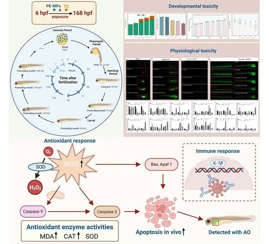
Microplastics Enhance the Toxic Effects of Tetracycline on the Early Development of Zebrafish in a Dose-Dependent Manner

College of Marine Sciences, South China Agricultural University, Guangzhou 510642, China
*
Authors to whom correspondence should be addressed.
Fishes 2025, 10(4), 150; https://doi.org/10.3390/fishes10040150
Submission received: 17 February 2025 / Revised: 21 March 2025 / Accepted: 25 March 2025 / Published: 27 March 2025
(This article belongs to the Special Issue Effects of Nanoplastics and Microplastics on Fish Health)

Abstract

Microplastic pollution in the environment has greatly increased due to the widespread use of plastics. Antibiotics and microplastic are common contaminants, especially in aquaculture. Microplastics could act as antibiotic vectors that raise the potential of their ecotoxicological effects. In this work, we conducted several analyses of biomarker responses to examine the developmental toxicity and toxicological endpoints that polyethylene microplastics (PE-MPs) and tetracycline antibiotics (TC) induced in zebrafish (Danio rerio) embryos/larvae. The results suggested that TC-PE-MPs induced significant physiological perturbations, including attenuated spontaneous cardiac contractions, cardiotoxicity, a dose-dependent elevation in mortality, and a marked reduction in body length, accompanied by morphological alterations. The mechanistic analysis revealed that ROS accumulation triggered enzymatic activity changes, which further induced aberrant vascular development, robust inflammatory responses, and dysregulated gene expression. These findings demonstrate that PE coexistence potentiates TC’s toxicological effects, with combined exposure inducing developmental toxicity during critical organogenesis stages in zebrafish. Overall, the current research demonstrated the detrimental effects of TC-PE-MPs on early fish development, suggesting potential environmental risks.
Key Contribution: PE-MPs can enhance TC toxicity to induce early developmental disorders in zebrafish.

Graphical Abstract

1. Introduction

Society has become progressively dependent on plastics since mercantile production began in approximately 1950 [1]. Plastic products have excellent properties such as a light load, robustness and durability, multi-functionality, and low production cost [2,3]. Currently, plastics can be categorized into about 45 types [4], of which polyethylene (PE), polystyrene (PS), polycarbonate (PC), polypropylene (PP), and polyvinyl chloride (PVC) are the most widely used. According to their size, there are five classes to be categorized: nanoplastics (NPs) are less than 1 μm, microplastics (MPs) are sized 1 μm–5 mm, medium plastics are sized 5 mm–5 cm, macroplastics and megaplastics are larger sizes [5,6]. MPs are extremely sustained and omnipresent in the environment [7,8]. They can be categorized into primary and secondary MPs based on the source of origin [9]. Primary MPs are intentionally manufactured by industry, which are small in size and are usually intended for use in industrial products or daily necessities (facial cleansers, cosmetics, etc.), and are the main constituents of environmental MPs; secondary MPs are produced by the weathering/degradation of plastic residues in the environment by physical and biological factors [10,11,12]. Plastic bottles and bags, worn-out tires, fishing nets, and plastic containers are the main sources of secondary MPs [13,14,15]. Large amounts of MPs are found in the soil environment, sediments, water environments, and even in the air we breathe [16,17,18].
In recent years, MPs in the micrometer range have become a research hotspot due to their potential toxicity in aquatic environments [19]. Notably, the primary origins of MPs in the aquatic environment are terrestrial plastics, coastal tourism debris, shipping and fishery debris, aquaculture equipment, and atmospheric deposition [20,21]. Numerous studies have revealed that aquatic organisms often ingest microplastics that critically affect them because of their uniquely large surfaces and high hydrophobicity, and MPs can adsorb compounds on their surfaces, which increases the possible toxic effects on aquatic biota [22,23,24]. Aquatic organisms transfer ingested MPs along the food chain and may eventually accumulate in the human body, thus posing a greater threat to aquatic ecosystems and human health, which has aroused serious public concern [25].
Since the 1940s, when antibiotics were first introduced into clinical practice, they have become essential in animal husbandry and aquaculture, driven by the ever-growing demand for animal-based food [26]. Antibiotics, on the other hand, are difficult to degrade and break down in nature due to their unique chemical structures [27]. Tetracyclines (TCs) are the most common antibiotic drugs in the world. TCs rank first in usage in China [28]. Nonetheless, only a low proportion of TCs can be metabolized or absorbed in vivo, while as many as 75% of TCs are commonly released from excreta in an active form [29,30]. Unfortunately, aquaculture production systems are often open-access, which results in the build-up of antibiotic residues in farmed and adjacent waters, wild fish, plankton, and sediments [31]. This suggests that beneficial bacteria in aquatic organisms, and even in the ecosystem and human body, may be influenced by antibiotic residues from aquaculture [32]. Studies of antibiotic residues in the environment are still restricted when compared to studies of antibiotic residues or resistance in the aquaculture environment. Additionally, there is currently no global database on antibiotic residues in the environment [33].
Given the prevalence of antibiotics and MPs in aquatic environments, their interactions are the focus of the current research. There are many studies showing that MPs can adsorb antibiotics in the aquatic environment. Guo et al. investigated the adsorption behavior and mechanism of sulfamethoxazole (SMX) on six common microplastics in the environment [34]. Li et al. confirmed that MPs can be used as vectors for antibiotics in the aquatic environment by studying the adsorption of five antibiotics to five MPs in both freshwater and seawater systems [19]. Weathered PS foam could be a vector for oxytetracycline in the environment [35]. The sorption process of TCs on PE has been investigated at the molecular level by molecular dynamic (MD) simulations [36]. Complex pollution systems consisting of MPs and antibiotic contaminants can be far more hazardous to aquatic ecosystems than individual contaminants [37]. The ecotoxicological interactions between antibiotics and MPs in aquatic organisms remain poorly understood. Previous studies have highlighted the potential for synergistic effects from such co-exposures. Liao et al. demonstrated that PS-MPs combined with TC promoted microbial colonization in the gill and intestinal tissues of medaka (Oryzias latipes), suggesting pathogenic risks via microbiome dysbiosis [38]. Han et al. reported synergistic immunotoxicity in hard-shell mussels (Mytilus coruscus) co-exposed to PS-MPs and OTC, characterized by ROS accumulation and downregulated immune-related gene expression [39]. Xu et al. further revealed suppressed detoxification gene expression in blue mussels exposed to PS-MP/TC mixtures, leading to gill lipid peroxidation and the potential facilitation of bacterial infiltration and antibiotic bioaccumulation [40]. Collectively, these findings underscore the complex molecular and cellular perturbations induced by antibiotic–MP co-exposures across aquatic taxa, which also resulted in vascular malformations, including ectopic budding of intersegmental vessels, surface ophthalmic vascular malformations, overgrowth of the normal main vein, and vascular disorders of the subintestinal venous plexus [41,42,43]. When organisms ingest MPs, the adsorbed antibiotics will migrate with MPs, and poisons potentially bioaccumulate and amplify across the food cycle [44,45].
Polyethylene (PE) is the plastic with the highest flux in life and is often found in aquatic environments [46,47]. Tetracycline (TC) is the most frequently utilized antibiotic in aquaculture [48], and 50–80% of the used TC is excreted into the aquatic environment through the urine and feces of aquatic organisms [49]. Such high PE and TC fluxes in aquatic environments have the potency to be detrimental to the aquaculture industry, which makes it necessary to fill the gaps in this part of the research. Current research on co-contamination of PE-MPs and TCs in aquaculture systems has primarily focused on antibiotic adsorption mechanisms, with no existing studies addressing their combined effects on aquatic organism growth. Therefore, the purpose of this study was to investigate the effect of TC-PE-MP co-presence on early developmental physiological toxicity in zebrafish and to provide a valuable reference with implications for the aquaculture industry.

2. Materials and Methods

2.1. Chemicals and Zebrafish Maintenance

Tetracycline (purity > 95%, CAS No. 60-54-8) was obtained from Macklin (Shanghai, China). Fluorescently labeled and non-fluorescently labeled micro-sized plastic PE spheres with sizes of 1–10 μm were purchased from Bessler Chromatography Technology Development Center (Tianjin, China). Twelve-month-old wild-type and fluorescent protein-s tagged adult zebrafish were reared in the laboratory in aerated tap water at 27 ± 1 °C.

2.2. Exposure Experiment and Sample Collection

The concentrations of PE-MPs were 10, 100, and 1000 μg/L [50]. The concentration of tetracycline was 200 μg/L [51]; the concentrations of the combined TC-PE exposure experiments were set as TC 200 μg/L + PE-MPs 10 μg/L, TC 200 μg/L + PE-MPs 100 μg/L, and TC 200 μg/L + PE-MPs 1000 μg/L, and zebrafish E3 embryo medium was used for the control group. All exposure solutions used zebrafish E3 embryo medium as the solvent. PE-MPs and TC were added into conical flasks at set concentration ratios, and the adsorption equilibrium was achieved according to Chen et al. [36].

2.3. PE-MP Fluorescence Intensity Measurement

The fertilized zebrafish embryos were exposed to 0 μg/L, 10 μg/L, 100 μg/L, or 1000 μg/L PE-MPs until the day of the endpoint (168 hpf), and the enrichment of green fluorescent PE-MPs in zebrafish larvae was observed under a fluorescence microscope (MZX81, Mingmei shot, Guangzhou, China) at an excitation wavelength of 488 nm (blue light).

2.4. Developmental Toxicity Endpoint Detection

Zebrafish embryos/larvae were monitored for development at the indicated time points [52] and used to evaluate the influence of combined exposure to TC-PE on early zebrafish development. The metrics tested included spontaneous movements (24 hpf), heartbeat frequency (48 hpf), hatchability (48 hpf and 72 hpf), mortality (24 hpf and 96 hpf), body length (168 hpf), and the frequency of other morphological malformations (96 hpf). The experimental conditions were maintained at 27 ± 1 °C.

2.5. Reactive Oxygen Species (ROS) Level Measurement

After exposure up to 168 hpf, ROS production was measured using dichloro-dihydrofluorescein diacetate (DCFH-DA, S0033S) supplied by Bain-Marie Biotechnology Research Institute (Jiangsu, China). Five zebrafish larvae were randomly selected from each treatment group and incubated with a 30 μM DCFH-DA working solution (prepared in phosphate-buffered saline, PBS) under dark conditions at room temperature for 30 min. After triple PBS washes, larvae were anesthetized with 0.001% MS-222 (Tricaine, Sigma Aldrich, Shanghai, China) and subsequently mounted in 4% methylcellulose gel for anatomical positioning. Fluorescence imaging was conducted using a microscope (MZX81, Mingmei shot, Guangzhou, China) (green fluorescence emission) and ROS levels were quantified. The integrated optical signal area was measured in order to quantify the fluorescence intensity of each larvae by Image J software (v1.8.0.112) [53].

2.6. Determination of the Oxidative Stress Index

The remaining supernatant was used for the determination of the activity of the antioxidant enzymes catalase (CAT, A007-1-1) and superoxide dismutase (SOD, A001-1) and the malonaldehyde (MDA, A003-1) content. Relevant chemical reagents were obtained from Nanjing Jiancheng Bioengineering Institute (Nanjing, China), and all operations were performed as instructed.

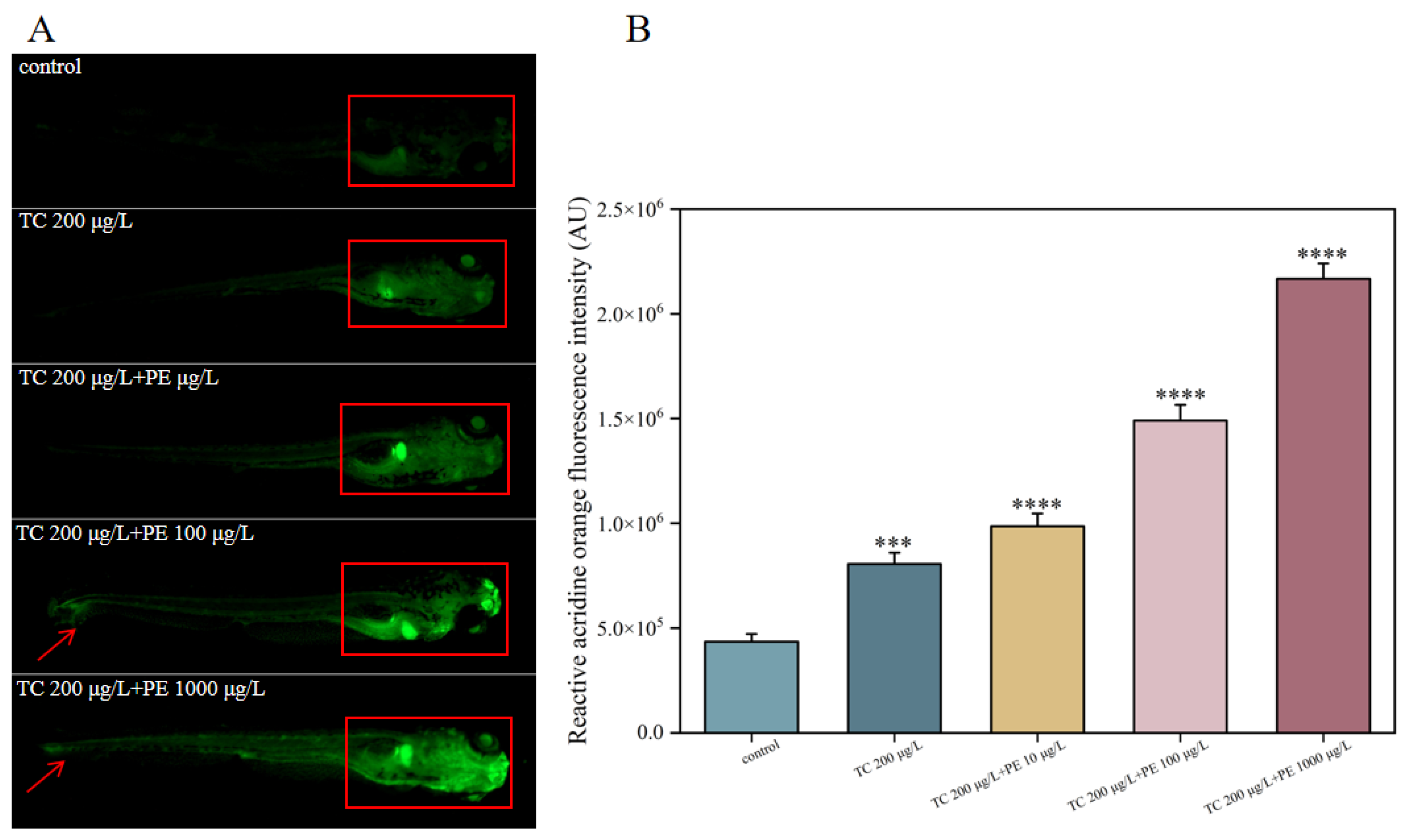
2.7. Acridine Orange (AO) Staining

Acridine Orange (AO) staining is a method that selectively stains nucleic acids for the detection of apoptosis [54]. AO staining was utilized to detect apoptotic signaling in zebrafish larvae. At the 168 hpf exposure endpoint, samples from each concentration group were incubated and transferred to 15 mL centrifuge tubes. Specimens were incubated with 5 mg/mL acridine orange (AO) solution under light-protected conditions for 30 min on an orbital shaker maintained at 28 °C. After triple rinses with PBS to remove the residual dye, the larvae were immobilized in 0.001% MS-222 (Tricaine, Sigma Aldrich, Shanghai, China) to prevent movement artifacts during imaging. Apoptotic cells were visualized under a fluorescence microscope (MZX81, Mingmei shot, Guangzhou, China) (green fluorescence emission), and the fluorescence intensity was subsequently quantified using ImageJ software [53].

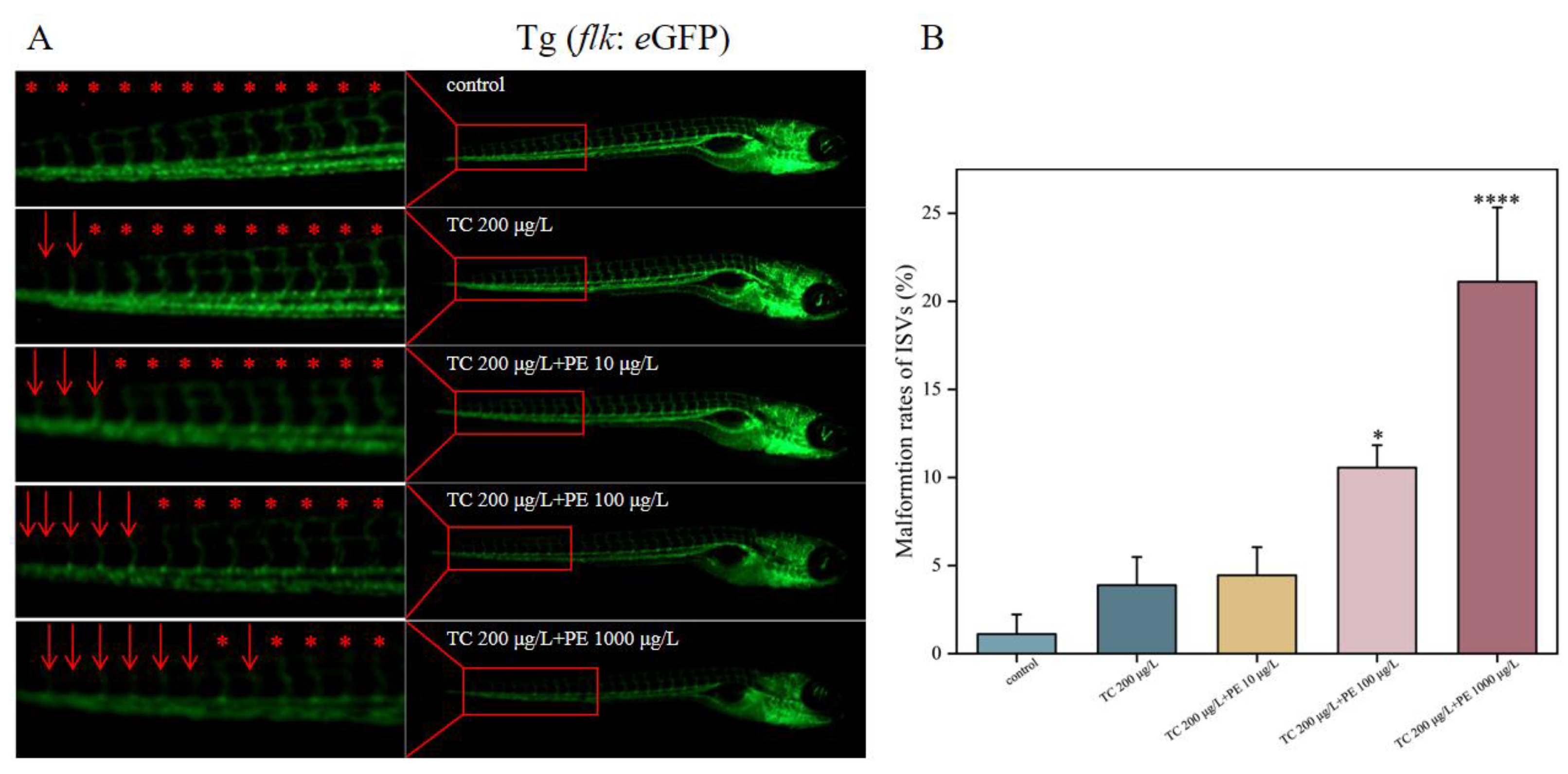
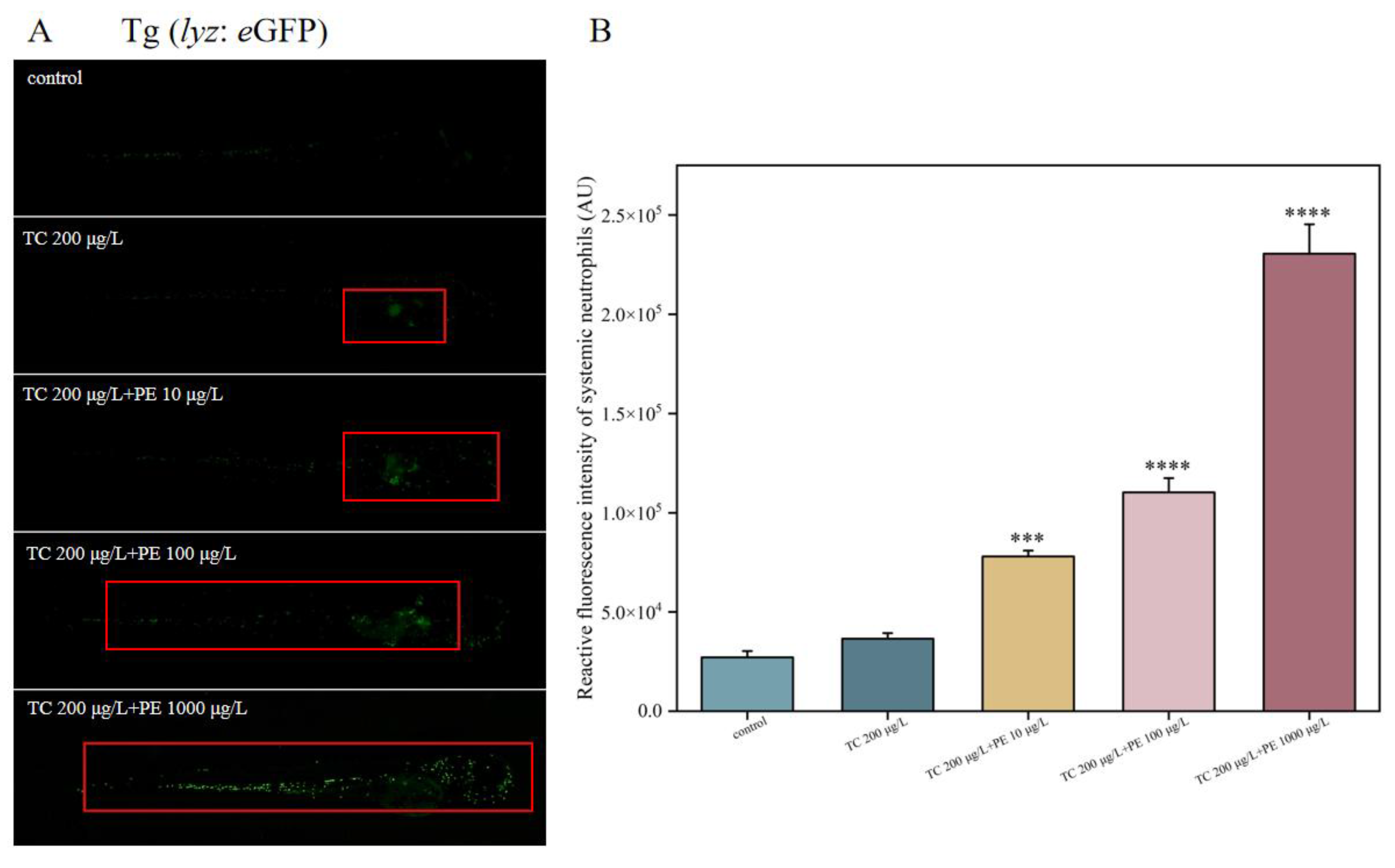
2.8. Zebrafish Expressing Fluorescent Markers to Detect Relevant Gene Expression

Zebrafish embryos expressing fluorescent proteins (Tg (flk: eGFP); Tg (lyz: eGFP)) were placed in the exposure solution. After reaching the exposure endpoint (168 hpf), the zebrafish larvae were anesthetized with 0.001% MS-222 (Tricaine, Sigma Aldrich, Shanghai, China) and the intensity of fluorescent proteins was observed under a fluorescence microscope so as to determine the influence of the exposure solution on the expression of relevant genes [41].

2.9. RT-qPCR

Relevant genes were selected from previous studies [51], RNA was extracted from zebrafish larvae, the 2−ΔΔCT method was utilized to normalize the respective expression levels of the mRNAs, and β-actin was used as an internal reference gene. RNA extraction reagent (9108), the reverse transcription kit (RR037), and the highly specific qPCR reagent TB Green (820S) were purchased from Baori Medical Technology (Beijing) Co., Ltd. (Beijing, China) All RT-qPCR procedures were performed according to the kit instructions. The primers used in this study are shown in Supplementary Table S1.

2.10. Statistical Analysis

Spontaneous movements, heart rate, body length, total protein content, CAT level, SOD level, MDA level, and fluorescence intensity were statistically analyzed by one-way ANOVA (SPSS 27.0 IBM, New York, USA). All data were presented as the means ± SEs (standard errors of the means). The data considered to display significant differences versus the control are marked as * p < 0.05, ** p < 0.01, *** p < 0.001, and **** p < 0.0001.

3. Results and Discussion

3.1. Polyethylene MP Accumulation in Zebrafish Larvae

Combined with the transparent nature of embryos and larvae, the uptake, localization, and excretion processes of MPs can be followed in real time by fluorescently labeled MPs [55]. The degree of microplastic accumulation in zebrafish larvae was detected by in vivo fluorescence microscopy, and the localization of PE-MP fluorescence was predominantly in the liver and intestine (Figure 1A). Compared to the control group, all experimental groups showed fluorescent signals of different intensities, with significantly enhanced fluorescent signals in the PE-MPs 100 μg/L concentration group and highly significantly enhanced fluorescent signals in the 1000 μg/L concentration group (Figure 1B). Lemoine et al. [56] investigated the effects of polyethylene MPs (PE-MPs) on the initial development of zebrafish; they observed consistent concentration-dependent MP accumulation in the gastrointestinal tract of the analyzed larvae. This is consistent with our findings.

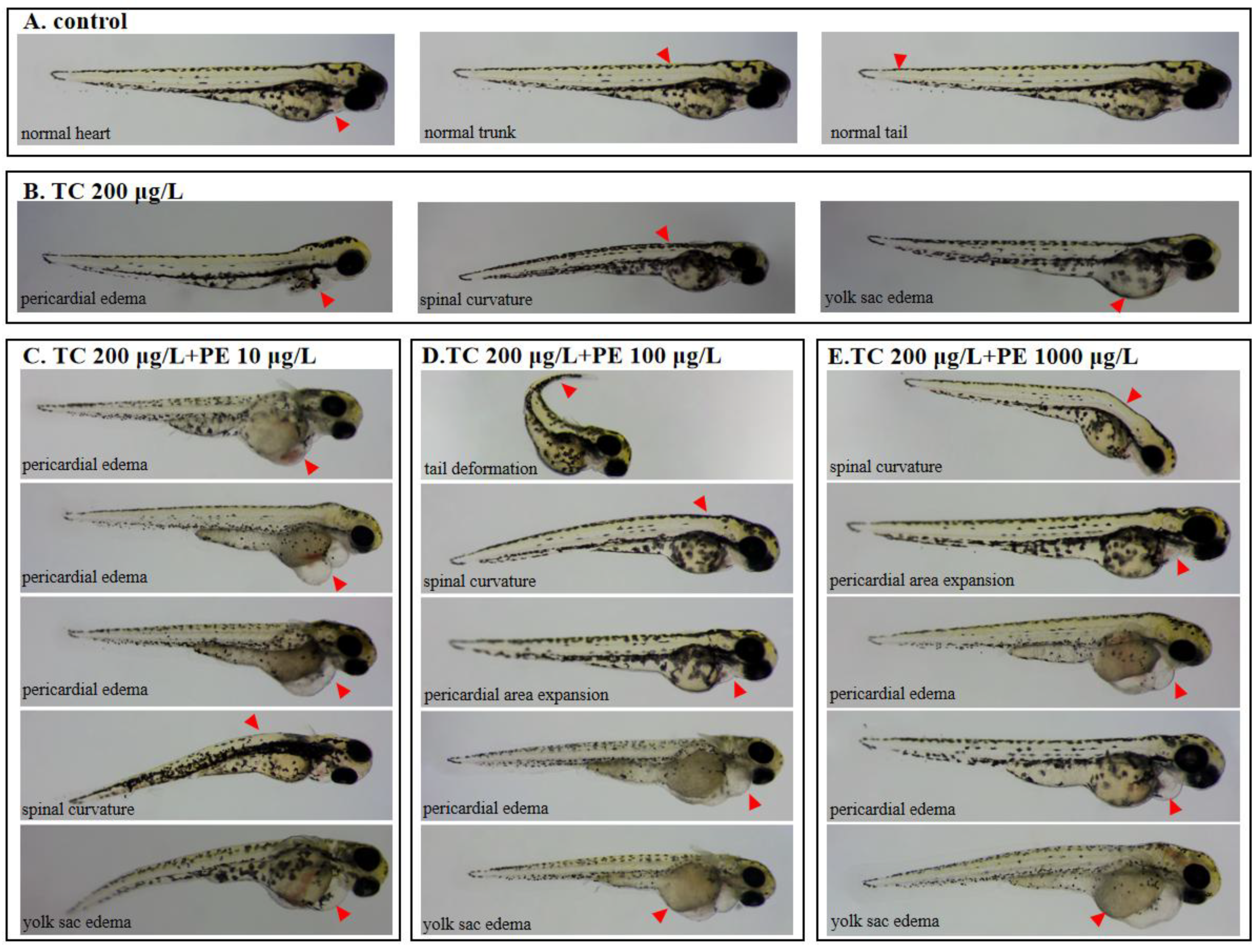
3.2. Exposure to PE-TC Induced Mortality and Malformation in Zebrafish

The developmental toxicity of TC-PE to zebrafish was studied in terms of malformations. Zebrafish embryos were treated until 72 hpf to observe the morphology of zebrafish larvae. The results showed that zebrafish larvae exposed to TC alone (TC 200 μg/L) exhibited slight malformations (Figure 2B), and we hypothesized that exposure to TC alone would only induce malformations in an individual part of the zebrafish or at a certain developmental stage. In contrast, co-exposed zebrafish larvae showed malformations of several parts during development, including embryonic pericardial edema, curvature of the pup spine, malformation of the caudal end of the spine, enlarged yolk sacs, and, as indicated by the arrows, a shorter body length (Figure 2C–E). According to the statistics, the malformation rates were not statistically significantly different in the TC-exposed group and were statistically significantly different in all combined-exposed groups (Figure 2F). These findings are similar to those observed for other well-known/widely used chemicals; for example, sodium benzoate causes similar deformities in zebrafish [57]. Notably, the triphenyl phosphate (TPHP) also induces pericardial edema within zebrafish [58]. The zebrafish heart, consisting of atria and ventricles, is the first organ to form and function during zebrafish embryogenesis. Exposure to MPs/NPs has been shown to have effects on the cardiovascular system and coagulation [59] and to suppress cardiac output and blood flow [43]. The results of this study indicate that combined TC-PE exposure has a blocking effect on early vascular development in zebrafish.
Mortality was counted at 24 hpf and 96 hpf (Figure 2G). Up to 24 hpf, there was no statistically significant difference in the mortality of zebrafish embryos between the TC-exposed group and the control group. The percentage of mortality increased significantly in a dose-dependent manner with combined exposure (p < 0.01, p < 0.001, and p < 0.0001). At 96 hpf, there were no deaths in the control and TC-exposed groups, but there was a statistically significant difference in deaths in the high-concentration combined treatment group (TC 200 μg/L + PE 1000 μg/L). In conclusion, PE that adsorbed TC may have a more dangerous toxicity. The results of the present study were lower compared to the levels of nanoparticles [60]. This may be because we used safer concentrations of microplastics and antibiotics in this experiment, bringing them closer to a real aquaculture environment.

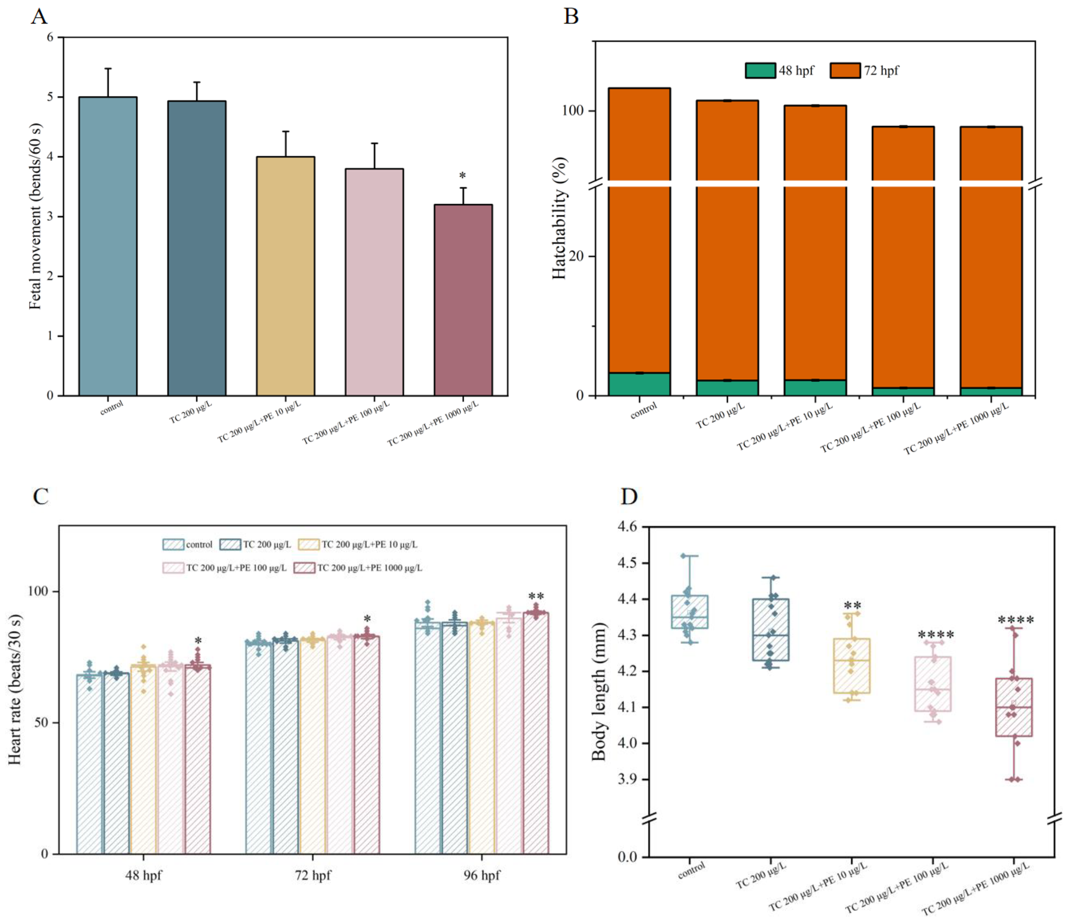
3.3. Developmental Toxicity of TC and PE Treatments in Zebrafish

To assess the influence of TC-PE on zebrafish development, four different indicators of developmental endpoints in zebrafish embryos/larvae were examined, including the spontaneous movement (fetal movement) at 24 hpf, hatchability at 48–72 hpf, the heartbeat rate at 48–96 hpf, and the body length at 168 hpf. The spontaneous movement at 24 hpf is the most commonly used sign, showing the initial development of the zebrafish with no involvement of the nervous system [61]. The heartbeat rate at 48 hpf is commonly used as a classical indicator of the developmental characteristics of the zebrafish cardiovascular system [62]. In early zebrafish development, body length is an important indicator of overall development.
Firstly, the influence of different doses of the exposure solution on the motility of zebrafish embryos was examined, and it was found that, in a dose-dependent manner, there was a decrease in the motility of zebrafish embryos in the combined exposure group (Figure 3A), but the difference in fetal movement of zebrafish embryos was statistically significant (p < 0.05) only for the high-concentration exposure (TC 200 μg/L + PE 1000 μg/L). Additionally, hatching rates during 48–72 hpf were dose-dependent, decreasing with increasing doses (Figure 3B). Our findings are consistent with those of known hazardous substances; for example, zebrafish fetal movements were significantly reduced after exposure to 2000 μg/L sulfamethazine (SMZ) or 50 mg/L PE [57,63].
The heart is an important functional organ in early zebrafish development [64], and the biventricular heart begins to contract at 26 hpf, divides at 48 hpf, and the vascular tree is fully developed at 72 hpf [65]. In 48–96 hpf zebrafish larvae, we investigated the effects on the heart rate. The results indicated no significant difference in heart rate in TC-exposed zebrafish compared to the controls at 48–96 hpf. In the combined exposure treatment group, the heart rate of zebrafish larvae was elevated and showed a positive correlation with the dose, with a statistically significantly different increase in the heart rate of the highest concentration group (TC 200 μg/L + PE 1000 μg/L) (p < 0.05, p < 0.05, and p < 0.01) (Figure 3C). Therefore, we conjecture that there may be a synergistic effect between the high concentration of PE in the presence of TC, which maximizes the adsorption of TC and thus amplifies the ecotoxicity of PE. Our study is consistent with previous studies by Xu et al. [66] that reported that both treatment with TC and combined exposure to TC + TiO2 increased the heart rate of zebrafish larvae. This study revealed that combined exposure to TC and PE led to a general trend of an increased heart rate, suggesting that co-contamination has an effect on cardiac activity and a synergistic deleterious effect on zebrafish embryos/larvae.
Zebrafish larval development is directly reflected in body length [67]. The body length of each group of zebrafish larvae was examined at 168 hpf. We emphasize that it was significantly altered in all groups receiving the combined treatment (p < 0.01, p < 0.0001, and p < 0.0001) (Figure 3D), suggesting that the toxicity of TC is enhanced when PE is present and that zebrafish larvae were more sensitive to the toxicity of the substance. Wang et al. [68] exposed zebrafish larvae to 4-epianhydrotetracycline (EATC), which resulted in a shortening of the larval body length. In this paper, zebrafish larvae exposed to tetracycline alone did not become significantly shorter in body length, probably because the concentration of tetracycline used in this paper was safer and the zebrafish larvae were not sensitive to its toxicity. Lin et al. [69] found that the body length of zebrafish larvae was also shortened by exposure to Cd and 4-epianhydrotetracycline (EATC). In the present study, we suggest that PE adsorbs TC, and zebrafish larvae ingest the compound, creating a false sense of satiety where microplastics accumulate in their digestive system, thus affecting their growth. It has been proposed that zebrafish may be associated with microplastic ingestion and pseudo-satiation during growth and development, which reduces feeding activity and disrupts energy and metabolic homeostasis [70,71].

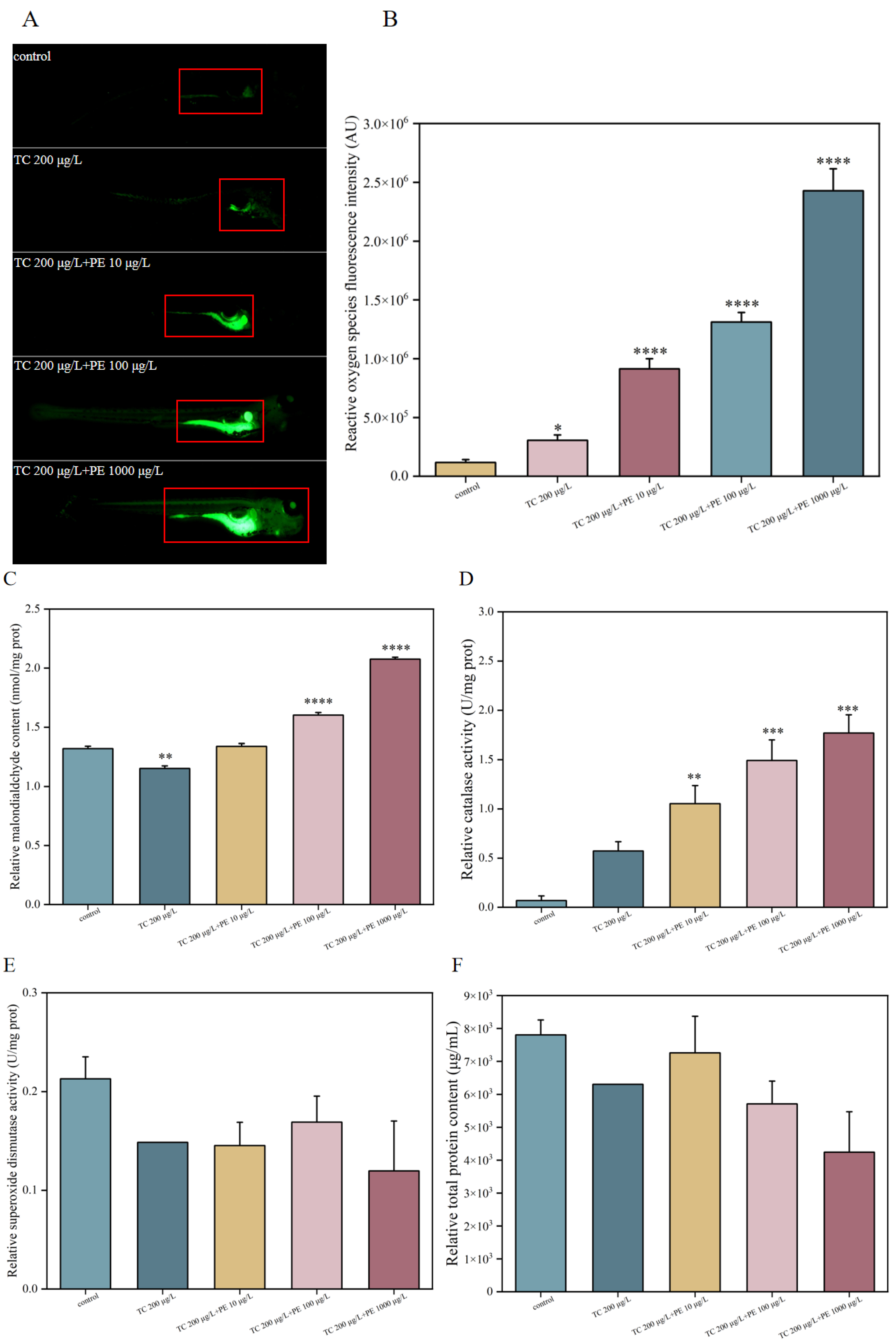
3.4. Oxidative Stress-Related Biochemical Indicators

In general, the level of ROS is an important indicator of body homeostasis. However, in order to protect itself from external stimulation, a live body will produce more ROS than usual when it is triggered by stress. The organism induces oxidative stress through the activation of ROS in the body, which results in the production of antioxidant enzymes, such as superoxide dismutase (SOD) and catalase (CAT), to scavenge harmful molecules and to maintain homeostasis [72]. It has been demonstrated that MPs/NPs affect antioxidant levels in zebrafish, interfering with ROS metabolism and resulting in oxidative stress, and that excessive ROS-induced oxidative stress causes damage to lipids, proteins, and DNA in organisms [73]. As a result, MDA, a typical oxidative stressor, is frequently used to assess an organism’s level of lipid peroxidation as well as oxidative DNA damage [74,75].
Here, ROS were assessed in the zebrafish larval model using the dichlorofluorescein diacetate (DCFH-DA) probe/reagent, and the results indicated a statistically significant rise in ROS levels across all treatment groups when compared to the control group. Notably, there was a significant dose-related increase in ROS levels in the coexposed groups, especially in the intestinal, hepatic, renal, and ocular regions (Figure 4A,B). It has been previously shown that TC exposure leads to excessive ROS production in zebrafish embryos [51,69] and that exposure to PE/PS-MNPs leads to increased ROS production [42,76,77]. Similar results have been observed for other hazardous substances, as high-dose SMZ stimulation causes severe oxidative stress in adult zebrafish [62]. The increase in ROS levels resulted in a dose-dependent increase in the MDA content of zebrafish larvae, a significant activation of antioxidant mechanisms, inducing a decrease in the activity of SOD, and a dose-dependent decrease in the total protein content (Figure 4C–F). By monitoring an increase in ROS generation in many areas of zebrafish larvae from different treatment groups, oxidative stress was validated. However, too strong oxidative stress can overconsume enzyme proteins, leading to a decrease in total protein content. Indeed, ROS production in different parts of the embryoid body closely matched the distribution of PE-MPs in these positions (i.e., liver, intestine, and kidney) (Figure 1A). Similarly, it was discovered that zebrafish exposed to PS-MPs/NPs had higher levels of CAT [78], and a comparable study found that SOD and CAT activities were significantly increased [79,80]. In contrast, Qiang et al. [81] found that MP exposure had no discernible impact on the expression of SOD in zebrafish larvae (96 hpf). It has been attributed that zebrafish larvae begin to feed after exposure at 96 hpf, and another reason for the altered profile of oxidative stress is low feeding behavior or limited food intake [79].

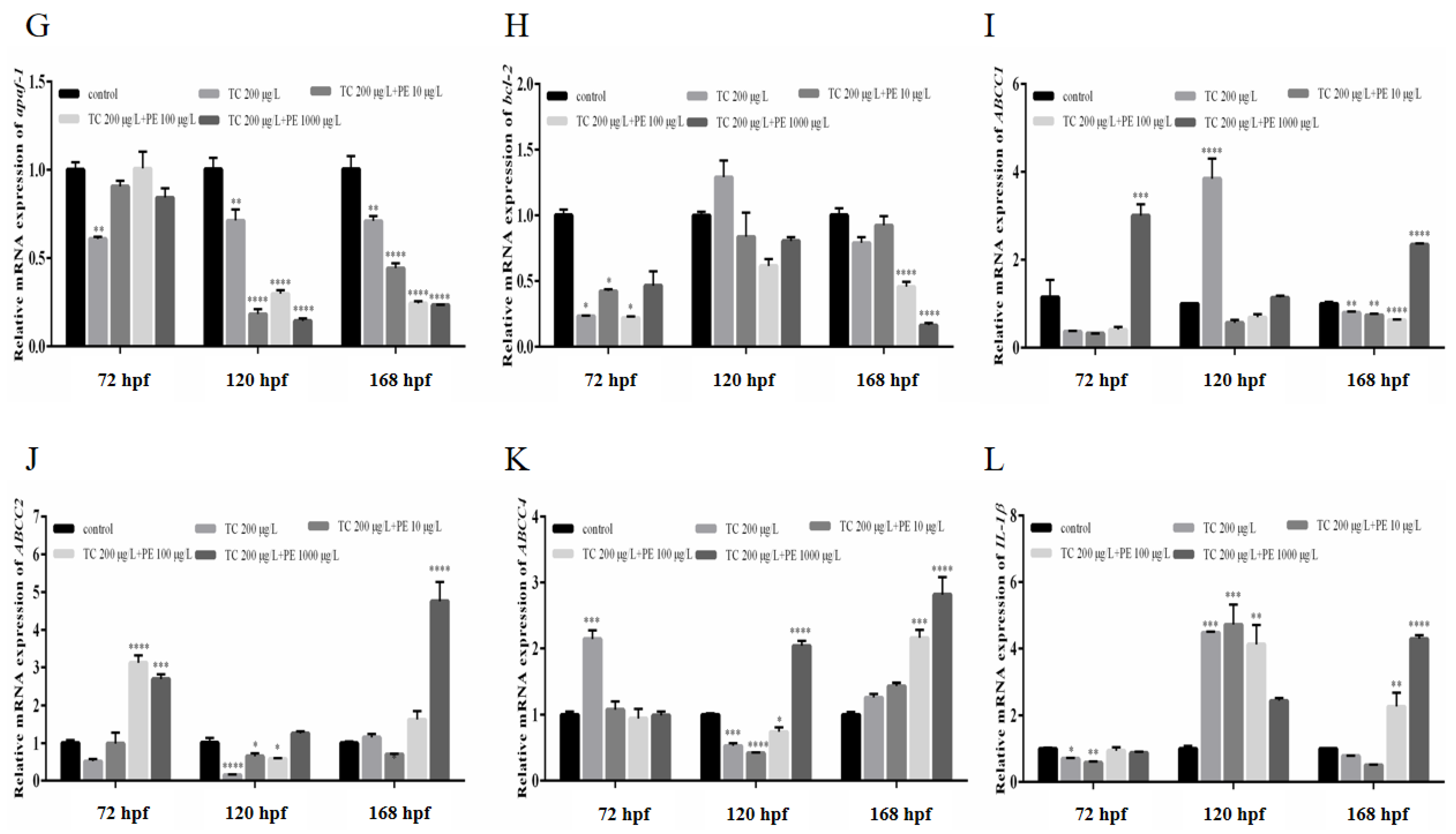
3.5. Effects of Combined TC-PE Exposure on Apoptosis in Zebrafish Larvae

After reaching the exposure endpoint (168 hpf), fluorescent signals were detected in certain regions of zebrafish larvae, such as the gastrointestinal tract, the caudal lateral region, and the eye region (Figure 5A). In Figure 1, PE-MPs can be observed in the gastrointestinal tract, which roughly coincides with the fluorescence of acridine orange (AO). The fluorescence intensity of AO labeling (which correlates with the process of cell death) was strong and statistically significantly different from that of controls following both individual and combined exposures (Figure 5B). This suggests that TC and TC-PE are present at the site and induce cytotoxicity.
AO labeling was localized in the caudal region, and weaker AO-labeled fluorescent spots were detected in the caudal region compared with the gastrointestinal tract (TC 200 μg/L + PE 100 μg/L, and TC 200 μg/L + PE 1000 μg/L). This result suggests that cytotoxicity may also exist in the caudal region. To the best of our knowledge, the caudal region is necessary for the swimming process of larvae. The current study’s findings imply that the reduced motility of the preceding embryos may be related to the cytotoxicity to which the region is exposed (Figure 3A), resulting in a possible impairment of larval swimming activity. However, the specific mechanism of cytological damage associated with these exposure conditions could not be determined. Studies have suggested that in other aquatic animals, such as fish and shellfish, PE-MP pollution leads to increased levels of ROS, apoptosis, and lipid peroxidation in their tissues, resulting in cytological damage [82]. Zhang et al. revealed that zebrafish larvae treated with tetracycline alone displayed a significant rise in the levels of ROS and apoptosis at 96 hpf, which occurred mainly in the caudal region [51]. We combined TC and PE to simulate the concentrations of pollutants in the water column and found that zebrafish larvae showed apoptosis at several sites when TC-PE exposure triggered ROS production and apoptosis, which led to oxidative stress, and the mechanisms underlying those consequences need to be studied. Nevertheless, additional dyes or molecular methods might be used in conjunction with this investigation.

3.6. TC and PE Exposure Induces Ectopic Sprouting of Intersegmental Blood Vessels (ISVs) in Zebrafish Larvae

To systematically evaluate the potential influence of exposure to antibiotics and microplastics on early vascular development in zebrafish larvae, several studies have used a Tg (flk: eGFP) line of zebrafish to dynamically follow the vascularization effects of MPs/NPs, where vascular endothelial cells display eGFP [42,77]. In this work, zebrafish embryos of the Tg (flk: eGFP) line were treated with different concentrations of exposure solutions up to 168 hpf, and different vascular phenotypes were found at early developmental stages (Figure 6). During zebrafish vascular development, ISVs first sprout in the dorsal aorta (DA) [83]. Sprouting ISVs (arrows) and ISV completion (asterisks) were determined (Figure 6A), and there was a dose-dependent increase in sprouting ISVs (aberrations in ISV completion) compared to the controls (Figure 6B).
Using this paradigm to examine in vivo angiogenesis, we found that zebrafish larvae exposed to both TC and PE had a greater percentage of ISV aberrations, with the number of ISV aberrations in the combined exposure group increasing in a dose-dependent manner. In addition, during the early stages of dorsal caudal fin formation [43], the diffusion of TC-PE in the bloodstream is relevant during angiogenesis. Sun et al. [43] treated zebrafish larvae with PE-NPs (100 and 200 μg/mL) and detected thrombus formation in the caudal region, revealing that PE-NP exposure inhibited angiogenesis. In the present study, the most important factor for angiogenesis, ISVs, was statistically significantly associated with an elevated count of aberrations in the medium- and high-dose groups of the combined exposure group. Our findings are consistent with known pollutants; for instance, vascular malformations were likewise caused when zebrafish are exposed to nanoplastic PE and PS of varying particle sizes [42]. In addition, exposure to MPs/NPs (400 nm and 1 μm) significantly impairs circulation in the caudal region [84], and some authors have attributed MP/NP-induced vascular toxicity to the local hypoxic microenvironment [85].

3.7. Different Forms of Exposure Cause Severe Inflammation in Zebrafish Larvae

Optically transparent zebrafish larvae provide an ideal platform to study inflammatory processes in an intact microenvironment. To detect the influences of TC and PE-MP exposure patterns on important immune cells, a zebrafish transgenic line with unique fluorescent labeling was developed for the real-time surveillance of possible immunotoxic actions in vivo. To track the dispersion of neutrophils at inflammatory loci, we present in this study a transgenic zebrafish (lyz: eGFP) line that is precisely tagged with the lysozyme C (lyz) promoter. We examined zebrafish embryos at 168 hpf and found that groups of neutrophils showed signals of different intensities (Figure 7). Fluorescence images showed that the exposed group alone showed a weaker fluorescent signal, while the fluorescent signal of the combined exposure group increased dose-dependently (Figure 7A), and the chemotaxis and recruitment of zebrafish embryo tail vein neutrophils were significantly increased (Figure 7B). Previous studies have shown that increased ROS production also stimulates apoptosis and triggers inflammation [86,87]. Neutrophils migrate in the liver in a dose- and size-dependent way, for instance, after being exposed to two PS-NPs with varying particle sizes, indicating that NPs with these sizes may cause an immediate inflammatory response [76]. Reactive oxygen species levels are increased and many cytokines are produced by neutrophils as part of the immune response in fish that are harmed by pollution or infections. Iftikhar et al. observed that zebrafish larvae exposed to 50 μg/L SMX expressed more pro-inflammatory cytokines, indicating that antibiotic levels in the environment could cause inflammation in zebrafish larvae [88]. Our study is consistent with previous studies in which antibiotics and microplastics resulted in increased neutrophil numbers in zebrafish larvae, suggesting that environmental levels of the TC and PE-MPs may induce inflammatory responses in healthy zebrafish larvae.

3.8. Impacts on Apoptotic and Inflammatory Gene Expression

The detection of inflammation-related genes (IL-1β), apoptosis-related genes (caspase-3, -6, -7, -9, bax, bcl-2, apaf-1, and aif) and multi-drug resistance genes (ABCC1, ABCC2, and ABCC4) expression levels in zebrafish larvae are shown in Figure 8.
Additionally crucial for pollutant-induced apoptosis is the caspase pathway [89]. Caspase-3 has been shown to be a key player in the downstream activation of the apoptotic pathway [90]. Apoptosis is often mediated by two caspase activation mechanisms that are connected to the caspase pathway. First, there is cytochrome c, and this dissociates from mitochondria and facilitates the activation of caspase-3 by forming complexes with cytochrome c/Apaf-1/caspase-9 in apoptotic vesicles, which ultimately results in apoptosis [91]. Apoptosis that is dependent on caspase-3 is necessary for zebrafish embryos to develop normally and contributes to the developmental period of zebrafish stress tolerance [88]. Usually, caspases-3/9 are activated in response to decreased bcl-2 expression [92]. In the present study, caspase-3, 6, 7, and 9 mRNA expression was upregulated most of the time in the medium–high-concentration group (TC 200 μg/L + PE 100 μg/L, TC 200 μg/L + PE 1000 μg/L) of the combined exposure group, and bcl-2 was downregulated most of the time in the medium–high-concentration group, and we conjectured that the combined exposure might activate the caspase pathway, triggering apoptosis (Figure 8A–E). The mRNA expression of related genes, such as apaf-1, bax, and aif, showed downregulation (Figure 8F–H). When considered collectively, these findings imply that TC and PE-induced apoptosis in the initial phases of zebrafish life may be primarily caused by the caspase-dependent apoptotic process.
In aquatic organisms, the presence of ABC transporters has been shown to be associated with multiple exogenous resistance (MXR) [93]. The expression levels of multi-drug resistance genes (ABCC1, ABCC2, and ABCC4) were in dynamic equilibrium (Figure 8I–K), with an upregulation in the combined exposure to a high concentration group (TC 200 μg/L + PE 1000 μg/L). Significant changes in mRNA expression after exposure to certain concentrations of TC and PE might indicate that the multidrug resistance system of zebrafish larvae was activated by transferring high concentrations of TC and PE via multidrug resistance-associated proteins.
To evaluate the immune response in zebrafish larvae, more significant immune-related transcripts, including inflammatory cytokines like IL-1β, were investigated. This gene was selected because research has shown that fish cytokines are impacted by antibiotic exposure [94]. When zebrafish larvae were evaluated for the levels of mRNA expression of the inflammation-related gene (IL-1β), the majority of the time nodes displayed an upregulation trend, as shown in Figure 8, with little trend in the exposure group alone and the coexposed low-concentration group (TC 200 μg/L + PE 10 μg/L), whereas inflammation is a dynamic phenomenon. The up-regulation of IL-1β, which is considered to be a key proinflammatory cytokine, may result in the production of ROS. This work expands on our knowledge of the fish early immune system and raises the possibility that exposure to microplastics and antibiotics may cause immune system disturbances in fish.

4. Conclusions

Tetracycline (TC) is the most regularly used antibiotic in marine aquaculture, while polyethylene (PE), the most popular plastic used in our life, is frequently detected in aquatic settings. In this study, we found that the combined use of PE enhanced the toxic impact of TC on zebrafish embryos/larvae in a dose-dependent manner, including both developmental toxicity (heart rate, locomotor ability, mortality, and the malformation rate) and physiological toxicity (oxidative stress, vascularity, the inflammatory response, and associated gene expression). The results imply that PE and TC may work together synergistically at lower effect levels, suggesting that synergistic toxic effects might happen at concentrations that are important to the environment. Additional research is needed to improve the understanding of the potential mechanisms of combined PE and TC contamination for a more complete assessment. The purpose of this study is to provide a more thorough understanding of potential environmental concerns by estimating the impacts of combinations of contaminants down to environmentally relevant quantities.

Supplementary Materials

The following supporting information can be downloaded at https://www.mdpi.com/article/10.3390/fishes10040150/s1: Table S1: The primer sequences of relative genes used in this study.

Author Contributions

Y.W., writing—reviewing and editing, writing—original draft preparation; Z.Z., conceptualization, methodology; R.Z., software; X.F., formal analysis; X.W., software; Y.H., investigation; H.G., supervision; M.Y., funding acquisition, project administration. All authors have read and agreed to the published version of the manuscript.

Funding

This project was supported by the National Natural Science Foundation of China, grant numbers 42377363 and 42177253; and the Guangdong Basic and Applied Basic Research Foundation, grant numbers 2022A1515010197, 2024A1515011401, and 2024A1515030201.

Institutional Review Board Statement

The animal study protocol was approved by the Experimental Animal Ethics Committee of South China Agricultural University (protocol code 2024G029, the approval date is 10 March 2024).

Data Availability Statement

The original contributions presented in the study are included in the article/Supplementary Materials; further inquiries can be directed to the corresponding author/s.

Conflicts of Interest

The authors declare that they have no known competing financial interests or personal relationships that could have appeared to influence the work reported in this paper.

References

  1. Hale, R.C.; Seeley, M.E.; Guardia, M.J.L.; Mai, L.; Zeng, E.Y. A Global Perspective on Microplastics. J. Geophys. Res. Ocean. 2020, 125, e2018JC014719. [Google Scholar] [CrossRef]
  2. Ivleva, N.P.; Wiesheu, A.C.; Niessner, R. Microplastic in Aquatic Ecosystems. Angew. Chem. Int. Ed. Engl. 2017, 56, 1720–1739. [Google Scholar] [CrossRef] [PubMed]
  3. Hammer, J.; Kraak, M.H.; Parsons, J.R. Plastics in the marine environment: The dark side of a modern gift. Rev. Environ. Contam. Toxicol. 2012, 220, 1–44. [Google Scholar] [CrossRef] [PubMed]
  4. Lin, L.; Zuo, L.Z.; Peng, J.P.; Cai, L.Q.; Fok, L.; Yan, Y.; Li, H.X.; Xu, X.R. Occurrence and distribution of microplastics in an urban river: A case study in the Pearl River along Guangzhou City, China. Sci. Total Environ. 2018, 644, 375–381. [Google Scholar] [CrossRef]
  5. Lebreton, L.; Slat, B.; Ferrari, F.; Sainte-Rose, B.; Aitken, J.; Marthouse, R.; Hajbane, S.; Cunsolo, S.; Schwarz, A.; Levivier, A.; et al. Evidence that the Great Pacific Garbage Patch is rapidly accumulating plastic. Sci. Rep. 2018, 8, 4666. [Google Scholar] [CrossRef]
  6. Gigault, J.; Halle, A.T.; Baudrimont, M.; Pascal, P.Y.; Gauffre, F.; Phi, T.L.; El Hadri, H.; Grassl, B.; Reynaud, S. Current opinion: What is a nanoplastic? Environ. Pollut. 2018, 235, 1030–1034. [Google Scholar] [CrossRef]
  7. Karim, M.E.; Sanjee, S.A.; Mahmud, S.; Shaha, M.; Moniruzzaman, M.; Das, K.C. Microplastics pollution in Bangladesh: Current scenario and future research perspective. Chem. Ecol. 2019, 36, 83–99. [Google Scholar] [CrossRef]
  8. Koehler, A.; Anderson, A.; Andrady, A.; Arthur, C.; Wyles, K. Sources, Fate and Effects of Microplastics in the Marine Environment: A Global Assessment; UNESCO: Paris, France, 2015. [Google Scholar]
  9. Tirkey, A.; Upadhyay, L.S.B. Microplastics: An overview on separation, identification and characterization of microplastics. Mar. Pollut. Bull. 2021, 170, 112604. [Google Scholar] [CrossRef]
  10. Andrady, A.L. The plastic in microplastics: A review. Mar. Pollut. Bull. 2017, 119, 12–22. [Google Scholar] [CrossRef]
  11. Abdurahman, A.; Li, S.; Li, Y.; Song, X.; Gao, R. Ecotoxicological effects of antibiotic adsorption behavior of microplastics and its management measures. Environ. Sci. Pollut. Res. Int. 2023, 30, 125370–125387. [Google Scholar] [CrossRef]
  12. da Costa, J.P.; Santos, P.S.M.; Duarte, A.C.; Rocha-Santos, T. (Nano)plastics in the environment-Sources, fates and effects. Sci. Total Environ. 2016, 566–567, 15–26. [Google Scholar] [CrossRef] [PubMed]
  13. Conkle, J.L.; Báez Del Valle, C.D.; Turner, J.W. Are We Underestimating Microplastic Contamination in Aquatic Environments? Environ. Manag. 2018, 61, 1–8. [Google Scholar] [CrossRef] [PubMed]
  14. Kovochich, M.; Liong, M.; Parker, J.A.; Oh, S.C.; Lee, J.P.; Xi, L.; Kreider, M.L.; Unice, K.M. Chemical mapping of tire and road wear particles for single particle analysis. Sci. Total Environ. 2021, 757, 144085. [Google Scholar] [CrossRef]
  15. Sahu, S.; Kaur, A.; Khatri, M.; Singh, G.; Arya, S.K. A review on cutinases enzyme in degradation of microplastics. J. Environ. Manag. 2023, 347, 119193. [Google Scholar] [CrossRef]
  16. Gallagher, A.; Rees, A.; Rowe, R.; Stevens, J.; Wright, P. Microplastics in the Solent estuarine complex, UK: An initial assessment. Mar. Pollut. Bull. 2016, 102, 243–249. [Google Scholar] [CrossRef]
  17. Zurub, R.E.; Cariaco, Y.; Wade, M.G.; Bainbridge, S.A. Microplastics exposure: Implications for human fertility, pregnancy and child health. Front. Endocrinol. 2023, 14, 1330396. [Google Scholar] [CrossRef]
  18. Sadri, S.S.; Thompson, R.C. On the quantity and composition of floating plastic debris entering and leaving the Tamar Estuary, Southwest England. Mar. Pollut. Bull. 2014, 81, 55–60. [Google Scholar] [CrossRef]
  19. Li, J.; Zhang, K.; Zhang, H. Adsorption of antibiotics on microplastics. Environ. Pollut. 2018, 237, 460–467. [Google Scholar] [CrossRef]
  20. Chen, G.; Li, Y.; Wang, J. Occurrence and ecological impact of microplastics in aquaculture ecosystems. Chemosphere 2021, 274, 129989. [Google Scholar] [CrossRef]
  21. Dong, H.; Chen, Y.; Wang, J.; Zhang, Y.; Zhang, P.; Li, X.; Zou, J.; Zhou, A. Interactions of microplastics and antibiotic resistance genes and their effects on the aquaculture environments. J. Hazard. Mater. 2021, 403, 123961. [Google Scholar] [CrossRef]
  22. Rubin, A.E.; Sarkar, A.K.; Zucker, I. Questioning the suitability of available microplastics models for risk assessment-A critical review. Sci. Total Environ. 2021, 788, 147670. [Google Scholar] [CrossRef] [PubMed]
  23. Kumar, R.; Manna, C.; Padha, S.; Verma, A.; Sharma, P.; Dhar, A.; Ghosh, A.; Bhattacharya, P. Micro(nano)plastics pollution and human health: How plastics can induce carcinogenesis to humans? Chemosphere 2022, 298, 134267. [Google Scholar] [CrossRef] [PubMed]
  24. Auta, H.S.; Emenike, C.U.; Fauziah, S.H. Distribution and importance of microplastics in the marine environment: A review of the sources, fate, effects, and potential solutions. Environ. Int. 2017, 102, 165–176. [Google Scholar] [CrossRef] [PubMed]
  25. Ragu Prasath, A.; Sudhakar, C.; Selvam, K. Microplastics in the environment: Types, sources, and impact on human and aquatic systems. Bioresour. Technol. Rep. 2025, 29, 102055. [Google Scholar] [CrossRef]
  26. Cherian, T.; Ragavendran, C.; Vijayan, S.; Kurien, S.; Peijnenburg, W.J.G.M. A review on the fate, human health and environmental impacts, as well as regulation of antibiotics used in aquaculture. Environ. Adv. 2023, 13, 100411. [Google Scholar] [CrossRef]
  27. Shao, Y.; Wang, Y.; Yuan, Y.; Xie, Y. A systematic review on antibiotics misuse in livestock and aquaculture and regulation implications in China. Sci. Total Environ. 2021, 798, 149205. [Google Scholar] [CrossRef]
  28. Chen, X.; Yang, Y.; Ke, Y.; Chen, C.; Xie, S. A comprehensive review on biodegradation of tetracyclines: Current research progress and prospect. Sci. Total Environ. 2022, 814, 152852. [Google Scholar] [CrossRef]
  29. Xu, L.; Zhang, H.; Xiong, P.; Zhu, Q.; Liao, C.; Jiang, G. Occurrence, fate, and risk assessment of typical tetracycline antibiotics in the aquatic environment: A review. Sci. Total Environ. 2021, 753, 141975. [Google Scholar] [CrossRef]
  30. Lundström, S.V.; Östman, M.; Bengtsson-Palme, J.; Rutgersson, C.; Thoudal, M.; Sircar, T.; Blanck, H.; Eriksson, K.M.; Tysklind, M.; Flach, C.F.; et al. Minimal selective concentrations of tetracycline in complex aquatic bacterial biofilms. Sci. Total Environ. 2016, 553, 587–595. [Google Scholar] [CrossRef]
  31. Lulijwa, R.; Rupia, E.J.; Alfaro, A.C. Antibiotic use in aquaculture, policies and regulation, health and environmental risks: A review of the top 15 major producers. Rev. Aquac. 2020, 12, 640–663. [Google Scholar] [CrossRef]
  32. Farid, M.U.; Choi, P.J.; Kharraz, J.A.; Lao, J.-Y.; St-Hilaire, S.; Ruan, Y.; Lam, P.K.S.; An, A.K. Hybrid nanobubble-forward osmosis system for aquaculture wastewater treatment and reuse. Chem. Eng. J. 2022, 435, 135164. [Google Scholar] [CrossRef]
  33. Chen, J.; Sun, R.; Pan, C.; Sun, Y.; Mai, B.; Li, Q.X. Antibiotics and Food Safety in Aquaculture. J. Agric. Food Chem. 2020, 68, 11908–11919. [Google Scholar] [CrossRef] [PubMed]
  34. Guo, X.; Chen, C.; Wang, J. Sorption of sulfamethoxazole onto six types of microplastics. Chemosphere 2019, 228, 300–308. [Google Scholar] [CrossRef] [PubMed]
  35. Zhang, H.; Wang, J.; Zhou, B.; Zhou, Y.; Dai, Z.; Zhou, Q.; Chriestie, P.; Luo, Y. Enhanced adsorption of oxytetracycline to weathered microplastic polystyrene: Kinetics, isotherms and influencing factors. Environ. Pollut. 2018, 243, 1550–1557. [Google Scholar] [CrossRef]
  36. Chen, Y.; Li, J.; Wang, F.; Yang, H.; Liu, L. Adsorption of tetracyclines onto polyethylene microplastics: A combined study of experiment and molecular dynamics simulation. Chemosphere 2021, 265, 129133. [Google Scholar] [CrossRef]
  37. Yu, Z.; Zhang, L.; Huang, Q.; Dong, S.; Wang, X.; Yan, C. Combined effects of micro-/nano-plastics and oxytetracycline on the intestinal histopathology and microbiome in zebrafish (Danio rerio). Sci. Total Environ. 2022, 843, 156917. [Google Scholar] [CrossRef]
  38. Liao, X.; Zhao, P.; Hou, L.; Adyari, B.; Xu, E.G.; Huang, Q.; Hu, A. Network analysis reveals significant joint effects of microplastics and tetracycline on the gut than the gill microbiome of marine medaka. J. Hazard. Mater. 2023, 442, 129996. [Google Scholar] [CrossRef]
  39. Han, Y.; Zhou, W.; Tang, Y.; Shi, W.; Shao, Y.; Ren, P.; Zhang, J.; Xiao, G.; Sun, H.; Liu, G. Microplastics aggravate the bioaccumulation of three veterinary antibiotics in the thick shell mussel Mytilus coruscus and induce synergistic immunotoxic effects. Sci. Total Environ. 2021, 770, 145273. [Google Scholar] [CrossRef]
  40. Xu, K.; Tang, Z.; Liu, S.; Liao, Z.; Xia, H.; Liu, L.; Wang, Z.; Qi, P. Effects of low concentrations copper on antioxidant responses, DNA damage and genotoxicity in thick shell mussel Mytilus coruscus. Fish. Shellfish. Immunol. 2018, 82, 77–83. [Google Scholar] [CrossRef]
  41. Dai, L.; Luo, J.; Feng, M.; Wang, M.; Zhang, J.; Cao, X.; Yang, X.; Li, J. Nanoplastics exposure induces vascular malformation by interfering with the VEGFA/VEGFR pathway in zebrafish (Danio rerio). Chemosphere 2023, 312, 137360. [Google Scholar] [CrossRef]
  42. Santos, A.L.; Rodrigues, L.C.; Rodrigues, C.C.; Cirqueira, F.; Malafaia, G.; Rocha, T.L. Polystyrene nanoplastics induce developmental impairments and vasotoxicity in zebrafish (Danio rerio). J. Hazard. Mater. 2024, 464, 132880. [Google Scholar] [CrossRef] [PubMed]
  43. Sun, M.; Ding, R.; Ma, Y.; Sun, Q.; Ren, X.; Sun, Z.; Duan, J. Cardiovascular toxicity assessment of polyethylene nanoplastics on developing zebrafish embryos. Chemosphere 2021, 282, 131124. [Google Scholar] [CrossRef]
  44. You, X.; Cao, X.; Zhang, X.; Guo, J.; Sun, W. Unraveling individual and combined toxicity of nano/microplastics and ciprofloxacin to Synechocystis sp. at the cellular and molecular levels. Environ. Int. 2021, 157, 106842. [Google Scholar] [CrossRef] [PubMed]
  45. Guo, X.; Cai, Y.; Ma, C.; Han, L.; Yang, Z. Combined toxicity of micro/nano scale polystyrene plastics and ciprofloxacin to Corbicula fluminea in freshwater sediments. Sci. Total Environ. 2021, 789, 147887. [Google Scholar] [CrossRef]
  46. Fred-Ahmadu, O.H.; Bhagwat, G.; Oluyoye, I.; Benson, N.U.; Ayejuyo, O.O.; Palanisami, T. Interaction of chemical contaminants with microplastics: Principles and perspectives. Sci. Total Environ. 2020, 706, 135978. [Google Scholar] [CrossRef]
  47. Dmytriw, A.A. The microplastics menace: An emerging link to environment and health. Sci. Total Environ. 2020, 707, 135558. [Google Scholar] [CrossRef]
  48. Suzuki, S.; Nakanishi, S.; Tamminen, M.; Yokokawa, T.; Sato-Takabe, Y.; Ohta, K.; Chou, H.Y.; Muziasari, W.I.; Virta, M. Occurrence of sul and tet(M) genes in bacterial community in Japanese marine aquaculture environment throughout the year: Profile comparison with Taiwanese and Finnish aquaculture waters. Sci. Total Environ. 2019, 669, 649–656. [Google Scholar] [CrossRef]
  49. Sarmah, A.K.; Meyer, M.T.; Boxall, A.B. A global perspective on the use, sales, exposure pathways, occurrence, fate and effects of veterinary antibiotics (VAs) in the environment. Chemosphere 2006, 65, 725–759. [Google Scholar] [CrossRef]
  50. Zhao, Y.; Qin, Z.; Huang, Z.; Bao, Z.; Luo, T.; Jin, Y. Effects of polyethylene microplastics on the microbiome and metabolism in larval zebrafish. Environ. Pollut. 2021, 282, 117039. [Google Scholar] [CrossRef]
  51. Zhang, Q.; Cheng, J.; Xin, Q. Effects of tetracycline on developmental toxicity and molecular responses in zebrafish (Danio rerio) embryos. Ecotoxicology 2015, 24, 707–719. [Google Scholar] [CrossRef]
  52. Zhao, X.; Liu, Z.; Zhang, Y.; Pan, Y.; Wang, T.; Wang, Z.; Li, Z.; Zeng, Q.; Qian, Y.; Qiu, J.; et al. Developmental effects and lipid disturbances of zebrafish embryos exposed to three newly recognized bisphenol A analogues. Environ. Int. 2024, 189, 108795. [Google Scholar] [CrossRef] [PubMed]
  53. Li, Y.; Chen, Q.; Liu, Y.; Bi, L.; Jin, L.; Xu, K.; Peng, R. High glucose-induced ROS-accumulation in embryo-larval stages of zebrafish leads to mitochondria-mediated apoptosis. Apoptosis 2022, 27, 509–520. [Google Scholar] [CrossRef] [PubMed]
  54. Xu, Y.; Peng, T.; Xiang, Y.; Liao, G.; Zou, F.; Meng, X. Neurotoxicity and gene expression alterations in zebrafish larvae in response to manganese exposure. Sci. Total Environ. 2022, 825, 153778. [Google Scholar] [CrossRef] [PubMed]
  55. Zhang, R.; Silic, M.R.; Schaber, A.; Wasel, O.; Freeman, J.L.; Sepúlveda, M.S. Exposure route affects the distribution and toxicity of polystyrene nanoplastics in zebrafish. Sci. Total Environ. 2020, 724, 138065. [Google Scholar] [CrossRef]
  56. LeMoine, C.M.R.; Kelleher, B.M.; Lagarde, R.; Northam, C.; Elebute, O.O.; Cassone, B.J. Transcriptional effects of polyethylene microplastics ingestion in developing zebrafish (Danio rerio). Environ. Pollut. 2018, 243, 591–600. [Google Scholar] [CrossRef]
  57. Tsay, H.J.; Wang, Y.H.; Chen, W.L.; Huang, M.Y.; Chen, Y.H. Treatment with sodium benzoate leads to malformation of zebrafish larvae. Neurotoxicol. Teratol. 2007, 29, 562–569. [Google Scholar] [CrossRef]
  58. Wiegand, J.; Avila-Barnard, S.; Nemarugommula, C.; Lyons, D.; Zhang, S.; Stapleton, H.M.; Volz, D.C. Triphenyl phosphate-induced pericardial edema in zebrafish embryos is dependent on the ionic strength of exposure media. Environment International 2023, 172, 107757. [Google Scholar] [CrossRef]
  59. Lett, Z.; Hall, A.; Skidmore, S.; Alves, N.J. Environmental microplastic and nanoplastic: Exposure routes and effects on coagulation and the cardiovascular system. Environ. Pollut. 2021, 291, 118190. [Google Scholar] [CrossRef]
  60. Asharani, P.V.; Lian Wu, Y.; Gong, Z.; Valiyaveettil, S. Toxicity of silver nanoparticles in zebrafish models. Nanotechnology 2008, 19, 255102. [Google Scholar] [CrossRef]
  61. Lu, J.; Wu, J.; Gong, L.; Cheng, Y.; Yuan, Q.; He, Y. Combined toxicity of polystyrene microplastics and sulfamethoxazole on zebrafish embryos. Environ. Sci. Pollut. Res. Int. 2022, 29, 19273–19282. [Google Scholar] [CrossRef]
  62. Tshering, G.; Plengsuriyakarn, T.; Na-Bangchang, K.; Pimtong, W. Embryotoxicity evaluation of atractylodin and β-eudesmol using the zebrafish model. Comp. Biochem. Physiol. C Toxicol. Pharmacol. 2021, 239, 108869. [Google Scholar] [CrossRef] [PubMed]
  63. Yan, Z.; Yang, Q.; Jiang, W.; Lu, J.; Xiang, Z.; Guo, R.; Chen, J. Integrated toxic evaluation of sulfamethazine on zebrafish: Including two lifespan stages (embryo-larval and adult) and three exposure periods (exposure, post-exposure and re-exposure). Chemosphere 2018, 195, 784–792. [Google Scholar] [CrossRef] [PubMed]
  64. Wu, L.; Shao, Y.; Hu, Z.; Gao, H. Effects of soluble sulfide on zebrafish (Danio rerio) embryonic development. Environ. Toxicol. Pharmacol. 2016, 42, 183–189. [Google Scholar] [CrossRef] [PubMed]
  65. Felker, A.; Prummel, K.D.; Merks, A.M.; Mickoleit, M.; Brombacher, E.C.; Huisken, J.; Panáková, D.; Mosimann, C. Continuous addition of progenitors forms the cardiac ventricle in zebrafish. Nat. Commun. 2018, 9, 2001. [Google Scholar] [CrossRef]
  66. Xu, L.; Yang, X.; He, Y.; Hu, Q.; Fu, Z. Combined exposure to titanium dioxide and tetracycline induces neurotoxicity in zebrafish. Comp. Biochem. Physiol. C Toxicol. Pharmacol. 2023, 267, 109562. [Google Scholar] [CrossRef]
  67. Yang, X.; Sun, Z.; Wang, W.; Zhou, Q.; Shi, G.; Wei, F.; Jiang, G. Developmental toxicity of synthetic phenolic antioxidants to the early life stage of zebrafish. Sci. Total Environ. 2018, 643, 559–568. [Google Scholar] [CrossRef]
  68. Wang, M.; Chen, X.; Zhang, R.; Zhao, J.; Yang, C.; Wu, L. Developmental toxicity and transcriptome analysis of 4-epianhydrotetracycline to zebrafish (Danio rerio) embryos. Sci. Total Environ. 2020, 734, 139227. [Google Scholar] [CrossRef]
  69. Lin, Y.; Yu, J.; Wang, M.; Wu, L. Toxicity of single and combined 4-epianhydrotetracycline and cadmium at environmentally relevant concentrations on the zebrafish embryos (Danio rerio). Environ. Pollut. 2023, 316, 120543. [Google Scholar] [CrossRef]
  70. Cormier, B.; Le Bihanic, F.; Cabar, M.; Crebassa, J.C.; Blanc, M.; Larsson, M.; Dubocq, F.; Yeung, L.; Clérandeau, C.; Keiter, S.H.; et al. Chronic feeding exposure to virgin and spiked microplastics disrupts essential biological functions in teleost fish. J. Hazard. Mater. 2021, 415, 125626. [Google Scholar] [CrossRef]
  71. Tarasco, M.; Gavaia, P.J.; Bensimon-Brito, A.; Cordelières, F.P.; Santos, T.; Martins, G.; de Castro, D.T.; Silva, N.; Cabrita, E.; Bebianno, M.J.; et al. Effects of pristine or contaminated polyethylene microplastics on zebrafish development. Chemosphere 2022, 303, 135198. [Google Scholar] [CrossRef]
  72. Zhang, Z.; Li, X.; Li, J.; Pan, Y.; Zhuang, Z.; Zhang, X.; Chen, C.; Liu, Y.; Zhang, L.; Luo, Y.; et al. The combined toxic effects of polystyrene microplastics and different forms of arsenic on the zebrafish embryos (Danio rerio). Sci. Total Environ. 2023, 887, 164017. [Google Scholar] [CrossRef] [PubMed]
  73. Wang, Y.; Pang, S.; Chen, Z.; Wang, J.; Liu, L.; Zhang, L.; Wang, F.; Song, M. Surface Modification Determines the Distribution and Toxicity of Quantum Dots during the Development of Early Staged Zebrafish. Environ. Sci. Technol. 2023, 57, 10574–10581. [Google Scholar] [CrossRef] [PubMed]
  74. Guo, Y.; Weck, J.; Sundaram, R.; Goldstone, A.E.; Louis, G.B.; Kannan, K. Urinary concentrations of phthalates in couples planning pregnancy and its association with 8-hydroxy-2′-deoxyguanosine, a biomarker of oxidative stress: Longitudinal investigation of fertility and the environment study. Environ. Sci. Technol. 2014, 48, 9804–9811. [Google Scholar] [CrossRef]
  75. Tugasworo, D.; Prasetyo, A.; Kurnianto, A.; Retnaningsih, R.; Andhitara, Y.; Ardhini, R.; Budiman, J. Malondialdehyde (MDA) and 8-hydroxy-2′-deoxyguanosine (8-OHdG) in ischemic stroke: A systematic review. Egypt. J. Neurol. Psychiatry Neurosurg. 2023, 59, 87. [Google Scholar] [CrossRef]
  76. Cheng, H.; Duan, Z.; Wu, Y.; Wang, Y.; Zhang, H.; Shi, Y.; Zhang, H.; Wei, Y.; Sun, H. Immunotoxicity responses to polystyrene nanoplastics and their related mechanisms in the liver of zebrafish (Danio rerio) larvae. Environ. Int. 2022, 161, 107128. [Google Scholar] [CrossRef]
  77. Huang, W.; Mo, J.; Li, J.; Wu, K. Exploring developmental toxicity of microplastics and nanoplastics (MNPS): Insights from investigations using zebrafish embryos. Sci. Total Environ. 2024, 933, 173012. [Google Scholar] [CrossRef]
  78. Wan, Z.; Wang, C.; Zhou, J.; Shen, M.; Wang, X.; Fu, Z.; Jin, Y. Effects of polystyrene microplastics on the composition of the microbiome and metabolism in larval zebrafish. Chemosphere 2019, 217, 646–658. [Google Scholar] [CrossRef]
  79. Lu, Y.; Zhang, Y.; Deng, Y.; Jiang, W.; Zhao, Y.; Geng, J.; Ding, L.; Ren, H. Uptake and Accumulation of Polystyrene Microplastics in Zebrafish (Danio rerio) and Toxic Effects in Liver. Environ. Sci. Technol. 2016, 50, 4054–4060. [Google Scholar] [CrossRef]
  80. Qiao, R.; Sheng, C.; Lu, Y.; Zhang, Y.; Ren, H.; Lemos, B. Microplastics induce intestinal inflammation, oxidative stress, and disorders of metabolome and microbiome in zebrafish. Sci. Total Environ. 2019, 662, 246–253. [Google Scholar] [CrossRef]
  81. Qiang, L.; Cheng, J. Exposure to microplastics decreases swimming competence in larval zebrafish (Danio rerio). Ecotoxicol. Environ. Saf. 2019, 176, 226–233. [Google Scholar] [CrossRef]
  82. Lee, J.H.; Kang, J.C.; Kim, J.H. Toxic effects of microplastic (Polyethylene) on fish: Accumulation, hematological parameters and antioxidant responses in Korean Bullhead, Pseudobagrus fulvidraco. Sci. Total Environ. 2023, 877, 162874. [Google Scholar] [CrossRef] [PubMed]
  83. Blum, Y.; Belting, H.G.; Ellertsdottir, E.; Herwig, L.; Lüders, F.; Affolter, M. Complex cell rearrangements during intersegmental vessel sprouting and vessel fusion in the zebrafish embryo. Dev. Biol. 2008, 316, 312–322. [Google Scholar] [CrossRef] [PubMed]
  84. Park, S.H.; Kim, K. Microplastics induced developmental toxicity with microcirculation dysfunction in zebrafish embryos. Chemosphere 2022, 286, 131868. [Google Scholar] [CrossRef] [PubMed]
  85. Duan, Z.; Duan, X.; Zhao, S.; Wang, X.; Wang, J.; Liu, Y.; Peng, Y.; Gong, Z.; Wang, L. Barrier function of zebrafish embryonic chorions against microplastics and nanoplastics and its impact on embryo development. J. Hazard. Mater. 2020, 395, 122621. [Google Scholar] [CrossRef]
  86. Lu, K.; Qiao, R.; An, H.; Zhang, Y. Influence of microplastics on the accumulation and chronic toxic effects of cadmium in zebrafish (Danio rerio). Chemosphere 2018, 202, 514–520. [Google Scholar] [CrossRef]
  87. Redza-Dutordoir, M.; Averill-Bates, D.A. Activation of apoptosis signalling pathways by reactive oxygen species. Biochim. Biophys. Acta 2016, 1863, 2977–2992. [Google Scholar] [CrossRef]
  88. Iftikhar, N.; Konig, I.; English, C.; Ivantsova, E.; Souders, C.L., 2nd; Hashmi, I.; Martyniuk, C.J. Sulfamethoxazole (SMX) Alters Immune and Apoptotic Endpoints in Developing Zebrafish (Danio rerio). Toxics 2023, 11, 178. [Google Scholar] [CrossRef]
  89. Xiong, Q.; Xie, P.; Li, H.; Hao, L.; Li, G.; Qiu, T.; Liu, Y. Involvement of Fas/FasL system in apoptotic signaling in testicular germ cells of male Wistar rats injected i.v. with microcystins. Toxicon 2009, 54, 1–7. [Google Scholar] [CrossRef]
  90. Liu, C.; Yu, K.; Shi, X.; Wang, J.; Lam, P.K.; Wu, R.S.; Zhou, B. Induction of oxidative stress and apoptosis by PFOS and PFOA in primary cultured hepatocytes of freshwater tilapia (Oreochromis niloticus). Aquat. Toxicol. 2007, 82, 135–143. [Google Scholar] [CrossRef]
  91. Gao, D.; Xu, Z.; Zhang, X.; Zhu, C.; Wang, Y.; Min, W. Cadmium triggers kidney cell apoptosis of purse red common carp (Cyprinus carpio) without caspase-8 activation. Dev. Comp. Immunol. 2013, 41, 728–737. [Google Scholar] [CrossRef]
  92. Morales-Cano, D.; Calviño, E.; Rubio, V.; Herráez, A.; Sancho, P.; Tejedor, M.C.; Diez, J.C. Apoptosis induced by paclitaxel via Bcl-2, Bax and caspases 3 and 9 activation in NB4 human leukaemia cells is not modulated by ERK inhibition. Exp. Toxicol. Pathol. 2013, 65, 1101–1108. [Google Scholar] [CrossRef] [PubMed]
  93. Kingtong, S.; Chitramvong, Y.; Janvilisri, T. ATP-binding cassette multidrug transporters in Indian-rock oyster Saccostrea forskali and their role in the export of an environmental organic pollutant tributyltin. Aquat. Toxicol. 2007, 85, 124–132. [Google Scholar] [CrossRef] [PubMed]
  94. Lin, T.; Yu, S.; Chen, Y.; Chen, W. Integrated biomarker responses in zebrafish exposed to sulfonamides. Environ. Toxicol. Pharmacol. 2014, 38, 444–452. [Google Scholar] [CrossRef] [PubMed]
Figure 1. Enrichment of PE-MPs in zebrafish larvae. (A) Fluorescence intensity distribution diagram; (B) fluorescence intensity distribution value of each group. The red box indicates the location of PE-MPs. The data considered to display significant differences versus the control are marked as * p < 0.05 and **** p < 0.0001. ND means not detected.
Fishes 10 00150 g001
Figure 2. Malformation and mortality rates in zebrafish. (A) Control larvae were normal in size and had no obvious malformations. (B) Larvae exposed to TC alone had a low rate of larval malformations. (CE) Morphology of malformed larvae exposed to a combination of TC-PE that had a high rate of malformations. (F,G) Malformation and mortality rate statistics. The red triangle indicates the location of the morphology of zebrafish larvae. The data considered to display significant differences versus the control are marked as * p < 0.05, ** p < 0.01, and **** p < 0.0001.
Fishes 10 00150 g002aFishes 10 00150 g002b
Figure 3. Related indicators of developmental toxicity in zebrafish larvae. (A) Motility of zebrafish embryos at 24 hpf. (B) Hatching statistics of zebrafish at 48–72 hpf; no statistically significant differences were observed. (C) Heart rate of zebrafish larvae at 48–96 hpf. (D) Zebrafish larval length at 168 hpf. The data considered to show significant differences versus the control are marked as * p < 0.05, ** p < 0.01, and **** p < 0.0001.
Fishes 10 00150 g003
Figure 4. Oxidative stress levels in the different treatment groups. (A) ROS fluorescent signal localization in zebrafish larvae. Fluorescence was detected in different parts of the small intestine, gallbladder, liver, and eyes. (B) ROS levels in zebrafish larvae, with considerably increased ROS plotted statistically. (C) The MDA levels of zebrafish larvae were all significantly elevated. (D) CAT levels were significantly elevated in a dose-dependent manner. (E) Decrease in the level of the antioxidant enzyme SOD. (F) Diminished total protein content in zebrafish larvae bodies. The red box indicates the ROS producing region of larvae. The data considered to show significant differences versus the control are marked as * p < 0.05, ** p < 0.01, *** p < 0.001, and **** p < 0.0001. No statistically significant differences (NS).
Fishes 10 00150 g004
Figure 5. Zebrafish larvae stained with acid orange after exposure for 168 hpf. (A) Apoptotic cells were mainly distributed in the head, gastrointestinal, and caudal regions; red arrows and boxes indicate apoptotic cells. (B) AO levels in zebrafish larvae. The red boxes show apoptosis in the gastrointestinal tract and the eye region, and the arrows show apoptosis in the caudal region of the larvae. The data considered to show significant differences versus the control are marked as *** p < 0.001, and **** p < 0.0001.
Fishes 10 00150 g005
Figure 6. TC and PE-MP exposure patterns induced intravascular malformations in zebrafish larvae. (A) Representative images of ISV vascular malformations in zebrafish larvae exposed for 168 hpf. (B) Rate of vascular malformations in zebrafish larvae. The arrows indicate ectopic sprouting ISVs and asterisks indicate fully formed ISVs. The data that were considered to show significant differences versus the control are marked as * p < 0.05, and **** p < 0.0001.
Fishes 10 00150 g006
Figure 7. The two pollutants caused different levels of inflammatory responses in zebrafish larvae. (A) Fluorescent images of inflammatory responses in zebrafish larvae. (B) Statistical graph of fluorescent signals of inflammatory responses in zebrafish larvae. The red box shows region where inflammatory cells are distributed. The data that were considered to show significant differences versus the control are marked as *** p < 0.001, and **** p < 0.0001.
Fishes 10 00150 g007
Figure 8. Control of the mRNA levels of related genes in model zebrafish larvae exposed to varying doses of TC-PE for different times. (AH) Apoptosis-related genes (caspase-3, -6, -7, -9, bax, bcl-2, apaf-1, and aif) expression levels in zebrafish larvae. (IK) multi-drug resistance genes (ABCC1, ABCC2, and ABCC4) expression levels in zebrafish larvae. (L) inflammation-related genes (IL-1β) expression levels in zebrafish larvae. The means ± SEs are used to express the data. The data that were considered to show significant differences versus the control are marked as * p < 0.05, ** p < 0.01, *** p < 0.001, and **** p < 0.0001.
Fishes 10 00150 g008aFishes 10 00150 g008b
Disclaimer/Publisher’s Note: The statements, opinions and data contained in all publications are solely those of the individual author(s) and contributor(s) and not of MDPI and/or the editor(s). MDPI and/or the editor(s) disclaim responsibility for any injury to people or property resulting from any ideas, methods, instructions or products referred to in the content.

