Article

Methyltransferase HsdM Regulates the Pathogenicity of Streptococcus agalactiae to Nile Tilapia (Oreochromis niloticus)

1
College of Animal Science and Technology, Guangxi University, Nanning 530004, China
2
Guangdong Provincial Key Laboratory of Aquatic Animal Disease Control and Healthy Culture, Key Laboratory of Control for Disease of Aquatic Animals of Guangdong Higher Education Institutes, Fisheries College, Guangdong Ocean University, Zhanjiang 524088, China
3
Key Laboratory of Fishery Drug Development, Ministry of Agriculture and Rural Affairs, Pearl River Fisheries Research Institute, Chinese Academy of Fishery Sciences, Guangzhou 510380, China
*
Authors to whom correspondence should be addressed.
Fishes 2025, 10(2), 86; https://doi.org/10.3390/fishes10020086
Submission received: 21 January 2025 / Revised: 13 February 2025 / Accepted: 17 February 2025 / Published: 19 February 2025
(This article belongs to the Special Issue Prevention and Control of Aquatic Animal Diseases)

Abstract

:
DNA methylation is a critical mechanism for regulating gene expression in bacteria and plays an essential role in bacterial pathogenesis. A mutant, WC1535ΔhsdM, lacking hsdM encoding a DNA methyltransferase was constructed using homologous recombination technology. The growth, hemolytic activity, and capsule formation of the mutant were analyzed. The dynamic distribution of the wild-type (WT) and mutant strains in tilapia tissues after artificial infection was determined. The adhesion, invasion, anti-phagocytic, and whole-blood survival abilities of the WT and mutant strains were analyzed. Tilapia were intraperitoneally injected with the WT or mutant strains, and the LD50 values were determined. The expression levels of the immune-related genes in tilapia were analyzed by qRT-PCR. The mutant showed faster growth during the logarithmic growth period (5–10 h) and lower hemolytic activity than the WT strain. Mutant loads in tilapia tissues were significantly lower than those of the WT strain. Mutant strain adhesion to epithelial cells was significantly reduced, it was more easily engulfed by macrophages, and it had decreased intracellular survival. The LD50 of the mutant was 2.06 times higher than that of the WT strain, indicating decreased pathogenicity. Expression levels of immune-related genes IL-1β, IL-6, IFN-γ, and TNF-α in tilapia induced by the mutant were lower than those by the WT strain. In conclusion, the WC1535ΔhsdM mutant exhibited an increased growth rate and decreased hemolytic activity, tissue colonization, and pathogenicity, indicating that HsdM could regulate S. agalactiae growth and pathogenicity. This study provides new insights into the pathogenesis of piscine S. agalactiae.
Key Contribution: HsdM modulates the growth rate of S. agalactiae, decreases its hemolytic activity, and reduces the pathogenicity of S. agalactiae in tilapia, thereby serving as an important virulence regulator.

1. Introduction

Streptococcus agalactiae (Group B Streptococcus, GBS) is a zoonotic pathogen that can infect various aquatic animals including tilapia (Oreochromis sp.) [1], golden pomfret (Trachinotus blochii) [2], and bighead carp (Aristichthys nobilis) [3]. Streptococcosis caused by GBS poses a serious threat to aquaculture development, especially in tilapia aquaculture. The predominant GBS serotypes responsible for tilapia streptococcosis are Ia and Ib in China [4]. GBS can resist phagocytosis by fish immune cells and evade host innate immunity, resulting in exophthalmia, meningitis, sepsis, and other clinical signs [1,5]. Phagocytosis is an important part of the host’s innate immunity, and phagocytes eliminate pathogenic bacteria from the host by internalizing and killing the pathogens. The major virulence factors of GBS include hemolysin, hyaluronidase, the C5a enzyme, capsular polysaccharide, fibrinogen-binding protein, and superoxide dismutase, which play important roles in the adhesion, invasion, and escape of GBS from host cells [6].
DNA carries genetic information in an organism and is responsible for the faithful transmission of genetic material across generations [7]. DNA methylation is the main epigenetic regulator in prokaryotic organisms and can assist bacteria in establishing restriction modification systems to resist the invasion of foreign DNA and control the expression of genes without changing the nucleotide sequence [8]. DNA methylation is a modification that transfers methyl groups from S-adenosyl-methionine (SAM) to DNA under the catalysis of DNA Methyltransferases. DNA methylation primarily involves 6-methyladenine (m6A) at N6 and 5-methylcytosine (m5C) at C5, of which m5C is mainly found in eukaryotes, whereas m6A is mainly found in prokaryotes.
Orphan DNA methyltransferases in bacteria can perform independent methylation functions. Common orphan DNA methyltransferases include DNA adenine methyltransferase (Dam), DNA cytosine methyltransferase (Dcm), and cell cycle-regulation methyltransferase (CcrM) [9]. In addition, the DNA methyltransferases YhdJ and HsdM are also involved in bacterial DNA adenine methylation. Dam was the first orphan methyltransferase identified in Escherichia coli, and its gene sequence is highly conserved in E. coli, Salmonella typhimurium, Yersinia pseudotuberculosis, and Vibrio cholera [10]. Dam is closely associated with bacterial pathogenicity. For example, the deletion of the dam gene in Salmonella enteritidis can significantly reduce the ability of the mutant to infect mice [11]. S. typhimurium with the deletion of dam reduced its pathogenicity, resulting in the LD50 of the mutant being more than 1000-fold higher than that of the wild-type (WT) strain [12].
The type I restriction modification (RM) system comprises restriction enzymes (HsdR), modified methyltransferase (HsdM), and specific subunits (HsdS). A typical type I RM system is a trimeric protein complex of 2R (HsdR) + 2M (HsdM) + S (HsdS), and each HsdS consists of two target recognition domains (TRD) [13,14]. HsdM in the type I RM system catalyzes the transfer of methyl groups from SAM to the exocyclic amino group of adenine to form the N6-methyladenosine (m6A) modification. The deletion of hsdM in Aeromonas veronii leads to the downregulation of the expression of key genes involved in flagella synthesis, resulting in a significant decrease in bacterial motility [15]. HsdM in the strain Mycobacterium tuberculosis 11826 is closely related to the methylated motifs of GTAYNNNNATC and affects drug resistance [16]. HsdM regulates the mycobacterial redox status and decreases bacterial susceptibility to isoniazid via DNA gene methylation [17]. The type I RM system is responsible for methylation in group A Streptococcus strains, and the inactivation of this system can promote plasmid transformation [18]. In the Streptococcus suis SS2 strain, HsdS in the type I RM system facilitates anti-phagocytosis and survival in the microglia and whole blood, enhances strain SS2’s survival ability against peroxidation environments, and reduces the production of tumor necrosis factor-alpha (TNF-α) and nitric oxide (NO) in the microglia [19].
In the present study, the DNA methyltransferase gene hsdM was predicted in the tilapia GBS strain WC1535 genome, whereas hsdR was lacking. The mutant WC1535ΔhsdM was constructed using homologous recombination technology. The growth rate, hemolytic activity, and capsule formation of the mutant WC1535ΔhsdM were evaluated. The survival ability of the mutant WC1535ΔhsdM in tilapia whole blood, cell adhesion ability, and anti-phagocytosis ability were analyzed. Furthermore, the pathogenicity of the mutant WC1535ΔhsdM and WT strain WC1535 in tilapia was determined by artificial infection testing, and the expression levels of the immune-related genes in the spleen and kidney tissues were analyzed by RT-qPCR.

2. Materials and Methods

2.1. Bacterial Strains and Cells

The Streptococcus agalactiae WC1535 strain isolated in 2015 from the brain of a moribund farmed tilapia in Wenchang city, Hainan Province, China, is an encapsulated, hemolytic, serotype Ia and ST7 strain [20]. Strain WC1535 (Genbank accession no. CP016501.2) was preserved in our laboratory and cultured in Brain Heart Infusion (BHI; Becton, Dickinson and Company, Sparks, MD, USA) or Columbia blood agar (Guangzhou Detgerm Microbiogical Science Ltd., Guangzhou, China). Mouse monocyte macrophages (RAW264.7) were cultured in RPMI-1640 medium supplemented with 10% fetal bovine serum at 37 °C and 5% CO2. Epithelioma papulosum cyprini (EPC) cells were cultured in Dulbecco’s Modified Eagle Medium (DMEM) medium containing 10% fetal bovine serum at 28 °C and 5% CO2 [21].

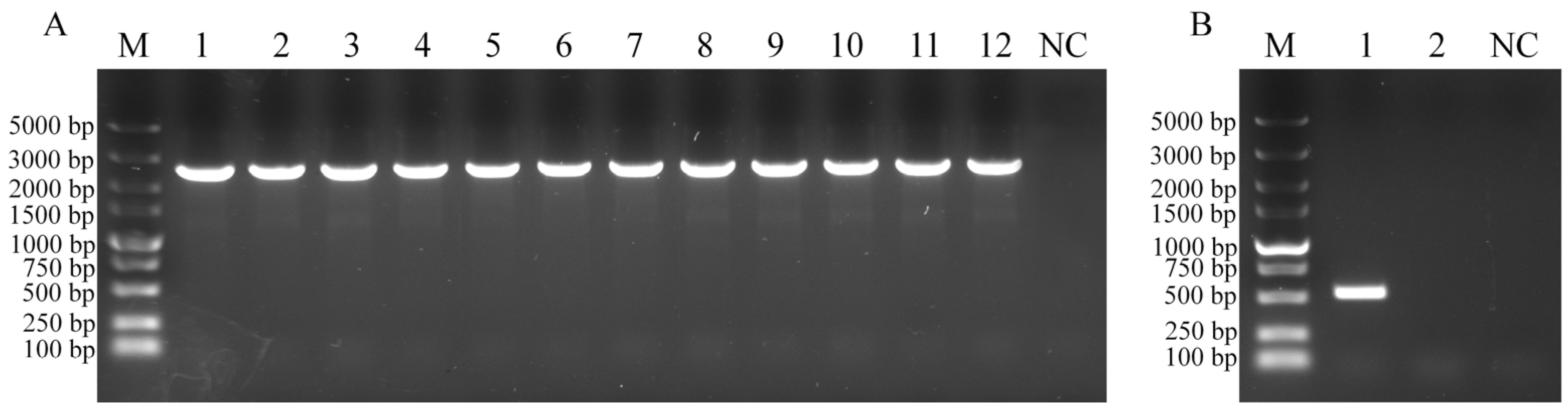
2.2. Construction of the hsdM Mutant

The suicide plasmid pSET4s-hsdMCm (Figure S1) was constructed as previously reported [20], and the mutant WC1535ΔhsdM was obtained by homologous recombination. Briefly, the pSET4s-hsdMCm plasmid was electrotransferred into WC1535 competent cells, streaked onto BHI agar (BHIA) plates with spectinomycin resistance (150 μg/mL), and inoculated at 28 °C. Monoclonal colonies were selected, inoculated into BHI broth, then diluted and streaked onto BHIA plates supplemented with chloramphenicol (10 μg/mL), and cultured at 37 °C. The mutant strain with hsdM deletion (WC1535ΔhsdM) was screened by PCR amplification using primers hsdMCm-dF/dR and hsdM-inF/R (Table 1). The mutant WC1535ΔhsdM was inoculated onto BHIA plates for 30 generations. Genomic DNA of the mutant WC1535ΔhsdM was extracted using a bacterial genomic DNA extraction kit (Guangzhou Magen Biotechnology, Co., Ltd., Guangzhou, China), the genetic stability of the mutant was detected by PCR amplification with primers hsdMCm-dF/dR, and the PCR products were sequenced. In addition, PCR amplification with the primers hsdM-inF and hsdM-inR was performed to further confirm whether the mutant contained hsdM, and PCR amplification using the primers Spe-dF and Spe-dR was conducted to confirm the presence of the spectinomycin resistance gene spe. The PCR amplification was as follows: pre-denaturation at 95 °C for 4 min, followed by 35 cycles of denaturation at 94 °C for 30 s, annealing at 52–55 °C for 30 s, and extension at 72 °C for 1.0–2.5 min, and final extension at 72 °C for 10 min.

2.3. Phenotype of Mutant WC1535ΔhsdM
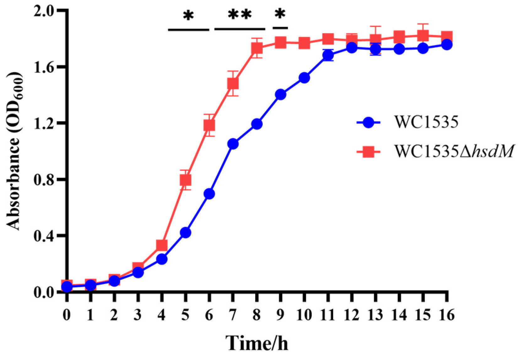
2.3.1. Growth Curve

The WT strain WC1535 and mutant WC1535ΔhsdM were streaked onto blood plates and cultured for 24 h at 28 °C, respectively. Single colonies were selected and inoculated into BHI broth and incubated overnight at 28 °C with shaking (180 r/min). The bacterial solution was adjusted to an OD600 of 0.8, and 1 mL of the bacterial solution was aspirated and inoculated into 100 mL of BHI medium (1:100). The bacteria were cultured at 28 °C with shaking at 180 r/min, and the OD600 value was measured every 1 h for 16 h. Bacterial growth curves of the WC1535 and mutant strains were obtained. Experiments were performed in triplicate.

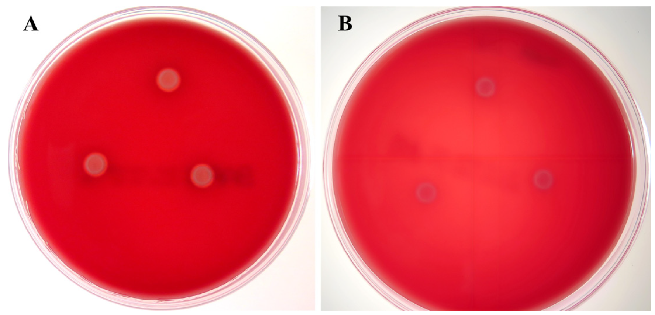
2.3.2. Hemolytic Activity Assay

Strains WC1535 and WC1535ΔhsdM were cultured in BHI broth at 28 °C for 12 h, and 2.5 μL of the bacterial solution was inoculated onto a blood plate and cultured at 37 °C for 48 h to observe the hemolytic activities of the colonies. Experiments were performed in triplicate.

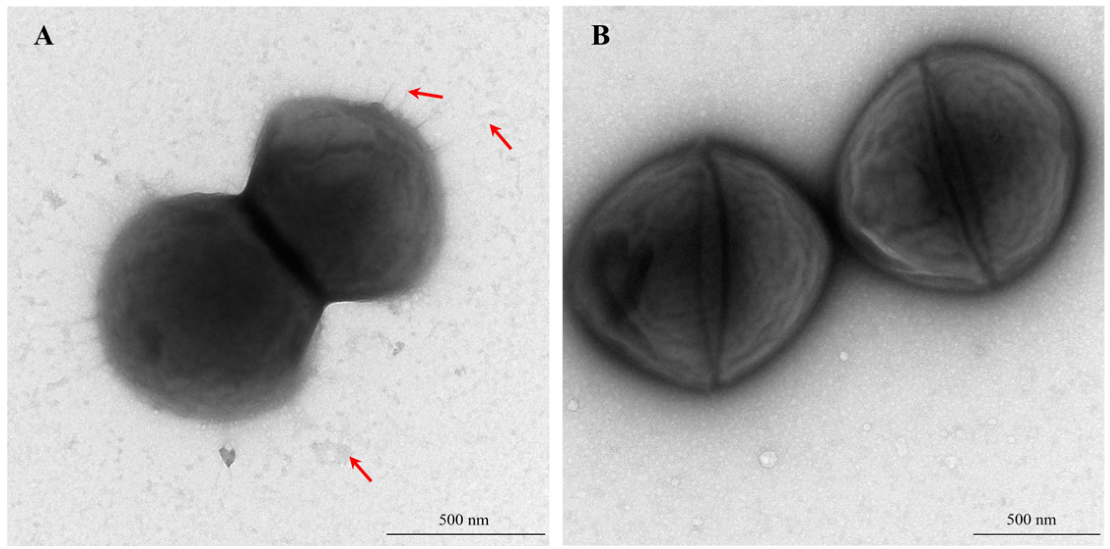
2.3.3. Capsular Staining and Transmission Electron Microscopy Observation

Strains WC1535 and WC1535ΔhsdM were inoculated onto BHIA plates and incubated at 28 °C for 48 h. The cells were then harvested, washed with phosphate-buffered saline (PBS, pH = 7.4), and resuspended. Capsule staining of both strains was performed using the Tyler method with a commercial capsule stain kit (Qingdao Rishui Bio-technologies Co., Ltd., Qingdao, China). The bacterial suspension was diluted 1:4 (v/v) with sterile PBS. After 30 min of incubation, 20 μL of the diluted suspension was pipetted onto a piece of tin foil. A copper grid with its support membrane surface was placed on the bacterial drop for 2 min. Excess bacterial suspension was absorbed away from the edges of the grid using filter paper. Then, 20 μL of 0.5% phosphotungstic acid staining solution was added to the tin foil. The copper grid with the carbon film surface was placed on the staining solution for 30–45 s. The excess staining solution was removed using filter paper, and the grid was dried at room temperature. Electron microscopy negative staining samples were prepared for both strains to observe their capsule characteristics.

2.4. Survival Assay in Whole Blood

The whole blood survival assay was performed as previously described [22]. Blood was collected from the tail veins of healthy Nile tilapia (Oreochromis niloticus) using an anticoagulant tube. Simultaneously, WC1535 and WC1535ΔhsdM bacterial solutions were adjusted to OD600 = 0.2 and then diluted to a concentration of 6 × 10³ colony-forming units (CFU)/mL. The diluted bacterial solutions (50 μL) were mixed with 450 μL of tilapia fresh tilapia blood, 100 μL of the mixed liquid was taken for bacterial counting, and the remaining samples were incubated at 28 °C for 2 h and 4 h with shaking at 100 r/min. The viability (as a proliferation factor) of WC1535 and WC1535ΔhsdM in whole blood was assessed based on the ratio of the bacterial numbers at the beginning and end of the experiment. The survival assay was performed in triplicate.

2.5. Cell Adhesion Assay

The cell adhesion assay was performed according to a previously described method [23]. Approximately 1 × 10⁵ EPC cells were inoculated into each well of 24-well plates and cultured at 28 °C until the cells covered the bottom of each well. The strains WC1535 and WC1535ΔhsdM strains were diluted in DMEM medium, respectively. Experimental groups were inoculated with 1 mL bacterial solutions (multiplicity of infection, MOI = 1:10) of WC1535 or WC1535ΔhsdM, respectively. The control group was inoculated with 1 mL DMEM medium. The DMEM medium was discarded after the cells were incubated at 28 °C in the presence of 5% CO2 for 2 h, and the cells were rinsed three times with Hanks’ balanced salt solution (HBSS) buffer. The 0.25% trypsin (200 μL/well) was added to each well to digest the cells and incubated for 5 min at 37 °C in a 5% CO2 cell incubator. A total of 0.8 mL of double-distilled water (ddH2O) was added to each well, and the plates were incubated at room temperature for 10 min. The cells were suspended and then transferred into sterilized 1.5 mL centrifuge tubes. The suspended cells were serially diluted 10-fold and plated on BHIA plates at appropriate dilutions for colony counting. The adhesion assay was performed in triplicate. Adhesion rate = number of colonies after lysis/number of bacteria added ×100.

2.6. Phagocytosis Assay

Approximately 1 × 10⁵ RAW264.7 cells were inoculated into each well of the 24-well cell culture plate and cultured at 28 °C until the cells covered the bottom of the well. The WC1535 and WC1535ΔhsdM strains were diluted in RPMI-1640 medium, respectively. The experimental groups were inoculated with 1 mL bacterial solutions (MOI = 1:10) of WC1535 and WC1535ΔhsdM, respectively. The control group was inoculated with 1 mL of RPMI-1640 medium. After 2 h of incubation, the cells were rinsed three times with sterile HBSS buffer, and extracellular bacteria were killed by treatment with a total of 1 mL of RPMI-1640 medium containing a 1% penicillin (100 U/mL), streptomycin (100 μg/mL), and amphotericin B (0.25 μg/mL) mixture and incubated at 37 °C in the presence of 5% CO2 for 1 h [24]. Subsequently, the cells were rinsed three times with PBS to remove the unbound bacteria and incubated at 37 °C for 0, 2, 4, 8, and 12 h, and then trypsin was added to each well to lyse the cells. A total of 0.8 mL of ddH2O was added to each well and the plates were incubated at room temperature for 10 min. The cells were suspended and then transferred into sterilized 1.5 mL centrifuge tubes. The suspended cells were serially diluted 10-fold and streaked onto BHIA plates for colony counting. The phagocytosis assay was performed in triplicate. The survival rate of bacteria within the macrophages at different time points was calculated using the following formula: Survival rate = CFU at N hours/CFU at 0 h. Phagocytic rate = CFU at 0 h/1 × 106 CFU.

2.7. Challenge Test

Juvenile Nile tilapia (N = 135, body weight, 8.74 ± 1.41 g) were obtained from a tilapia breeding farm in Guangdong province and maintained in recirculating aquaria for 2 weeks to allow them to acclimatize to the environment. The domestication conditions were maintained with water temperature at 30 ± 0.5 °C, dissolved oxygen > 5.5 mg/L, total ammonia concentrations < 0.05 mg/L, and pH = 7.2–7.8. Healthy Nile tilapia were randomly divided into 9 groups, including the WT group (4 tanks), Mutant group (4 tanks), and Control group (1 tank), with 15 fish in each group. The WC1535 and WC1535ΔhsdM strains were inoculated into BHI broth and cultured at 28 °C, bacteria were grown to an OD600 of 0.8 and then collected by centrifugation at 5000 r/min, and the bacterial concentrations were adjusted to 5.0 × 10⁸, 5.0 × 10⁷, 5.0 × 10⁶, and 5.0 × 10⁵ colony-forming unit (CFU)/mL with PBS, respectively. Fish in the WT group tanks were intraperitoneally injected with 0.1 mL of the WC1535 strain at the bacterial concentrations of 5.0 × 10⁸, 5.0 × 10⁷, 5.0 × 10⁶, and 5.0 × 10⁵ CFU/mL, respectively. Fish in the Mutant group tanks were intraperitoneally injected with 0.1 mL of the WC1535ΔhsdM strain at the bacterial concentrations of 5.0 × 10⁸, 5.0 × 10⁷, 5.0 × 10⁶, and 5.0 × 10⁵ CFU/mL, respectively. Each fish in the Control group was intraperitoneally injected with 0.1 mL of PBS. The water temperature was 30 ± 0.5 °C, and the dissolved oxygen was maintained at 6.0 ± 0.5 mg/L. Clinical signs and mortality were recorded daily for 2 weeks. The median lethal dose was calculated according to the modified Karber’s method [25]. This study was conducted in accordance with the internal ethics committee of the Pearl River Fisheries Research Institute, Chinese Academy of Fishery Sciences (LAEC-PRFRI-2024-07-20), approved on 20 July 2024.

2.8. Dynamic Distribution Assay

A total of 45 healthy tilapia (body weight, 17.48 ± 2.26 g) were selected for the dynamic distribution experiment. The WC1535 and WC1535ΔhsdM strains were adjusted to a concentration of 3.5 × 10⁷ CFU/mL with PBS and mixed in equal volume, and each fish was intraperitoneally injected with 0.1 mL of the mixed bacterial solution. The water temperature was 30 ± 0.5 °C and the dissolved oxygen was maintained at 6.0 ± 0.5 mg/L.
Brain, liver, spleen, and kidney tissues were obtained at 24, 48, 72, and 120 h post-infection (hpi). Three fish were collected at each time point, and the tissues were weighed. Subsequently, the tissues were homogenized and subjected to serial 10-fold dilutions that were streaked onto BHIA plates and BHIA supplemented with chloramphenicol (15 μg/mL) plates, respectively. Bacterial counts in the tissues were performed after 36 h of incubation at 28 °C. The dynamic distribution in different tissues was analyzed based on the bacterial loads of the WC1535 and WC1535ΔhsdM strains in each tissue.

2.9. Reverse-Transcription Quantitative Polymerase Chain Reaction (RT-qPCR) Assay

Eighty healthy tilapia (body weight, 27.15 ± 3.78 g) were selected for the RT-qPCR analysis. Each fish was intraperitoneally injected with 0.1 mL of the WC1535 or WC1535ΔhsdM strains at a dose of 25% LD50, respectively. The water temperature was 30 ± 0.5 °C and the dissolved oxygen was maintained at 6.0 ± 0.5 mg/L. Spleen and head kidney tissues were collected before infection and at 12, 24, 48, 72, and 120 hpi, respectively, and 4 samples were taken from each group at each time point. Total RNA in tissues was extracted using an RNA extraction kit (Vazyme Biotech Co., Ltd., Nanjiang, China) and immediately reverse transcribed into cDNA using a PrimeScript™ RT reagent kit with the DNA Eraser (Dalian Bao Biological Engineering Co., Ltd., Dalian, China). Gene expression was quantified by RT-qPCR using TB Green® Premix Ex Taq™ II FAST qPCR reagent (Dalian Bao Biological Engineering Co., Ltd., Dalian, China) under the following reaction conditions: 30 s at 95 °C, followed by 40 cycles of 95 °C for 5 s and 60 °C for 30 s. The analyzed genes were interleukin-1β (IL-1β), interleukin-6 (IL-6), gamma-interferon gene (IFN-γ), and TNF-α, while β-actin and elongation factor 1α (EF-1α) genes (Table 2) were used as reference genes and Ct values were normalized [26]. These data were analyzed using the 2−ΔΔCt method [27].

2.10. Statistics

Statistical analysis was performed using Graphpad Prism 9.5 software (GraphPad Software, Inc., San Dego, CA, USA). The statistical significances in the growth, cell adhesion assay, intracellular survival assay, anti-macrophage phagocytosis assay, dynamic distribution assay, and qRT-PCR were determined using Student’s t-test. The data were expressed as mean ± SD. p values as indicated here: * for p < 0.05; ** for p < 0.01; and *** for p < 0.001.

3. Results

3.1. Construction of WC1535ΔhsdM

Gel electrophoresis of the PCR products with double-exchange PCR detection showed positive target bands (2460 bp) (Figure 1A), and the products were sequenced, revealing that hsdM was deleted. Furthermore, gel electrophoresis of the PCR products with the primers hsdM-inF/inR showed a positive band for hsdM in the WT strain, whereas no band was observed in the mutant WC1535ΔhsdM (Figure 1B). PCR detection of the spectinomycin resistance gene showed that the spe was absent in the mutant; moreover, WC1535ΔhsdM was not resistant to spectinomycin, indicating that the suicide plasmid in the mutant had been eliminated.
Furthermore, after 30 generations of passages on BHIA plates, the mutant was subjected to PCR detection and sequencing analysis, which indicated that the mutant was genetically stable and that no reversion mutation occurred.

3.2. Biological Characteristics of WC1535ΔhsdM

3.2.1. Growth Curves

The growth rate of WC1535ΔhsdM was significantly higher than that of WC1535, particularly from 5 to 9 h, and the mutant reached a stable growth phase faster (Figure 2). There was no significant difference in the OD600 between the mutant and WT strains during the stable growth phase.

3.2.2. Hemolytic Activity

Obvious hemolytic circles were observed on the Colombian blood plates cultured with WC1535 and cultured for 48 h, indicating that the WC1535 strain was β-hemolytic (Figure 3). However, no hemolytic circles were observed on the Colombian blood plates inoculated with the mutant WC1535ΔhsdM for 48 h, suggesting that the hemolytic ability of the mutant was weakened.

3.2.3. Bacterial Morphology and Capsule Characteristics

Numerous filamentous structures and granular materials were covered on the WT strain, whereas none of them were observed on the surface of the mutant WC1535ΔhsdM (Figure 4). Capsule staining revealed no significant difference in the capsule between the WC1535 and WC1535ΔhsdM (Figure S2).

3.3. Survival in Whole Blood

The results of the whole-blood survival assay showed that the CFUs of strains WC1535 and WC1535ΔhsdM significantly decreased after incubation for 2 and 4 h, whereas there was no significant difference between the WT and the mutant (Figure S3).

3.4. Cell Adhesion and Anti-Macrophage Phagocytosis

The cell adhesion assay revealed that the adhesion rate of strain WC1535 to the EPC cells was 32.7%, whereas the mutant was 17.0%. The adhesion rate of the WC1535ΔhsdM strain to EPC cells was significantly reduced compared to that of the WT strain by 52.0% (Figure 5A).
The phagocytosis assay (removed the unbound bacteria and incubated at 37 °C for 0 h) showed that the phagocytosis rate of strain WC1535 was 1.87%, whereas that of the mutant was 4.07%. The anti-phagocytic ability of WC1535ΔhsdM was significantly lower than that of WC1535 (p < 0.05) (Figure 5B). The intracellular survival assay showed that the survival rate of the WC1535ΔhsdM was significantly lower than that of the WT strain after 2–4 h (p < 0.01) (Figure 5C).

3.5. Pathogenicity

The results of the artificial infection experiment showed that the cumulative mortality of tilapia in the high-concentration infection group of the WT and mutant strains was high, among which the mortality of the WT strain was 100% and that of the mutant was 80%. The LD50 of the WT strain and the mutant strains to tilapia were 6.3 × 10⁷ CFU/mL and 1.3 × 10⁸ CFU/mL, respectively, indicating that the pathogenicity of the mutant to tilapia was significantly decreased.

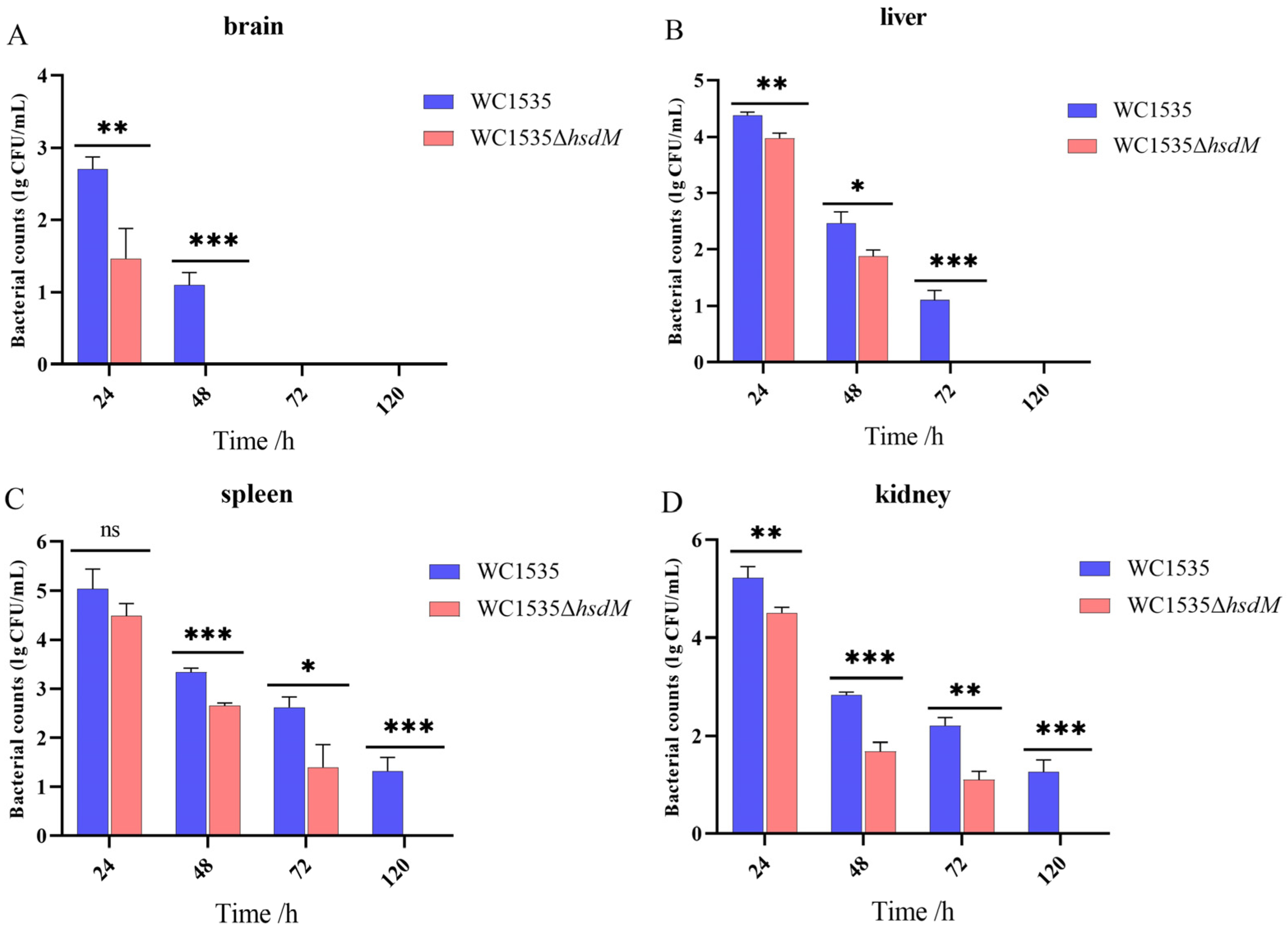
3.6. Dynamic Distribution In Vivo

The dynamic distribution assay revealed that the mutant WC1535ΔhsdM persisted in tilapia brain tissue for 24 to 48 h, whereas the WT strain maintained its presence for over 48 h (Figure 6A). Notably, the bacterial load of the WC1535ΔhsdM in brain tissue was significantly lower than that of WC1535. In liver tissue, the WC1535ΔhsdM survived for 48–72 h, whereas WC1535 persisted for 72–120 h, and the bacterial load of the WC1535ΔhsdM in liver tissue was significantly lower than that of the WC1535 strain (Figure 6B). In spleen tissue, the WC1535ΔhsdM strain survived for 72–120 h, whereas the WC1535 strain was still isolated after 120 h (Figure 6C). The bacterial load of the WC1535ΔhsdM strain in spleen tissue was significantly lower than that of WC1535 during the 48 to 120 h period. In kidney tissue, the WC1535ΔhsdM strain survived for 72–120 h, whereas the WC1535 strain persisted beyond 120 h, and the bacterial load of WC1535ΔhsdM in kidney tissue was significantly lower than that of WC1535 from 24–120 h (Figure 6D). In summary, the number of WC1535ΔhsdM surviving in the brain, liver, spleen, and kidney tissues of tilapia was significantly lower than that of the WT strain, and the persistence time of WC1535ΔhsdM was shorter than that of WC1535.

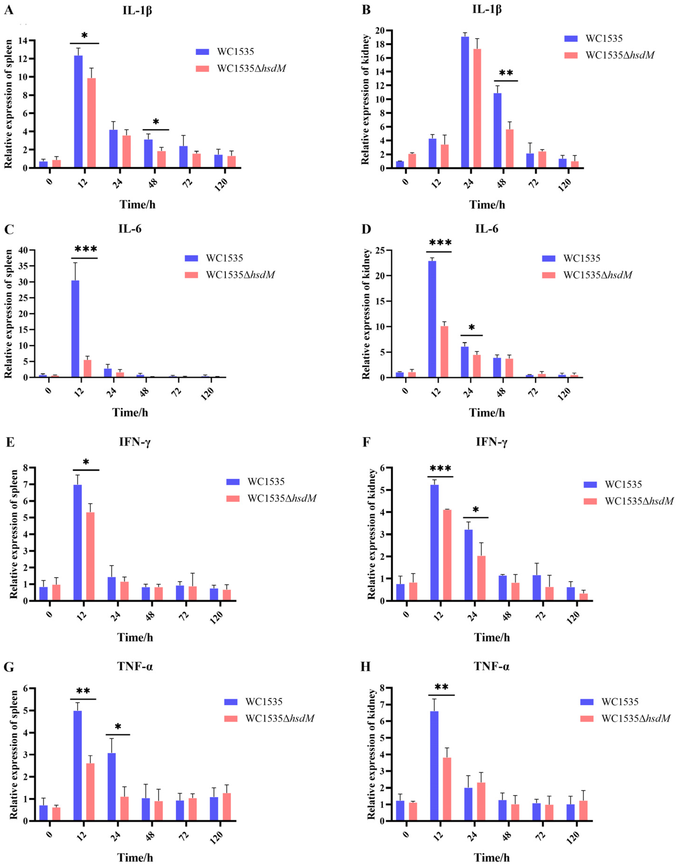
3.7. Expression Levels of Immune-Related Genes

The expression levels of immune-related genes in the spleen and kidney of tilapia post-infection with the mutant and WT strains were analyzed using RT-qPCR. The RT-qPCR results indicated that the expression levels of IL-1β in the spleen infected with WC1535ΔhsdM were significantly lower than those in the spleen infected with the WT strain at 12 and 48 hpi (p < 0.05). Furthermore, the expression levels of IL-1β in the head kidney infected with WC1535ΔhsdM were significantly lower than those in the head kidney infected with the WT strain at 48 hpi (p < 0.01) (Figure 7A,B). The expression levels of IL-6 in the spleen and head kidney infected with WC1535ΔhsdM were significantly lower than in those infected with the WT strain at 12 hpi (p < 0.001), and a similar trend of IL-6 in the head kidney was found at 24 h (p < 0.05) (Figure 7C,D). The expression levels of IFN-γ in the spleen infected with WC1535ΔhsdM were significantly lower than those in the spleen infected with the WT strain at 12 hpi (p < 0.05), and the expression levels of IFN-γ in the head kidney tissue were significantly lower than those in the head kidney infected with the WT strain at 12 (p < 0.001) and 24 hpi (p < 0.05) (Figure 7E,F). The expression levels of TNF-α in the head kidney and spleen infected with WC1535ΔhsdM were significantly lower than in those infected with the WT strain at 12 hpi (p < 0.01), and a similar trend of TNF-α in the head kidney was found at 12 hpi (p < 0.01) (Figure 7G,H). In summary, the expression levels of IL-1β, IL-6, IFN-γ, and TNF-α in the spleen and kidney tissues of tilapia infected with WC1535ΔhsdM decreased, especially in the early stage of infection (12–24 h).

4. Discussion

DNA methylation is one of the most important epigenetic modifications in prokaryotes. DNA methyltransferases, such as HsdM, Dam, and Dcm, play key roles in the methylation modification of bacterial genomic DNA, regulating essential biological processes, including growth, metabolism, virulence, and antibiotic resistance [9,28,29]. DNA methylation, modified by the RM system, directly or indirectly regulates the expression of virulence genes involved in bacterial adhesion and colonization [30]. The type I RM system is conserved in group A Streptococcus strains, along with variable type II systems, whereas many GBS strains harbor HsdM and lack HsdR in the type I RM system [18]. However, limited information is available regarding the function of HsdM in GBS [18]. In this study, the GBS strain WC1535, a representative strain of molecular serotype Ia and ST7 isolated from tilapia, was selected for the deletion of hsdM (protein_id: WP_000818330.1). We constructed the mutant WC1535ΔhsdM by knocking out hsdM from the WC1535 strain. The effects of HsdM on growth were determined by comparing the growth rates of the mutant WC1535ΔhsdM and its parental strain WC1535 in BHI broth at 28 °C. The WC1535ΔhsdM had a faster growth rate in the logarithmic phase. This is inconsistent with previous reports [17]; for example, hsdM gene deletion in wild-type Mycobacterium bovis BCG Pasteur (BCG) did not affect mycobacterial growth in vitro, and deletion of the hsdM gene did not affect the growth of group A Streptococcus [18]; however, the growth of M. tuberculosis 11826ΔhsdM was significantly slower than that of its parental strain [16]. Therefore, we hypothesized that hsdM affects the growth of GBS, with potentially distinct regulatory mechanisms for different strains.
The hemolytic activity assay showed that the hemolytic activity of WC1535ΔhsdM decreased significantly, and almost no hemolysis circle was observed. The synthesis of β-hemolysin or cell-hemolysin is closely related to acetyl-CoA and propionyl-CoA [31]. We hypothesized that HsdM mediates the biosynthesis of β-hemolysin or cell-hemolysin by regulating acetyl-CoA levels. This hypothesis is supported by our experiment, which showed that the expression levels of pflA and pflB, which encode formate C-acetyltransferase and the pyruvate formate lyase-activating enzyme, were significantly reduced in WC1535ΔhsdM (according to the transcriptome data). Therefore, we hypothesized that HsdM reduces the hemolytic activity of GBS by regulating the expression of pflA and pflB, resulting in attenuated virulence. In addition, hsdM deletion had no effect on the GBS capsule or survival in tilapia whole blood.
The type I RM system regulates the adhesion ability of bacteria to cells and their intracellular survival. For example, Streptococcus pyogenes mutant ΔRSM with deletion of hsdM/R/S genes was constructed, which consequently lacked DNA methylation and exhibited attenuated survival in human neutrophils and reduced adherence to human epithelial cells [30]. HsdS in S. suis contributes to anti-phagocytosis and survival in adverse host environments by regulating gene expression and reducing the secretion of TNF-α and NO by phagocytes [19]. Similar to the previous reports, the adhesion ability of WC1535ΔhsdM to EPC cells was significantly reduced compared with its parental strain, indicating that HsdM could regulate the adhesion ability of GBS to cells. Furthermore, the anti-macrophage phagocytosis and survival ability in phagocytic cells by WC1535ΔhsdM were significantly reduced compared to those of the WT strain, indicating that HsdM could enhance the resistance of GBS to macrophage killing. In addition, the challenge test revealed that HsdM improved the virulence of GBS in tilapia, and the dynamic distribution assay showed that HsdM promoted GBS survival in the brain, liver, spleen, and kidney tissues. These results indicated that HsdM regulates the adhesion and invasion ability of GBS to cells and their pathogenicity in tilapia. We hypothesized that HsdM may affect the bacterial surface components involved in host interactions.
To explore the regulatory mechanism of HsdM in the pathogenesis of GBS, RT-qPCR was used to analyze the expression levels of IL-1β, IL-6, IFN-γ, and TNF-α in spleen and kidney tissues infected with WC1535ΔhsdM. The results showed that the expression of IL-1β, IL-6, IFN-γ, and TNF-α was significantly reduced in the Mutant group compared with that in the WT group from 12 to 24 hpi, revealing that attenuated mutant WC1535ΔhsdM could decrease the expression of host inflammatory factor genes. Similar to our findings, attenuated strain S. suis Δhp1330 induced lower production/mRNA expression levels of pro-inflammatory factors IL-1β and TNF-α in the blood of mice and in RAW264.7 cells in vitro [32]. In S. pneumoniae, the regulatory gene hsdSA regulates the production of extracellular vesicles, resulting in the inability of lysosomes to clear intracellular bacteria and prolonging the survival of S. pneumoniae in macrophages [29]. Moreover, our findings suggested that HsdM contributes to the GBS strain upregulating the expression levels of host inflammatory factors, including IL-1β, IL-6, IFN-γ, and TNF-α, thereby causing an excessive inflammatory response that is detrimental to the host. These findings improve our understanding of the excessive inflammation caused by virulent GBS.

5. Conclusions

A deletion of hsdM in the tilapia GBS strain was constructed and designated as WC1535ΔhsdM. This mutant exhibited an increased growth rate, reduced hemolytic activity, adherence ability, and survival within macrophages and tilapia tissues, and attenuated virulence in tilapia. We demonstrated that HsdM can influence the hemolytic activity of GBS, leading to attenuated bacterial virulence. HsdM is not only a critical virulence regulator of GBS but also plays an important role in modulating the growth of GBS. This study provides new insight into GBS pathogenesis. However, the mechanism underlying the influence of HsdM on GBS pathogenesis requires further investigation.

Supplementary Materials

The following supporting information can be downloaded at: https://www.mdpi.com/article/10.3390/fishes10020086/s1, Figure S1: Map of the pSET4S-hsdMCm; Figure S2: Capsule staining of strains WC1535 and WC1535ΔhsdM; Figure S3: Survival ability of strains WC1535 and WC1535ΔhsdM in tilapia whole blood.

Author Contributions

Conceptualization, D.Z. and D.J.; methodology, D.Z. and D.J.; software, D.J. and X.M.; validation, D.J. and M.Y.; formal analysis, D.Z. and D.J.; investigation, X.M. and B.W.; resources, D.Z. and X.M.; data curation, Y.R. and M.Y.; writing—original draft preparation, D.J.; writing—review and editing, D.Z., M.Y. and X.M.; visualization, Y.R. and B.W.; supervision, Y.R.; project administration, D.Z. and M.Y.; funding acquisition, D.Z. and X.M. All authors have read and agreed to the published version of the manuscript.

Funding

This research was funded by the Guangdong Basic and Applied Basic Research Foundation, grant numbers 2021A1515010956 and 2024A1515011641; the Research Fund Program of Guangdong Provincial Key Laboratory of Aquatic Animal Disease Control and Healthy culture, grant number FBEA2022ZD01; and the Guangzhou Science and Technology Plan Project, grant number 2023A04J0714.

Institutional Review Board Statement

This study was conducted in accordance with the internal ethics committee of the Pearl River Fisheries Research Institute, Chinese Academy of Fishery Sciences (LAEC-PRFRI-2024-07-20), approved on 20 July 2024.

Informed Consent Statement

Not applicable.

Data Availability Statement

All data supporting findings of this study are available from the corresponding author upon request.

Acknowledgments

We would like to express our gratitude to Ouqin Chang for her assistance in capsule staining and transmission electron microscopy observation.

Conflicts of Interest

The authors declare no conflicts of interest.

References

  1. Piamsomboon, P.; Srisuwatanasagul, S.; Kongsonthana, K.; Wongtavatchai, J. Streptococcus agalactiae infection caused spinal deformity in juvenile red tilapia (Oreochromis sp.). J. Fish Dis. 2022, 45, 603–606. [Google Scholar] [CrossRef] [PubMed]
  2. Chong, S.M.; Wong, W.K.; Lee, W.Y.; Tan, Z.B.; Tay, Y.H.; Teo, X.H.; Chee, L.D.; Fernandez, C.J. Streptococcus agalactiae outbreaks in cultured golden pomfret, Trachinotus blochii (Lacepede), in Singapore. J. Fish Dis. 2017, 40, 971–974. [Google Scholar] [CrossRef] [PubMed]
  3. Zhang, D.F.; Ke, X.; Liu, L.; Lu, M.; Shi, C.; Liu, Z. Streptococcus agalactiae from tilapia (Oreochromis sp.) transmitted to a new host, bighead carp (Aristichthys nobilis), in China. Aquac. Int. 2018, 26, 885–897. [Google Scholar] [CrossRef]
  4. Zhang, D.; Liu, Z.; Ren, Y.; Wang, Y.; Pan, H.; Liang, D.; Bei, W.; Chang, O.; Wang, Q.; Shi, C. Epidemiological characteristics of Streptococcus agalactiae in tilapia in China from 2006 to 2020. Aquaculture 2022, 549, 885–897. [Google Scholar] [CrossRef]
  5. Abdallah, E.S.H.; Metwally, W.G.M.; Abdel-Rahman, M.A.M.; Albano, M.; Mahmoud, M.M. Streptococcus agalactiae infection in Nile tilapia (Oreochromis niloticus): A review. Biology 2024, 13, 914. [Google Scholar] [CrossRef] [PubMed]
  6. Liu, Y.; Liu, J. Group B Streptococcus: Virulence factors and pathogenic mechanism. Microorganisms 2022, 10, 2483. [Google Scholar] [CrossRef]
  7. Burton, N.O.; Greer, E.L. Multigenerational epigenetic inheritance: Transmitting information across generations. Semin. Cell Dev. Biol. 2022, 127, 132–212. [Google Scholar] [CrossRef] [PubMed]
  8. Lyko, F. The DNA methyltransferase family: A versatile toolkit for epigenetic regulation. Nat. Rev. Genet. 2018, 19, 81–92. [Google Scholar] [CrossRef] [PubMed]
  9. Sánchez-Romero, M.A.; Casadesús, J. The bacterial epigenome. Nat. Rev. Microbiol. 2020, 18, 7–20. [Google Scholar] [CrossRef] [PubMed]
  10. Casadesús, J.; Low, D. Epigenetic gene regulation in the bacterial world. Microbiol. Mol. Biol. Rev. 2006, 70, 830–856. [Google Scholar] [CrossRef]
  11. Heusipp, G.; Fälker, S.; Schmidt, M.A. DNA adenine methylation and bacterial pathogenesis. Int. J. Med. Microbiol. 2007, 297, 1–7. [Google Scholar] [CrossRef] [PubMed]
  12. Heithoff, D.M.; Sinsheimer, R.L.; Low, D.A.; Mahan, M.J. An essential role for DNA adenine methylation in bacterial virulence. Science 1999, 284, 967–970. [Google Scholar] [CrossRef] [PubMed]
  13. Willemse, N.; Schultsz, C. Distribution of type I restriction–modification systems in Streptococcus suis: An outlook. Pathogens 2016, 5, 62. [Google Scholar] [CrossRef] [PubMed]
  14. Croix, M.D.S.; Vacca, I.; Kwun, M.J.; Ralph, J.D.; Bentley, S.D.; Haigh, R.; Croucher, N.J.; Oggioni, M.R. Phase-variable methylation and epigenetic regulation by type I restriction–modification systems. FEMS Microbiol. Rev. 2017, 41, S3–S15. [Google Scholar] [CrossRef] [PubMed]
  15. Chen, T.; Li, H.; Wang, D.; Tang, Y.Q.; Tang, H.Q.; Ma, X.; Liu, Z. The effect of DNA methyltransferase hsdM gene on the motility of Aeromonas veronii. J. Biol. 2022, 39, 28–32. (In Chinese) [Google Scholar] [CrossRef]
  16. Chu, H.; Hu, Y.; Zhang, B.; Sun, Z.; Zhu, B. DNA Methyltransferase HsdM induce drug resistance on Mycobacterium tuberculosis via multiple effects. Antibiotics 2021, 10, 1544. [Google Scholar] [CrossRef] [PubMed]
  17. Hu, X.; Zhou, X.; Yin, T.; Chen, K.; Hu, Y.; Zhu, B.; Mi, K. The mycobacterial DNA methyltransferase HsdM decreases intrinsic isoniazid susceptibility. Antibiotics 2021, 11, 1323. [Google Scholar] [CrossRef]
  18. DebRoy, S.; Shropshire, W.C.; Tran, C.N.; Hao, H.; Gohel, M.; Galloway-Peña, J.; Hanson, B.; Flores, A.R.; Shelburne, S.A.; Fey, P.D. Characterization of the type I restriction modification system broadly conserved among group A Streptococci. mSphere 2021, 6, e00799-21. [Google Scholar] [CrossRef] [PubMed]
  19. Xu, B.; Zhang, P.; Li, W.; Liu, R.; Tang, J.; Fan, H. hsdS, belonging to the type I restriction-modification system, contributes to the Streptococcus suis serotype 2 survival ability in phagocytes. Front. Microbiol. 2017, 8, 1524. [Google Scholar] [CrossRef] [PubMed]
  20. Zhang, D.; Ke, X.; Liu, Z.; Cao, J.; Su, Y.; Lu, M.; Gao, F.; Wang, M.; Yi, M.; Qin, F. Capsular polysaccharide of Streptococcus agalactiae is an essential virulence factor for infection in Nile tilapia (Oreochromis niloticus Linn.). J. Fish Dis. 2019, 42, 293–302. [Google Scholar] [CrossRef]
  21. Fijan, N.; Sulimanović, D.; Bearzotti, M.; Muzinić, D.; Zwillenberg, L.O.; Chilmonczyk, S.; Vautherot, J.F.; Kinkelin, P. Some properties of the Epithelioma papulosum cyprini (EPC) cell line from carp Cyprinus carpio. Ann. Virol. (Inst. Pasteur) 1983, 134, 207–220. [Google Scholar] [CrossRef]
  22. Li, H.; Cao, J.; Han, Q.; Li, Z.; Zhuang, J.; Wang, C.; Wang, H.; Luo, Z.; Wang, B.; Li, A. Protease SfpB plays an important role in cell membrane stability and immune system evasion in Streptococcus agalactiae. Microb. Pathog. 2024, 192, 106683. [Google Scholar] [CrossRef] [PubMed]
  23. Song, H.; Yang, B.; Kang, Y.; Cong, W. Critical roles of VipB protein on virulence and oxidative stress tolerance in Aeromonas veronii. J. Fish Dis. 2023, 46, 487–497. [Google Scholar] [CrossRef] [PubMed]
  24. Pei, X.; Liu, M.; Zhou, H.; Fan, H. Screening for phagocytosis resistance-related genes via a transposon mutant library of Streptococcus suis serotype 2. Virulence 2020, 11, 825–838. [Google Scholar] [CrossRef]
  25. Li, Q.; Yu, X.; Ye, L.; Hou, T.; Liu, Y.; Liu, G.; Wang, Q.; Zhang, D. Hypermucoviscous multidrug-resistant Klebsiella variicola strain LL2208 isolated from Chinese longsnout catfish (Leiocassis longirostris): Highly similar to human K. variicola strains. Pathogens 2024, 13, 647. [Google Scholar] [CrossRef] [PubMed]
  26. Vandesompele, J.; De Preter, K.; Pattyn, F.; Poppe, B.; Van Roy, N.; De Paepe, A.; Speleman, F. Accurate normalization of real-time quantitative RT-PCR data by geometric averaging of multiple internal control genes. Genome Biol. 2002, 3, research0034.1. [Google Scholar] [CrossRef] [PubMed]
  27. Livak, K.J.; Schmittgen, T.D. Analysis of relative gene expression data using real-time quantitative PCR and the 2−ΔΔCT Method. Methods 2001, 25, 402–408. [Google Scholar] [CrossRef]
  28. Ma, J.; Zhao, H.; Mo, S.; Li, J.; Ma, X.; Tang, Y.; Li, H.; Liu, Z. Acquisition of type I methyltransferase via horizontal gene transfer increases the drug resistance of Aeromonas veronii. Microb. Genom. 2023, 9, 001107. [Google Scholar] [CrossRef] [PubMed]
  29. Wang, L.; Liu, M.; Qi, Y.; Wang, J.; Shi, Q.; Xie, X.; Zhou, C.; Ma, L. hsdSA regulated extracellular vesicle-associated PLY to protect Streptococcus pneumoniae from macrophage killing via LAPosomes. Microbiol. Spectr. 2024, 12, e0099523. [Google Scholar] [CrossRef]
  30. Nye, T.M.; Jacob, K.M.; Holley, E.K.; Nevarez, J.M.; Dawid, S.; Simmons, L.A.; Watson, M.E. DNA methylation from a type I restriction modification system influences gene expression and virulence in Streptococcus pyogenes. PLoS Pathog. 2019, 15, e1007841. [Google Scholar] [CrossRef]
  31. Huang, Q.Q.; Liu, S.L.; Huang, J.H.; Wang, F.; Zhao, Z.C.; Deng, H.W.; Lin, C.; Guo, W.L.; Zhong, Z.H.; Li, J.L.; et al. Transcriptome analysis of tilapia Streptococcus agalactiae in response to baicalin. Genes Genom. 2025, 47, 37–46. [Google Scholar] [CrossRef] [PubMed]
  32. Zhang, Q.; Huang, J.; Yu, J.; Xu, Z.; Liu, L.; Song, Y.; Sun, X.; Zhang, A.; Jin, M. HP1330 contributes to Streptococcus suis virulence by inducing toll-like receptor 2- and ERK1/2-dependent pro-inflammatory responses and influencing in vivo S. suis loads. Front. Immunol. 2017, 8, 869. [Google Scholar] [CrossRef]
Figure 1. Detection of PCR products of WC1535ΔhsdM by gel electrophoresis. (A) Gel electrophoresis of double-exchange PCR productions. M: DL5000 marker; 1–12: mutant WC1535ΔhsdM; NC: negative control. (B) Gel electrophoresis of PCR products of the hsdM gene in mutant WC1535ΔhsdM and wild-type WC1535. M: DL5000 marker; 1: wild-type strain WC1535; 2: mutant WC1535ΔhsdM; NC: negative control.
Fishes 10 00086 g001
Figure 2. Growth curves of WC1535ΔhsdM and wild-type strains. *, p < 0.05; **, p < 0.01.
Fishes 10 00086 g002
Figure 3. Hemolytic activity of strains WT (A) and WC1535ΔhsdM (B).
Fishes 10 00086 g003
Figure 4. Morphological features of strains WC1535 (A) and WC1535ΔhsdM (B). The bacterial morphology of strains was observed using transmission electron microscopy (HT7800; Hitachi, Tokyo, Japan). Bar = 500 nm; red arrow, filamentous extracellular products and granular materials.
Fishes 10 00086 g004
Figure 5. Adhesion and anti-phagocytosis experiments. (A) The ability of GBS strain to adhere EPC cells; (B) Anti-RAW264.7 phagocytosis; (C) intracellular survival ability in RAW264.7 cells. *, p < 0.05; **, p < 0.01; ***, p < 0.001.
Fishes 10 00086 g005
Figure 6. Dynamic distribution of strains WC1535ΔhsdM and WC1535 in tilapia tissues: (A) brain; (B) liver; (C) spleen; (D) kidney. *, p < 0.05; **, p < 0.01; ***, p < 0.001; ns, indicates no significant difference.
Fishes 10 00086 g006
Figure 7. Expression levels of inflammatory factor-related genes in tilapia spleen and kidney tissues. (A) IL-1β in spleen; (B) IL-1β in head kidney; (C) IL-6 in spleen; (D) IL-6 in head kidney; (E) IFN-γ in spleen; (F) IFN-γ in head kidney; (G) TNF-α in spleen; (H) TNF-α in head kidney. *, p < 0.05; **, p < 0.01; ***, p < 0.001.
Fishes 10 00086 g007
Table 1. Primers of PCR amplification for mutant WC1535ΔhsdM.
PrimerSequence (5′-3′)Length (bp)Annealing Temperature (°C)
hsdMCm-dFTTAAACTTCAGGGAGCAGGATC246053
hsdMCm-dRAACTCTCCGTCGCTATTGTAACC
hsdM-inFCTGCTATGGAATCTGGAACAC52255
hsdM-inRTAATAGTCGTAGGGATGCTTG
Spe-dFTTGGTACTTACATGTTTGGATC61252
Spe-dRCTCCAAGATAACTACGAACTGC
Table 2. Primers for RT-qPCR analysis.
PrimerSequence (5′-3′)Annealing Temperature (°C)Genbank Accession No.
IL-1β-FCGCTCCAGTTTGACCTTCTTA60XM_019365842.2
IL-1β-RTGATGCTACCTTGTGATGTAACT
IFN-γ-FCCAACAACTCAGGCTCGCTA60KF294754.1
IFN-γ-RTGCTCATGGTAGCGGTGTTT
TNF-α-FAGGGTGATCTGCGGGAATACT60AY428948.1
TNF-α-RGCCCAGGTAAATGGCGTTGT
IL-6-FACAGAGGAGGCGGAGATG60XM_019350387.2
IL-6-RGCAGTGCTTCGGGATAGA
EF-1α-FCAGGGCATCCATCAACAAGA60NM_001279647.1
EF-1α-RGCATAAGCCAGTCCTTGAGTATAG
β-actin-FGCGGAATCCACGAAACCACC60XM_003443127.5
β-actin-RCTGTCAGCGATGCCAGGGTA
Disclaimer/Publisher’s Note: The statements, opinions and data contained in all publications are solely those of the individual author(s) and contributor(s) and not of MDPI and/or the editor(s). MDPI and/or the editor(s) disclaim responsibility for any injury to people or property resulting from any ideas, methods, instructions or products referred to in the content.

