Article

Danger! Stay Alert: The Role of Skein Cells in the Evolution of the Sea Lamprey (Petromyzon marinus, Linnaeus 1758)

by
Alessio Alesci
1,*,
Sebastian Marino
1,2,
Stefania Fiorentino
1,
Anthea Miller
3,*,
Simon Palato
1,
Sergio Famulari
1,
Giorgia Pia Lombardo
1,
Roberto Ferreira Artoni
4 and
Eugenia Rita Lauriano
1
1
Department of Chemical, Biological, Pharmaceutical and Environmental Sciences, University of Messina, 98166 Messina, Italy
2
Science, Technology and Society Class, University School for Advanced Studies—IUSS Pavia, 27100 Pavia, Italy
3
Department of Veterinary Sciences, University of Messina, 98166 Messina, Italy
4
Department of Structural and Molecular Biology and Genetic, State University of Ponta Grossa, Ponta Grossa 84030-900, Brazil
*
Authors to whom correspondence should be addressed.
Fishes 2025, 10(12), 605; https://doi.org/10.3390/fishes10120605
Submission received: 6 November 2025 / Revised: 20 November 2025 / Accepted: 24 November 2025 / Published: 26 November 2025
(This article belongs to the Section Biology and Ecology)

Abstract

When it comes to predation, alarm signals enable individuals to assess risks and modulate their behavior accordingly. These signals, often chemical in aquatic environments, can be recognized across species boundaries and are typically released through injury-induced mechanisms in response to predation. While extensively documented in teleosts, particularly those possessing epidermal club cells, such mechanisms remain poorly understood in more basal vertebrates, such as lampreys, which possess unique epidermal structures called skein cells. The present study investigated the potential role of skein cells in the immune and alarm signaling systems of the sea lamprey (Petromyzon marinus), given their strategic location in the skin and distinctive ultrastructural characteristics, such as polarized nuclei and tonofilament-rich basal cytoplasm. Although originally misidentified as club cells, skein cells may be involved in mucus secretion and the release of compounds such as sialic acid and glycoconjugates, which provide defense against aquatic pathogens. This study employed histomorphological analysis, immunoperoxidase labeling, confocal microscopy, bioinformatics, and quantitative and statistical analysis to investigate the hypothesis that skein cells contribute to anti-predator defense via the release of alarm substances. These findings provide new insights into the evolutionary origins and functional diversity of chemical signaling in the early vertebrate.
Key Contribution: Skein cells express TLR2, vimentin, α-tubulin, Muc2, and Piscidin-1; Skein cells may act as alarm cells in lampreys; Lamprey skein TLR2, vimentin, α-tubulin, Muc2 share domains with vertebrate proteins.

1. Introduction

Warning signals, which are released in the aftermath of a predator attack, function as reliable public information and reveal high-risk areas. The utility of this information may extend beyond species boundaries, which is an advantage of heterospecifics that can recognize and respond appropriately to signals. The acquisition of heterospecific signal responsiveness may be explained by non-mutually exclusive hypotheses, which suggest that the phenomenon may be phylogenetically conserved following evolution in a common ancestor and/or learned during periods of shared risk [1]. In water, information regarding predation risk often manifests as damage-induced alarm signals. These chemicals are unintentionally released into the environment following injury and elicit antipredator responses in conspecifics [2,3,4,5]. Alarm signals are expressed by major aquatic taxa in freshwater and marine environments, including echinoderms [6], mollusks [7], crustaceans [8], arachnids [9], insects [10], and fish [11]. Furthermore, they facilitate the learned recognition of predators, including the relative level of risk posed by individual predator species [12], changes in the relative threat across ontogeny [13], and labeling of risky habitats [14]. During an attack, these and other chemical compounds can be perceived together as a mixture that can provide both an indication of predation and the identity of the species involved [15,16,17].
Lampreys and myxines are considered to be among the most primitive members of the vertebrate lineage, with an estimated divergence time from gnathostomes of 615 million years ago [18]. Lampreys represent the most ancient group of vertebrates, with approximately 40 species that occupy both marine and freshwater environments. In general, lampreys are approximately 30 cm in length; however, Petromyzon marinus (Linnaeus, 1758) has been recorded to reach a maximum length of 1.20 m [19]. Its body is elongated and resembles that of eels. The mouth is disk-shaped, and the tongue is replete with keratin teeth, which are used to penetrate the skin of the prey and extract blood, body fluids, and tissues. Lampreys are fish belonging to the Agnatha group, which lack true jaws and paired fins. The mouth is round and equipped with numerous small teeth and suckers [20]. Lampreys have become well-established model organisms in various scientific disciplines because of their relatively simple anatomy and physiology [21]. In particular, the sea lamprey (P. marinus) has become a popular model organism in many research fields [22,23].
Several studies have highlighted the presence of an alarm signal released following damage and the related anti-predator behavior in sea lamprey [24,25], which reduces exposure to predation risk by spatially avoiding the source of the signal or accelerating movement to reduce the time spent in the risk area [26,27]. Initially, it was hypothesized that the exclusive possession of an alarm signal upon damage, stored in epidermal club cells, was limited to members of the fish superorder Ostariophysi [28]. However, club cell homologs that display similar antipredator responses have been identified in several non-Ostariophysi lineages [3,29], including those that lack homologous club cells altogether or during ontogenetic stages in which club cells are absent [30]. A notable distinction of lamprey skin is the absence of club cells, with the presence of skein, granule, and mucous cells [31]. Initially, skein cells were identified as club cells and were considered homologous to the cells of the same name described in teleosts. However, Lane and Whitear (1980) subsequently renamed them skein cells based on their marked ultrastructural differences [32]. Skein cells are characterized by their polarization, with the nucleus located in the apical cytoplasm and numerous spirally arranged tonofilaments in the basal cytoplasm of the cell. These tonofilaments are intimately associated with the basement membrane through numerous hemidesmosomes [33]. Despite the pivotal phylogenetic position of lampreys, the function of epidermal skein cells remains largely unclear. Although data on their actual role are still very poor, these cells are probably involved in maintaining skin tone, and their contractions facilitate the excretion of mucus and other substances, such as sialic acid and glycoconjugates [31,34].
These compounds have been demonstrated to provide protection against pathogens in both teleosts [35] and lampreys [36]. Despite the separate evolution of the adaptive immune systems of gnathostomes and lampreys, they exhibit profound similarities [37].
The release of alarm signals in lampreys may be associated with protective responses against aquatic pathogens, a pattern also observed in teleosts and possibly shaped by selective pressures related to immune defense [14,38].
Given the paucity of information in the current literature regarding the function of skein cells in sea lampreys, this study aims to (1) determine the morphological characteristics and position of skein cells in the adult sea lamprey epidermis; (2) investigate the possible role of these cells related to innate immunity and alarm signaling using a multi-marker immunolabeling approach; and (3) discuss the evolutionary implications of these findings for epidermal defense in early vertebrates. This hypothesis is grounded in their distinctive morphology and strategic localization within the skin, suggesting a specialized function that remains to be elucidated further.

2. Materials and Methods

2.1. Specimen Collection

A total of 30 skin samples from 15 specimens of sea lamprey (P. marinus) were obtained from our laboratory’s histotheca and processed following standard light microscopy techniques to create durable preparations for paraffin block storage.

2.2. Tissue Preparation and Histological Analysis

Skin samples were fixed in 10% formalin solution in 0.1 M phosphate-buffered saline (pH 7.4) for 2 h. Subsequently, they were dehydrated in graded ethanol (from 100% to 50%) and rinsed in xylene before being embedded in Paraplast® (McCormick Scientific LLC, St. Louis, MO, USA). Serial slices (5 μm thick) were obtained using a rotary microtome (LEICA 2065 Supercut, Nussloch, Germany). The sections were collected, dried, and stained with Hematoxylin and Eosin (05-06002; 05-10007 Bio-Optica Milano S.p.A., Milano, Italy), Alcian Blue pH 2.5—PAS (04-163802 Bio-Optica Milano S.p.A., Milano, Italy), and toluidine blue (05-M23001 Bio-Optica Milano S.p.A., Milano, Italy) for optical microscopy evaluation.

2.3. Rationale for Antibody Selection

The marker proteins were selected based on their well-established relevance to the immune function and structural characterization of skein cells. TLR2 was chosen as a key pattern recognition receptor involved in immune activation [39], Muc2 as a marker of mucous secretory activity [40], vimentin and alpha-tubulin to characterize cytoskeletal and structural features [41,42], and Piscidin-1 as an antimicrobial peptide indicative of alarm signaling [43]. Collectively, these markers allow a comprehensive evaluation of both the immunological role and signaling functions of skein cells following an insult, ensuring that our analyses accurately reflect their biological activity. Furthermore, it has been demonstrated in the literature that all antibodies tested are involved in immune and defense functions [44,45,46].

2.4. Peroxidase Immunohistochemistry

The presence of Piscidin-1 (GenScript, Piscataway, NJ, USA; dilution 1:100; source: rabbit) was assessed using immunohistochemical techniques with an optical microscope. The slides were incubated for 60 min at room temperature with goat anti-rabbit IgG-peroxidase conjugate (Sigma-Aldrich, St. Louis, MO, USA; dilution 1:100; source: goat) after being treated with the antibody for an entire night in a humidified chamber. The sections were incubated for 10–15 min at room temperature in a solution containing 0.02% diaminobenzidine (DAB) and 0.015% hydrogen peroxide to assess peroxidase activity. The slices were then rinsed with PBS, dehydrated, mounted, and examined using an Alexasoft TP3100A CMOS digital camera (Firenze, Italy) and a Zeiss Axioskop 2 Plus microscope (Oberkochen, Germany).

2.5. Immunofluorescence

The slices were first deparaffinized in two washes of xylene and rehydrated in graded ethanol (100–30°). Subsequently, a 2.5% bovine serum albumin (BSA) solution was added to the sections for 20 min. Sections were incubated overnight at 4 °C in a humid chamber with primary antibodies against TLR2, α-tubulin, Muc2, and Vimentin. The next day, an Alexa Fluor 594 donkey anti-rabbit IgG TRITC conjugate (Molecular Probes, Invitrogen, Eugene, OR, USA, 1:300) and an Alexa Fluor 488 donkey anti-mouse IgG FITC conjugated secondary antibody (Molecular Probes, Invitrogen, Eugene, OR, USA, 1:300) were used. FluoromountTM (Diagnostic BioSystems Inc., Pleasanton, CA, USA) was used to mount the coverslips on the sections to prevent photobleaching. The preparation of all antibodies used for immunoperoxidase and immunofluorescence was performed by diluting the antibody in phosphate-buffered saline (PBS), an aqueous salt solution containing sodium chloride, sodium phosphate, and potassium chloride. To validate the specificity of the antibodies, one negative and one positive control were used. The negative control was performed by omitting the primary antibody, and the sections, in this case, did not show specific fluorescence. The positive control was performed by testing each antibody on rat skin, showing the antigen–antibody reaction by immunofluorescence. Table 1 and Table 2 present the information regarding the primary and secondary antibodies used in this study.

2.6. Confocal Microscopy

The sections were carefully examined, and photographs were taken using a Zeiss LSM DUO confocal laser-scanning microscope equipped with a META module (Carl Zeiss MicroImaging GmbH, Jena, Germany). Two Helium-Neon (543 nm) and two argon (458 nm) lasers included in this microscope were employed, with scanning speeds of 1 min and 2 s and up to 8 averages. The images were improved using Zen 2011 software (LSM 700; Zeiss Software 1.0, Oberkochen, Germany). Digital photo cropping was performed using Adobe Photoshop CC (Adobe Systems, San Jose, CA, USA) to create a montage.

2.7. Quantitative and Statistical Image Analysis

Fiji (ImageJ ver. 2.9.0) software was used to evaluate the intensity of morphological and histochemical staining. Quantitative analysis was performed on at least 10 independent fields of view. After converting the images to 8-bit, a threshold was applied, and regions of interest (ROIs) were manually identified to distinguish the three layers of the epidermis: basal, intermediate, and superficial. Subsequently, the software’s ‘Measure’ function was used to analyze the mean gray value, which evaluated the average intensity of staining in the area of interest. Higher values indicate a lower intensity. Next, a repeated measures ANOVA with Tukey post hoc comparison was applied to the three different stains to assess whether there were significant differences in the staining intensity between the three layers of the epidermis.
To quantitatively assess the colocalization between markers, confocal images were first acquired under identical imaging settings to avoid variability due to acquisition parameters. The images were converted to a 16-bit format and split into individual channels. Background subtraction was performed to minimize noise, and regions of interest (ROIs) were selected to ensure that the analyses were restricted to representative areas of the epidermis. Colocalization analysis was performed using the Coloc 2 plugin in Fiji (ImageJ), which calculates Pearson’s correlation coefficient (PCC) as a measure of the degree of pixel-by-pixel correlation between fluorescence intensities in the two channels. The analysis was repeated across multiple independent fields of view, each containing skein cells, under the same experimental conditions.
The same software was used to count the immunopositive skein cells. The confocal microscopy and immunoperoxidase images were converted to 8-bit, and threshold and background subtraction were applied to better highlight the cells positive for the various antibodies. Subsequently, the program ‘Analyze particles’ function was used to count the cells in at least 20 different fields per antibody. Subsequently, ANOVA was performed to examine the existence of statistically significant differences between the number of immunoreactive skein cells [47].

2.8. In Silico Comparison of Muc2, TLR2, Vimentin and α-Tubulin Protein Domains

To detect vimentin, α-tubulin, TLR2, and Muc2 orthologs in the genome of the sea lamprey Petromyzon marinus, we used data deposited in GenBank (GCA_010993605.1). After identifying the orthologous proteins, we used the InterProScan v98.0 web server to functionally annotate both the reference and newly identified P. marinus protein orthologs to describe and compare their functional domains [48].

3. Results

3.1. Histology and Histochemistry

Histologically, our results show thick skin consisting of an epidermis and an underlying dermis. The multilayered epidermis can be divided into three layers: superficial, intermediate, and basal layers. The most superficial layers consist of cuboidal cells, larger in size and predominantly round in appearance. The cells of the intermediate layer are always round but vary in size, whereas those of the basal layer appear slightly prismatic. A discontinuous and very thin cuticle is present, which is sometimes difficult to distinguish, and many small, flattened cells have been observed covering the free surface in some areas (Figure 1A,B). Three cell populations are identified: mucous, granular, and skein cells. Mucous cells are the most abundant cell type in the epidermis of P. marinus. These cells are arranged in layers, extending from the basal part to the free surface of the epidermis. Those in contact with the basal lamina, known as the basal layer, contain an ovoid-shaped nucleus that progressively rounds off in upper layers. Concurrently, the cytoplasm of the cells increases in volume and becomes more acidophilic. Mucocytes exhibit an elongated morphology in the basal layer, rounded morphology in the middle layers, and flattened morphology in the upper layers. Typically, granular cells are distinguished by their rounded shape and the presence of a dense and small nucleus, in contrast to mucocytes, which are located centrally or slightly eccentrically. Furthermore, the cytoplasm is characterized by clarity and the presence of numerous bluish granules dispersed throughout. These cells are predominantly located in the intermediate and superficial layers of the epidermis. Skein cells exhibit an elongated morphology, with the basal part in contact with the basal lamina and the apical part protruding towards the surface. The cytoplasm of these cells exhibits a slight eosinophilic quality, accompanied by twisted internal filaments extending along the entire cell body, except for the perinuclear region (Figure 1A–D).
By employing the AB/PAS histochemistry method, it is observed that the most superficial mucous cells exhibited intense blue staining, in contrast to the basal cells, which demonstrated a more subtle staining pattern. Furthermore, mucopolysaccharides have been identified in the underlying dermis, specifically between the muscle fibers. The staining of mucous cells becomes more intense as it progresses from the basal to the superficial layer. Notably, granular and skein cells exhibit weak staining with AB/PAS (Figure 1C,D). A similar phenomenon is observed in the Toluidine Blue staining, where a decrease in intensity was noted in the basal layers and an increase in the superficial layers (Figure 1E,F).
To confirm the different staining intensities of the three layers (basal, intermediate, and superficial) of the epidermis, quantitative and statistical digital image analyses were performed. The results (Table 3) confirm that in AB/PAS staining, the superficial layer was significantly more intensely stained than the intermediate (p = 0.006) and basal layers (p = 0.009), while no significant differences were observed between the basal and intermediate layers (p = 0.391). In contrast, Toluidine blue staining showed that the basal layer differed significantly from both the intermediate (p < 0.001) and surface layers (p = 0.001), while no significant differences were found between the intermediate and surface layers (p = 0.363). No significant differences were observed in the H/E morphological staining.

3.2. Immunofluorescence and Confocal Microscopy

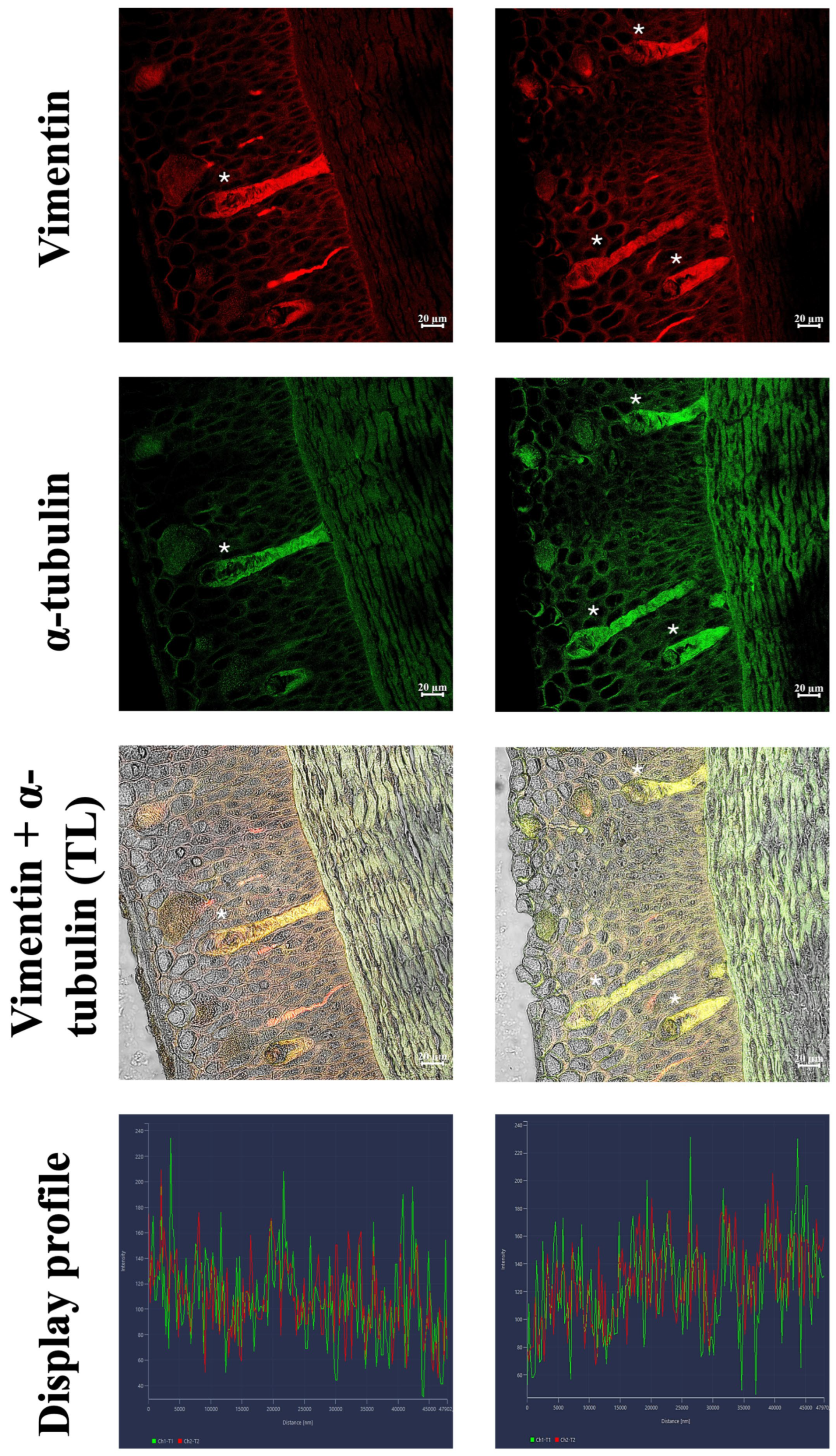
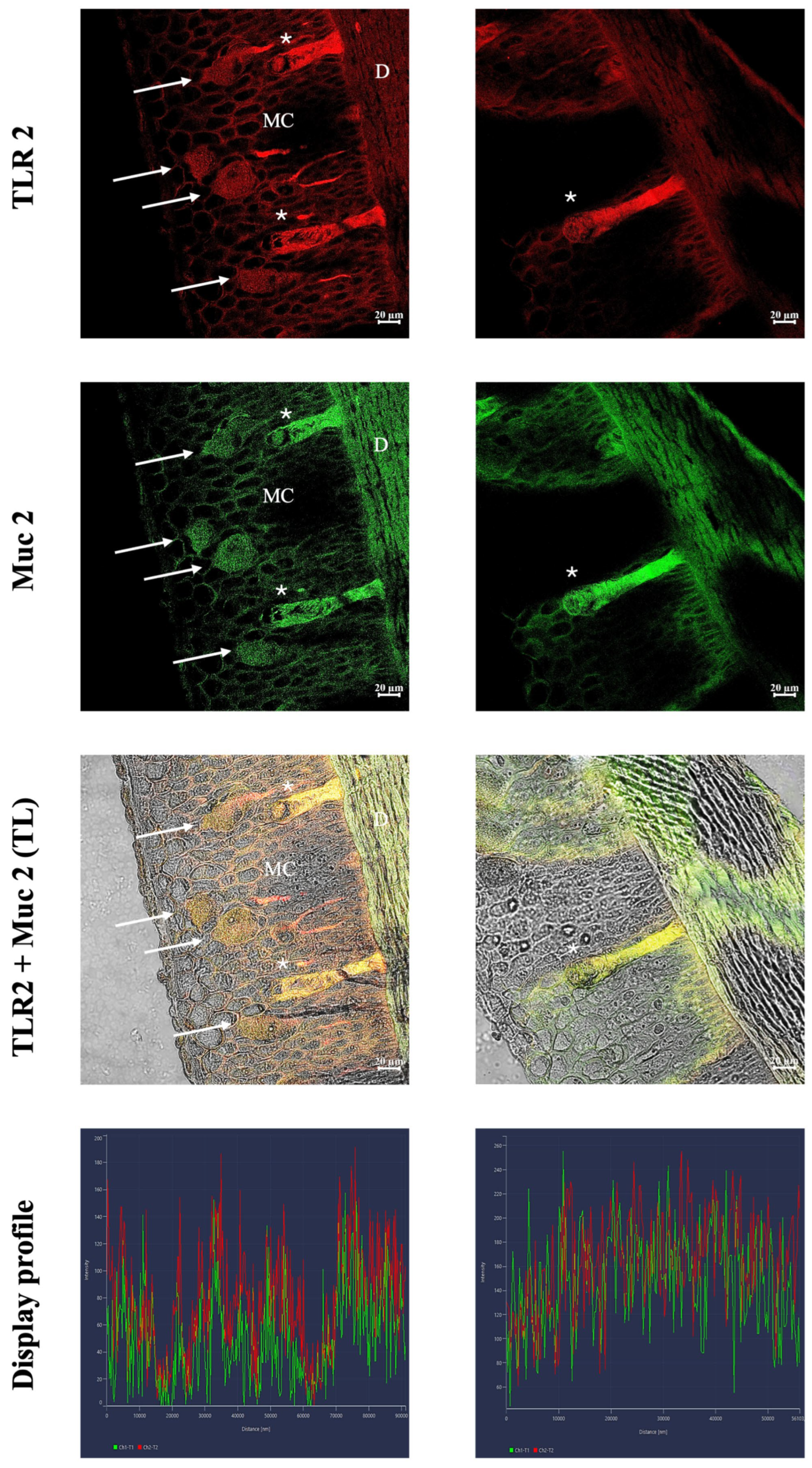
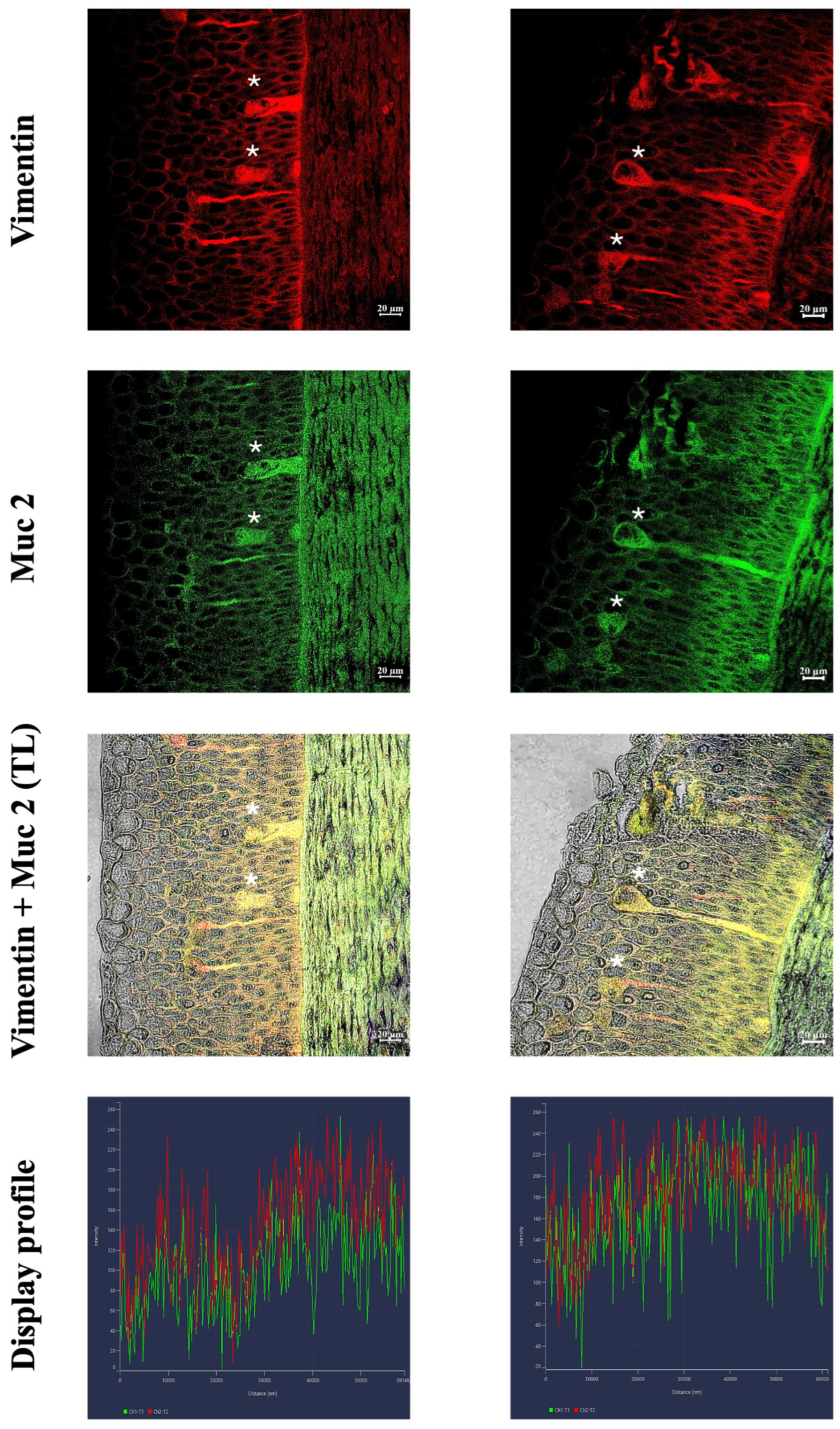
Immunoreactive skin cells are identified using confocal microscopy for all tested antibodies. In particular, immunoreactive keratinocytes and strong colocalization of antibodies are found by testing Vimentin and Tubulin (Figure 2). Colocalization is also observed between TLR2 and Muc2. In addition to skein cells positive for both antibodies, a slight positivity is noted in some granular and mucous cells of the basal layer. Furthermore, the mucous cells of the basal layer are immunoreactive for Muc2, in contrast to the superficial cells. Furthermore, a certain degree of reactivity is also appreciable in the dermis, between the muscle fibers, as highlighted by AB/PAS staining (Figure 3). Finally, the colocalization of Vimentin and Muc2 further confirm the reactivity of the skein cells for the tested antibodies (Figure 4).
Quantitative image analysis confirmed the colocalization of the investigated markers within the skein cells. Pearson’s correlation coefficient (PCC) yielded a mean R value of 0.85 ± 0.03 for the colocalization of TLR2 and Muc2 across ten independent fields of view. For the colocalization of vimentin and Muc2, the mean PCC R value was 0.81 ± 0.03 across 10 independent fields of view. The mean PCC R value for the colocalization between vimentin and α-tubulin was 0.75 ± 0.02 across 10 independent fields of view. These results indicate a consistent and robust degree of overlap between the fluorescence signals, in agreement with the qualitative observations obtained via confocal microscopy.

3.3. Immunoperoxidase

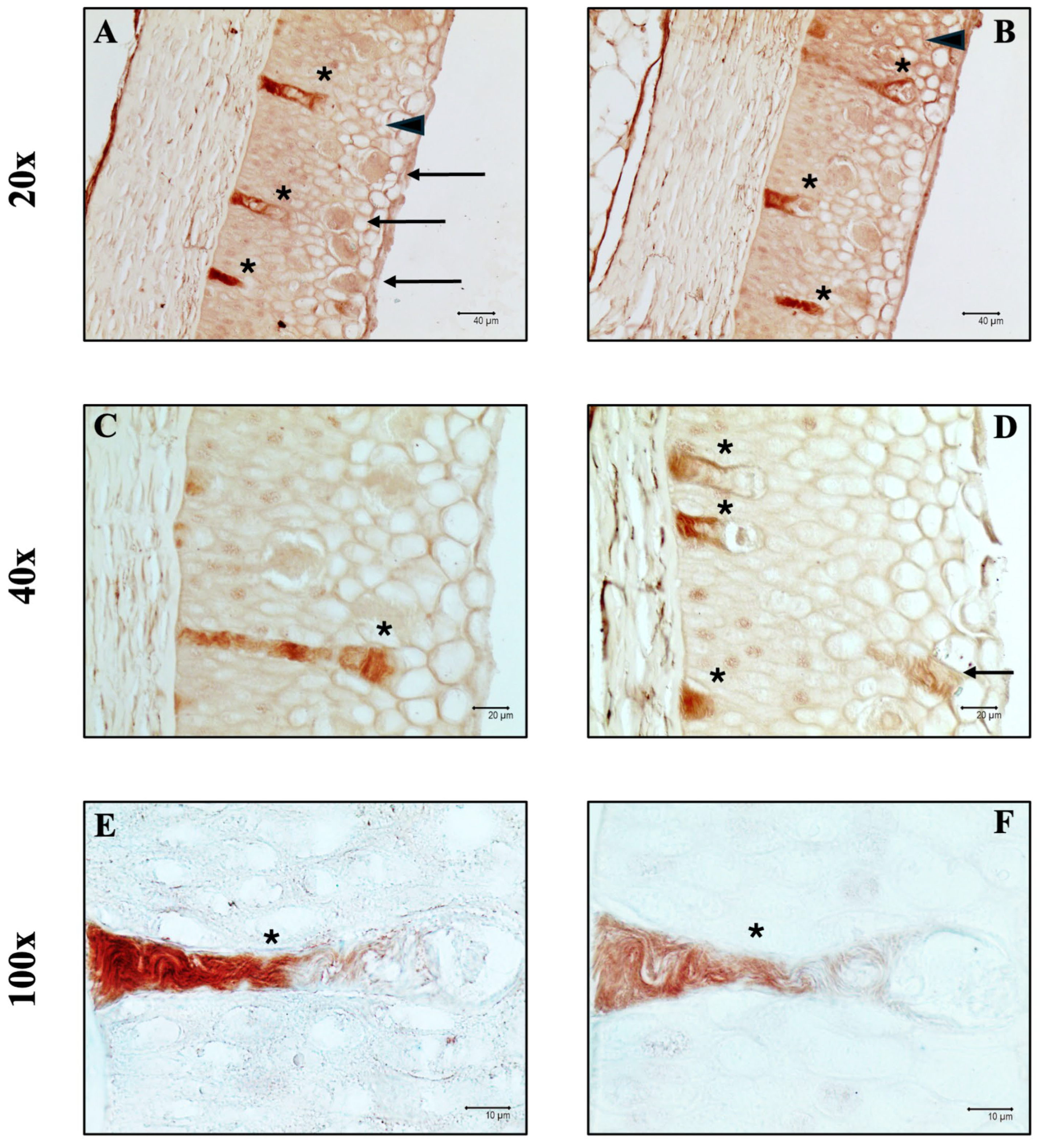
Immunoperoxidase staining revealed the presence of Piscidin1-positive skein cells, with stronger positivity observed at the base and cell body compared to the apical portion. Furthermore, examination of the superficial layer reveals that a small number of granular and mucous cells exhibit a slight degree of reactivity to the antibody (Figure 5).
Quantitative and statistical analyses of the skein cells immunoreactive to the various antibodies tested were performed, demonstrating that there were no significant differences in the number of immunopositive skein cells among the different antibodies (F(4) = 0.14; p = 0.97) (Table 4).

3.4. In Silico Analyses

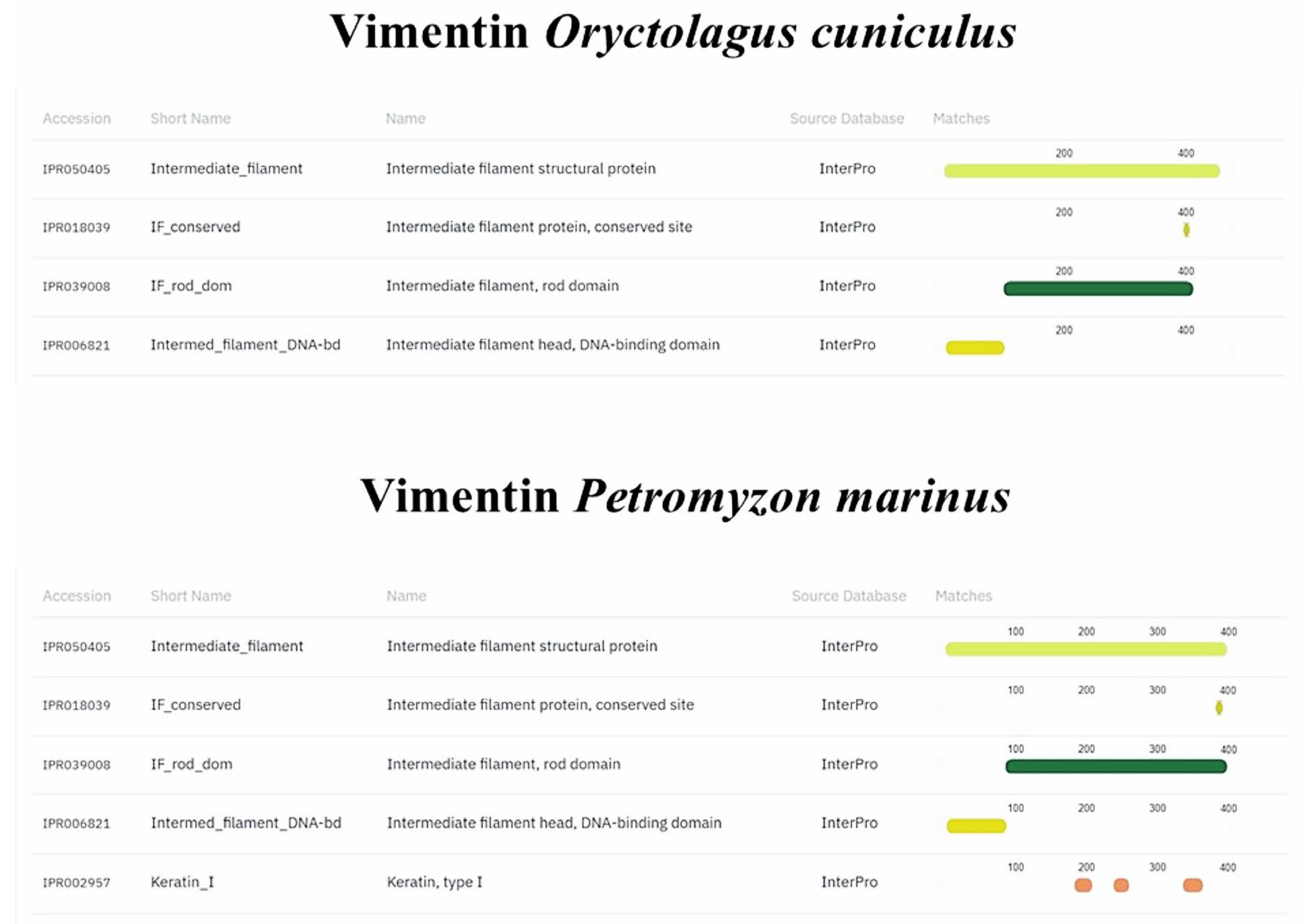
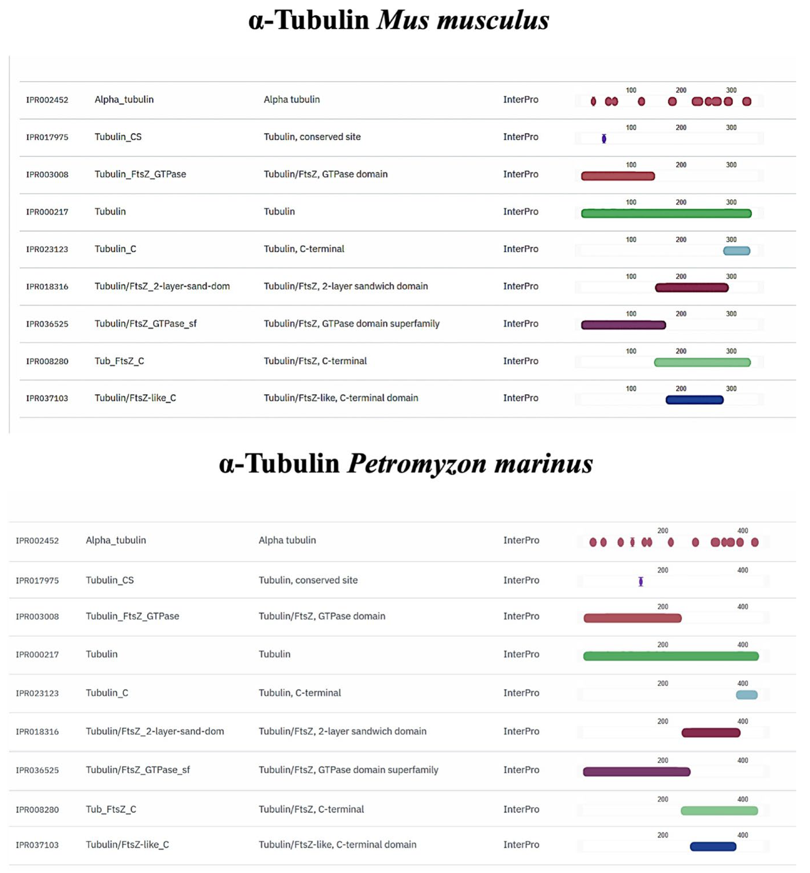
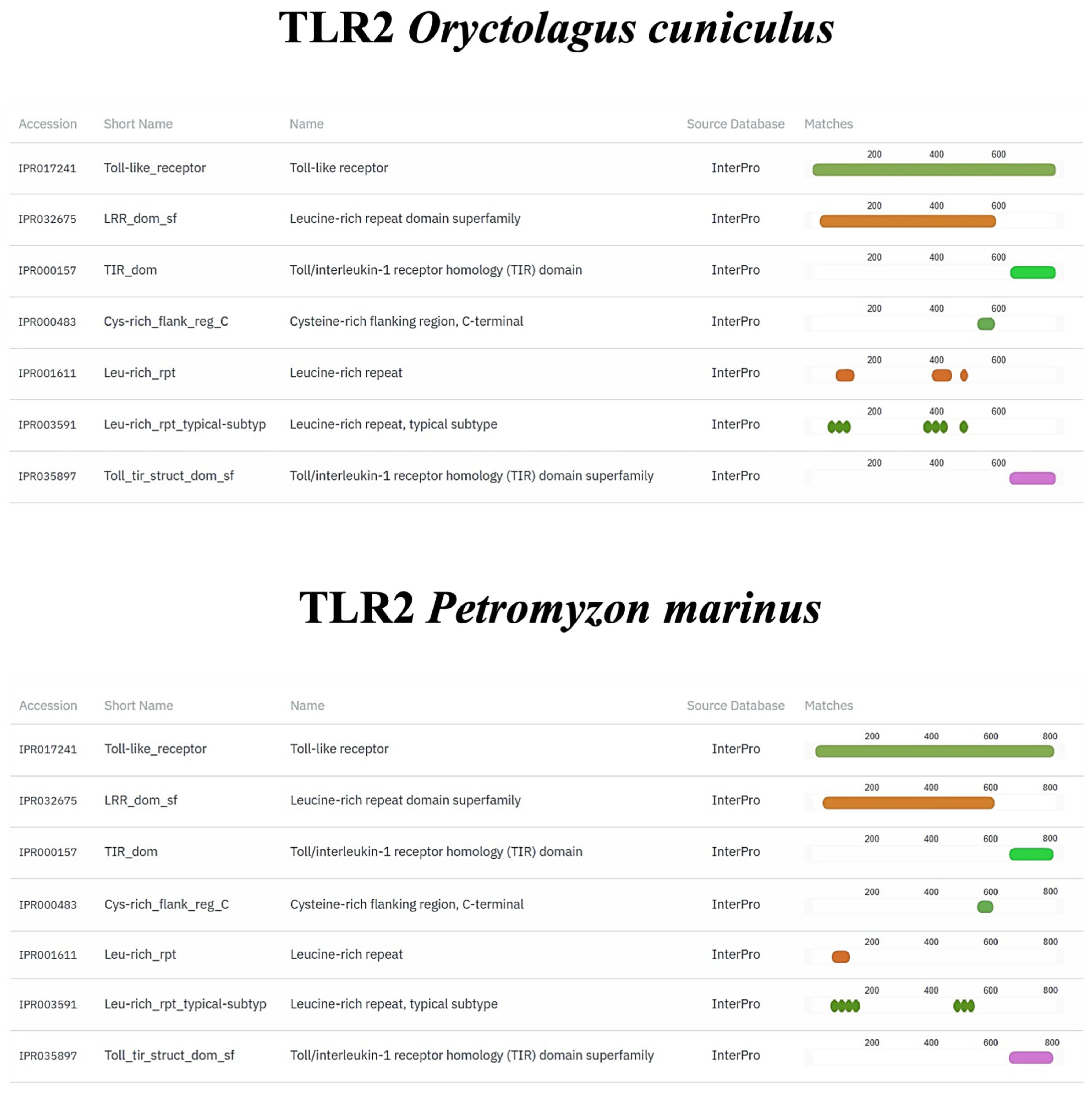
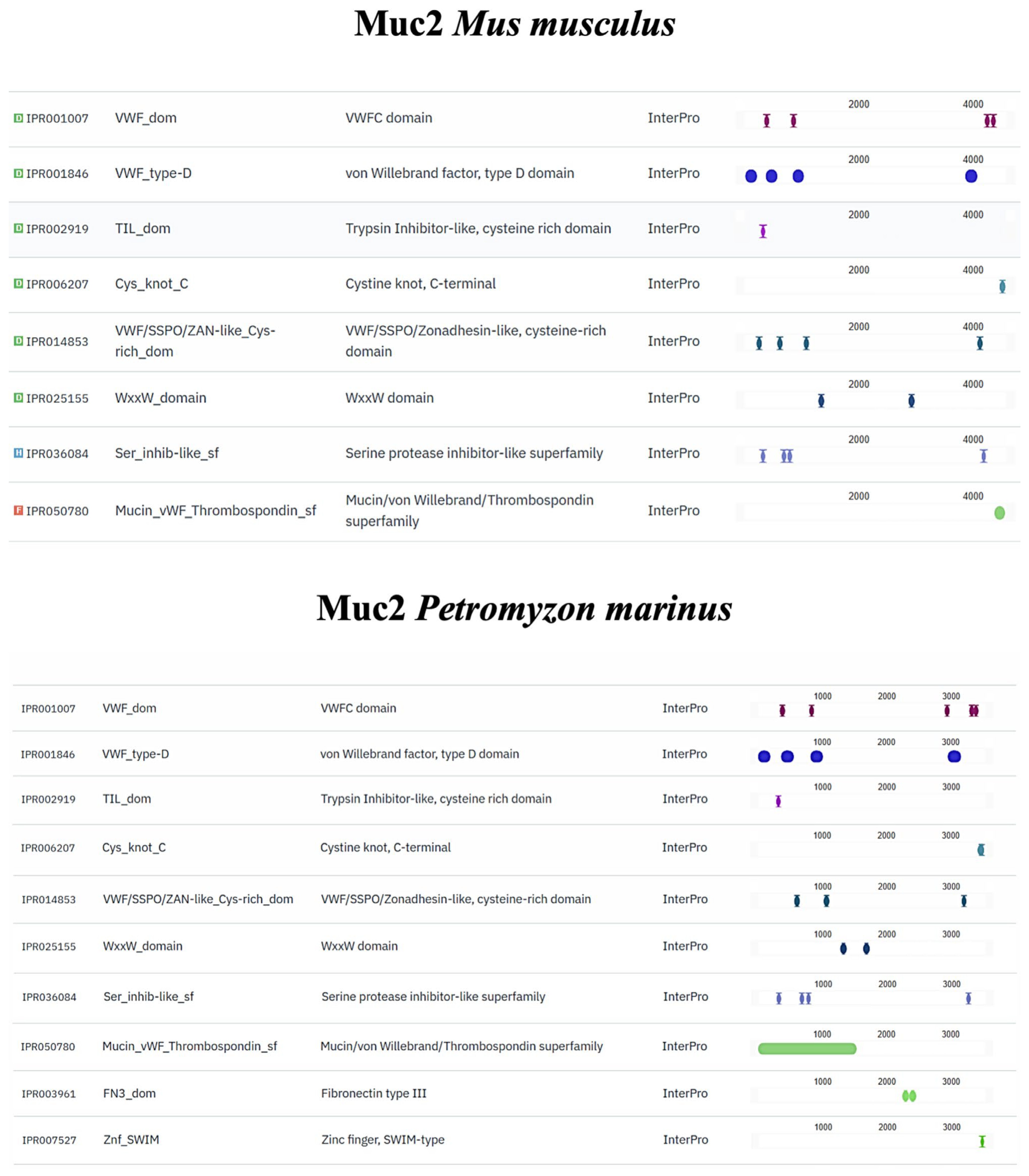
Vimentin, α-tubulin, TLR2, and Muc2 orthologs were found in the P. marinus genome. Our analyses revealed the presence of one vimentin orthologous gene that encodes a 401 aa protein. The results of the protein functional annotation step showed a significant similarity in the number and structure of the conserved protein domains, with the orthologous protein having an additional domain (keratin, type I, IPR002957) (Figure 6). The P. marinus α-tubulin ortholog and the reference α-tubulin of M. musculus shared the same number and types of protein domains (Figure 7). A similar pattern can also be observed in TLR2 orthologs (Figure 8). Regarding Muc2 protein prediction, we found that the P. marinus ortholog shares eight of the ten protein domains present in M. musculus, with the addition of two domains exclusively found in P. marinus (fibronectin type III IPR003961; Zinc finger, SWIM-type IPR007527) (Figure 9). Fibronectin type-III (FN3) domain is both the largest and the most common of the fibronectin subdomains, containing a conserved β-sandwich fold, similar to that of Ig-like domains [49]. These domains can be found in different animal protein families, such as cell-surface receptors, molecules from the extracellular matrix, and enzymes [49]. FN3 domain has been shown to be crucial in the regulation of Toll-like receptor 4 function during Xenopus embryos development [50]. Other studies demonstrated that this domain can be involved in skeletal and cartilaginous development, wound healing, and tumorigenesis [51]. The Zinc finger SWIM-type domain acts as a stable scaffold and can be found in proteins involved in gene transcription, cytoskeletal organization, cell adhesion, protein folding, and chromatin remodeling [52]. Therefore, it is possible to speculate that the presence of these additional domains in Lamprey Muc2 could link this mucin to immune or signaling processes. Although the homology of the entire sequence is not very high for some antibodies, in silico analysis of orthologs revealed significant conservation of functional domains responsible for antigen recognition and antigen–antibody binding.

4. Discussion

4.1. Alarm Signaling: An Evolutionary Perspective

Alarm signals are chemical mixtures released from damaged fish tissues after injury, which warn nearby conspecifics and closely related taxa of the risk of predation [53,54]. Recognizing and responding appropriately to danger is essential for survival and success in risky environments; both excessive caution and risk-taking have maladaptive consequences [55]. In variable environments with novel risks, animals can directly learn about new threats, such as predators, when they occur in conjunction with aversive events, such as physical injury or pursuit [56]. Given the risks of direct learning, it is often advantageous to learn to respond to novel threats using social information, where the cues that evoke defensive responses come from conspecifics [57]. Animals have been shown to form associations between novel stimuli and conspecific cues that indicate a threat, such as chemical cues released during predation or the defensive responses of experienced conspecifics [56,58]. The innate capacity to detect alarm signals has been demonstrated in several studies [59,60]. Furthermore, the analysis of signal dispersion patterns in the environment has revealed the locations of risks for conspecifics and closely related species that may share similar predators [1]. When an unfamiliar predator’s scent is accompanied by alarm signals, prey can learn to associate the predator’s scent with danger and avoid it in the future [54].
Several studies have highlighted the ability of sea lampreys to release and recognize alarm signals [1,54,61,62], although it is unclear where they are released. Hume et al. (2017) analyzed the response of sea lamprey (Petromyzon marinus) to alarm signals released by five cofamilial species and two distantly related outgroups, highlighting that an alarm signal released following damage is at least partially conserved within Petromyzontidae and that sea lamprey perceives attacks by predators directed at closely related taxa [1]. Furthermore, cross-reactivity to damage-released compounds has been reported in two lamprey species, the silver lamprey (Ichthyomyzon unicuspis, C. L. Hubbs & Trautman, 1937) [63] and Pacific lamprey (Entosphenus tridentatus, Richardson, 1836) [64], suggesting that this may be a characteristic shared by all Northern Hemisphere lampreys (Petromyzontidae) that would allow them to avoid predators that attack lamprey species. The alarm response of the sea lamprey is elicited by tissues throughout the body but is strongest when originating from the skin [63], suggesting that the alarm signal originates from the skin. Throughout ontogeny, the epidermis of lampreys increases in thickness from the pro-larval stage to spawning migration, and in sea lampreys, this is primarily due to an increase in the number of mucous cell layers [31]. The mucous cell layers of the sea lamprey during spawning migration consist of three distinct types based on the differentiation process that causes them to migrate from the base to the surface of the epidermis: basal, mid-epidermal, and superficial layers [31]. Superficial mucous cells located in the upper layers of the epidermis contain sialic acid and a variety of other glycoconjugates similar to those found in mucus [31].

4.2. Morphological and Histochemical Profile of Skein Cells Within the Petromyzon marinus Epidermis

Histologically, our results showed that the skin was composed of a stratified epidermis and an underlying dermis. The epidermis is divided into three main layers: superficial, intermediate, and basal. Within the epidermis, three cell populations can be identified: mucous, granular, and skein. Mucous cells are the most abundant cell type and are arranged in layers from the basal lamina to the surface. Those in contact with the basal lamina, called the basal layer, have an ovoid nucleus that becomes more rounded in the upper layers. The mucous cells in the intermediate and superficial layers had cytoplasm that increased in volume and was more acidophilic. The shape of the mucous cells changed from elongated in the basal layer to rounded and finally flattened in the more superficial parts. Granular cells are rounded, with small and dense nuclei, and are mainly found in the intermediate and superficial layers. Their cytoplasm was clear and rich in scattered bluish granules. Skein cells have an elongated morphology, with the basal part in contact with the basal lamina and the apical part facing the surface and extending into the upper layers. They have a slightly eosinophilic cytoplasm with twisted internal filaments along the entire cell body, except in the perinuclear zone.
Histochemically, AB/PAS allows for the distinction between glycoconjugates containing acidic carbohydrates (blue-stained) and neutral ones (red-stained). The combination of both techniques produces a purple color when the carbohydrates are co-expressed. In the epidermis of Petromyzon, an intense blue color was observed in the most superficial mucous cells, whereas the basal cells were stained more lightly. Traces of mucopolysaccharides were also evident in the underlying dermis between the muscle fibers. The staining of mucous cells became more intense from the base to the surface. In addition, granular and skein cells showed very weak AB/PAS staining. Toluidine Blue staining confirmed this distribution, which was less marked in the basal layers and more evident in the superficial layers. This was corroborated by quantitative and statistical analyses of the staining intensity. Our histological results are consistent with those reported in the literature [31]. Interestingly, and in agreement with Alonso et al. (2017) [31], we found that mucus cells were more reactive to AB/PAS in the most superficial layer, indicating a higher concentration of acidic glycoproteins. Furthermore, traces of mucopolysaccharides were scattered among the underlying muscle fibers.
This study focused on skein cells, whose actual role is still little known. Given their previous association with teleost club cells and the data found in the literature regarding the starting site of the alarm signal, we speculate that skein cells are involved in the defense system of the sea lamprey, playing an active role in the release of chemical mediators of the alarm. To corroborate our thesis, optical and confocal immunohistochemistry analyses were conducted using specific antibodies directed against vimentin, alpha-tubulin, Toll-Like Receptor 2, mucin 2, and Piscidin1.
In fish, vimentin, a type III intermediate filament protein, is found in several cell types, including club cells, which are specialized in releasing chemical alarm signals that warn other fish of potential danger. The presence of vimentin suggests that it may play a role in the structure and function of these cells, potentially contributing to their ability to release alarm signals [65,66]. Furthermore, this protein is involved in immune function and inflammation, acting at the intracellular level, influencing immune cell behavior, and at the extracellular level, participating in immune responses and interactions [41,44,67,68]. Our results show vimentin-immunoreactive skein cells, suggesting that these cells may mediate immune responses in the skin of sea lampreys. Granular cells that were mildly positive for vimentin were also observed.

4.3. Evidence for the Possible Functional Role of Skein Cells: Immunity and Alarm Signaling

In fish, epidermal club cells, which release chemical alarm signals, do not directly use tubulin for their primary function of releasing these signals. While tubulin is a component of microtubules, which are essential for various cellular processes, including cell shape and movement, club cells primarily rely on specialized vesicles for the synthesis and secretion of alarm substances. Microtubules are involved in the transport of these vesicles into the cell but not in the direct release of alarm substances [65,69,70]. However, tubulin also plays a role in immune function by influencing various aspects of immune cell behavior and their interactions. In particular, tubulin involvement in the cytoskeleton is crucial for cellular interactions during antigen recognition and presentation [45,71,72]. Using confocal microscopy, we highlighted skein cells that were strongly immunoreactive to alpha-tubulin, confirming their identity not only by morphology but also by the filamentous skein content inside them. Furthermore, this study adds to our hypothesis regarding the defensive role of these cells. Interestingly, but not surprisingly, the reaction with both antibodies, vimentin and tubulin, showed a clear colocalization of these proteins in the skein cells. A slight positivity was also found in the granular cells, while a more marked reactivity was present in the muscle fibers of the dermis, as reported in the literature [73].
In fish, epidermal club cells express TLR2 [74]. TLRs are a class of recognition receptors (PRRs) that play a crucial role in the innate immune response of fish by recognizing pathogen-associated molecular patterns (PAMPs) [65,75]. We have previously demonstrated the presence of TLR2 in club cells of zebrafish (Danio rerio Hamilton, 1822) skin [74]. TLR2, a major player in the innate immune response, is an evolutionarily conserved receptor in all metazoans. In previous studies, we demonstrated its presence in the immune and structural cells of invertebrates [68,76,77,78,79], protochordates [80], cartilaginous fish [81], bony fish [82], amphibians [83], reptiles [84], and mammals [40]. Our confocal microscopy results showed that skein cells were strongly reactive for TLR2, further corroborating our hypothesis on their immune role.
In fish, club and mucous cells are involved in the production and release of mucus, which forms a protective layer on the skin [40,65,70]. Interestingly, we noted Muc2-reactive skein cells and slightly positive granular cells. Furthermore, the mucous cells of the innermost layer were positive, suggesting that these cells are more concentrated in Muc2, while in the more superficial ones, the literature reports a prevalence of sialic acid and acid glycoconjugates [31]. Furthermore, the muscle fibers of the dermis appeared to be well-reactive to the tested antibody, as noted histologically with AB/PAS staining. Furthermore, the colocalization of TLR2 and Muc2 suggests a strong defensive link between these cells. It has been demonstrated that TLRs act by influencing mucus secretion and that this interaction confers an evolutionarily conserved sentinel role to mucus cells [43,81,85,86]. The further colocalization of Vimentin and Muc2 has strengthened what has been debated so far.
The quantitative colocalization analysis further strengthens the evidence for the functional association of the investigated proteins within skein cells. The mean Pearson’s correlation coefficients indicate a robust degree of spatial overlap between the markers, a value consistent with strong colocalization according to established thresholds in confocal imaging studies. This quantitative result supports our qualitative observations and provides objective confirmation that skein cells integrate both structural and immunological components. In particular, the colocalization of immune-related markers suggests that skein cells are not only structural elements of the epidermis but also active participants in immune surveillance and alarm signaling, potentially indicating that skein cells act as important contributors to epidermal specialization and evolutionary adaptation in agnathan immunity.
Fish club cells possess antimicrobial peptides in their cytoplasm [65]. We previously demonstrated the presence of Piscidin1 in zebrafish club cells [73] and in the goblet cells of Eptatretus cirrathus (J. R. Forster, 1801) [43]. Piscidin is a broad-spectrum antimicrobial peptide that is evolutionarily conserved in fish [87]. Our optical microscopy data revealed skein cells that were highly reactive to piscidin, further supporting our hypothesis regarding their immune role.
Consistent with these observations, our findings indicate that the number of skein cells immunopositive for the various antibodies tested does not differ significantly, suggesting a comparable expression profile across the markers analyzed.
To further corroborate the data obtained by optical and confocal microscopy, an in silico investigation was performed, which demonstrated the evolutionary conservation of the tested antibodies produced in mammals on P. marinus.
Interestingly, the staining obtained with all antibodies allowed us to appreciate the presence of a skein inside the cell when photographed at a higher magnification. Furthermore, we did not notice any difference in positivity between the shorter and longer skein cells that deepened towards the most superficial layer of the skin.

5. Conclusions

Since all the antibodies tested are proteins involved in immune functions, we can affirm that skein cells play a role in the internal defense system of sea lampreys. Furthermore, because the same antibodies that we tested and found in the skein cells are present on the surface or in the cytoplasm of the club cells, we can speculate that these cells are involved in the production and release of alarm chemicals into the external environment. These new data provide deeper knowledge of the biology of these primitive animals, shedding light on new aspects of their immune system and, above all, laying the foundations for further studies on the role of skein cells in the evolution and adaptation of this agnathous vertebrate.
These new data provide deeper knowledge of the biology of these primitive animals, shedding light on new aspects of their immune system and, above all, laying the foundations for further studies on the role of skein cells in the evolution and adaptation of this agnathous vertebrate.

Author Contributions

Conceptualization, A.A.; investigation, A.A., S.M., S.F. (Stefania Fiorentino), A.M., S.P., S.F. (Sergio Famulari), G.P.L., R.F.A. and E.R.L.; data curation, A.A., S.M., S.F. (Stefania Fiorentino), A.M., S.P., S.F. (Sergio Famulari), G.P.L., R.F.A. and E.R.L.; writing—original draft preparation, A.A.; writing—review and editing, A.A., R.F.A. and E.R.L.; visualization, S.M.; supervision, A.A. and E.R.L. All authors have read and agreed to the published version of the manuscript.

Funding

This research received no external funding.

Institutional Review Board Statement

No live animals were subjected to experimental manipulation or invasive procedures in the course of this research, and thus ethical review and approval by an Institutional Review Board were not required.

Informed Consent Statement

Not applicable.

Data Availability Statement

The original contributions presented in this study are included in this article; further inquiries can be directed to the corresponding authors.

Conflicts of Interest

The authors declare no conflicts of interest.

References

  1. Hume, J.B.; Wagner, M. A Death in the Family: Sea Lamprey (Petromyzon marinus) Avoidance of Confamilial Alarm Cues Diminishes with Phylogenetic Distance. Ecol. Evol. 2018, 8, 3751–3762. [Google Scholar] [CrossRef]
  2. Barreto, R.E.; Miyai, C.A.; Sanches, F.H.C.; Giaquinto, P.C.; Delicio, H.C.; Volpato, G.L. Blood Cues Induce Antipredator Behavior in Nile Tilapia Conspecifics. PLoS ONE 2013, 8, e54642. [Google Scholar] [CrossRef] [PubMed]
  3. Ferrari, M.C.O.; Elvidge, C.K.; Jackson, C.D.; Chivers, D.P.; Brown, G.E. The Responses of Prey Fish to Temporal Variation in Predation Risk: Sensory Habituation or Risk Assessment? Behav. Ecol. 2010, 21, 532–536. [Google Scholar] [CrossRef]
  4. Kicklighter, C.E.; Germann, M.; Kamio, M.; Derby, C.D. Molecular Identification of Alarm Cues in the Defensive Secretions of the Sea Hare Aplysia Californica. Anim. Behav. 2007, 74, 1481–1492. [Google Scholar] [CrossRef]
  5. Mathuru, A.S.; Kibat, C.; Cheong, W.F.; Shui, G.; Wenk, M.R.; Friedrich, R.W.; Jesuthasan, S. Chondroitin Fragments Are Odorants That Trigger Fear Behavior in Fish. Curr. Biol. 2012, 22, 538–544. [Google Scholar] [CrossRef] [PubMed]
  6. Majer, A.P.; Trigo, J.R.; Duarte, L.F.L. Evidence of an Alarm Signal in Ophiuroidea (Echinodermata). Mar. Biodivers. Rec. 2009, 2, e102. [Google Scholar] [CrossRef]
  7. Daleo, P.; Alberti, J.; Avaca, M.S.; Narvarte, M.; Martinetto, P.; Iribarne, O. Avoidance of Feeding Opportunities by the Whelk Buccinanops Globulosum in the Presence of Damaged Conspecifics. Mar. Biol. 2012, 159, 2359–2365. [Google Scholar] [CrossRef]
  8. Chemical Cues and Reducing the Risk of Predation. In Chemical Communication in Crustaceans; Springer: New York, NY, USA, 2010; pp. 355–370. ISBN 978-0-387-77100-7.
  9. Persons, M.H.; Walker, S.E.; Rypstra, A.L.; Marshall, S.D. Wolf Spider Predator Avoidance Tactics and Survival in the Presence of Diet-Associated Predator Cues (Araneae: Lycosidae). Anim. Behav. 2001, 61, 43–51. [Google Scholar] [CrossRef]
  10. Llandres, A.L.; Gonzálvez, F.G.; Rodríguez-Gironés, M.A. Social but Not Solitary Bees Reject Dangerous Flowers Where a Conspecific Has Recently Been Attacked. Anim. Behav. 2013, 85, 97–102. [Google Scholar] [CrossRef]
  11. Brown, C.; Laland, K.; Krause, J. (Eds.) Fish Cognition and Behavior, 1st ed.; Wiley: Hoboken, NJ, USA, 2011; ISBN 978-1-4443-3221-6. [Google Scholar]
  12. Gherardi, F.; Mavuti, K.M.; Pacini, N.; Tricarico, E.; Harper, D.M. The Smell of Danger: Chemical Recognition of Fish Predators by the Invasive Crayfish Procambarus Clarkii: Chemical Recognition of Fish Predators by Invasive Crayfish. Freshw. Biol. 2011, 56, 1567–1578. [Google Scholar] [CrossRef]
  13. Johnston, B.R.; Molis, M.; Scrosati, R.A. Predator Chemical Cues Affect Prey Feeding Activity Differently in Juveniles and Adults. Can. J. Zool. 2012, 90, 128–132. [Google Scholar] [CrossRef]
  14. Chivers, D.P.; Smith, R.J.F. Chemical Alarm Signalling in Aquatic Predator-Prey Systems: A Review and Prospectus. Écoscience 1998, 5, 338–352. [Google Scholar] [CrossRef]
  15. Faulkner, A.E.; Holstrom, I.E.; Molitor, S.A.; Hanson, M.E.; Shegrud, W.R.; Gillen, J.C.; Willard, S.J.; Wisenden, B.D. Field Verification of Chondroitin Sulfate as a Putative Component of Chemical Alarm Cue in Wild Populations of Fathead Minnows (Pimephales promelas). Chemoecology 2017, 27, 233–238. [Google Scholar] [CrossRef]
  16. Sorensen, P.W.; Wisenden, B.D. (Eds.) Fish Pheromones and Related Cues, 1st ed.; Wiley: Hoboken, NJ, USA, 2014; ISBN 978-0-8138-2386-7. [Google Scholar]
  17. Schoeppner, N.M.; Relyea, R.A. When Should Prey Respond to Consumed Heterospecifics? Testing Hypotheses of Perceived Risk. Copeia 2009, 2009, 190–194. [Google Scholar] [CrossRef]
  18. dos Reis, M.; Thawornwattana, Y.; Angelis, K.; Telford, M.J.; Donoghue, P.C.J.; Yang, Z. Uncertainty in the Timing of Origin of Animals and the Limits of Precision in Molecular Timescales. Curr. Biol. 2015, 25, 2939–2950. [Google Scholar] [CrossRef] [PubMed]
  19. Almeida, P.R.; Arakawa, H.; Aronsuu, K.; Baker, C.; Blair, S.-R.; Beaulaton, L.; Belo, A.F.; Kitson, J.; Kucheryavyy, A.; Kynard, B.; et al. Lamprey Fisheries: History, Trends and Management. J. Great Lakes Res. 2021, 47, S159–S185. [Google Scholar] [CrossRef]
  20. Marchiori, C.H.; Santana, M.V.D.O.; Malheiros, K.D.P. Ecological and Medicinal Importance of Lampreys (Cyclostomata: Petromyzontiformes: Petromyzontidae). Middle East. Res. J. Microbiol. Biotechnol. 2024, 4, 34–49. [Google Scholar] [CrossRef]
  21. Docker, M.F. (Ed.) Lampreys: Biology, Conservation and Control; Springer: Dordrecht, The Netherlands, 2015; Volume 1, ISBN 978-94-017-9305-6. [Google Scholar]
  22. Fish & Fisheries Series. Available online: https://link.springer.com/series/5973 (accessed on 8 July 2025).
  23. Green, S.A.; Bronner, M.E. The Lamprey: A Jawless Vertebrate Model System for Examining Origin of the Neural Crest and Other Vertebrate Traits. Differentiation 2014, 87, 44–51. [Google Scholar] [CrossRef] [PubMed]
  24. Imre, I.; Di Rocco, R.T.; Belanger, C.F.; Brown, G.E.; Johnson, N.S. The Behavioural Response of Adult Petromyzon marinus to Damage-released Alarm and Predator Cues. J. Fish Biol. 2014, 84, 1490–1502. [Google Scholar] [CrossRef]
  25. Wagner, C.M.; Stroud, E.M.; Meckley, T.D. A Deathly Odor Suggests a New Sustainable Tool for Controlling a Costly Invasive Species. Can. J. Fish Aquat. Sci. 2011, 68, 1157–1160. [Google Scholar] [CrossRef]
  26. Hume, J.B.; Meckley, T.D.; Johnson, N.S.; Luhring, T.M.; Siefkes, M.J.; Wagner, C.M. Application of a Putative Alarm Cue Hastens the Arrival of Invasive Sea Lamprey (Petromyzon marinus) at a Trapping Location. Can. J. Fish. Aquat. Sci. 2015, 72, 1799–1806. [Google Scholar] [CrossRef]
  27. Luhring, T.M.; Meckley, T.D.; Johnson, N.S.; Siefkes, M.J.; Hume, J.B.; Wagner, C.M. A Semelparous Fish Continues Upstream Migration When Exposed to Alarm Cue, but Adjusts Movement Speed and Timing. Anim. Behav. 2016, 121, 41–51. [Google Scholar] [CrossRef]
  28. Pfeiffer, W. The Distribution of Fright Reaction and Alarm Substance Cells in Fishes. Copeia 1977, 1977, 653. [Google Scholar] [CrossRef]
  29. Brönmark, C.; Hansson, L.-A. (Eds.) Chemical Ecology in Aquatic Systems; Oxford University Press: Oxford, UK, 2012; ISBN 978-0-19-958309-6. [Google Scholar]
  30. Carreau-Green, N.D.; Mirza, R.S.; MartÍnez, M.L.; Pyle, G.G. The Ontogeny of Chemically Mediated Antipredator Responses of Fathead Minnows Pimephales promelas. J. Fish Biol. 2008, 73, 2390–2401. [Google Scholar] [CrossRef]
  31. Rodríguez-Alonso, R.; Megías, M.; Pombal, M.A.; Molist, P. Morphological and Functional Aspects of the Epidermis of the Sea Lamprey Petromyzon marinus throughout Development. J. Fish Biol. 2017, 91, 80–100. [Google Scholar] [CrossRef] [PubMed]
  32. Lane, E.B.; Whitear, M. Skein Cells in Lamprey Epidermis. Can. J. Zool. 1980, 58, 450–455. [Google Scholar] [CrossRef]
  33. Akat, E.; Yenmiş, M.; Pombal, M.A.; Molist, P.; Megías, M.; Arman, S.; Veselỳ, M.; Anderson, R.; Ayaz, D. Comparison of Vertebrate Skin Structure at Class Level: A Review. Anat. Rec. 2022, 305, 3543–3608. [Google Scholar] [CrossRef] [PubMed]
  34. Li, S.; Zhao, Z.; Li, Q.; Li, J.; Pang, Y. Lamprey Wound Healing and Regenerative Effects: The Collaborative Efforts of Diverse Drivers. IJMS 2023, 24, 3213. [Google Scholar] [CrossRef]
  35. Ángeles Esteban, M. An Overview of the Immunological Defenses in Fish Skin. ISRN Immunol. 2012, 2012, 1–29. [Google Scholar] [CrossRef]
  36. Tsutsui, S.; Nakamura, O.; Watanabe, T. Lamprey (Lethenteron japonicum) IL-17 Upregulated by LPS-Stimulation in the Skin Cells. Immunogenetics 2007, 59, 873–882. [Google Scholar] [CrossRef]
  37. Rast, J.P.; Buckley, K.M. Lamprey Immunity Is Far from Primitive. Proc. Natl. Acad. Sci. USA 2013, 110, 5746–5747. [Google Scholar] [CrossRef]
  38. Chivers, D.P.; Zhao, X.; Brown, G.E.; Marchant, T.A.; Ferrari, M.C.O. Predator-Induced Changes in Morphology of a Prey Fish: The Effects of Food Level and Temporal Frequency of Predation Risk. Evol. Ecol. 2008, 22, 561–574. [Google Scholar] [CrossRef]
  39. Pergolizzi, S.; Fumia, A.; D’Angelo, R.; Mangano, A.; Lombardo, G.P.; Giliberti, A.; Messina, E.; Alesci, A.; Lauriano, E.R. Expression and Function of Toll-like Receptor 2 in Vertebrate. Acta Histochem. 2023, 125, 152028. [Google Scholar] [CrossRef]
  40. Dash, S.; Das, S.K.; Samal, J.; Thatoi, H.N. Epidermal Mucus, a Major Determinant in Fish Health: A Review. IJVR 2018, 19, 72–81. [Google Scholar] [CrossRef]
  41. Arrindell, J.; Desnues, B. Vimentin: From a Cytoskeletal Protein to a Critical Modulator of Immune Response and a Target for Infection. Front. Immunol. 2023, 14, 1224352. [Google Scholar] [CrossRef]
  42. Ilan-Ber, T.; Ilan, Y. The Role of Microtubules in the Immune System and as Potential Targets for Gut-Based Immunotherapy. Mol. Immunol. 2019, 111, 73–82. [Google Scholar] [CrossRef] [PubMed]
  43. Alesci, A.; Pergolizzi, S.; Savoca, S.; Fumia, A.; Mangano, A.; Albano, M.; Messina, E.; Aragona, M.; Lo Cascio, P.; Capillo, G.; et al. Detecting Intestinal Goblet Cells of the Broadgilled Hagfish Eptatretus Cirrhatus (Forster, 1801): A Confocal Microscopy Evaluation. Biology 2022, 11, 1366. [Google Scholar] [CrossRef] [PubMed]
  44. Suprewicz, Ł.; Zakrzewska, M.; Okła, S.; Głuszek, K.; Sadzyńska, A.; Deptuła, P.; Fiedoruk, K.; Bucki, R. Extracellular Vimentin as a Modulator of the Immune Response and an Important Player during Infectious Diseases. Immunol. Cell Biol. 2024, 102, 167–178. [Google Scholar] [CrossRef]
  45. Martín-Cófreces, N.B.; Sánchez-Madrid, F. Sailing to and Docking at the Immune Synapse: Role of Tubulin Dynamics and Molecular Motors. Front. Immunol. 2018, 9, 1174. [Google Scholar] [CrossRef] [PubMed]
  46. Birchenough, G.M.H.; Nyström, E.E.L.; Johansson, M.E.V.; Hansson, G.C. A Sentinel Goblet Cell Guards the Colonic Crypt by Triggering Nlrp6-Dependent Muc2 Secretion. Science 2016, 352, 1535–1542. [Google Scholar] [CrossRef]
  47. Mcdonald, J.H.; Dunn, K.W. Statistical Tests for Measures of Colocalization in Biological Microscopy. J. Microsc. 2013, 252, 295–302. [Google Scholar] [CrossRef] [PubMed]
  48. Paysan-Lafosse, T.; Blum, M.; Chuguransky, S.; Grego, T.; Pinto, B.L.; Salazar, G.A.; Bileschi, M.L.; Bork, P.; Bridge, A.; Colwell, L.; et al. InterPro in 2022. Nucleic Acids Res. 2023, 51, D418–D427. [Google Scholar] [CrossRef] [PubMed]
  49. Leahy, D.J.; Hendrickson, W.A.; Aukhil, L.; Erickson, H.P. Structure of a Fibronectin Type III Domain from Tenascin Phased by MAD Analysis of the Selenomethionyl Protein. Science 1992, 258, 987–991. [Google Scholar] [CrossRef]
  50. Kim, H.-S.; McKnite, A.; Xie, Y.; Christian, J.L. Fibronectin Type III and Intracellular Domains of Toll-like Receptor 4 Interactor with Leucine-Rich Repeats (Tril) Are Required for Developmental Signaling. MBoC 2018, 29, 523–531. [Google Scholar] [CrossRef]
  51. Bencharit, S.; Cui, C.B.; Siddiqui, A.; Howard-Williams, E.L.; Sondek, J.; Zuobi-Hasona, K.; Aukhil, I. Structural Insights into Fibronectin Type III Domain-Mediated Signaling. J. Mol. Biol. 2007, 367, 303–309. [Google Scholar] [CrossRef]
  52. Laity, J.H.; Lee, B.M.; Wright, P.E. Zinc Finger Proteins: New Insights into Structural and Functional Diversity. Curr. Opin. Struct. Biol. 2001, 11, 39–46. [Google Scholar] [CrossRef] [PubMed]
  53. Feder, M.E.; Wagner, C.M. Building a Natural Repellent: Effects of Varying Alarm Cue Exposure on Swim Activity and Spatial Avoidance in an Invasive Fish. Conserv. Physiol. 2025, 13, coaf028. [Google Scholar] [CrossRef]
  54. Mensch, E.L.; Dissanayake, A.A.; Nair, M.G.; Wagner, C.M. Sea Lamprey Alarm Cue Comprises Water- and Chloroform- Soluble Components. J. Chem. Ecol. 2022, 48, 704–717. [Google Scholar] [CrossRef]
  55. Peckarsky, B.L.; Abrams, P.A.; Bolnick, D.I.; Dill, L.M.; Grabowski, J.H.; Luttbeg, B.; Orrock, J.L.; Peacor, S.D.; Preisser, E.L.; Schmitz, O.J.; et al. Revisiting the Classics: Considering Nonconsumptive Effects in Textbook Examples of Predator–Prey Interactions. Ecology 2008, 89, 2416–2425. [Google Scholar] [CrossRef]
  56. Fan, R.; Reader, S.M.; Sakata, J.T. Alarm Cues and Alarmed Conspecifics: Neural Activity during Social Learning from Different Cues in Trinidadian Guppies. Proc. R. Soc. B 2022, 289, 20220829. [Google Scholar] [CrossRef]
  57. McRae, T.R. A Review of Squirrel Alarm-Calling Behavior: What We Know and What We Do Not Know about How Predator Attributes Affect Alarm Calls. Anim. Behav. Cogn. 2020, 7, 168–191. [Google Scholar] [CrossRef]
  58. Danchin, É.; Giraldeau, L.-A.; Valone, T.J.; Wagner, R.H. Public Information: From Nosy Neighbors to Cultural Evolution. Science 2004, 305, 487–491. [Google Scholar] [CrossRef]
  59. Lucon-Xiccato, T.; Di Mauro, G.; Bisazza, A.; Bertolucci, C. Alarm Cue-Mediated Response and Learning in Zebrafish Larvae. Behav. Brain Res. 2020, 380, 112446. [Google Scholar] [CrossRef]
  60. Poisson, A.; Valotaire, C.; Borel, F.; Bertin, A.; Darmaillacq, A.-S.; Dickel, L.; Colson, V. Embryonic Exposure to a Conspecific Alarm Cue Triggers Behavioural Plasticity in Juvenile Rainbow Trout. Anim. Behav. 2017, 133, 35–45. [Google Scholar] [CrossRef]
  61. Feder, M.E.; Wisenden, B.D.; Luhring, T.; Wagner, C. Speed Kills? Migrating Sea Lamprey Increase Speed When Exposed to an Antipredator Cue but Make Worse Short-Term Decisions. J. Great Lakes Res. 2024, 50, 102398. [Google Scholar] [CrossRef]
  62. Hume, J.B.; Luhring, T.M.; Wagner, C.M. Push, Pull, or Push–Pull? An Alarm Cue Better Guides Sea Lamprey towards Capture Devices than a Mating Pheromone during the Reproductive Migration. Biol. Invasions 2020, 22, 2129–2142. [Google Scholar] [CrossRef]
  63. Bals, J.D.; Wagner, C.M. Behavioral Responses of Sea Lamprey (Petromyzon marinus) to a Putative Alarm Cue Derived from Conspecific and Heterospecific Sources. Behaviour 2012, 149, 901–923. [Google Scholar] [CrossRef]
  64. Byford, G.J.; Wagner, C.M.; Hume, J.B.; Moser, M.L. Do Native Pacific Lamprey and Invasive Sea Lamprey Share an Alarm Cue? Implications for Use of a Natural Repellent to Guide Imperiled Pacific Lamprey into Fishways. N. Am. J. Fish. Manag. 2016, 36, 1090–1096. [Google Scholar] [CrossRef]
  65. Pandey, S.; Stockwell, C.A.; Snider, M.R.; Wisenden, B.D. Epidermal Club Cells in Fishes: A Case for Ecoimmunological Analysis. IJMS 2021, 22, 1440. [Google Scholar] [CrossRef]
  66. Goodall, J.; Rincón-Camacho, L.; Pozzi, A.G. Epidermal Club Cells in the Cardinal Tetra (Paracheirodon Axelrodi): Presence, Distribution, and Relationship to Antipredator Behavior. Zoology 2024, 164, 126170. [Google Scholar] [CrossRef] [PubMed]
  67. Su, L.; Pan, P.; Yan, P.; Long, Y.; Zhou, X.; Wang, X.; Zhou, R.; Wen, B.; Xie, L.; Liu, D. Role of Vimentin in Modulating Immune Cell Apoptosis and Inflammatory Responses in Sepsis. Sci. Rep. 2019, 9, 5747. [Google Scholar] [CrossRef] [PubMed]
  68. Alesci, A.; Fumia, A.; Mastrantonio, L.; Marino, S.; Miller, A.; Albano, M. Functional Adaptations of Hemocytes of Aplysia Depilans (Gmelin, 1791) and Their Putative Role in Neuronal Regeneration. Fishes 2024, 9, 32. [Google Scholar] [CrossRef]
  69. Elliott, D. The SKIN|Functional Morphology of the Integumentary System in Fishes. In Encyclopedia of Fish Physiology; Elsevier: Amsterdam, The Netherlands, 2011; pp. 476–488. [Google Scholar]
  70. Chia, J.S.M.; Wall, E.S.; Wee, C.L.; Rowland, T.A.J.; Cheng, R.-K.; Cheow, K.; Guillemin, K.; Jesuthasan, S. Bacteria Evoke Alarm Behaviour in Zebrafish. Nat. Commun. 2019, 10, 1–13. [Google Scholar] [CrossRef] [PubMed]
  71. Hua, S.; Chen, F.; Gou, S. Microtubule Inhibitors Containing Immunostimulatory Agents Promote Cancer Immunochemotherapy by Inhibiting Tubulin Polymerization and Tryptophan-2,3-Dioxygenase. Eur. J. Med. Chem. 2020, 187, 111949. [Google Scholar] [CrossRef]
  72. Lucas, L.; Cooper, T.A. Insights into Cell-Specific Functions of Microtubules in Skeletal Muscle Development and Homeostasis. Int. J. Mol. Sci. 2023, 24, 2903. [Google Scholar] [CrossRef] [PubMed]
  73. Alesci, A.; Albano, M.; Savoca, S.; Mokhtar, D.M.; Fumia, A.; Aragona, M.; Lo Cascio, P.; Hussein, M.M.; Capillo, G.; Pergolizzi, S.; et al. Confocal Identification of Immune Molecules in Skin Club Cells of Zebrafish (Danio Rerio, Hamilton 1882) and Their Possible Role in Immunity. Biology 2022, 11, 1653. [Google Scholar] [CrossRef]
  74. Yu, J.; Liu, X.; Yang, N.; Wang, B.; Su, B.; Fu, Q.; Zhang, M.; Tan, F.; Li, C. Characterization of Toll-like Receptor 1 (TLR1) in Turbot (Scophthalmus maximus L.). Fish Shellfish. Immunol. 2021, 115, 27–34. [Google Scholar] [CrossRef]
  75. Alesci, A.; Di Paola, D.; Marino, S.; De Gaetano, F.; Albano, M.; Morgante, S.; Rigano, G.; Giuffrè, L.; Kotanska, M.; Spanò, N.; et al. Exploring the Internal Defense System of Cerastoderma glaucum (Bruguière, 1789) Exposed to Pristine Microplastics: The Sentinel Role of Haemocytes as Biomarkers. Fishes 2024, 9, 241. [Google Scholar] [CrossRef]
  76. Alesci, A.; Di Paola, D.; Fumia, A.; Marino, S.; D’Iglio, C.; Famulari, S.; Albano, M.; Spanò, N.; Lauriano, E.R. Internal Defense System of Mytilus galloprovincialis (Lamarck, 1819): Ecological Role of Hemocytes as Biomarkers for Thiacloprid and Benzo[a]Pyrene Pollution. Toxics 2023, 11, 731. [Google Scholar] [CrossRef]
  77. Alesci, A.; Capillo, G.; Fumia, A.; Albano, M.; Messina, E.; Spanò, N.; Pergolizzi, S.; Lauriano, E.R. Coelomocytes of the Oligochaeta Earthworm Lumbricus terrestris (Linnaeus, 1758) as Evolutionary Key of Defense: A Morphological Study. Zool. Lett. 2023, 9, 5. [Google Scholar] [CrossRef]
  78. Alesci, A.; Marino, S.; Miller, A.; Giliberti, A.; Rigano, G.; Giuffrè, L.; Fernandes, J.M.O.; Savoca, S.; Capillo, G. Role of Mucous Cells in the Defence System of the Cnidarian Actinia equina (Linnaeus, 1758): The Keystone of Evolution. Acta Zool. 2024, 106, azo.12523. [Google Scholar] [CrossRef]
  79. Alesci, A.; Pergolizzi, S.; Lo Cascio, P.; Capillo, G.; Lauriano, E.R. Localization of Vasoactive Intestinal Peptide and Toll-like Receptor 2 Immunoreactive Cells in Endostyle of Urochordate Styela plicata (Lesueur, 1823). Microsc. Res. Tech. 2022, 85, 2651–2658. [Google Scholar] [CrossRef]
  80. Alesci, A.; Capillo, G.; Fumia, A.; Messina, E.; Albano, M.; Aragona, M.; Lo Cascio, P.; Spanò, N.; Pergolizzi, S.; Lauriano, E.R. Confocal Characterization of Intestinal Dendritic Cells from Myxines to Teleosts. Biology 2022, 11, 1045. [Google Scholar] [CrossRef] [PubMed]
  81. Alesci, A.; Capillo, G.; Mokhtar, D.M.; Fumia, A.; D’Angelo, R.; Lo Cascio, P.; Albano, M.; Guerrera, M.C.; Sayed, R.K.A.; Spanò, N.; et al. Expression of Antimicrobic Peptide Piscidin1 in Gills Mast Cells of Giant Mudskipper Periophthalmodon schlosseri (Pallas, 1770). Int. J. Mol. Sci. 2022, 23, 13707. [Google Scholar] [CrossRef]
  82. Lombardo, G.P.; Miller, A.; Aragona, M.; Messina, E.; Fumia, A.; Kuciel, M.; Alesci, A.; Pergolizzi, S.; Lauriano, E.R. Immunohistochemical Characterization of Langerhans Cells in the Skin of Three Amphibian Species. Biology 2024, 13, 210. [Google Scholar] [CrossRef] [PubMed]
  83. Alesci, A.; Marino, S.; Di Fresco, D.; Miller, A.; Saccardi, L.; Famulari, S.; Albano, M.; Di Paola, D.; Spanò, N.; Lauriano, E.R. Evolution of Snake Skin: Role of Cutaneous Tactile Corpuscles in Hierophis viridiflavus (Lacépède, 1789). Acta Zool. 2024, 106, azo.12499. [Google Scholar] [CrossRef]
  84. Miller, A.; Lombardo, G.P.; Guerrera, M.C.; Messina, E.; Marino, S.; Pellicanò, F.; Kotanska, M.; Pergolizzi, S.; Alesci, A.; Lauriano, E.R. Immunohistochemistry of the Nasal Cavity-associated Lymphoid Tissue in the Dolphin (Stenella coeruleoalba, Meyen 1833). Microsc. Res. Tech. 2024, 87, 2103–2112. [Google Scholar] [CrossRef]
  85. McGuckin, M.A.; Hasnain, S.Z. Goblet Cells as Mucosal Sentinels for Immunity. Mucosal Immunol. 2017, 10, 1118–1121. [Google Scholar] [CrossRef]
  86. Knoop, K.A.; Newberry, R.D. Goblet Cells: Multifaceted Players in Immunity at Mucosal Surfaces. Mucosal Immunol. 2018, 11, 1551–1557. [Google Scholar] [CrossRef]
  87. Alesci, A.; Marino, S.; D’Iglio, C.; Morgante, S.; Miller, A.; Rigano, G.; Ferri, J.; Fernandes, J.M.O.; Capillo, G. Investigating Development and Defense Systems in Early Reproductive Stages of Male and Female Gonads in Black Scorpionfish Scorpaena porcus (Linnaeus, 1758). Biology 2024, 13, 587. [Google Scholar] [CrossRef]
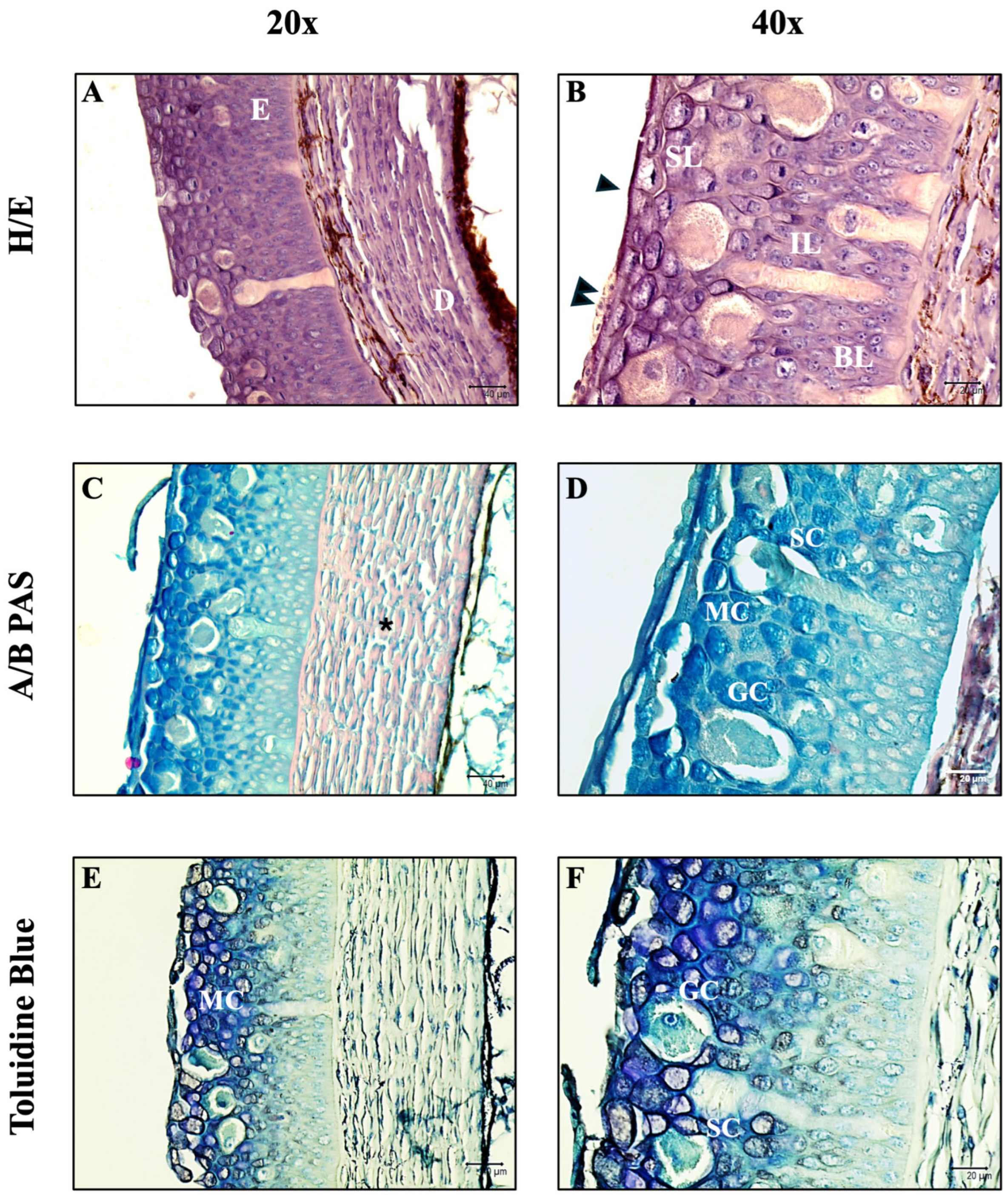
Figure 1. (AF) Histologically, there is evidence of an epidermis (E) subdivided into three layers (superficial, SL; intermediate, IL; and basal, BL) and an underlying dermis (D). Three cell populations are evident: mucous cells (MC), granular cells (GC), and skein cells (SC). The morphology of mucous cells and their content type changes from the basal to the superficial layer, as evidenced by AB/PAS staining. Traces of acidic mucopolysaccharides are evident in the dermis (*). Skein cells are easily recognizable by their typical elongated morphology, starting from the basal layer and moving towards the superficial layer. In addition, a thin cuticle (arrowhead) is present covering almost the entire epidermis, and where it is missing, flattened epithelial cells (double arrowhead) are observed. Furthermore, the staining intensity was measured for all stains used, highlighting a statistically significant difference between the surface and basal layers of the AB/PAS and Toluidine Blue histochemical stains, confirming the variation in staining intensity in the three layers.
Fishes 10 00605 g001
Figure 2. Skein cells (*) immunoreactive for vimentin (red) and α-tubulin (green) are evident. These cells extend from the basal to the superficial layer and show antibody positivity in their basal portion, cell body, and apical perinuclear portion. Antibody colocalization (yellow) demonstrates the presence of both molecules in these cells, as evidenced by the display profile. TL = Transmitted Light.
Fishes 10 00605 g002
Figure 3. Skein cells (*) immunoreactive for TLR2 (red) and Muc2 (green) are evident. These cells extend from the basal to the superficial layer, and show antibody positivity both in their basal portion, cell body, and apical perinuclear portion. Antibody colocalization (yellow) demonstrates the presence of both molecules in these cells, as evidenced by the display profile. In addition, a slight positivity is also appreciable for some mucous cells (MC) and some granular cells (arrows), for both antibodies tested. Also the underlying dermis (D) is visible. TL = Transmitted Light.
Fishes 10 00605 g003
Figure 4. Skein cells (*) immunoreactive for vimentin (red) and Muc2 (green) are evident. These cells extend from the basal to the superficial layer, and show antibody positivity both in their basal portion, cell body, and apical perinuclear portion. Antibody colocalization (yellow) demonstrates the presence of both molecules in these cells, as evidenced by the display profile. TL = Transmitted Light.
Fishes 10 00605 g004
Figure 5. (AF) Immunoperoxidase. Skein cells (*) immunoreactive to Piscidin1 are evident. These cells extend from the basal to the superficial layer, and show antibody positivity both in their basal portion, cell body, and apical perinuclear portion. In addition, a slight positivity is also appreciable for some mucous cells (arrowhead) and some granular cells (arrows) for both antibodies tested.
Fishes 10 00605 g005
Figure 6. Structural and functional protein domains of Petromyzon marinus vimentin ortholog and Oryctolagus cuniculus vimentin protein predicted by InterProScan.
Fishes 10 00605 g006
Figure 7. Structural and functional protein domains of Petromyzon marinus α-tubulin ortholog and Mus musculus α-tubulin protein predicted by InterProScan.
Fishes 10 00605 g007
Figure 8. Structural and functional protein domains of Petromyzon marinus TLR2 ortholog and Oryctolagus cuniculus TLR2 protein predicted by InterProScan.
Fishes 10 00605 g008
Figure 9. Structural and functional protein domains of Petromyzon marinus Muc2 ortholog and Mus musculus Muc2 protein predicted by InterProScan.
Fishes 10 00605 g009
Table 1. Details and information about the antibodies used in this study.
AntibodySupplierDilution (µL/µL)Animal Source/Clone
Piscidin-1GenScript, Piscataway, NJ, USA1:200Rabbit
TLR2Thermo Fisher Scientific, Waltham, MA, USA1:200Rabbit
α-tubulinSigma-Aldrich, Saint Louis, MO, USA1:300Mouse, clone
6-11B-1
Muc2Santa Cruz Biotechnology, Dallas, TX, USA1:200Mouse, clone F-2
VimentinSigma-Aldrich, St. Louis, MO, USA1:200Rabbit
Alexa Fluor 488 donkey anti-mouse IgG FITC conjugatedMolecular Probes, Invitrogen, Waltham, MA, USA1:300Donkey
Alexa Fluor 594 donkey anti-rabbit IgG TRITC conjugatedMolecular Probes, Invitrogen, Waltham, MA, USA1:300Donkey
Table 2. Antibodies used in this study, including their supplier-reported immunogen sequences or regions, the corresponding P. marinus orthologs (NCBI accession numbers), the closest host species accession numbers, and the percentage of sequence homology.
AntibodyImmunogen Sequence/Region from the Supplier CompanyP. marinus Ortholog
Accession
(NCBI)
Host Accession (Closest Species)Sequence Homology
TLR2a.a. 180–196, 353–370, and 473–489XP_032835900.1XP_058522725.134.53%
α-tubulin15 S dynein fraction from sea urchinsperm axonemeXP_032815810.1NP_001019507.184.70%
Muc2a.a. C-terminus 4880–5179XP_075913103.1NP_076055.435.28%
Vimentina.a. 63–90 (includes Ser82)XP_032807738.1BAE29426.170.75%
Table 3. Mean gray area value and SDs of the three layers of the epidermis of P. marinus. A lower value indicates a major intensity of staining. (N = 90).
BasalIntermediateSuperficial
H/E156.916 ± 21.956150.512 ± 27.501147.945 ± 22.427
AB/PAS157.534 ± 18.372142.850 ± 27.938111.701 ± 26.746
Toluidin Blue189.518 ± 16.514162.610 ± 25.070156.510 ± 26.332
Table 4. Number of immunopositive skein cells for the different antibodies tested. (N = 100).
No. of Immunopositive Skein Cells
Vimentin+58
α-tubulin+60
TLR2+63
Muc2+57
Piscidin-1+61
Disclaimer/Publisher’s Note: The statements, opinions and data contained in all publications are solely those of the individual author(s) and contributor(s) and not of MDPI and/or the editor(s). MDPI and/or the editor(s) disclaim responsibility for any injury to people or property resulting from any ideas, methods, instructions or products referred to in the content.