Article

Trace Element Accumulation and Oxidative Stress in Three Populations of the European Eel Anguilla anguilla L. from Southern Italy

Department of Biology, Ecology and Earth Sciences, University of Calabria, 87036 Rende, Italy
*
Author to whom correspondence should be addressed.
These authors contributed equally to this work.
Fishes 2025, 10(10), 517; https://doi.org/10.3390/fishes10100517 (registering DOI)
Submission received: 3 September 2025 / Revised: 7 October 2025 / Accepted: 8 October 2025 / Published: 11 October 2025
(This article belongs to the Special Issue The Impact of Contamination on Fishes)

Abstract

The European eel (Anguilla anguilla), a catadromous species currently listed as Critically Endangered by the IUCN, is undergoing a severe continental decline. Among the multiple contributing factors, chemical contamination of aquatic environments—particularly by heavy metals—plays a major role. This study analyzed the concentrations of 16 trace elements in the muscle tissue of A. anguilla specimens collected from three ecologically distinct sites in Southern Italy: an estuary (Foce del Crati), a lagoon (Laghi di Gizzeria) and a stream (torrente Raganello). Correlations between trace element accumulation and the onset of oxidative stress were also examined. To assess eel health status, oxidative biomarkers were also analyzed in heart, liver, and gill tissues. Statistical analysis among populations revealed significant differences in the bioaccumulation of 10 of the 16 elements, with Cd and As being of particular concern. No significant correlations were found between these two elements and oxidative biomarkers, but Spearman analysis identified both positive and negative correlations with other elements varying by the site of collection. Oxidative biomarkers also showed site- and tissue-specific variation. In particular, SOD activity was highest in the liver and varied across sites; LPO and protein carbonyl levels were generally lower in eels from the Crati River, although heart values deviated from this trend, highlighting tissue-specific response patterns. These results underscore the complex interplay between chemical contamination and the physiology of the European eel, emphasizing the influence of environmental context in modulating tissue-specific oxidative responses.
Key Contribution: The correlation between trace metals and oxidative stress onset in the European eel from Southern Italy reveals a spatial variability in both contaminant profiles and redox biomarkers, which may reflect combined effects of local exposure and habitat-specific factors.

1. Introduction

The European eel Anguilla anguilla (L.) is a widely distributed catadromous amphihaline species that colonizes fresh, brackish and coastal waters of Europe, including the Mediterranean Basin. This long-living, slow-growing species represents an important link in the flow of organic matter between marine and inland waters and plays a major part in marine and freshwater ecosystems, both as predator and prey species [1]. However, since the 1980s, the European eel has suffered a 90–95% decline in its population and, within 50 years, has turned from one of the most abundant freshwater fish into an endangered species [2] listed as Critically Endangered on the IUCN’s Red List [3]. To support the recovery of the European eel stock, in 2007, the European Union adopted the Council Regulation (EC) No. 1100/2007 (the ‘Eel Regulation’), which obliges EU Member States to have an eel management plan in place. Despite several preservation measures put in place to comply with this regulation, European eel recruitment remains low throughout its geographical range and its stock status remains critical [4]. A combination of several factors has been proposed to contribute to this decline: climate change, overfishing, habitat degradation or loss, water and sediment pollution, diseases, and predators. Particularly, water pollution, resulting from the occurrence of harmful substances, such as persistent organic pollutants and heavy metals, may have an important impact on eel physiology at all life stages.
Pollutants can disturb the immune, nervous, and endocrine systems, thereby negatively affecting cellular and organ functions, impairing reproduction, and compromising migration [5]. Particularly, heavy metals have been shown to disturb eel physiology at different biological levels. For example, exposure to Pb affected the immune system by increasing lymphocytes number [6]; Cu was reported to influence the endocrine function by decreasing plasma cortisol and T3 levels, and to induce genotoxicity in blood, as revealed by erythrocytic nuclear abnormalities [7]; Cd has been shown to inhibit the activity of carbonic anhydrase and Na+-K+-ATPase enzymes in the intestine and gills, thus altering both acid–base balance and osmoregulation [8]. Heavy metal bioaccumulation is also associated with increased free radical concentrations within the cytosol. These oxidative forms may damage all components of the cell, including proteins, lipids, and DNA, thus increasing the risk of cellular dysfunction, and/or death. Animals may prevent oxidative damage by activating enzymatic and non-enzymatic systems [i.e., superoxide dismutase (SOD), catalase (CAT), and glutathione S-transferase (GST)] that maintain a reduced state within cells. However, when the balance between oxidant/antioxidant is shifted in favor of oxidants, oxidative stress occurs, and it contributes to the onset of physiological alterations. In this context, the measurements of oxidative products and antioxidant systems are used as indices to evaluate the animal health status [9] and applied for environmental biomonitoring of pollution exposure [5].
To the best of our knowledge, very few studies have analyzed the effects of heavy metal pollution on the oxidative status of the European eel, with information mainly referring to laboratory studies in which the effects of single metals have been investigated [10]. To fulfill this gap, this study aims to assess the potential correlation between heavy metal and oxidative stress onset in the European eel by analyzing trace element concentrations and oxidative stress biomarkers in the skeletal muscle, heart, liver, and gills, to obtain a comprehensive picture of the general health status of specimens from different habitats in Southern Italy.

2. Materials and Methods

2.1. Sampling Collection

Sampling was carried out in October 2019 at three different sites of confirmed presence of the species in Calabria (Figure 1): an estuary (Foce del Crati, Site 1), a stream (torrente Raganello, Site 2) and a lagoon (Laghi di Gizzeria, Site 3). Crati River estuary (Ionian Sea, northern Calabria) is the estuary of the largest river in Calabria, draining an extensive agricultural and urbanized basin; this estuarine mouth represents a productive but contaminant-exposed transitional habitat. Raganello Stream (Ionian side, northern Calabria) is a mountain stream within the Pollino National Park, flowing through limestone gorges; upper reaches are relatively pristine, while downstream sections may be influenced by agriculture and small settlements before reaching the Ionian Sea. Gizzeria coastal lakes (Tyrrhenian Sea, central Calabria) are small coastal brackish lagoons on the Tyrrhenian side, influenced by both marine intrusion and freshwater inputs; surrounding agriculture and tourism activities contribute to variable nutrient and contaminant loads. Although the three sampling sites are located at comparable latitudes in Calabria, they encompass contrasting ecological settings: a large river estuary (Crati), coastal brackish lagoons (Gizzeria), and a mountain stream (Raganello). These distinct habitats differ in hydrology, salinity, and anthropogenic influence, providing a natural gradient for assessing metal accumulation and oxidative stress in eels.
A total of 23 specimens of A. anguilla were sampled, distributed among sites as reported in Table 1. Eel sampling was carried out following the standardized national protocol issued by ISPRA (2016) for monitoring A. anguilla populations in Italian water bodies by using electrofishing [11]. The electrofishing unit was powered by a rechargeable 24 V lithium battery able to generate both direct current (DC, 650 W) and pulsed current, adjustable between 20 and 200 pulses per second (peak voltage 115–565 V, maximum power 1200 W), with wide flexibility in regulating voltage, pulse frequency, and output power.
After sampling, eels were transferred to the laboratory of Organ and System Physiology at the University of Calabria and, after anesthetization in tricaine methanesulfonate (MS222; Sigma–Aldrich, Milan, Italy), biometric information was collected (Table 1). Animals were dissected ventrally, from the cloacal opening up to the cardiac cavity, and the following tissues were isolated: heart, liver, gills, and muscle. Samples were stored at −80 °C until further analysis.
Animal care and experimental procedures were performed in accordance with the European and Italian laws and approved by the Ministry of Health of Italy (N. 42/2019-PR).

2.2. Trace Elements Analysis

The analysis consists of the determination of concentrations of 16 trace elements: Aluminum (Al), Arsenic (As), Cadmium (Cd), Cobalt (Co), Copper (Cu), Manganese (Mn), Molybdenum (Mo), Nickel (Ni), Zinc (Zn), Selenium (Se), Strontium (Sr), Lead (Pb), Chromium (Cr), Iron (Fe57), Barium (Ba) and Bismuth (Bi). This analysis was conducted on 20 specimens (10 from Estuary, 4 from Stream and 6 from Lagoon) using the inductively coupled plasma mass spectrometer (ICP-MS, ElanDRCe model, PerkinElmer inc.; Shelton, CT, USA).
Prior to analysis, muscle tissue samples were chemically digested according to Gallo et al. [12], using 12 mL of ultra-pure (65–69%) nitric acid (HNO3, Merck KGaa, Darmstadt, Germany) for each sample under microwave-assisted conditions to obtain clear solutions; this stage was necessary because the ICP-MS instrument can only analyze samples in solution form. After the acid digestion, the solutions obtained were temporarily stored in sterile containers suitably labeled at a temperature of approximately 4 °C. The accuracy of the method has been evaluated thanks to the Certified Reference Material (CRM); in this case it was the Lobster Hepatopancreas (Tort-3; National Research Council Canada). Muscle samples and the CRM were processed following the same analytical protocol. The only exception was the drying step (24 h under a laminar-flow hood at room temperature; hood model KS-12 class II, Herasafe, Thermo Fisher Scientific, Langenselbold, Germany), which was applied exclusively to the muscle samples because the CRM was already supplied in powdered form. During ICP-MS measurements, blanks (prepared with ultrapure water and 1% HNO3) and calibration standards were analyzed at the beginning and end of each run and after every 6–8 samples to routinely verify analytical accuracy, precision, and detection limits. The amount of muscle needed was about 1.0 g of fresh weight and, after drying, we used 0.1 g of dry weight.
The Trace Element Pollution Index (TEPI) was calculated following the equation
TEPI = (Cf1 × Cf2…Cfn)1/n
as stated by Richir & Gobert [13], where Cf is the normalized mean concentration of the trace element—calculated as the mean concentration of each TE per location divided by the overall mean of that TE—and n is the total number of examined potentially toxic elements [14].

2.3. Measurement of Oxidative Stress Biomarkers

For each animal (N = 10 Estuary; N = 7 Stream; N = 6 Lagoon), tissue evaluation of oxidative stress biomarkers was performed on liver, heart, gills, and muscle samples, as detailed in [15]. Samples were homogenized in cold Tris/HCl buffer (100 mM; pH 7.2; Sigma–Aldrich, Milan, Italy), containing a mixture of protease inhibitors (Sigma–Aldrich, Milan, Italy). An aliquot of homogenates was immediately used to assess lipid peroxidation (LPO); the remaining part was centrifuged at 5000× g for 5 min at 4 °C and the supernatant tested for protein oxidation and SOD enzyme activity. Protein concentration in the supernatant was determined with the Bradford method by using a commercial kit (Bio-Rad Laboratories S.r.l., Milan, Italy), and bovine serum albumin (BSA) as a standard.

2.3.1. Lipid Peroxidation

LPO was assessed by measuring the content of malondialdehyde (MDA), the major end-product of lipid oxidation, using the 2-thiobarbituric acid (TBA)-reacting substances (TBARS) assay [16]. A reaction mixture, prepared by mixing equal volumes of sample homogenate (10% w/v), 0.8% TBA (Sigma–Aldrich, Milan, Italy), and 20% trichloroacetic acid (TCA) (Carlo Erba Reagents, Milan, Italy), was boiled in a water bath at 100 °C for 10 min and then centrifuged at 7000 rpm for 10 min. The absorbance of the supernatant was measured at 540 nm, and the MDA content was calculated using an extinction coefficient of 156,000 M−1 cm−1 and expressed as μM per gram of tissue.

2.3.2. Protein Oxidation

The levels of oxidatively modified protein (OMP) were evaluated by measuring carbonyl group content according to the 2,4-dinitrophenylhydrazine (DNPH) method [17]. Aliquots of the supernatant were first incubated at room temperature for 1 h with 10 mM DNPH (Sigma–Aldrich, Milan, Italy) in 2 M HCl (PanReac Applichem ITW Reagents, Monza, Italy) and then precipitated with 2 volumes of TCA. The solution was centrifuged for 20 min at 7000 rpm; the pellet was washed thrice with ethanol-ethyl acetate (Sigma–Aldrich, Milan, Italy) (1:1; v/v) to remove DNPH excess and dissolved in 6M guanidine (Sigma–Aldrich, Milan, Italy). The concentration of carbonyl groups was measured spectrophotometrically at 370 nm (aldehydic derivatives) and at 430 nm (ketonic derivatives) using an extinction coefficient of 22,000 M−1 cm−1. Results were expressed as nmol per mg protein.

2.3.3. Superoxide Dismutase (SOD) Activity

SOD activity was assessed according to the pyrogallol method [18] by monitoring the SOD-dependent inhibition of pyrogallol auto-oxidation at pH 8.2. The reaction was prepared by combining 0.5 mL of tissue homogenate (prepared as detailed in 2.3) with 50 mM Tris-HCl (Sigma–Aldrich, Milan, Italy), 1 mM EDTA (Sigma–Aldrich, Milan, Italy), and 0.2 mM pyrogallol (J.T.Baker® Avantor Performance Materials, Gliwice, Poland, in a final volume of 2 mL. The reaction was monitored spectrophotometrically at 420 nm for 5 min at 25 °C. One unit of SOD was defined as the amount of the enzyme that inhibits 50% of pyrogallol auto-oxidation. Results were expressed in U/mg protein.

2.4. Data Analysis

For the ecotoxicological comparison of samples, Kruskal–Wallis test was carried out using the PAST software (version: 4.15; Øyvind Hammer, Oslo, Norway). The same program was used to carry out the correlation of Spearman rs. The Spearman R correlation coefficients were used to assess the relationships between trace metal concentrations and oxidative stress parameters in muscle tissue of eels from different sampling sites. Muscle tissue was chosen because it is commonly used as a reference for contaminant monitoring and reflects medium-term accumulation patterns due to its relatively low turnover rate. This slow turnover allows contaminants to accumulate over time, potentially enhancing the tissue’s susceptibility to ROS generation. As an exploratory step, Principal Component Analysis (PCA) was conducted using RStudio (Version 2025.05.1+513; Posit Software, PBC; Boston, MA, USA). The working matrix was first base-10 logarithmically transformed (log10). The analysis was subsequently performed on the resulting log10-transformed and standardized matrix using R’s prcomp function. Full details and results of this analysis are reported in the Supplementary Materials.
For oxidative stress, statistical analyses were performed using a mixed-effects model (REML) using GraphPad Prism software, version 10.4.1 (GraphPad Software Ltd., La Jolla, CA, USA). Tissue was treated as the within-subject factor, site as the between-subject factor, and individual animals were included as a random effect to account for repeated measures. Tukey’s post hoc test was used for multiple comparisons. Data were expressed as mean ± sem of absolute values and statistical significance was accepted at p < 0.05.

3. Results

3.1. Trace Element Concentrations in Muscle Tissue of Eels from Different Sites

The ecotoxicological investigation produced the mean values (with standard deviation) of each element per population (Table 2). The site that showed the highest concentrations was the Estuary—in 9 elements out of 16 (56.2%)—followed by the Stream—in 5 elements out of 16 (31.3%)—and finally by the Lagoon—in 2 elements out of 16 (12.5%).
Across all sites, the elements with the highest mean concentrations were iron (Fe), zinc (Zn), and nickel (Ni). Site-specific differences became evident thereafter (Table 3): the fourth-ranked element was aluminum (Al) in estuary eels, selenium (Se) in lagoon eels, and strontium (Sr) in stream eels. Although these occur at lower mean concentrations than the top three, their absolute levels remain non-negligible and likely reflect natural background variability among habitats.
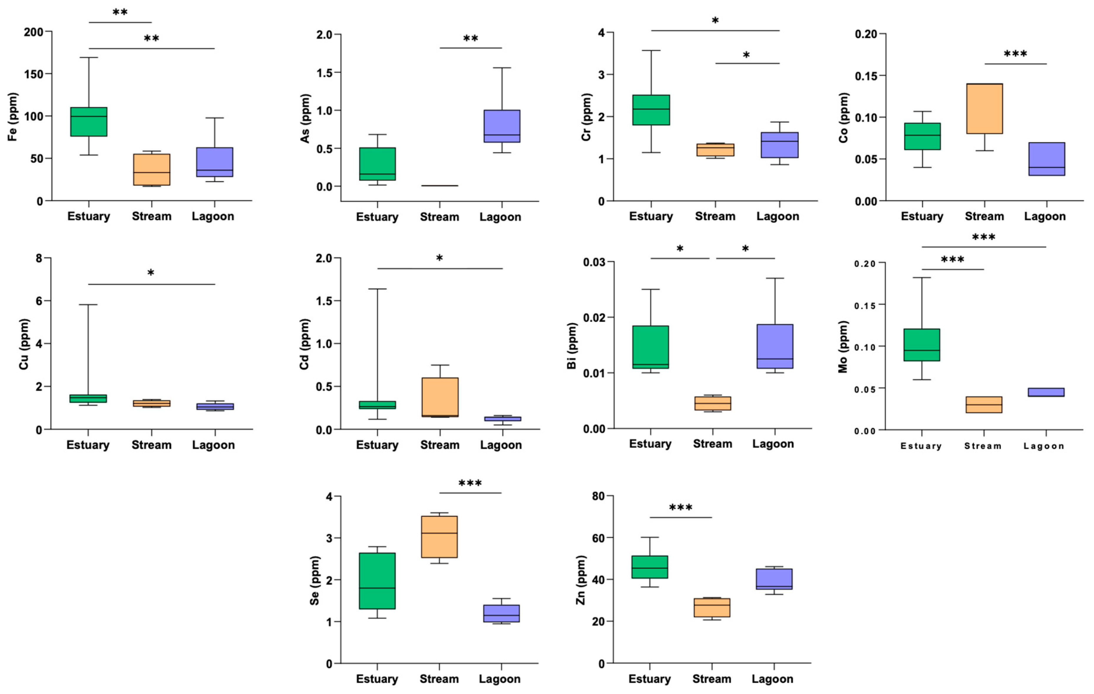
The statistical analysis performed using the Kruskal–Wallis test revealed significant differences for 10 out of 16 elements (Figure 2). The only elements that did not show significant differences among sites were Ni, Sr, Mn, Ba, Pb, and Al. Notably, arsenic exhibited a highly significant difference (p < 0.001) between eels from the lagoon and the stream. Other significant differences (p < 0.001) were observed for Mo (estuary vs. stream), Se (stream vs. lagoon), and Zn (estuary vs. stream). Lastly, Cd—the only legally regulated element for fish muscle to show significance—was significantly different (p < 0.01) between eels from the estuary and the lagoon.
The Trace Elements Pollution Index (TEPI) values were 0.87 for the estuary, 0.85 for the stream, and 0.91 for the lagoon. Although only three sites were considered, a relative contamination scale was constructed by partitioning the TEPI values into quartiles to define threshold ranges. According to this classification, the lagoon (Site 3) exhibited the highest level of impact, followed by the estuary (Site 1), whereas the stream was the least affected site.

3.2. Tissue Evaluation of the Oxidative Status

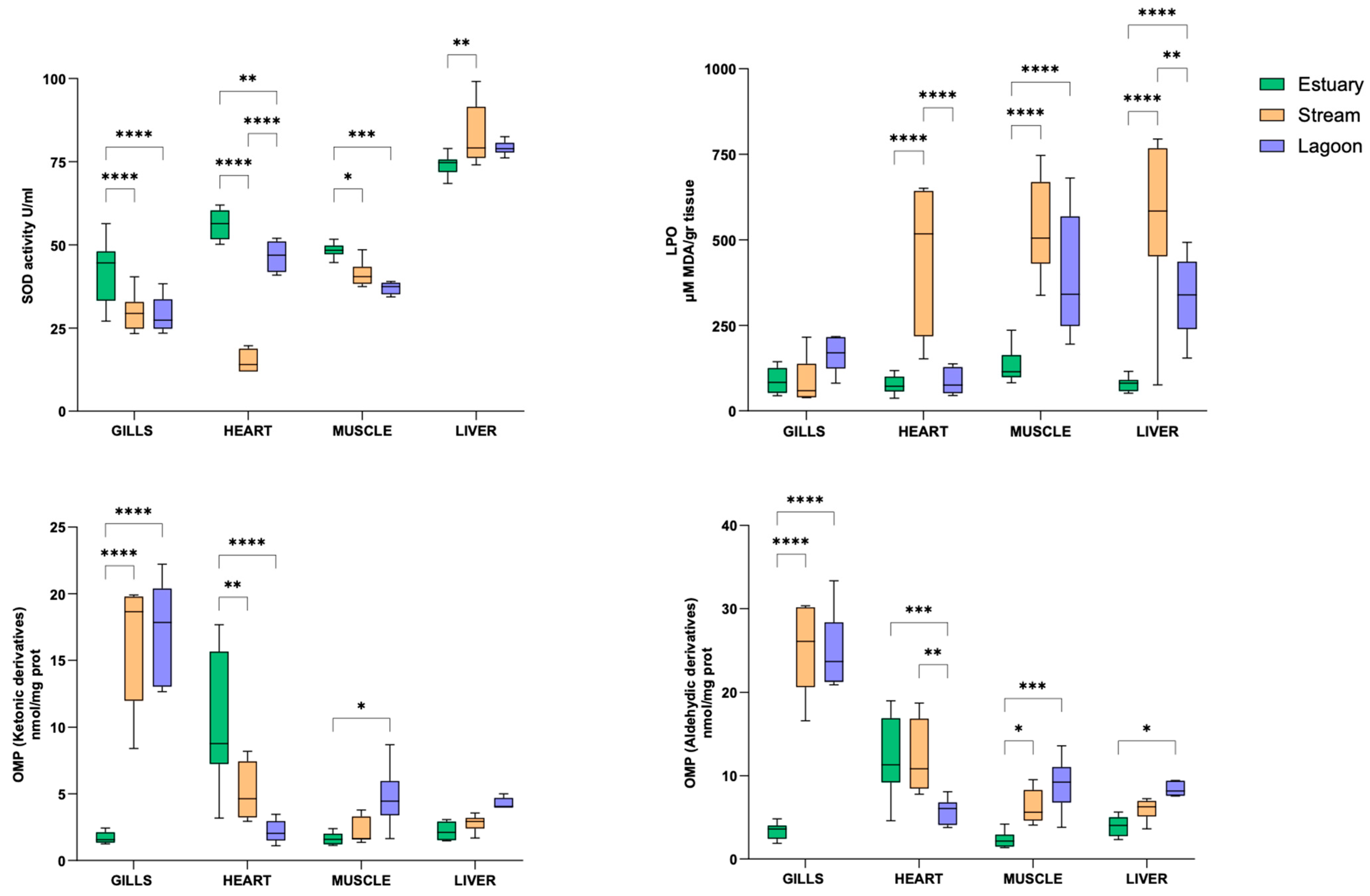
For each animal, the oxidative status was evaluated by measuring the levels of LPO, protein carbonylation, and SOD activity in tissue homogenates from gills, heart, skeletal muscle, and liver. An overview of the tissue levels of oxidative biomarkers in animals from different sites is shown in Figure 3.
The activity of the antioxidant enzyme SOD varied significantly among sites [F(2.0, 20.0) = 37.94), p < 0.0001] and tissues [F(2.3, 43.64) = 358.4), p < 0.0001]. The highest activity was measured in the liver, while similar values were recorded in all other tissues. For each tissue, a greater SOD activity was detected in eels from the Estuary, except for the liver, in which lower values than those recorded in eels from the Stream and the Lagoon were observed.
The analysis of lipid oxidation products showed site [F(2.0, 20.0) = 59.51), p < 0.0001]- and tissue [F(2.35, 44.57) = 20.82), p < 0.0001]-dependent variations. Within tissues, eels from the Estuary consistently exhibited lower LPO values compared to those from the other sites. The only exception was in the heart, in which similar LPO levels were detected in individuals from the Estuary and the Lagoon.
Protein carbonyl levels—both aldehydic and carboxylic derivatives—followed a similar trend in the gills, muscle, and liver, with lower values recorded in eels from the Estuary and higher levels in those from the Lagoon and the Stream [carboxylic derivatives: sites F(2.0, 20.0) = 12.12), p < 0.0004; tissues F(1.8, 31.8) = 64.03, p < 0.0001—aldehydic derivatives: sites F(2.0, 20.0) = 40.47), p < 0.0001; tissues F(1.9, 34.2) = 89.07, p < 0.0001]. Notably, the heart deviated from this pattern, showing the lowest levels of both aldehydic and ketonic carbonyl derivatives in eels from the Lagoon.

3.3. Correlation Analysis

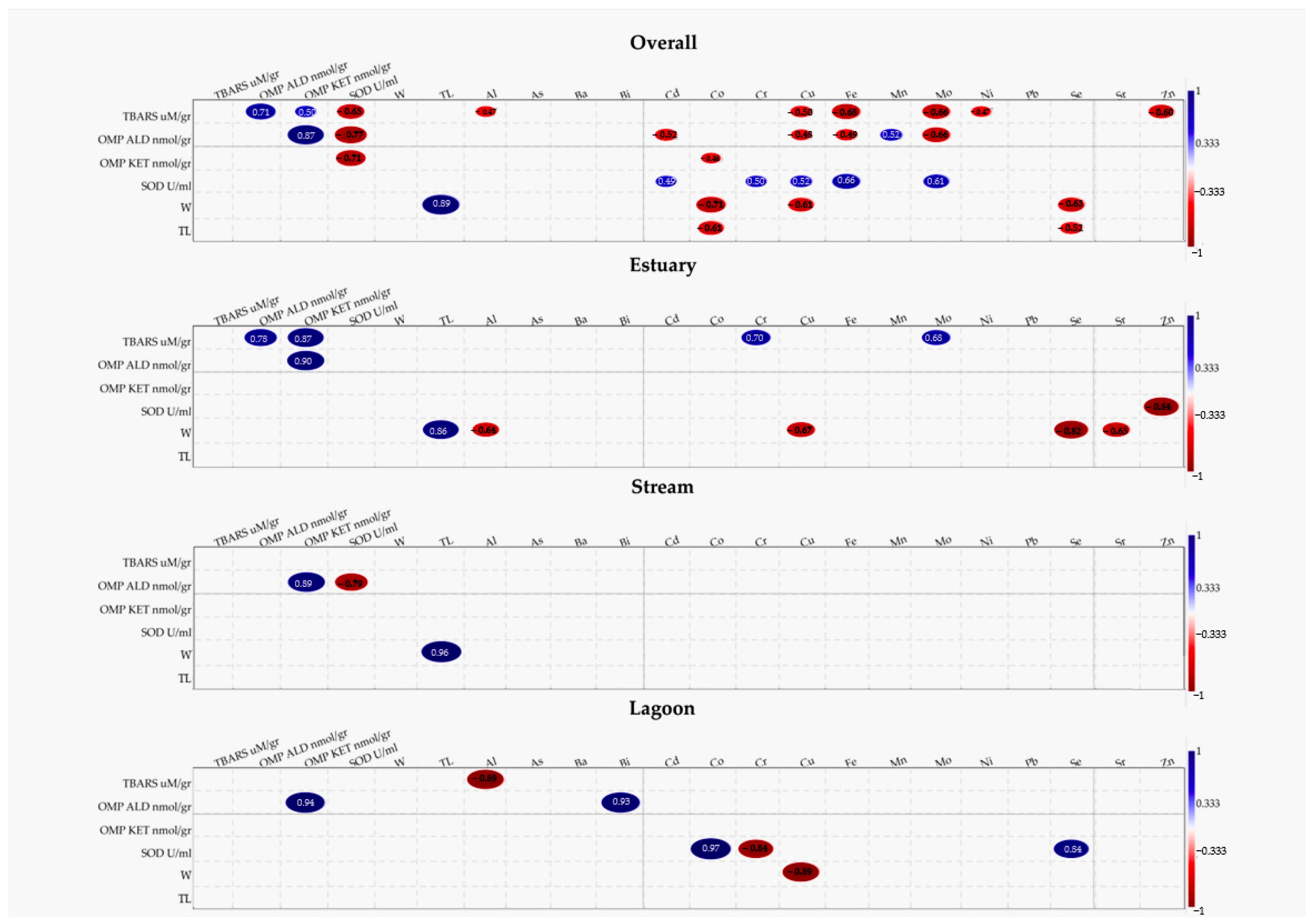
The Spearman R correlation coefficients were used to assess the relationships between trace metal concentrations and oxidative stress parameters in muscle tissue of eels from different sampling sites. Significant correlations for each site are shown in Figure 4.
Correlation analysis did not reveal any significant positive relationships between element concentrations and specimen size (W or TL). Considering the entire dataset irrespective of population origin, significant negative correlations were observed between size (both W and TL) and Co and Se, while weight was also negatively correlated with Cu. At the population level, no significant correlations were detected for Site 2 (Stream); in Site 1 (Estuary), W showed negative correlations with Al, Cu, Se, and Sr; and in Site 3 (Lagoon), a single negative correlation was observed between W and Cu.
In the Estuary, positive significant correlations between LPO, OMP, Cr, and Mo were found, while SOD activity negatively correlated with Zn. In the Stream, SOD negatively correlated with OMP; any significant correlations with trace elements were found. In the Lagoon, positive correlations were found between OMP and Bi, and between SOD, Co, and Se. A negative correlation was identified between LPO and Al, and SOD and Cr.

4. Discussion

This study provides new evidence on trace metal accumulation in the European eel from southern Italy, revealing significant associations with oxidative stress markers and habitat differences.

4.1. Trace Elements Accumulation

The ecotoxicological evaluation of trace element concentrations in eels from the three investigated sites revealed notable spatial variability, suggesting potential differences in contamination exposure among locations [19]. Eels collected from the estuarine area exhibited the highest mean concentrations for several elements, including Al, Cr, Cu, Fe, Ni, and Zn, which might indicate greater exposure in this environment, possibly due to its function as a depositional zone for natural and anthropogenic inputs [19], as commonly observed in transitional waters [20]. Such a pattern could be influenced by hydrodynamic conditions and upstream discharges reported for similar systems [19,21]. However, as no complementary analyses of sediments, water chemistry, or hydrology were conducted, these explanations remain hypothetical.
Although eels from the estuary exhibited higher concentrations for some individual elements, those from the lagoon showed the highest Trace Elements Pollution Index (TEPI) value, suggesting a broader, though less extreme, accumulation of contaminants. Even further environmental data are needed to confirm this possibility, and we suggest that this trend could relate to the semi-enclosed morphology of lagoon environments, which may favor pollutant retention due to limited water exchange and longer residence times [21].
Although eels from the stream exhibited lower overall elemental concentrations, they showed comparatively higher levels of certain elements, notably Mn and Se. It is possible that these patterns are related to catchment-derived inputs from lithogenic sources or agricultural runoff, as often reported for river systems [22], though additional evidence would be needed to validate this hypothesis. The TEPI value for the stream was the lowest among the three locations, suggesting a comparatively reduced cumulative trace element burden; however, the presence of Cd and Co at elevated levels in some specimens indicates that continued monitoring remains important given their ecotoxicological relevance [22].
Fe, Zn, and Ni were consistently among the most abundant elements in eels across all sites, likely reflecting both their natural geochemical background and widespread anthropogenic use [20,23]. The Kruskal–Wallis test indicated significant inter-site differences for 10 elements in eels, supporting the notion of site-related variability [24]. To further explore potential co-exposure patterns, we also performed a Principal Component Analysis (PCA). While this exploratory approach highlighted some correlation blocks among elements (Supplementary Figure S1, Tables S1 and S2), the proportion of variance explained by the first components was limited, and therefore single-element analyses based on non-parametric methods were retained as the most informative strategy for this dataset. Notably, eels from the lagoon exhibited the highest As concentrations, whereas Se levels peaked in individuals from the stream. Mo and Bi concentrations were also relatively elevated in eels from both the estuary and the lagoon, suggesting spatial variation in elemental accumulation patterns among habitats.
No significant positive correlations were observed between element concentrations and eel size (total length and weight), suggesting that bioaccumulation does not necessarily increase with growth under the conditions examined. On the contrary, significant negative correlations observed for Co, Se, and Cu at the whole-dataset level may point to growth dilution effects [25] or possible sublethal effects on somatic development. At the site-specific level, no significant correlations were detected in stream specimens. In contrast, negative associations between weight and Al, Cu, Se, and Sr were observed in the estuary, while in the lagoon, only a single negative correlation emerged between weight and Cu. These patterns may reflect local bioavailability and individual physiological responses; however, given the limited scope of this study, no definitive conclusions can be drawn regarding the underlying mechanisms. Overall, these findings highlight the value of combining element-specific analyses with integrative indices such as TEPI to effectively characterize contamination in biota. The distinct profiles observed among eels from the three sites suggest complex interactions between natural processes and anthropogenic influences [24].

4.2. Eel Oxidative Status

To provide an integrated view of the health status of eels from different populations, oxidative stress biomarkers—namely SOD activity, LPO, and protein carbonyls—were evaluated in metabolically distinct target tissues (i.e., gills, liver, heart, and muscle).
As the primary enzymatic defense against reactive oxygen species, SOD catalyzes the dismutation of superoxide radicals into hydrogen peroxide and oxygen, thereby mitigating oxidative damage [26]. In the present study, SOD activity exhibited clear tissue-specific patterns, with the liver displaying the highest activity across all individuals. Our finding is in line with previous reports in A. anguilla and A. marmorata, showing a predominant expression of the Cu/ZnSOD mRNA in the liver compared to other tissues [27,28]. This may be linked to the prominent role of this tissue in detoxification and metabolic regulation, which makes it a primary site for ROS generation, thereby requiring a constitutive high expression of the antioxidant systems [29,30]. Regarding markers of oxidative damage, i.e., protein (OMP) and lipid (TBARS) oxidation products, data analysis did not reveal substantial differences among the examined tissues. These patterns may therefore reflect site-specific environmental influences and/or intrinsic tissue-specific variability within populations.
Animals from site 1 show comparable TBARS levels in all tissues. The same was observed for OMP, with the exception of the heart, in which a major variability was noted. Although literature reporting basal levels of oxidative damage markers in eels is limited—and often based on differing methodologies or expressed with non-comparable units—data from yellow eels (A. anguilla) collected in northwestern Portuguese estuaries with different contamination levels [31] show LPO values comparable to those here detected in the Estuary population. Of note, in the same population, SOD activity was generally higher than that measured in eels from other sites, suggesting a greater enzymatic antioxidant response. Such constitutively elevated activity may contribute to maintaining low ROS levels, thereby limiting the formation of oxidative damage products. The enhanced antioxidant activity detected in site 1 could be linked to a continuous and more variable contaminant exposure in this riverine system, in which agricultural runoff, urban discharges, or sediment-associated pollutants may trigger oxidative stress responses [32].
Compared to animals from the Estuary, eels sampled in the Lagoon exhibited a more pronounced oxidative status. This was particularly evident in the liver, in which both antioxidant activity and oxidation products were found to be relatively high, suggesting a state of oxidative imbalance in which the upregulated antioxidant response is unable to fully counteract ROS production. High levels of protein carbonylation were also detected in muscle and gills, associated with either higher LPO and lower SOD levels. This suggested a tissue-specific protein damage that may occur either in parallel with lipid peroxidation or as a result of a reduced antioxidant capacity (as indicated by a low SOD activity). Of note, protein carbonylation itself can irreversibly modify and inhibit the activity of antioxidant enzymes, including SOD [33,34], thus worsening the redox imbalance. Evidence in the European eel also revealed a different behavior of oxidative biomarkers depending on water habitat [35]. Indeed, a decreased antioxidant activity, together with elevated LPO levels, was detected in eels grown in a brackish pond with respect to animals cultured in river or sea water. This suggests that local environmental conditions (e.g., natural or artificial habitat, dynamic or closed system, different water salinity) may be associated with increased oxidative pressure.
Eels from the Stream showed a tissue oxidative status like that detected in animals from the Lagoon, with the exception of the heart, in which a lower SOD activity and higher oxidative products were observed. This tissue-specific response may be associated with environmental differences between riverine and brackish habitats. In fact, in lotic systems (e.g., mountain rivers), fish are continuously exposed to mechanical stress due to current flow, requiring increased swimming effort and hence elevated basal cardiac output [36,37]. Notably, a negative correlation between SOD activity and oxidative products has also been identified at muscle level, suggesting that a chronic energetic demand can stimulate mitochondrial ROS production in metabolically active tissues.

4.3. Correlation Between Oxidative Markers and TE Accumulation

The interplay between biomarkers of oxidative stress and TE accumulation provides key insights into how environmental contamination influences cellular homeostasis in eels.
In the muscle of eels from the Estuary, SOD activity negatively correlated with Zn, one of the most abundant metals detected in this site. As a structural and catalytic cofactor of the Cu/Zn-SOD enzyme, Zn plays an important regulatory role in the oxidative stress responses. However, the negative correlation we observed suggests that Zn accumulation does not directly trigger a proportional upregulation of SOD activity in these animals. One plausible explanation is that individuals with inherently higher or more efficient antioxidant capacity—reflected by higher baseline SOD activity, as described above—better tolerate Zn deposition, possibly via enhanced membrane integrity [38], or elevating metallothionein expression [39]. Alternatively, a capability of the muscle tissue to sequester a non-reactive form of the metal, thus avoiding a significant oxidative stress response, may be hypothesized. In fact, metals may be stored in forms that are not redox-active, for example, bound to intracellular proteins (e.g., metallothioneins) or precipitated as insoluble granules, thereby limiting their capacity to generate ROS and to stimulate antioxidant enzymes [40].
A positive correlation was observed between oxidative products and Mo and Cr. Mo can contribute indirectly to ROS production as a cofactor of enzymes, such as xanthine oxidase, which generate ROS as by-products of their catalytic activity; Cr may directly induce ROS, particularly in its hexavalent form, through intracellular reduction that produces reactive intermediates. Although literature lacks conclusive evidence on the role of Mo in oxidative damage in fish, studies on Carassius auratus [41] and Labeo rohita [42] reported that Cr exposure clearly associated with elevated levels of oxidative biomarkers. This may also support the negative correlation between SOD and Cr observed in eels from the Lagoon. Of note, in these animals, SOD positively correlated with Se and Co. Co, particularly in its ionic forms, may indirectly promote ROS generation through redox interactions and mitochondrial stress. Se, as a cofactor of selenoenzymes, plays a key role in modulating antioxidant defenses in aquatic organisms [43,44], and several studies reported an enhancement in the activity of antioxidant enzymes -including SOD- following dietary Se supplementation [45]. The enhanced activation of antioxidant systems may play a role in coping with the more pronounced oxidative status observed in these animals. Although the role of Co in directly modulating SOD activity is less well established, some evidence also suggests an influence of Co on antioxidant defenses [46].
Of interest is the negative correlation between TBARS and Al observed in the muscle of animals from the Lagoon. Evidence in fish usually reports a positive increase in LPO in response to Al exposure. An explanation for the negative correlation we observed may lie in the chemical properties of Al. Aluminum has a high affinity for phosphate and carboxyl groups, allowing it to bind directly to phospholipids and membrane-associated proteins [47]. This binding reduces membrane fluidity, which in turn may alter the accessibility of Fe2+ or ROS to lipid acyl chains, thereby modulating TBARS formation. However, under specific conditions (pH, phospholipid compositions, presence of Fe2+), Al can act either as a prooxidant (facilitating lipid peroxidation via Fe2+) or as an antioxidant (inhibiting TBARS formation) [48]; this suggests that dose- and tissue-specific effects may justify the patterns observed in the present study.
No correlation between oxidative markers and TEs was observed in the muscle of eels from the Stream. This supports our previous hypothesis that in this population, abiotic factors, such as current flow, oxygenation, or temperature, may influence the tissue oxidative status more than TE accumulation. This highlights the necessity to consider habitat characteristics when interpreting oxidative stress biomarkers.

5. Conclusions

This study provides the first integrated assessment of trace element accumulation and oxidative stress responses in European eels from different habitats in Southern Italy, contributing to a better understanding of the ecological context affecting this critically endangered species. The results indicate spatial variability in both contaminant profiles and redox biomarkers. The correlations between oxidative stress markers and trace elements indicate complex tissue-specific interactions rather than simple dose–response relationships. Although these patterns should be interpreted cautiously given the lack of complementary environmental data, overall, the results support the usefulness of integrating chemical and biomarker measurements in biomonitoring programs and emphasize the importance of broader environmental characterization and long-term monitoring to enhance data interpretation. From a conservation perspective, understanding the interplay between contaminant exposure and physiological stress is essential for developing management strategies aimed at improving habitat quality and reducing additional pressures on declining eel populations.

Limitations of the Study

While this study provides valuable insights into the variability of contaminant profiles and redox biomarkers across different habitats, the results warrant careful interpretation. In field-based research, numerous environmental and physiological factors can influence animal homeostasis, potentially confounding the relationship between metal concentrations and antioxidant responses [49]. Moreover, in catadromous species undertaking long-distance migrations, this complexity is further amplified by potential exposure to ROS-generating substances that may induce oxidative stress prior to capture. Accounting for all these factors is particularly difficult to achieve under non-controlled field conditions, and this represents a general limitation of this type of study. Addressing these aspects requires more targeted and controlled investigations, which fall beyond the scope of the present work.

Supplementary Materials

The following supporting information can be downloaded at: https://www.mdpi.com/article/10.3390/fishes10100517/s1, Figure S1, Principal Component Analysis (PCA) of the 16 trace elements measured in the muscle of A. anguilla from the three study sites; Table S1, Eigenvalues and proportion of variance explained by each principal component (PC) derived from the PCA of trace element concentrations in A. anguilla muscle tissue; Table S2: Loadings of the 16 trace elements on the first three principal components (PC1–PC3) obtained from the PCA of muscle samples from A. anguilla.

Author Contributions

Conceptualization, S.I. and E.S.; methodology, M.F., A.C., S.G., C.M. and D.B.; formal analysis, G.G. and F.L.L.; investigation, M.F., S.G., G.G., F.L.L. and E.S.; data curation, A.C., G.G. and F.L.L.; writing—original draft preparation, M.F. and S.G.; writing—review and editing, A.G., M.C.C., D.B., E.S. and S.I.; supervision, E.S. and M.F.; project administration, S.I.; funding acquisition, S.I. All authors have read and agreed to the published version of the manuscript.

Funding

This research was funded by Parco Nazionale del Pollino, grant number “SOS fish: benessere, salute e salvaguardia della fauna ittica del Parco Nazionale del Pollino” CUP_D43C17000320005”.

Institutional Review Board Statement

Animal care and experimental procedures were performed in accordance with the European and Italian laws and approved by the Ministry of Health of Italy (protocol code N. 42/2019-PR, date 15 January 2019).

Informed Consent Statement

Not applicable.

Data Availability Statement

The original contributions presented in this study are included in the article. Further inquiries can be directed to the corresponding author.

Conflicts of Interest

The authors declare no conflicts of interest.

Abbreviations

The following abbreviations are used in this manuscript:
IUCNInternational Union for Conservation of Nature
LPOLipid Peroxidation
OMPOxidatively Modified Protein
TEPITrace Elements Pollution Index
SODSuperoxide Dismutase

References

  1. Dekker, W. Slipping Though Our Hands–Population Dynamics of the European Eel; Wageningen University and Research: Wageningen, The Netherlands, 2004. [Google Scholar]
  2. Altmayer, A. Addressing the Critical State of European Eel Stocks; European Parliament: Strasbourg, France, 2024.
  3. Jacoby, D.M.P.; Gollock, M.J. Anguilla Anguilla. In The IUCN Red List of Threatened Species; Version 2014.3; IUCN: Gland, Switzerland, 2014. [Google Scholar]
  4. ICES. Joint EIFAAC/ICES/GFCM Working Group on Eels, and Country Reports 2018–2019; ICES Scientific Report; ICES: Copenhagen, Denmark, 2019. [Google Scholar]
  5. Geeraerts, C.; Belpaire, C. The Effects of Contaminants in European Eel: A Review. Ecotoxicology 2010, 19, 239–266. [Google Scholar] [CrossRef] [PubMed]
  6. Santos, M.A.; Hall, A. Influence of Inorganic Lead on the Biochemical Blood Composition of the Eel, Anguilla anguilla L. Ecotoxicol. Environ. Saf. 1990, 20, 7–9. [Google Scholar] [CrossRef]
  7. Oliveira, M.; Serafim, A.; Bebianno, M.J.; Pacheco, M.; Santos, M.A. European Eel (Anguilla Anguilla L.) Metallothionein, Endocrine, Metabolic and Genotoxic Responses to Copper Exposure. Ecotoxicol. Environ. Saf. 2008, 70, 20–26. [Google Scholar] [CrossRef]
  8. Lionetto, M.G.; Maffia, M.; Cappello, M.; Giordano, M.E.; Storelli, C.; Schettino, T. Effect of Cadmium on Carbonic Anhydrase and Na+-K+-ATPase in Eel, Anguilla Anguilla, Intestine and Gills. Comp. Biochem. Physiol. Part A Mol. Integr. Physiol. 1998, 120, 89–91. [Google Scholar] [CrossRef]
  9. Beaulieu, M. Oxidative Status: A General but Overlooked Indicator of Welfare across Animal Species? BioEssays 2024, 46, 2300205. [Google Scholar] [CrossRef]
  10. Ahmad, I.; Oliveira, M.; Pacheco, M.; Santos, M.A. Anguilla Anguilla L. Oxidative Stress Biomarkers Responses to Copper Exposure with or without β-Naphthoflavone Pre-Exposure. Chemosphere 2005, 61, 267–275. [Google Scholar] [CrossRef]
  11. Stoch, F.; Genovesi, P. Manuali per Il Monitoraggio Di Specie e Habitat Di Interesse Comunitario (Direttiva 92/43/CEE) in Italia: Specie Animali. In Serie Manuali e Linee Guida, 141/2016; ISPRA: Roma, Italy, 2016. [Google Scholar]
  12. Gallo, S.; Nania, G.; Caruso, V.; Zicarelli, G.; Leonetti, F.L.; Giglio, G.; Fedele, G.; Romano, C.; Bottaro, M.; Mangoni, O.; et al. Bioaccumulation of Trace Elements in the Muscle of the Blackmouth Catshark Galeus Melastomus from Mediterranean Waters. Biology 2023, 12, 951. [Google Scholar] [CrossRef] [PubMed]
  13. Richir, J.; Gobert, S. A Reassessment of the Use of Posidonia Oceanica and Mytilus Galloprovincialis to Biomonitor the Coastal Pollution of Trace Elements: New Tools and Tips. Mar. Pollut. Bull. 2014, 89, 390–406. [Google Scholar] [CrossRef]
  14. Marengo, M.; Fullgrabe, L.; Fontaine, Q.; Boissery, P.; Cancemi, M.; Lejeune, P.; Gobert, S. Ecological and Human Health Risk Assessment of Potentially Toxic Element Contamination in Waters of a Former Asbestos Mine (Canari, Mediterranean Sea): Implications for Management. Environ. Monit. Assess. 2022, 195, 150. [Google Scholar] [CrossRef] [PubMed]
  15. Filice, M.; Reinero, F.R.; Cerra, M.C.; Faggio, C.; Leonetti, F.L.; Micarelli, P.; Giglio, G.; Sperone, E.; Barca, D.; Imbrogno, S. Contamination by Trace Elements and Oxidative Stress in the Skeletal Muscle of Scyliorhinus Canicula from the Central Tyrrhenian Sea. Antioxidants 2023, 12, 524. [Google Scholar] [CrossRef]
  16. Tkachenko, H.; Grudniewska, J. Evaluation of Oxidative Stress Markers in the Heart and Liver of Rainbow Trout (Oncorhynchus Mykiss Walbaum) Exposed to the Formalin. Fish Physiol. Biochem. 2016, 42, 1819–1832. [Google Scholar] [CrossRef]
  17. Levine, R.L.; Garland, D.; Oliver, C.N.; Amici, A.; Climent, I.; Lenz, A.-G.; Ahn, B.-W.; Shaltiel, S.; Stadtman, E.R. [49] Determination of Carbonyl Content in Oxidatively Modified Proteins. In Methods in Enzymology; Academic Press: New York, NY, USA, 1990; Volume 186, pp. 464–478. ISBN 0076-6879. [Google Scholar]
  18. Marklund, S.; Marklund, G. Involvement of the Superoxide Anion Radical in the Autoxidation of Pyrogallol and a Convenient Assay for Superoxide Dismutase. Eur. J. Biochem. 1974, 47, 469–474. [Google Scholar] [CrossRef] [PubMed]
  19. Bourillon, B.; Feunteun, E.; Acou, A.; Trancart, T.; Teichert, N.; Belpaire, C.; Dufour, S.; Bustamante, P.; Aarestrup, K.; Walker, A.; et al. Anthropogenic Contaminants Shape the Fitness of the Endangered European Eel: A Machine Learning Approach. Fishes 2022, 7, 274. [Google Scholar] [CrossRef]
  20. Di Duca, F.; Montuori, P.; De Rosa, E.; De Simone, B.; Russo, I.; Nubi, R.; Triassi, M. Assessing Heavy Metals in the Sele River Estuary: An Overview of Pollution Indices in Southern Italy. Toxics 2024, 12, 38. [Google Scholar] [CrossRef] [PubMed]
  21. Oujidi, B.; El Bouch, M.; Tahri, M.; Layachi, M.; Boutoumit, S.; Bouchnan, R.; Ouahidi, H.; Bounakhla, M.; El Ouamari, N.; Maanan, M.; et al. Seasonal and Spatial Patterns of Ecotoxicological Indices of Trace Elements in Superficial Sediments of the Marchica Lagoon Following Restoration Actions during the Last Decade. Diversity 2021, 13, 51. [Google Scholar] [CrossRef]
  22. Hossain, M.M.; Jahan, I.; Dar, M.A.; Dhanavade, M.J.; Mamtaz, A.F.B.; Maxwell, S.J.; Han, S.; Zhu, D. A Review of Potentially Toxic Elements in Sediment, Water, and Aquatic Species from the River Ecosystems. Toxics 2025, 13, 26. [Google Scholar] [CrossRef]
  23. Chen, Y.; Yang, Q.; Liang, Y.; Cui, X.; Bu, D. Accumulation Characteristics of Trace Elements in Fish from High-Altitude Lakes and Risk Assessment: A Case Study of Nam Co Lake. Biol. Trace Elem. Res. 2025. [Google Scholar] [CrossRef]
  24. De Donato, C.; Barca, D.; Milazzo, C.; Santoro, R.; Giglio, G.; Tripepi, S.; Sperone, E. Is Trace Element Concentration Correlated to Parasite Abundance? A Case Study in a Population of the Green Frog Pelophylax Synkl. Hispanicus from the Neto River (Calabria, Southern Italy). Parasitol. Res. 2017, 116, 1745–1753. [Google Scholar] [CrossRef]
  25. Saidon, N.B.; Szabó, R.; Budai, P.; Lehel, J. Trophic Transfer and Biomagnification Potential of Environmental Contaminants (Heavy Metals) in Aquatic Ecosystems. Environ. Pollut. 2024, 340, 122815. [Google Scholar] [CrossRef]
  26. Wang, Y.; Branicky, R.; Noë, A.; Hekimi, S. Superoxide Dismutases: Dual Roles in Controlling ROS Damage and Regulating ROS Signaling. J. Cell Biol. 2018, 217, 1915–1928. [Google Scholar] [CrossRef]
  27. Ferrão, L.; Blanes-García, M.; Pérez, L.; Asturiano, J.F.; Morini, M. Superoxidase Dismutases (SODs) in the European Eel: Gene Characterization, Expression Response to Temperature Combined with Hormonal Maturation and Possible Migratory Implications. Comp. Biochem. Physiol. Part A Mol. Integr. Physiol. 2024, 290, 111590. [Google Scholar] [CrossRef]
  28. Wang, X.; Wang, L.; Ren, Q.; Yin, S.; Liang, F.; Jia, Y. Two Superoxide Dismutases (SODs) Respond to Bacterial Challenge Identified in the Marbled Eel Anguilla Marmorata. Aquaculture 2016, 451, 316–325. [Google Scholar] [CrossRef]
  29. Livingstone, D.R. Contaminant-Stimulated Reactive Oxygen Species Production and Oxidative Damage in Aquatic Organisms. Mar. Pollut. Bull. 2001, 42, 656–666. [Google Scholar] [CrossRef] [PubMed]
  30. Regoli, F.; Giuliani, M.E.; Benedetti, M.; Arukwe, A. Molecular and Biochemical Biomarkers in Environmental Monitoring: A Comparison of Biotransformation and Antioxidant Defense Systems in Multiple Tissues. Aquat. Toxicol. 2011, 105, 56–66. [Google Scholar] [CrossRef]
  31. Guimarães, L.; Gravato, C.; Santos, J.; Monteiro, L.S.; Guilhermino, L. Yellow Eel (Anguilla Anguilla) Development in NW Portuguese Estuaries with Different Contamination Levels. Ecotoxicology 2009, 18, 385–402. [Google Scholar] [CrossRef]
  32. Pacheco, M.; Santos, M.A. Biotransformation, Genotoxic, and Histopathological Effects of Environmental Contaminants in European Eel (Anguilla Anguilla L.). Ecotoxicol. Environ. Saf. 2002, 53, 331–347. [Google Scholar] [CrossRef]
  33. Fabbri, E. Pharmaceuticals in the Environment: Expected and Unexpected Effects on Aquatic Fauna. Ann. N. Y. Acad. Sci. 2015, 1340, 20–28. [Google Scholar] [CrossRef]
  34. Fukai, T.; Ushio-Fukai, M. Superoxide Dismutases: Role in Redox Signaling, Vascular Function, and Diseases. Antioxid. Redox Signal. 2011, 15, 1583–1606. [Google Scholar] [CrossRef]
  35. Elsawy, M.; Abdel-Hay, A.-H.M.; Abozeid, A.; Mohamed, R.; Shukry, M.; Khalafalla, M. Effect of Habitat and Water Salinity on Hematological, Biochemical, Immunological and Stress Parameters in European Eels (Anguilla Anguilla). Biol. Bull. 2024, 50, S708–S716. [Google Scholar] [CrossRef]
  36. Farrell, A.P.; Eliason, E.J.; Sandblom, E.; Clark, T.D. Fish Cardiorespiratory Physiology in an Era of Climate changeThe Present Review Is One of a Series of Occasional Review Articles That Have Been Invited by the Editors and Will Feature the Broad Range of Disciplines and Expertise Represented in Our Editorial Advisory Board. Can. J. Zool. 2009, 87, 835–851. [Google Scholar] [CrossRef]
  37. Gamperl, A.K.; Farrell, A.P. Cardiac Plasticity in Fishes: Environmental Influences and Intraspecific Differences. J. Exp. Biol. 2004, 207, 2539–2550. [Google Scholar] [CrossRef]
  38. Oteiza, P.I. Zinc and the Modulation of Redox Homeostasis. Free Radic. Biol. Med. 2012, 53, 1748–1759. [Google Scholar] [CrossRef] [PubMed]
  39. Marreiro, D.D.N.; Cruz, K.J.C.; Morais, J.B.S.; Beserra, J.B.; Severo, J.S.; De Oliveira, A.R.S. Zinc and Oxidative Stress: Current Mechanisms. Antioxidants 2017, 6, 24. [Google Scholar] [CrossRef]
  40. Briffa, J.; Sinagra, E.; Blundell, R. Heavy Metal Pollution in the Environment and Their Toxicological Effects on Humans. Heliyon 2020, 6, e04691. [Google Scholar] [CrossRef]
  41. Velma, V.; Tchounwou, P.B. Oxidative Stress and DNA Damage Induced by Chromium in Liver and Kidney of Goldfish, Carassius auratus. Biomark. Insights 2013, 8, BMI.S11456. [Google Scholar] [CrossRef]
  42. Kumari, K.; Khare, A.; Dange, S. The Applicability of Oxidative Stress Biomarkers in Assessing Chromium Induced Toxicity in the Fish Labeo Rohita. BioMed Res. Int. 2014, 2014, 782493. [Google Scholar] [CrossRef]
  43. Li, Z.-M.; Wang, X.-L.; Jin, X.-M.; Huang, J.-Q.; Wang, L.-S. The Effect of Selenium on Antioxidant System in Aquaculture Animals. Front. Physiol. 2023, 14, 1153511. [Google Scholar] [CrossRef] [PubMed]
  44. Mazza, R.; Gattuso, A.; Imbrogno, S.; Boukhzar, L.; Leo, S.; Mallouki, B.Y.; Filice, M.; Rocca, C.; Angelone, T.; Anouar, Y.; et al. Selenoprotein T as a New Positive Inotrope in the Goldfish, Carassius auratus. J. Exp. Biol. 2019, 222, jeb201202. [Google Scholar] [CrossRef] [PubMed]
  45. Thangarani, A.J.; Felix, N.; Suresh, A.V.; Shanmugam, S.A.; Kathirvelpandian, A.; Suresh, E.; Ramya, R.; Ranjan, A.; Prabu, E.; Suman, T.Y. Impact of Selenium Supplementation on Antioxidant Enzyme Activity and Liver Gene Expression in Juvenile Pangasius Catfish (Pangasius Pangasius). J. Fish Biol. 2025, 107, 7–22. [Google Scholar] [CrossRef]
  46. Kubrak, O.I.; Rovenko, B.M.; Husak, V.V.; Vasylkiv, O.Y.; Storey, K.B.; Storey, J.M.; Lushchak, V.I. Goldfish Exposure to Cobalt Enhances Hemoglobin Level and Triggers Tissue-Specific Elevation of Antioxidant Defenses in Gills, Heart and Spleen. Comp. Biochem. Physiol. Part C Toxicol. Pharmacol. 2012, 155, 325–332. [Google Scholar] [CrossRef]
  47. Jones, D.L.; Kochian, L.V. Aluminum Interaction with Plasma Membrane Lipids and Enzyme Metal Binding Sites and Its Potential Role in Al Cytotoxicity. FEBS Lett. 1997, 400, 51–57. [Google Scholar] [CrossRef] [PubMed]
  48. Oteiza, P.I.; Fraga, C.G.; Keen, C.L. Aluminum Has Both Oxidant and Antioxidant Effects in Mouse Brain Membranes. Arch. Biochem. Biophys. 1993, 300, 517–521. [Google Scholar] [CrossRef] [PubMed]
  49. Costantini, D.; Rowe, M.; Butler, M.W.; McGraw, K.J. From Molecules to Living Systems: Historical and Contemporary Issues in Oxidative Stress and Antioxidant Ecology. Funct. Ecol. 2010, 24, 950–959. [Google Scholar] [CrossRef]
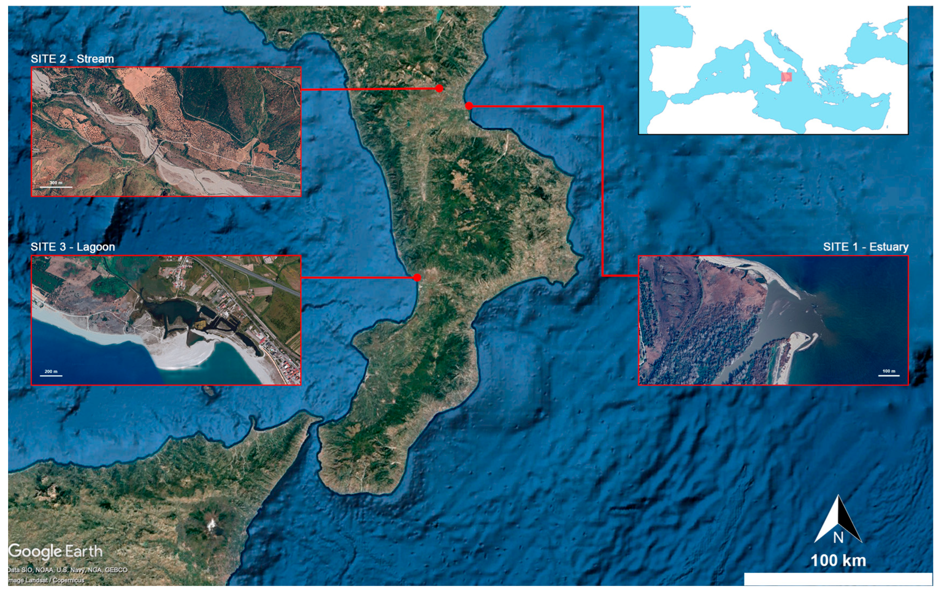
Figure 1. Location of the three sampling sites in Calabria (Southern Italy). In order, starting from the northernmost point, the three sampling sites are indicated: Torrente Raganello (mountain stream—Site 2), Foce del Crati (river estuary—Site 1), and Laghi di Gizzeria (coastal brackish lagoons—Site 3). The inset shows the position of Calabria within the Mediterranean basin.
Fishes 10 00517 g001
Figure 2. Kruskal–Wallis test results, shown as box plots and standard error, only for the elements that exhibited significant difference according to the Bonferroni correction during Dunn’s post hoc test (*, 0.01 ≤ p ≤ 0.001; **, 0.001 ≤ p ≤ 0.0001; ***, p < 0.0001).
Fishes 10 00517 g002
Figure 3. Markers of oxidative status measured in gills, heart, muscle, and liver extracts of A. anguilla from different sampling sites. Data are presented using box plots (*, p < 0.05; **, p < 0.01; ***, p ≤ 0.001; ****, p ≤ 0.0001).
Fishes 10 00517 g003
Figure 4. Significant correlations (p < 0.05) between trace metal concentrations and oxidative parameters in muscle tissue of A. anguilla from different sites.
Fishes 10 00517 g004
Table 1. Summary of the sample stock.
SitesWeight (g)
Mean ± SD
Total Length (cm)
Mean ± SD
N
Estuary145.4 ± 146.840.4 ± 11.110
Stream119.4 ± 97.039.2 ± 12.17
Lagoon104.3 ± 16.336.7 ± 2.76
The specimens are divided by localities. Mean weight with standard deviation, mean total length with standard deviation, and number of individuals are reported.
Table 2. Trace Elements concentration (mean and standard deviation) for site, expressed in ppm (mg/kg) for dry weight.
EstuaryStreamLagoonKWp
Al3.68 ± 1.642.19 ± 0.242.50 ± 0.644.5540.1
As0.26 ± 0.250.000.80 ± 0.3913.550.001
Ba0.27 ± 0.140.15 ± 0.040.16 ± 0.084.510.1
Bi0.01 ± 0.000.000.01 ± 0.019.330.01
Cd0.40 ± 0.440.30 ± 0.290.12 ± 0.049.050.01
Co0.08 ± 0.020.12 ± 0.040.05 ± 0.028.330.015
Cr2.22 ± 0.661.23 ± 0.161.36 ± 0.369.920.01
Cu1.84 ± 1.411.21 ± 0.161.06 ± 0.179.050.01
Fe98.8 ± 33.335.5 ± 20.045.7 ± 27.411.760.003
Mn1.39 ± 0.352.19 ± 0.582.98 ± 2.247.0050.03
Mo0.10 ± 0.030.03 ± 0.010.04 ± 0.0115.490.0004
Ni17.3 ± 33.86.94 ± 11.94.61 ± 8.065.8690.05
Pb0.07 ± 0.030.04 ± 0.010.09 ± 0.093.260.2
Se1.90 ± 0.653.05 ± 0.521.19 ± 0.2411.580.003
Sr2.30 ± 0.872.03 ± 0.524.03 ± 2.312.760.25
Zn46.4 ± 8.0126.8 ± 4.7938.8 ± 5.3811.890.003
Table 3. Concentration ranking for site.
Site 1Fe > Zn > Ni > Al > Sr > Cr > Se > Cu > Mn > Cd > Ba > As > Mo > Co > Pb > Bi
Site 2Fe > Zn > Ni > Se > Al = Mn > Sr > Cr > Cu > Cd > Ba > Co > Pb > Mo > Bi > As
Site 3Fe > Zn > Ni > Sr > Mn > Al > Cr > Se > Cu > As > Cd > Ba > Pb > Co > Mo > Bi
Disclaimer/Publisher’s Note: The statements, opinions and data contained in all publications are solely those of the individual author(s) and contributor(s) and not of MDPI and/or the editor(s). MDPI and/or the editor(s) disclaim responsibility for any injury to people or property resulting from any ideas, methods, instructions or products referred to in the content.