Review

The Allium cepa Model: A Review of Its Application as a Cytogenetic Tool for Evaluating the Biosafety Potential of Plant Extracts

by
Daniela Nicuță
1,
Luminița Grosu
2,
Oana-Irina Patriciu
2,
Roxana-Elena Voicu
1 and
Irina-Claudia Alexa
2,*
1
Department of Biology, Faculty of Sciences, “Vasile Alecsandri” University of Bacău, 157, Calea Mărășești, 600115 Bacău, Romania
2
Department of Chemical and Food Engineering, Faculty of Engineering, “Vasile Alecsandri” University of Bacău, 157, Calea Mărășești, 600115 Bacău, Romania
*
Author to whom correspondence should be addressed.
Methods Protoc. 2025, 8(4), 88; https://doi.org/10.3390/mps8040088 (registering DOI)
Submission received: 30 May 2025 / Revised: 7 July 2025 / Accepted: 23 July 2025 / Published: 2 August 2025
(This article belongs to the Special Issue Feature Papers in Methods and Protocols 2025)

Abstract

In establishing the safety or tolerability profile of bioactive plant extracts, it is important to perform toxicity studies using appropriate, accessible, and sustainable methods. The Allium cepa model is well known and frequently used for accurate environmental risk assessments, as well as for evaluating the toxic potential of the bioactive compounds of plant extracts. The present review focuses on this in vivo cytogenetic model, highlighting its widespread utilization and advantages as a first assessment in monitoring the genotoxicity and cytotoxicity of herbal extracts, avoiding the use of animals for testing. This plant-based assay allows for the detection of the possible cytotoxic and genotoxic effects induced on onion meristematic cells. The outcomes of the Allium cepa assay are comparable to other tests on various organisms, making it a reliable screening test due to its simplicity in terms of implementation, as well as its high sensitivity and reproducibility.

1. Introduction

In recent years, research on plant extracts has continuously increased, highlighting their importance and use in various fields, including the food, pharmaceutical, and cosmetic industries [1]. An inexhaustibly rich source of bioactive phytocompounds, plant extracts may have multiple therapeutic and pharmacological benefits for human health [2,3].
The biomolecules extracted from plants have excellent potential for replacing synthetic drugs. Plant-based extracts are perceived as an alternative or as a complement to classical medicine, their integration in various treatments being a sustainable strategy [3,4,5,6,7,8].
However, in addition to the extraction, analysis, or isolation of certain bioactive compounds, researchers have been concerned with the toxicity of herbal extracts. As they are a complex mixture of compounds, and due to the possible interaction between certain compounds, the plant extracts are not completely without side effects or toxicity [9]. Even though some extracts are considered beneficial, if consumed in large quantities, they can be toxic. The toxicity of herbal extracts depends on several factors, including the extraction method, chemical composition, dosage, and interactions with other substances.
Therefore, researchers consider it imperative and of great importance to evaluate the cytotoxic potential of plant extracts, to ensure that their use is harmless. Various in vivo and in vitro assay are used for establishing the safety or tolerability profiles of bioactive plant extracts; toxicological methods frequently involve brine shrimp, cell lines or animals [10,11]. Before performing very elaborate, long-term and expensive tests on cell lines and knowing that toxicity assessment involving animal experiments is limited by ethical and economic reasons, more accessible and sustainable assays can be carried out as an initial rapid and low-cost approach in the evaluation of the toxicity.
Higher plants are representative systems that are known to be sensitive indicators to the action of toxic agents. Materials of plant origin, such as whole plants, seeds, organs and tissues, have proven to be suitable for monitoring cytotoxicity when exposed to different chemical agents [12,13].
Plant-based monitoring systems rely on microscopic observations of aberrations occurring during mitosis and the subsequent effects on chromosomes. Therefore, plants such as Allium cepa, Allium sativum, Lactuca sativa, Sinapis alba, Triticum aestivum, Vicia faba, Zea mays, etc., possess excellent characteristics for monitoring and screening the cytotoxicity of different agents [12,13,14,15,16,17,18]. The advantages of the plant-based toxicity system are numerous: the ease of obtaining the material, its storage and handling, simplicity and rapidity of implementation, high sensitivity and reproducibility [19,20,21,22,23].
In search of such a method for evaluating the potential toxicity of some plant extracts, following a careful and meticulous bibliographic study, our research team turned its attention to the Allium cepa model [24,25].
Due to our successful application of this plant-based system, the present article constitutes an exploratory review of the Allium cepa model: in particular, its use as a cytogenetic tool for assessing the biosafety potential of plant extracts. We highlight the advantages it offers.
Moreover, this review aims to highlight the differences between the experimental protocols and the influences of several factors related to both the extraction process for the tested samples and the experimental conditions of the Allium test itself on the level of cytogenotoxicity of plant extracts. These aspects are less frequently discussed in the very few reviews already published [21,23,26]. Additionally, this systematic review intends to provide an overview of the plant species studied using this model.

2. The Allium cepa Model: General Considerations

The Allium cepa test is an in vivo experimental model used to evaluate DNA damage (clastogenic and/or aneugenic effects) by identifying chromosomal aberrations and disorders occurring in the mitotic cycle, due to the action of various mutagenic agents.
Since 1938, when it was used for the first time by Levan [27] when studying the effect of colchicine on the mitosis of onions, the methodology of the Allium cepa assay has undergone continuous improvements, making it appropriate for many applications. Important contributions to the development of this bioassay were made by Grant [28,29], Fiskesjö [30,31], Rank and Nielsen [32] (Figure 1). They reported that, when compared to other test materials, Allium material can produce similar results. It has also been demonstrated that this model has high sensitivity and can be used as a standard method for environmental monitoring [22,23,33,34].
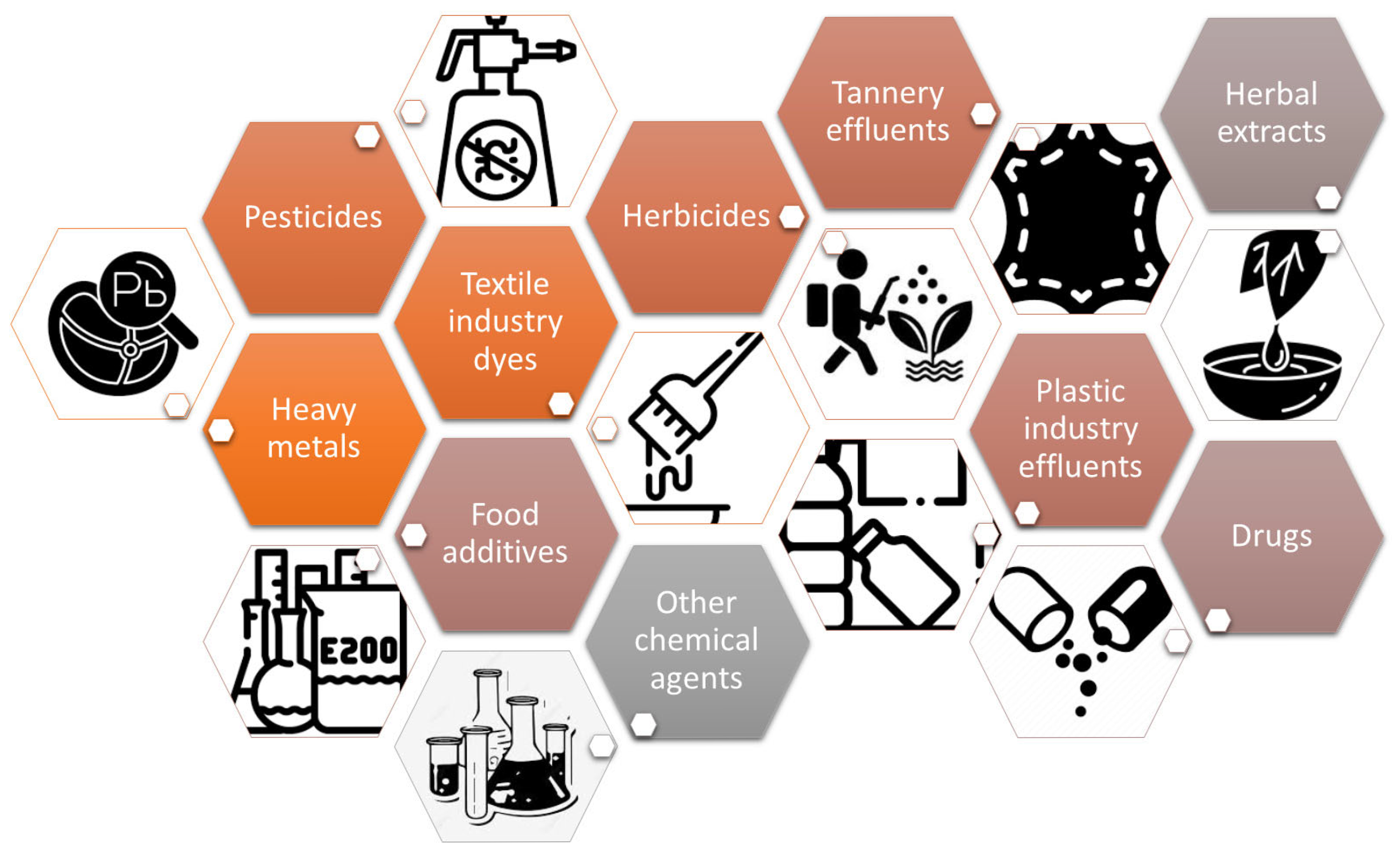
Over the years, the Allium cepa model has been used, with good results, in the detection of a wide variety of pollutants and chemical agents (Figure 2).
Several comprehensive reviews concerning the application of the Allium cepa as environmental monitoring assay have been reported [21,22,33,34,35]. Research describing the use of A. cepa tests in the investigation of heavy metal accumulation in soil, surface water and sediment, industrial wastewater, groundwater, vegetables, etc., are noteworthy [33,34,35,36,37,38].
The A. cepa bioassay was successfully applied to effluents from the tannery, textile and plastic industries [39,40,41,42,43]. Several studies are related to the cytotoxicity of some herbicides [44], pesticides [45,46,47,48], fungicides [49], insecticides [50], or other chemical agents [51,52]. Moreover, the potential cytotoxicities of therapeutic drugs (e.g., doxorubicin, erlotibin, metalodrugs, nevirapine, etc.) [53,54,55], food additives (e.g., saccharin, potassium metabisulphite) and animal feeds additives (e.g., urea) [56,57] were tested using the Allium cepa assay. Additionally, the Allium cepa model was reported “as a ‘warning’ bioindicator in detecting the genotoxicity of medicinal plants” [21,26].

3. Basic Principles of the Allium cepa Test and Protocol

Toxicity studies based on the Allium test are performed on onion roots, which, when exposed to different substances, can indicate their potential cytotoxic or genotoxic effect on organisms.
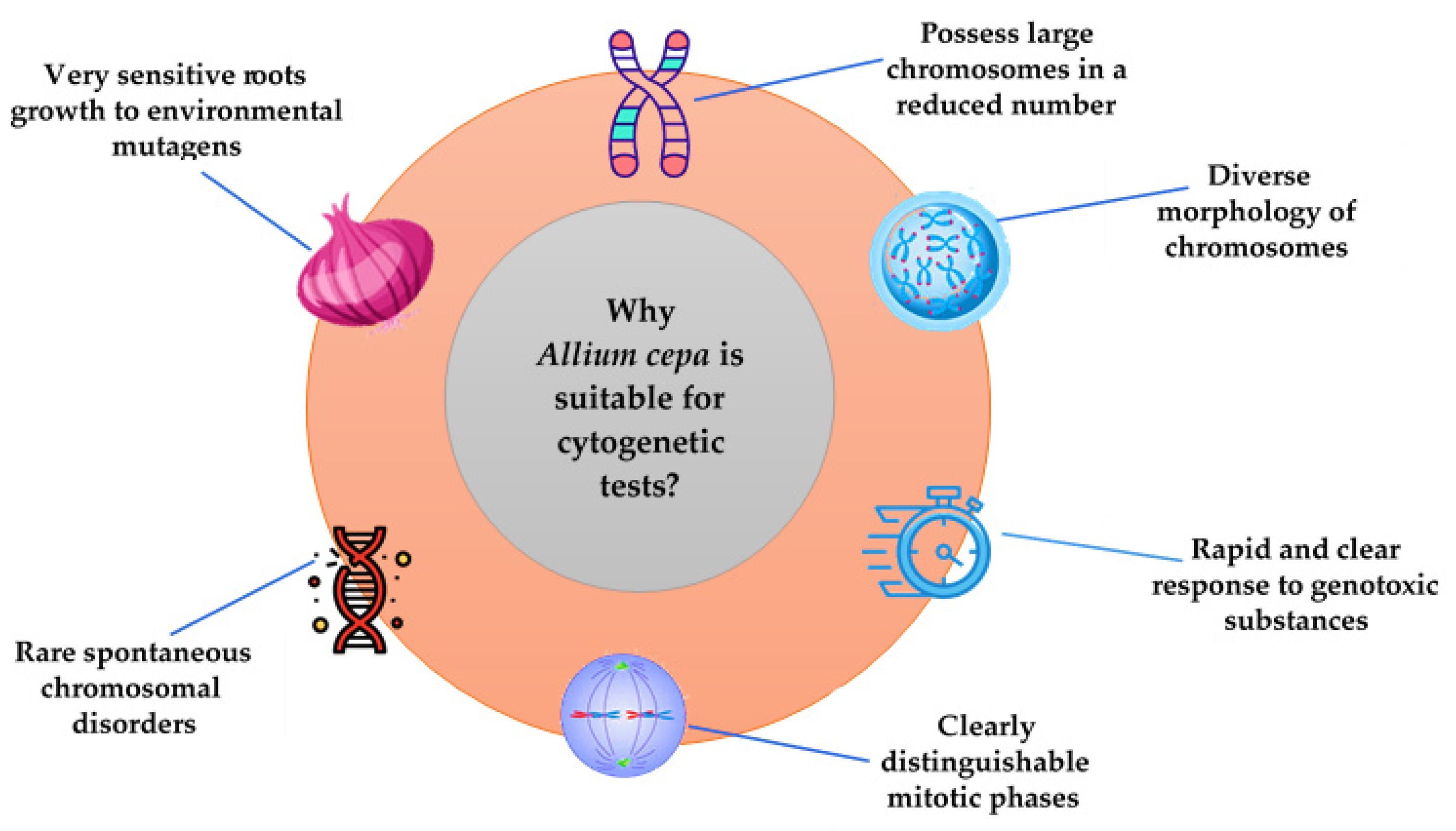
Onions are considered appropriate for toxicological evaluations because the roots grow rapidly and their tips contain cells in various phases of cell division, showing a clear and rapid response to genotoxic substances; moreover, spontaneous chromosomal damage rarely occurs. Due to the presence of distinct cells, large chromosomes in a reduced number (2n = 2x = 16), and a stable karyotype, it is easy to identify the possible chromosomal lesions and mitotic cycle disorders under a microscope [21,22,23]. The reduced number of chromosomes (8 pairs) compared with other species (e.g., wheat Triticum aestivum: 2n = 42) simplifies their tracking and identification during cell division, highlighting possible mutations that can affect the number of chromosomes such as polyploidy (3x, 4x, etc.) or aneuploidy (2n + 1, 2n − 1).
Some characteristics of the onion that make it suitable for cytogenetic tests are presented in Figure 3.
Since 1938, when Levan [27] described the first protocol for the Allium cepa test, the recommended plant material is represented by the onion bulbs with rapidly growing root tips used in most reported studies. The cells of the root tip actively divide, being the first to come into contact with substances in the environment. Therefore, toxic effects on mitosis and chromosome behavior during the division phases can be observed [58]. Buds from germinated seeds can also be used for the same purpose, as mentioned in other research [59,60]; alternatively, seeds are first germinated in water until the roots are about 2 mm in height [61,62].
The onion bulbs used for testing should be of similar size (approximately 1.5–2.0 cm in diameter) and not exposed to herbicide or fungicide treatments [19,63]. Generally, between three and five onion bulbs are needed for each sample (including the control) to obtain roots. According to the protocol proposed by Tedesco and Laughinghouse [21] as a standard experiment for the Allium test, it is recommended to use five different sets of bulbs: one for the negative control, one for the positive control with a known genotoxic agent, and three groups for different concentrations of the test agent. Some authors initially use a larger number of bulbs to test their germination rate, placing them in water for two or four days. Subsets of three or five bulbs which showed the best root growth are then chosen to be exposed to the test solutions [54,64,65,66,67]. It is recommended that the bulbs be lightly scraped in the lower area (primary root ring), to favor the emergence of new roots [19,68]. Many studies report that onion bulbs are initially placed in distilled or tap water (if potable) in narrow glass or plastic containers (50 mL) [21]. Only the area where the roots will form is submerged in water. The water should be renewed every day until the roots grow to a certain length. Root growth varies in time (two to four days), depending on the temperature conditions in which the onion bulbs are stored (a room or growth room/chamber at 22 ± 2 °C) [35]. If onion bulbs are placed in a growth chamber, a controlled photoperiod (18 h/6 h light/dark) can also be ensured. When the roots reach the appropriate length (0.5–2 cm), the onion bulbs may be transferred to the flasks containing the different test extracts and only the base of each bulb should be immersed/suspended in the extract. The exposure time of onion roots in the tested plant extracts may vary: 24 h [69,70], 48 h [65,71], 72 h [68], 96 h [67]. Sabini et al. [72] reported two and five days, as well as two days followed by three days with water (reversion) of exposure.
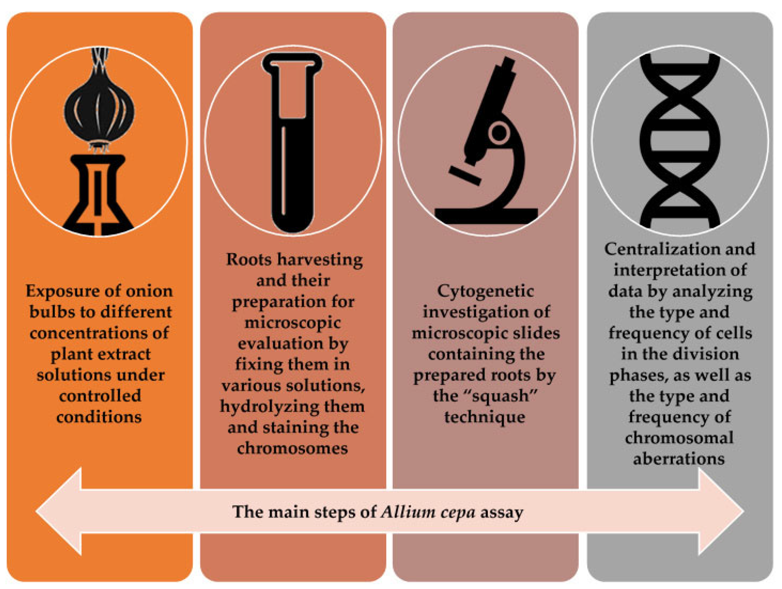
The main steps for the Allium cepa protocol are presented in Figure 4.
In the standard Allium testing protocol, normal tap water [30,73,74,75], or distilled water [76] are used as the negative control sample. It seems that, compared to tap water, distilled water used as a negative control leads to a statistically significant inhibition of mitosis in the apical cell of the onion [75]. Several studies report the use of a positive control sample, in addition to a negative control sample. Various substances are used as positive controls, namely: glyphosate [70,77,78,79,80,81], cyclophosphamide [71], methotrexate [82,83], paracetamol [72,84], methyl methane sulfonate [85], ethyl methane sulfonate [69,73,74], copper sulfate [86,87], lead nitrate [88], hydrogen peroxide [89], sodium azide [90], dimethyl sulfoxide [91]. Glyphosate, most commonly used as a positive control, is a chemical compound known for its extremely cytogenotoxic effect; it is used as herbicide for a broad spectrum of weeds [92,93]. When the essential oil samples are tested, ethanol or methanol is used as positive control [94,95].
At the end of the exposure period, onion roots treated with the test solutions, including the control, are harvested for cytogenetic preparations. Each plant has its own biological clock for mitotic division; therefore, the root harvesting should be performed when the meristematic cells are actively dividing [96]. The timing of root cutting is therefore a crucial step to identify as many cells as possible in the different phases of mitotic division. The duration of mitosis in roots of Allium cepa L. is about 4 h (prophase—2 h, metaphase—40 min, anaphase + telophase − 1 hrs and 20 min) [97]. According to Sangur et al. [96], a high frequency of metaphase cells coincides with a high value of the mitotic index. Therefore, it is advisable to harvest the roots at the time of metaphase unfolding within the mitotic division. Well-growing roots with an average length of 1–2 cm are the best candidates for cytogenetic studies, while exceptionally long or short roots are removed [21,35].
After harvesting, the roots should be immersed in a fixative solution, which has the role of instantly coagulating the cellular constituents. In this way, the cells are rapidly fixed in the phase of mitotic division at that time. A mixture of alcohol and acetic acid can be used for this step, namely, Clarke’s fixative (ethanol:glacial acetic acid, 3:1 v/v) for 90 min [60,76], Farmer solution (ethanol:glacial acetic acid, 3:1 v/v) [24,25,98], Carnoy’s solution (ethanol:glacial acetic acid, 3:1 v/v) [99], or a solution of acetic acid:methanol, 1:3 v/v [19]. Fixation is achieved by keeping the harvested roots in fixative solutions for 12–18 h at 4 °C in the refrigerator. Until microscopic preparations are made, the roots can be preserved in 70% ethanol in the refrigerator for several months [70,100]. In the protocol described by Wierzbicka [63], fixation can be performed in a solution containing hydrochloric acid (glacial acetic acid (45%):HCl (1N), 9:1 v/v) by immersing the roots for 5 min at 50 °C. Therefore, together with fixation, hydrolysis of onion roots also occurs [28].
The hydrolysis step is performed with the aim of dissolving the pectocelluloses from the cell wall, allowing the dye used for coloration to penetrate the cell and the chromosomes in the nucleus. In this regard, root hydrolysis can be performed at room temperature with 1N HCl for 20–30 min, or at 60 °C (in a water bath) for 5–10 min. Thus, the root tissue softens, and the dye is able to penetrate the cells, reaching the nucleus to stain the genetic material. Then, the roots are gently rinsed with distilled water, avoiding damage to the hydrolyzed tips. This step is very important because, if HCl is not removed by washing the roots, staining is compromised and the cells cannot be identified under the microscope [68,80,101].
Root coloring is accomplished by treating the roots with different dyes, allowing the microscopic observation of various cell categories in interphase or division. Thus, aceto-orcein solution (2%) [66,67,69,94,101], acetocarmine (2%) [64,68,89] or lacto-propionic orcein [102] ensure a rapid staining in few hours and a good contrast of the chromosomes, which explains their frequent use in the mentioned studies. The cytogenetic preparations must be visualized immediately, because their discoloration occurs over time, which represents a disadvantage of using these staining solutions. For the accuracy of cytogenetic preparations, a staining step with Schiff reagent [55,103] or carbol fuchsin solution [24] is recommended in cytogenetic research, which allow for a very good visualization of nuclei, chromatin and chromosomes in metaphase or anaphase. The microscopic preparations are persistent, and the roots remain stained for a long time (one to two years) if kept in the refrigerator. However, the need for a carefully controlled acid hydrolysis and the longer root staining time (three to five days in the refrigerator) could constitute disadvantages of using these types of coloring agents. Vicentini et al. [104] reported that the roots were fixed and stained with Feulgen reaction.
After root staining, preparation of microscope slides for cell analysis is carried out via the “squash” technique, consisting first in placing one to two onion roots on a microscope slide, in a drop of acetic acid aqueous solution. By cutting the tips of the roots (approximately 1–2 mm) with a scalpel, only the area containing the meristematic cells where mitosis occurs is preserved for microscopic analysis. Then, a coverslip is placed over the tissue and the plant material is crushed by pressing. The cells are thus arranged in a single plane, without overlapping, so that they can be observed individually under the microscope [35,70]. For each bulb, from the harvested and stained roots, two or three slides are prepared for analysis and cell counting under a microscope [19,80,105]. Yekeen et al. [106] reported that, for the fixation and preservation of the samples, the slides can be immersed in liquid nitrogen, thus allowing their subsequent evaluation. For the same purpose, the edges of the slide can be sealed by applying transparent nail polish, so that the cytogenetic preparation can be analyzed even after several days [28,38,72].
One of the objectives of cytogenetic research is to analyze the number and behavior of chromosomes in dividing cells, under the influence of different types of plant extracts.
The analysis of cytogenetic preparations is performed using the 40× and 100× objectives of an optical microscope, which can be equipped with a camera and connected to a computer [67,68,107].
Each microscopic slide is examined to identify and quantify the different cell types present in the microscopic fields. For a correct evaluation, it is recommended to investigate 1000–5000 cells/microscopic slide/per tested sample, as well as in the control samples [30,38,108]. Few authors reported the analysis of a larger number of cells (over 5000) [59,60,66].
The most important cytogenetic parameters calculated based on microscopic observations are the mitotic index (MI) and the proportion of chromosomal abnormalities (CA). In some reported studies, in addition to these parameters, the index of each phase of mitosis (PI) is also calculated [25,76,102,109], as well as the limit value of cytotoxicity (LCV) [25,36,40,76]. The calculation formulas of these parameters are detailed in Figure 5.
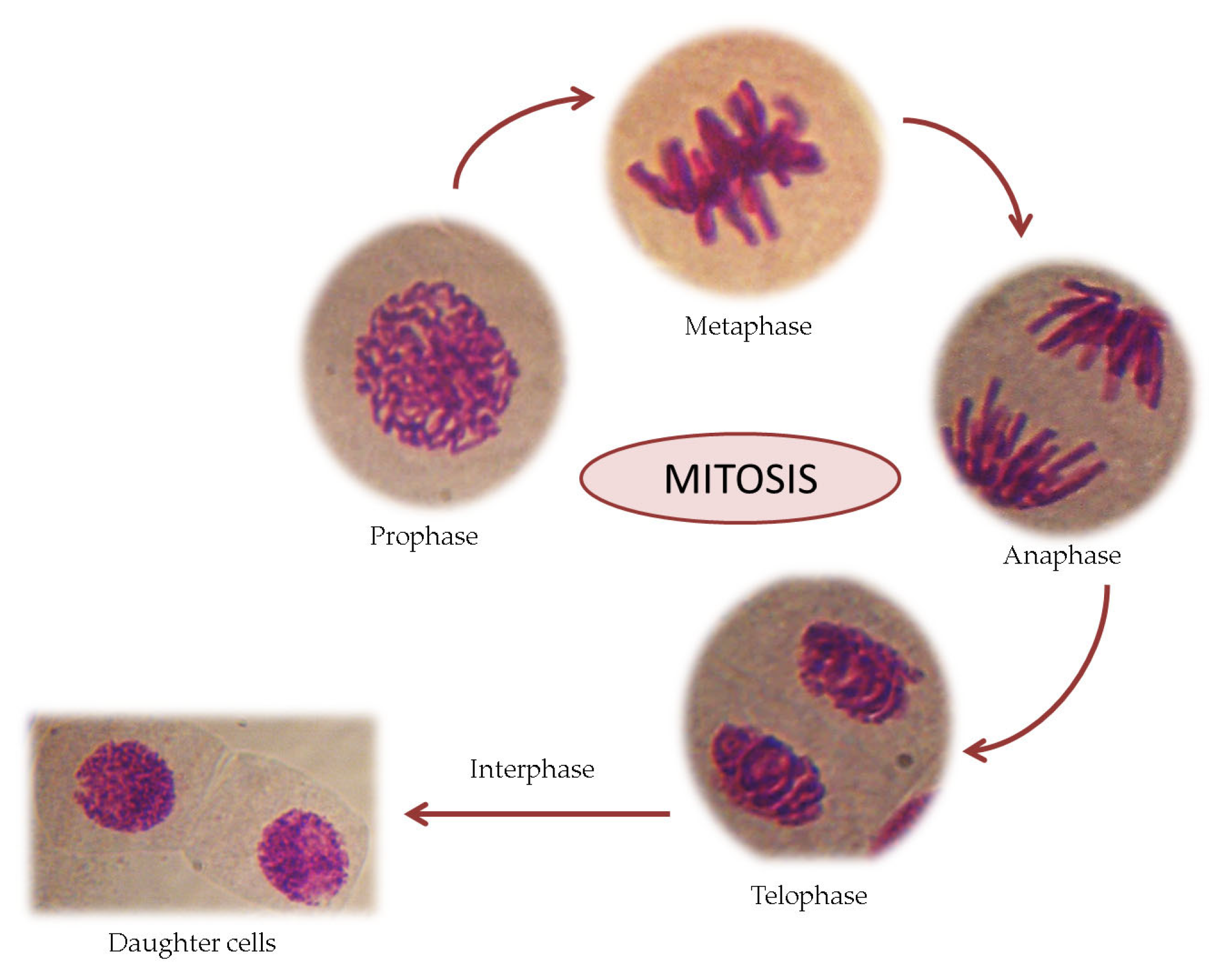
The mitotic index is a tool used to measure the percentage of dividing cells from each phase of mitosis out of the total cells observed in a microscopic sample. A correct identification of all phases of cell division in Allium cepa L. roots is essential. The mitotic cell division comprises regular phases such as prophase, metaphase, anaphase and telophase (see Figure 6). Interphase is a stage in which genetic material is replicated, which takes place over a longer period of the cell cycle compared to mitosis. The most numerous cells are found in the interphase.
Therefore, the number of cells analyzed under the microscope is important for calculating the mitotic index (MI) as accurately as possible. If, in the samples tested with different plant extracts, the MI value is high, this indicates intense cellular activity, and the tissue is actively growing, since it is not affected. If, on the contrary, the MI value is low (compared to the control sample), it indicates the inhibition of this parameter, which can be interpreted as cell death, a delay in the kinetics of cell proliferation, or cellular damage [76], due to the cytotoxic or genotoxic effect of the plant extracts [58,67,68,69,76]. The MI may vary in different roots of the same plant, but the average data are fairly stable [21]. Additionally, the reduced number of cells per division phase (PI) indicates an inhibitory effect on the division process due to tested extracts [65]. Some authors reported that, if the MI value decreases by 50% compared to the control, it is considered the limit value for cytotoxicity (LVC), but, if it drops below 22–25%, it can be lethal to organisms [25,36,40,76].
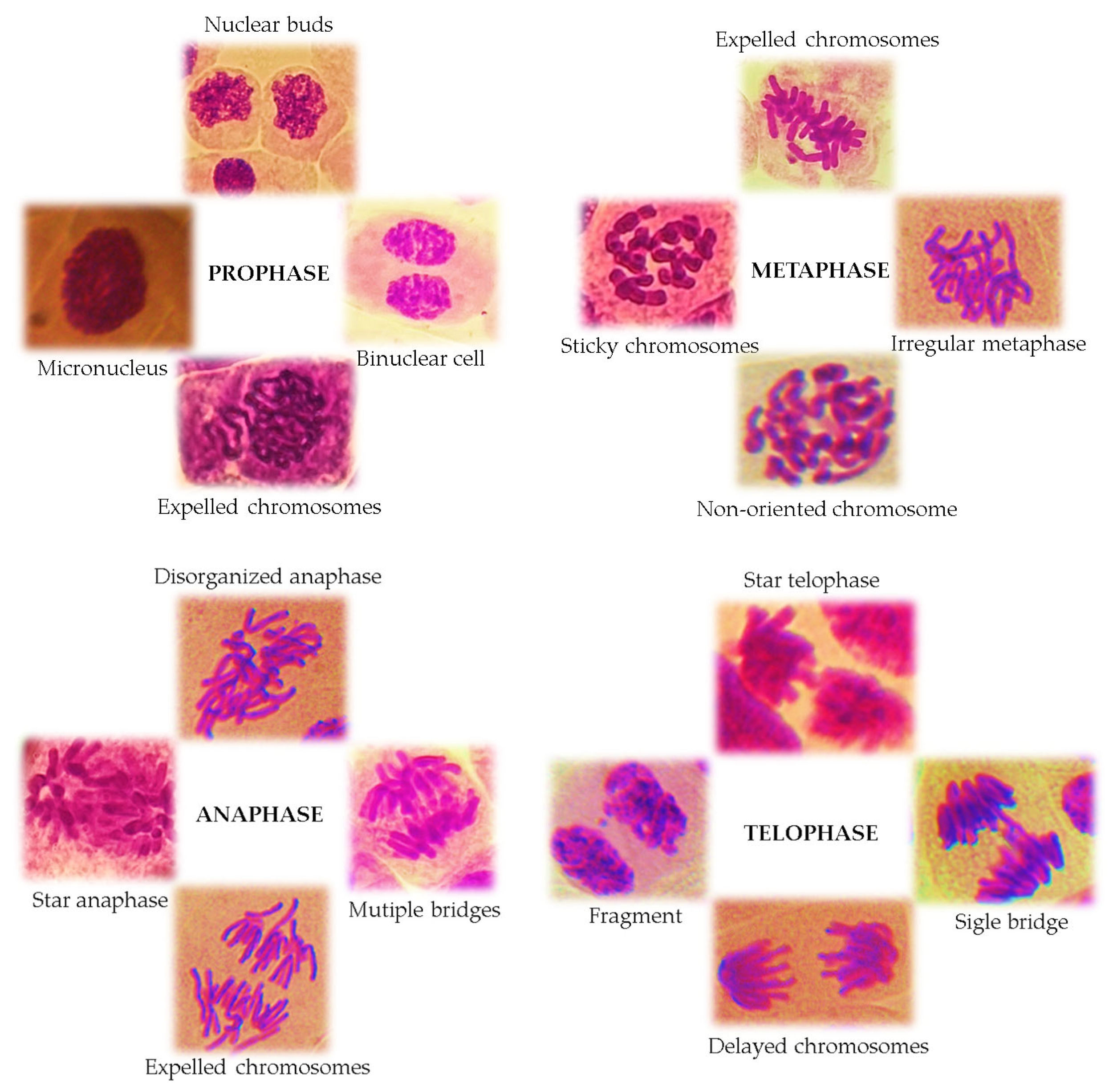
Chromosomal aberrations (CAs) are changes in the structure and number of chromosomes and can be observed in all stages of mitosis. The most remarkable cells can be highlighted; especially in anaphase and telophase, CAs appear in the form of chromosome bridges, chromosome losses and fragments, chromosome delays, disorganized and multipolar anaphases (star anaphase), and c-mitosis. A series of chromosomal aberrations can also be highlighted in the metaphase as a result of the expulsion of whole chromosomes from the metaphase plate, as well as chromosome breaks, chromosome fragments, and irregular metaphases [110]. Chromosomal aberrations are caused by the breakage of chromosome fragments, the unbalanced exchange of chromatid segments, or damage to the mitotic spindle.
Antiproliferative capacity and genotoxic potential on cell division in A. cepa L. of different plant extracts are due to the interaction of various chemical components, present in high concentrations, which cause inhibitory effects on the cell cycle [77,108]. Toxic substances can also affect interphase cells by blocking DNA and protein synthesis in the nucleus [71]. This is evidenced by the presence of micronucleus in interphase and prophase cells. The micronucleus has a similar structure but a reduced size compared to the main nucleus. In the interphase daughter cells, a series of nuclear abnormalities can be identified, such as lobulated nuclei, nuclei with nuclear buds, polynucleated cells or minicells.
Some types of chromosomal aberrations in Allium cepa L. roots induced by various cytotoxic and genotoxic agents are presented in Figure 7.
Within the Allium cepa test, in addition to microscopic parameters, macroscopic parameters can be also measured [30]. The most important macroscopic parameter is the root growth length RGL (the average root length for each sample) [67]. Thus, the percentage of root growth inhibition in the tested extracts compared to the control can be calculated [98,101]. Additionally, the EC50 (effective concentration at which root growth is 50% of the control) can be calculated. Inhibition of root elongation greater than 20% was considered evidence of toxicity, based on standard phytotoxicity tests [54]. Other macroscopic root growth parameters, e.g., restriction of leaf growth, can also be evaluated to estimate the toxicity index (turgescence, consistency, color change, root tip shape, presence of swellings, hooks, twists, or necroses) [30,57,98,102,111].
After centralizing the data, the results can be analyzed using various statistical methods such as chi-square (χ2) [84,112,113,114] or one-way analysis of variance (ANOVA) [61,91] followed by Tukey’s test [16,115,116], Bonferroni test [87], etc.

4. Results of the Cytotoxic and Genotoxic Evaluation of Plant Extracts Using the Allium cepa Model: Literature Review

Early research from 1999 was found in the Web of Science database on the assessment of the cytotoxicity of plant extracts. Aqueous extracts of Allophylus edulis leaves obtained by decoction, a plant widely used in folk medicine in Argentina, were studied in terms of cytotoxicity and genotoxicity [102]. Yajía et al. reported an important decrease in the MI compared to the control, indicating a significant statistical correlation between the mitotic index (MI) and the root growth length (RGL) and chromosome aberrations (CAs).
After the 2000s, the number of researchers who started to use the Allium cepa model as a tool in the toxicological evaluation of plant extracts has continuously increased.
Table 1 presents the cytotoxic and genotoxic studies using Allium cepa assay on various plants.

5. Discussion

Most studies highlight that a gradual decrease in MI is significantly correlated with an increasing concentration of the tested extracts or with a longer exposure time, indicating the interference of plant extracts in the progression of the cell division cycle. Moreover, an increase in CA compared with the negative control sample may be detected in the highest concentrations.
The presence of cells with different chromosomal aberrations proves the clastogenic or aneugenic effects of some tested plant extracts [16,66,109,120,142,149]. As previously mentioned, clastogenic aberrations cause changes in the structure of chromosomes, which can be observed at all stages of mitosis, especially in the anaphase, telophase and metaphase of cell division. Plant extracts can induce physiological aberrations (stickiness, c-mitosis and stray chromosomes), which are more frequent than clastogenic aberrations (breaks and bridges) [66]. Thus, in numerous studies, the presence of cells with sticky chromosomes [65,66,67,68], anaphase–telophase with bridges [25,64,76,109,118], laggard chromosomes [117], disordered (irregular) anaphase–telophase chromosome fragments [24,66,69,76], vagrant chromosomes [98,117], c-mitosis [98,108,128], binuclear cell [109,127,135], and enucleated (ghost) cells [120] was frequently detected.
Some extracts can also induce aneugenic changes (adhesion, subsequent segregation, multipolarity, chromosome loss) in A. cepa L. roots, causing variations in the number of chromosomes in the meristematic cells. The distinction between a clastogenic effect (damage to the structure of chromosomes) and an aneugenic one (a change in the number of chromosomes) can be made by analyzing the sizes of micronuclei that appear in prophase or interphase cells. If the micronuclei are large, this can be considered an aneugenic effect, since they are formed from entire chromosomes, and, if they are reduced in size, they only contain chromosome fragments [109].
If both the MI and the CA percentages are significantly different in relation to the control at the concentrations tested, a usage warning can be made, revealing possible harmful effects on human health [24,88,118,119,128]. If mild cytotoxicity with minor chromosomal aberrations is revealed, it demonstrates that the extracts in question are safe for consumption [77,104,124,129].
The cytotoxic and genotoxic effects of tested extracts can be related to the phytochemicals present in the species and to their synergetic effect [149,152]. The concentration of metabolites in the extracts can be influenced by the extraction method [118]. Thus, phytochemical screening and the quantification of bioactive compounds present in the tested crude extracts performed using various spectrophotometric methods are important [24,90,119,124]. Simultaneous tests of entire extracts as such, as well as of certain compounds from the extracts (e.g., citral and limonene from Citrus aurantiifolia essential oil), were reported by Fagodia et al. [122]. Often, in studies involving essential oils, the observed effects are explained on the basis of their main compounds. Pawlowski et al. [149] state that, even though the major compound of the essential oils from Schinus molle and Schinus terebinthifolius is α-pinene, the results observed on cell division in onion meristematic cells may be due to both major and minor compounds, rather than a single compound, probably acting synergistically. The presence of monoterpenes or the combined synergistic effect of different monoterpenes in essential oils on the mitotic index of A. cepa L. has also been reported in other studies [85,152], without mentioning the specific action of a particular compound. In the study of Cavalcante et al. [87], it was not the entire extract that was tested on onion roots, but rather 2-oleyl-1,3-dipalmitoyl-glycerol, a separate compound from Platonia insignis extract.
The findings of the studies showed that the level of cytogenotoxicity of plant extracts on onion meristematic cells can be influenced by several factors related to both the extraction process for the tested samples and the experimental conditions of the Allium test itself.
Parameters such as the plant material conditioning (state), solvent, temperature, time, and extraction method influence the content of extracts in active principles, which may lead to different results in terms of cytotoxicity and genotoxicity.
In the extraction process, the plant material (aerial part, leaf, stem, inflorescence, rhizome, bark, etc.) can be used both fresh [85,88,94,112,119,145] and dried, crushed [137] or in powder form [66,82,120]. Drying is carried out using classical methods (oven) or modern methods (freeze drying) [116].
Concerning the solvent, water is the most frequently employed solvent, resulting in aqueous extracts either by infusion, decoction or maceration. Hot water is often used, but so is cold water [59,72]. The aqueous extracts (infusion, most often) are prepared as they would be made at home by the general population [24,64,68,98]. Studies are also reported on hydroalcoholic [82,84,118,131], methanolic [67,69,76,89,124], hexanic [87] or dichloromethane [61] extracts. The essential oils obtained from various plants are also subjected to toxicological research using the Allium test [79,85,91,94,95,115,116,119,122]. The cytotoxicity and genotoxicity effect of different concentrations of latex from Hancornia speciose or Jatropha curcas L. are reported [129,133].
Furthermore, in several studies, comparative cytotoxic and genotoxic evaluations are performed between aqueous extracts and alcoholic extracts [23,118,123,134,137] or between aqueous extracts and essential oils [70,81,94] from the same plant source.
The experimental conditions of the Allium test can vary. Usually, different concentrations/dilutions of the studied extracts are tested, and exposure can occur at different times.
Some studies reported that the onion roots were immersed directly in plant extracts without any dilution [24,64,68,98] or the tested concentrations were established according to doses that are recommended in alternative medicinal usage by the general public [141].
In parallel with the Allium cepa assay, several studies analyze cytotoxicity using tests on animal cells (e.g., bone marrow of rat cells) [104,109,136], human cells (e.g., lymphocytes) [91], tumor cell lines [138], and brine shrimp (Artemia salina) [87,132,138]. There are some studies that report tests on different weeds [79,85,122] to establish the phytotoxicity of the tested plant extracts. In addition to the A. cepa assay, recent research on Platonia insignis extracts has reported tests on different insects or aquatic crustaceans’ species [87]. Most often, the results concerning the toxicology of plant extracts using different kinds of tests are similar to those obtained with the Allium test.
Additionally, the examination of antigenotoxicity can establish the potential protective effect of plant extracts against alterations or mutations induced in the genetic material by various compounds. The antigenotoxic effect of some herbal extracts has been tested in relation to the genotoxicity induced by mutagenic substances such as hydrogen peroxide [120,141], methyl methanesulfonate [109,125] or lead nitrate [150] on onion meristematic cells.

6. Conclusions

The side effects associated with the consumption of plants are essentially influenced by the dosage and frequency of their use. Since bioactive compounds and their interactions can lead to toxic effects, there is a constant need for scientific knowledge regarding the efficacy and safety of medicinal herbs. It is important to establish, with accuracy, what the ideal and safe concentrations are for the use of these plants.
The present review is intended as proof that the Allium cepa model constitutes an important cytogenetic tool in the process of evaluating the biosafety potential of herbal extracts. This plant-based assay allows for the detection of the possible cytotoxic and genotoxic effects induced on onion meristematic cells, as a prompt step in the evaluation process.
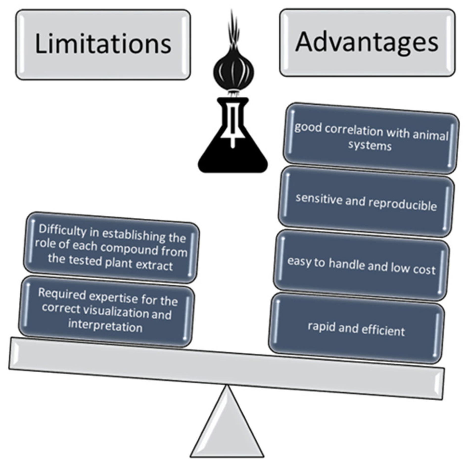
The Allium cepa model is not a perfect tool, presenting some limitations (Figure 8). Considering that plants extracts represent a complex mixture of biomolecules, it is difficult to establish the selective influence of each compound on cell division. Some of them may have cytotoxic and/or genotoxic effects, while others may possess cytoprotective and/or antigenotoxic properties. Moreover, the use of the test requires expertise in the correct visualization and interpretation of mitotic phases and chromosomal abnormalities. Based on the research reviewed, it most often emerged that, for a more in-depth study of the cytotoxicity of plant extracts, other tests must be performed, as a complement to the Allium test.
Nevertheless, the Allium cepa bioassay is a successful test that is used because of its many advantages: simplicity in implementation, cheapness, high sensitivity, and reproducibility. Moreover, the results provided by this biomarker are in good correlation with other accessible systems like animals, cell lines, etc. The ecological significance of using this model, which can be considered environmentally friendly, must also be highlighted. The advantages of the Allium test are far greater than the disadvantages presented.
It is concluded that the Allium cepa test is a very convenient tool, especially in the preliminary screening of the cytotoxic and genotoxic effects of various plants frequently used in traditional medicine, from which aspects related to their biosafety potential can be established.

7. Future Directions

Although the Allium cepa test cannot completely replace other cytotoxicity tests, combining it with the analysis, identification and quantitative determination of bioactive compounds in the analyzed plant extracts could lead to clearer information about the causal compounds of the observed effects.
The application of the Allium cepa model, however, indicates significant scientific discoveries, and new adaptations of the test, as well as its standardization, could lead to countless possibilities for its use, avoiding other types of more laborious tests, including those on animals.
In recent years, cytotoxicity research has tended to use and develop sustainable testing systems such as the Allium cepa assay, with the aim of completely or partially replacing a large percentage of sophisticated experiments involving high costs and environmentally unfriendly methods.

Author Contributions

Conceptualization, D.N., L.G., O.-I.P., R.-E.V. and I.-C.A.; methodology, D.N., L.G., O.-I.P., R.-E.V. and I.-C.A.; software, I.-C.A.; writing—original draft preparation, D.N., L.G., O.-I.P., R.-E.V. and I.-C.A.; writing—review and editing, D.N., L.G., O.-I.P. and I.-C.A.; visualization, D.N., L.G., O.-I.P., R.-E.V. and I.-C.A.; supervision, D.N. and I.-C.A. All authors have read and agreed to the published version of the manuscript.

Funding

This research received no external funding.

Data Availability Statement

Not applicable.

Conflicts of Interest

The authors declare no conflicts of interest.

References

  1. El Allaoui, H.; El Ahmadi, K.; El Abdouni, A.; Dira, I.; El Bastrioui, M.; Bouhrim, M.; Eto, B.; Shahat, A.A.; Herqash, R.N.; Haboubi, K. Trends and Insights in Medicinal Plant Extract Research: A Ten-Year Bibliometric and Visualization Study. Horticulturae 2024, 10, 1163. [Google Scholar] [CrossRef]
  2. Proestos, C. The Benefits of Plant Extracts for Human Health. Foods 2020, 9, 1653. [Google Scholar] [CrossRef]
  3. Almasri, R.S.; Bedir, A.S.; Al Raish, S.M. Comprehensive Ethnopharmacological Analysis of Medicinal Plants in the UAE: Lawsonia inermis, Nigella sativa, Ziziphus spina-christi, Allium cepa, Allium sativum, Cymbopogon schoenanthus, Matricaria aurea, Phoenix dactylifera, Portulaca oleracea, Reichardia tingitana, Salvadora persica, Solanum lycopersicum, Trigonella foenum-graecum, Withania somnifera, and Ziziphus lotus. Nutrients 2025, 17, 411. [Google Scholar] [CrossRef]
  4. Srivastava, A.; Srivastava, P.; Pandey, A.; Khanna, V.K.; Pant, A.B. Phytomedicine: A Potential Alternative Medicine in Controlling Neurological Disorders. In New Look to Phytomedicine; Khan, M.S.S., Ahmad, I., Chattopadhyay, D., Eds.; Academic Press: Cambridge, MA, USA; Elsevier: Amsterdam, The Netherlands, 2019; pp. 625–655. [Google Scholar] [CrossRef]
  5. Chaachouay, N.; Zidane, L. Plant-Derived Natural Products: A Source for Drug Discovery and Development. Drugs Drug Candidates 2024, 3, 184–207. [Google Scholar] [CrossRef]
  6. Soni, V.; Raizada, P.; Singh, P.; Cuong, H.N.; Saini, A.; Saini, R.V.; Le, Q.V.; Nadda, A.K.; Le, T.T.; Nguyen, V.H. Sustainable and green trends in using plant extracts for the synthesis of biogenic metal nanoparticles toward environmental and pharmaceutical advances: A review. Environ. Res. 2021, 202, 111622. [Google Scholar] [CrossRef]
  7. Sarkar, A.; Sarkar, D.; Poddar, K. Plant metabolites as new leads to drug discovery: Approaches and challenges. In Medicinal Plants: Chemistry, Pharmacology, and Therapeutic Applications, 1st ed.; Swamy, M.K., Patra, J.K., Rudramurthy, G.R., Eds.; CRC Press: Boca Raton, FL, USA, 2019; pp. 61–70. [Google Scholar] [CrossRef]
  8. Puri, V.; Nagpal, M.; Singh, I.; Singh, M.; Dhingra, G.A.; Huanbutta, K.; Dheer, D.; Sharma, A.; Sangnim, T. A Comprehensive Review on Nutraceuticals: Therapy Support and Formulation Challenges. Nutrients 2022, 14, 4637. [Google Scholar] [CrossRef]
  9. Anywar, G.; Kakudidi, E.; Byamukama, R.; Mukonzo, J.; Schubert, A.; Oryem-Origa, H.; Jassoy, C. A Review of the Toxicity and Phytochemistry of Medicinal Plant Species Used by Herbalists in Treating People Living With HIV/AIDS in Uganda. Front. Pharmacol. 2021, 12, 615147. [Google Scholar] [CrossRef]
  10. Hamidi, M.R.; Jovanova, B.; Panovska, T.K. Toxicological evaluation of the plant products using Brine Shrimp (Artemia salina L.) model. Maced. Pharm. Bull. 2014, 60, 9–18. [Google Scholar] [CrossRef]
  11. Ghica, A.; Tănase, M.L.; Niculițe, C.M.; Tocilă, A.; Popescu, L.; Luță, E.A.; Olaru, O.T.; Popovici, V.; Balaci, T.D.; Duțu, L.E.; et al. In Vitro Toxicity Evaluation of Some Plant Extracts and Their Potential Application in Xerosis cutis. Cosmetics 2024, 11, 124. [Google Scholar] [CrossRef]
  12. Mulaszynska, J.; Juchimiuk, J. Plant genotoxicity: A molecular cytogenetic approach in plant bioassays. Arh. Hig. Rada Toksikol. 2005, 56, 177–184. [Google Scholar] [PubMed]
  13. Turkez, H.; Arslan, M.E.; Ozdemir, O. Genotoxicity testing: Progress and prospects for the next decade. Expert Opin. Drug Metab. Toxicol. 2017, 13, 1089–1098. [Google Scholar] [CrossRef]
  14. Morozesk, M.; Bonomo, M.M.; Souza, I.D.C.; Rocha, L.D.; Duarte, I.D.; Martins, I.O.; Dobbss, L.B.; Carneiro, M.T.; Fernandes, M.N.; Matsumoto, S.T. Effects of humic acids from landfill leachate on plants: An integrated approach using chemical, biochemical and cytogenetic analysis. Chemosphere 2017, 184, 309–317. [Google Scholar] [CrossRef]
  15. Onisan, E.; Sarac, I.; Petolescu, C.; Horablaga, M.N.; Mate, C.; Simina, A.; Camen, D.; Ganea, M.; Ardelean, D.R.; Călugar, L.; et al. Application of the Allium Test in Toxicity Studies of Lead and Copper: A Cytological Perspective. Appl. Sci. 2025, 15, 1491. [Google Scholar] [CrossRef]
  16. Şuţan, N.A.; Fierăscu, I.; Fierăscu, R.; Ionica, D.; Soare, L.C. Phytochemical analysis and in vitro assessment of Polystichum setiferum extracts for their cytotoxic and antimicrobial activities. Caryologia 2019, 72, 53–61. [Google Scholar] [CrossRef]
  17. Klein, P.; Chauvey, L.; Kallerhoff, J.; Pinelli, E.; Morard, M.; Silvestre, J. A Tool Derived from the Vicia faba Micronucleus Assay, to Assess Genotoxicity, Cytotoxicity or Biostimulation of Novel Compound used in Agriculture. Agronomy 2021, 11, 321. [Google Scholar] [CrossRef]
  18. Palm, E.R.; Guidi Nissim, W.; Adamcová, D.; Podlasek, A.; Jakimiuk, A.; Vaverková, M.D. Sinapis alba L. and Triticum aestivum L. as biotest model species for evaluating municipal solid waste leachate toxicity. J. Environ. Manag. 2022, 302, 114012. [Google Scholar] [CrossRef] [PubMed]
  19. Banti, C.N.; Hadjikakou, S.K. Evaluation of Genotoxicity by Micronucleus Assay In vitro and by Allium cepa test in vivo. Bio-protocol 2019, 9, e3311. [Google Scholar] [CrossRef]
  20. Santos, C.L.V.; Pourrut, B.; Ferreira de Oliveira, J.M.P. The use of comet assay in plant toxicology: Recent advances. Front. Genet. 2015, 6, 216. [Google Scholar] [CrossRef]
  21. Tedesco, B.S.; Laughinghouse, H.D., IV. Bioindicator of genotoxicity: The Allium cepa test. In Environmental Contamination; Srivastava, J.K., Ed.; IntechOpen Limited: London, UK, 2012; pp. 137–156. [Google Scholar] [CrossRef]
  22. Leme, D.M.; Marin-Morales, M.A. Allium cepa test in environmental monitoring: A review on its application. Mutat. Res. 2009, 682, 71–81. [Google Scholar] [CrossRef]
  23. Ciobanu, D.G. A Review About Phytotoxicity with a Focus on the Allium test. Biostudent 2019, 2, 65–74. Available online: https://cbg.uvt.ro/wp-content/uploads/2021/10/A-review-about-phytotoxicity-with-a-focus-on-the-Allium-testBIOSTUDENT_December2019_Ciobanu_65-74.pdf (accessed on 15 November 2024).
  24. Nicuță, D.; Grosu, L.; Alexa, I.-C.; Fînaru, A.-L. Sustainable Characterization of Some Extracts of Origanum vulgare L. and Biosafety Evaluation Using Allium cepa Assay. Horticulturae 2024, 10, 504. [Google Scholar] [CrossRef]
  25. Grosu, L.; Ferenț, E.; Nicuță, D.; Alexa, I.-C. Approach regarding the biosafety evaluation of black and red currant pomace extracts using Allium cepa test. Ovidius Univ. Ann. Chem. 2024, 35, 126–136. [Google Scholar] [CrossRef]
  26. de Melo, E.C.; da Silva Pinheiro, R.; Costa, B.S.; de Lima, R.M.T.; Dias, A.C.S.; de Jesus Aguiar dos Santos, T.; de Nascimento, M.L.L.B.; de Castro e Sousa, J.M.; Islam, M.T.; de Carvalho Melo Cavalcante, A.A.; et al. Allium cepa as a toxicogenetic investigational tool for plant extracts: A systematic review. Chem. Biodivers. 2024, 21, e202401406. [Google Scholar] [CrossRef] [PubMed]
  27. Levan, A. The effect of colchicine on root mitoses in Allium. Hereditas 1938, 24, 471–486. [Google Scholar] [CrossRef]
  28. Grant, W.F. Chromosome aberration assays in allium: A report of the U.S. Environmental Protection Agency Gene-Tox Program. Mutat. Res. 1982, 99, 273–291. [Google Scholar] [CrossRef] [PubMed]
  29. Grant, W.F. The present status of higher plant bioassays for detection of environmental mutagens. Mutat. Res. 1994, 310, 175–185. [Google Scholar] [CrossRef]
  30. Fiskesjö, G. The Allium test as a standard in environmental monitoring. Hereditas 1985, 102, 99–112. [Google Scholar] [CrossRef]
  31. Fiskesjö, G. The Allium test—A potential standard protocol of assessment of environmental toxicity. In Environmental Toxicology and Risk Assessment; Gorsuch, J.W., Dwyer, F.J., Ingorsoll, C.G., La Point, T.W., Eds.; American Society for Testing and Materials: Philadelphia, PA, USA, 1993; Volume 2, pp. 331–345. [Google Scholar]
  32. Rank, J.; Nielsen, M.H. Evaluation of the Allium anaphase-telophase test in relation to genotoxicity screening of industrial wastewater. Mutat. Res. 1994, 312, 17–24. [Google Scholar] [CrossRef]
  33. Rasgele, P.G. The use of Allium cepa L. assay as bioindicator for the investigation of genotoxic effects of industrial waste water. Arch. Environ. Prot. 2021, 47, 3–8. [Google Scholar] [CrossRef]
  34. Iqbal, M.; Abbas, M.; Nisar, J.; Nazir, A. Bioassays based on higher plants as excellent dosimeters for ecotoxicity monitoring: A review. Chem. Int. 2019, 5, 1–80. Available online: https://ssrn.com/abstract=3407325 (accessed on 16 April 2025).
  35. Bonciu, E.; Firbas, P.; Fontanetti, C.S.; Wusheng, J.; Karaismailoğlu, M.C.; Liu, D.; Menicucci, F.; Pesnya, D.S.; Popescu, A.; Romanovsky, A.V.; et al. An evaluation for the standardization of the Allium cepa test as cytotoxicity and genotoxicity assay. Caryologia 2018, 71, 191–209. [Google Scholar] [CrossRef]
  36. Wijeyaratne, W.M.D.N.; Wadasinghe, L.G.Y.J.G. Allium cepa Bio Assay to Assess the Water and Sediment Cytogenotoxicity in a Tropical Stream Subjected to Multiple Point and Nonpoint Source Pollutants. J. Toxicol. 2019, 2019, 5420124. [Google Scholar] [CrossRef] [PubMed]
  37. Cauich-Suárez, L.Y.; Sánchez-Sánchez, V.E.; Moreno-Ortiz, G.; Noreña-Barroso, E.; Rodríguez-Fuentes, G. Bioassays with Allium cepa for the Monitoring of Toxicity in the Groundwater of Yucatan, Mexico. Appl. Sci. 2024, 14, 11428. [Google Scholar] [CrossRef]
  38. Sabeen, M.; Mahmood, Q.; Bhatti, Z.A.; Faridullah; Irshad, M.; Bilal, M.; Hayat, M.T.; Irshad, U.; Akbar, T.A.; Arslan, M.; et al. Allium cepa assay based comparative study of selected vegetables and the chromosomal aberrations due to heavy metal accumulation. Saudi J. Biol. Sci. 2020, 27, 1368–1374. [Google Scholar] [CrossRef]
  39. Taychew, A.; Kerisew, B. Assessment of Cytotoxicity and Genotoxicity Potential of Effluents from Bahir Dar Tannery Using Allium cepa. Adv. Public Health. 2022, 2022, 5519304. [Google Scholar] [CrossRef]
  40. Wijeyaratne, W.M.D.N.; Wickramasinghe, P.G.M.U. Chromosomal Abnormalities in Allium cepa Induced by Treated Textile Effluents: Spatial and Temporal Variations. J. Toxicol. 2020, 2020, 8814196. [Google Scholar] [CrossRef]
  41. Hassan, G.M.; Yassein, A.A.M. Cytogenotoxicity Evaluation of Water Contaminated with Some Textile Azo Dyes Using Rapd Markers and Chromosomal Aberrations of Onion (Allium cepa) Root Cells. Egypt. J. Genet. Cytol. 2014, 43, 39–57. [Google Scholar] [CrossRef]
  42. Mohammed, J.S.; Mustapha, Y.; Him, M.A.; Danladi, Z.N. Assessment of Cytogenotoxicity of Plastic Industrial Effluent Using Allium cepa Root Tip Cells. Int. J. Cell Biol. 2023, 2023, 5161017. [Google Scholar] [CrossRef]
  43. Pathiratne, A.; Hemachandra, C.K.; De Silva, N. Efficacy of Allium cepa test system for screening cytotoxicity and genotoxicity of industrial effluents originated from different industrial activities. Environ. Monit. Assess. 2015, 187, 730. [Google Scholar] [CrossRef]
  44. Datcu, A.-D.; Ciobanu, D.-G.; Boros, B.-V.; Ostafe, V.; Ianovici, N. A new approach for phytotoxicity testing using Allium cepa bulbs. Rom. Biotechnol Lett. 2020, 25, 1488–1494. [Google Scholar] [CrossRef]
  45. Camilo-Cotrim, C.F.; Bailão, E.F.L.C.; Ondei, L.S.; Carneiro, F.M.; Almeida, L.M. What can the Allium cepa test say about pesticide safety? A review. Environ. Sci. Pollut. Res. 2022, 29, 48088–48104. [Google Scholar] [CrossRef]
  46. Feretti, D.; Zerbini, I.; Zani, C.; Ceretti, E.; Moretti, M.; Monarca, S. Allium cepa chromosome aberration and micronucleus tests applied to study genotoxicity of extracts from pesticide-treated vegetables and grapes. Food Addit. Contam. 2007, 24, 561–572. [Google Scholar] [CrossRef]
  47. Kuchy, A.H.; Wani, A.A.; Kamili, A.N. Cytogenetic effects of three commercially formulated pesticides on somatic and germ cells of Allium cepa. Environ. Sci. Pollut. Res. 2016, 23, 6895–6906. [Google Scholar] [CrossRef]
  48. Bianchi, J.; Fernandes, T.C.C.; Marin-Morales, M.A. Induction of mitotic and chromosomal abnormalities on Allium cepa cells by pesticides imidacloprid and sulfentrazone and the mixture of them. Chemosphere 2016, 144, 475–483. [Google Scholar] [CrossRef] [PubMed]
  49. Çıldır, D.S.; Liman, R. Cytogenetic and genotoxic assessment in Allium cepa exposed to imazalil fungicide. Environ. Sci. Pollut. Res. 2020, 27, 20335–20343. [Google Scholar] [CrossRef] [PubMed]
  50. Rodríguez, Y.A.; Christofoletti, C.A.; Pedro, J.; Bueno, O.C.; Malaspina, O.; Ferreira, R.A.; Fontanetti, C.S. Allium cepa and Tradescantia pallida bioassays to evaluate effects of the insecticide imidacloprid. Chemosphere 2015, 120, 438–442. [Google Scholar] [CrossRef]
  51. Animasaun, D.A.; Adedibu, P.A.; Afolabi, S.O.; Abdulkareem, K.A.; Ibrahim, S.; Krishnamurthy, R. Hazard assessment and cytogenotoxic effect of different concentrations of mercury chloride sterilant using the Allium cepa assay. Discov. Toxicol. 2024, 1, 2. [Google Scholar] [CrossRef]
  52. Khalil, A.M.; Salman, W.K.; Al-Qaoud, K.M. Preliminary evaluation of acute cytogenotoxicity of a novel phenylboronic acid derivative; 2- (bromoacetamido) phenylboronic acid using the Allium cepa chromosome aberrations assay. Caryologia 2016, 70, 34–41. [Google Scholar] [CrossRef]
  53. Das, D.; Mitra, P.K.; Gupta, S. Evaluation of cytotoxicity indiced by the anti-cancerous drugs doxorubicin and erlotinib in Allium cepa assay for eco-safety monitoring. Cytologia 2021, 86, 19–199. [Google Scholar] [CrossRef]
  54. Alias, C.; Feretti, D.; Viola, G.V.C.; Zerbini, I.; Bisceglie, F.; Pelosi, G.; Zani, C.; Aflatox group. Allium cepa tests: A plant-based tool for the early evaluation of toxicity and genotoxicity of newly synthetized molecules. Mutat. Res. Genet. Toxicol. Environ. Mutagen. 2023, 889, 503654. [Google Scholar] [CrossRef]
  55. Diniz, J.S.; Souza-Silva, G.D.; De Souza, C.R.; De Paula Freitas, L.A.; Souki Parreira, A.L.; Rocha Pena, B.; Mol, M.P.G.; Silveira, M.R. Cytotoxicity, genotoxicity, and mutagenicity of the active pharmaceutical ingredient nevirapine and a nevirapine-based drug on the plant species Allium cepa. Ann. Environ. Sci. Toxicol. 2023, 7, 025–033. [Google Scholar] [CrossRef]
  56. Bhuia, M.S.; Siam, M.S.H.; Ahamed, M.R.; Roy, U.K.; Hossain, M.I.; Rokonuzzman, M.; Islam, T.; Sharafat, R.; Bappi, M.H.; Mia, M.N.; et al. Toxicity Analysis of Some Frequently Used Food Processing Chemicals Using Allium cepa Biomonitoring System. Biology 2023, 12, 637. [Google Scholar] [CrossRef]
  57. Rosculete, E.; Olaru, A.L.; Rosculete, C.A.; Bonciu, E. Assessment of cytological effects of food preservative potassium metabisulphite to Allium cepa. Am. J. Plant Sci. 2020, 11, 11–23. [Google Scholar] [CrossRef]
  58. Tkachuk, N.; Zelena, L. An onion (Allium cepa L.) as a test plant. Biota. Hum. Technol. 2022, 3, 50–59. [Google Scholar] [CrossRef]
  59. Dragoeva, A.P.; Koleva, V.P.; Nanova, Z.D.; Kaschieva, M.Z. Allelopathy of cold water extracts from Origanum vulgare ssp. vulgare L. J. Agric. Chem. Environ. 2014, 3, 144–150. [Google Scholar] [CrossRef]
  60. Dragoeva, A.P.; Koleva, V.P.; Nanova, Z.D.; Kaschieva, M.Z.; Yotova, I.R. Allelopathic and cytotoxic activity of Origanum vulgare ssp. vulgare growing wild in Bulgaria. Chem. Bulg. J. Sci. Educ. 2014, 23, 914–924. Available online: https://www.researchgate.net/publication/287350745 (accessed on 20 March 2025).
  61. Valente, P.M.; Valente, V.M.M.; Silva, M.C.; dos Reis, L.B.; Silva, F.D.; Praça-Fontes, M.M. Phytotoxicity and cytogenotoxicity of Dionaea muscipula Ellis extracts and its major compound against Lactuca sativa and Allium cepa. Biologia 2022, 77, 2975–2988. [Google Scholar] [CrossRef]
  62. Felicidade, I.; Lima, J.D.; Pesarini, J.R.; Monreal, A.C.; Mantovani, M.S.; Ribeiro, L.R.; Oliveira, R.J. Mutagenic and antimutagenic effects of aqueous extract of rosemary (Rosmarinus officinalis L.) on meristematic cells of Allium cepa. Genet. Mol. Res. 2014, 13, 9986–9996. [Google Scholar] [CrossRef]
  63. Wierzbicka, M. An improved method of preparing onion bulbs for Allium test. Acta Soc. Bot. Pol. 1987, 56, 43–53. [Google Scholar] [CrossRef]
  64. Akinboro, A.; Bakare, A.A. Cytotoxic and genotoxic effects of aqueous extracts of five medicinal plants on Allium cepa Linn. J. Ethnopharmacol. 2007, 112, 470–475. [Google Scholar] [CrossRef]
  65. Aşkin Celik, T.; Aslantürk, O.S. Evaluation of Cytotoxicity and Genotoxicity of Inula viscosa Leaf Extracts with Allium Test. J. Biomed. Biotechnol. 2010, 2010, 189252. [Google Scholar] [CrossRef]
  66. Chukwujekwu, J.C.; Van Staden, J. Cytotoxic and genotoxic effects of water extract of Distephanus angulifolius on Allium cepa Linn. S. Afr. J. Bot. 2014, 92, 147–150. [Google Scholar] [CrossRef]
  67. Ihegboro, G.O.; Alhassan, A.J.; Ononamadu, C.J.; Owolarafe, T.A.; Sule, M.S. Evaluation of the biosafety potentials of methanol extracts/fractions of Tapinanthus bangwensis and Moringa oleifera leaves using Allium cepa model. Toxicol. Rep. 2020, 7, 671–679. [Google Scholar] [CrossRef] [PubMed]
  68. Owolarafe, T.A.; Salawu, K.; Ihegboro, G.O.; Ononamadu, C.J.; Alhassan, A.J.; Wudil, A.M. Investigation of cytotoxicity potential of different extracts of Ziziphus mauritiana (Lam) leaf Allium cepa model. Toxicol. Rep. 2020, 7, 816–821. [Google Scholar] [CrossRef] [PubMed]
  69. Yuet Ping, K.; Darah, I.; Yusuf, U.K.; Yeng, C.; Sasidharan, S. Genotoxicity of Euphorbia hirta: An Allium cepa Assay. Molecules 2012, 17, 7782–7791. [Google Scholar] [CrossRef] [PubMed]
  70. Kuhn, A.W.; de Freitas, J.M.B.; Funk, N.L.; de Oliva Araujo, L.C.; Frescura, V.; do Canto-Dorow, T.S.; da Silva, C.B.; Andriolo, J.L.; Hister, C.; Tedesco, S. Cytogenotoxicity of Basil (Ocimum basilicum ‘Basilicão’) by Allium cepa Test under Saline Stress Induction. Braz. Arch. Biol. Technol. 2024, 67, e24231031. [Google Scholar] [CrossRef]
  71. Akinboro, A.; Baharudeen, I.; Mohamed, K. Evaluation of cytogenotoxic and antimutagenic potency of water extract of Centella asiatica Linn. using the Allium cepa assay. Int. Food Res. J. 2016, 23, 2449–2452. Available online: http://www.ifrj.upm.edu.my (accessed on 28 March 2025).
  72. Sabini, M.C.; Cariddi, L.N.; Escobar, F.M.; Bachetti, R.A.; Sutil, S.B.; Contigiani, M.S.; Zanon, S.M.; Sabini, L.I. Evaluation of Cytogenotoxic Effects of Cold Aqueous Extract from Achyrocline satureioides by Allium cepa L test. Nat. Prod. Commun. 2011, 6, 995–998. [Google Scholar] [CrossRef]
  73. Dey, A.; Hazra, A.K.; Mukherjee, A.; Nandy, S.; Pandey, D.K. Chemotaxonomy of the ethnic antidote Aristolochia indica for aristolochic acid content: Implications of anti-phospholipase activity and genotoxicity study. J. Ethnopharmacol. 2021, 266, 113416. [Google Scholar] [CrossRef]
  74. Dey, A.; Hazra, A.K.; Nandy, S.; Kaur, P.; Pandey, D.K. Selection of elite germplasms for industrially viable medicinal crop Bacopa monnieri for bacoside A production: An HPTLC-coupled chemotaxonomic study. Ind. Crops Prod. 2020, 158, 112975. [Google Scholar] [CrossRef]
  75. Şuţan, N.A.; Fierăscu, I.; Fierăscu, R.C.; Manolescu, D.Ş.; Soare, L.C. Comparative analytical characterization and in vitro cytogenotoxic activity evaluation of Asplenium scolopendrium L. leaves and rhizome extracts prior to and after Ag nanoparticles phytosynthesis. Indust. Crops Prod. 2016, 83, 379–386. [Google Scholar] [CrossRef]
  76. Ouzid, Y.; Kaci-Boudiaf, M.N.; Zeghouini, A.; Madi, A.-O.; Smail-Saadoun, N.; Houali, K. Antimitotic and genotoxic effect of methanolic extracts of leaves of Peganum harmala L. on the meristematic cells of Allium cepa L. Bioagro 2023, 35, 97–104. [Google Scholar] [CrossRef]
  77. Souza, L.F.B.; Laughinghouse, H.D., IV; Pastori, T.; Tedesco, M.; Kuhn, A.W.; Canto-Dorow, T.S.; Tedesco, S.B. Genotoxic potential of aqueous extracts of Artemisia verlotorum on the cell cycle of Allium cepa. Int. J. Environ. Stud. 2010, 67, 871–877. [Google Scholar] [CrossRef]
  78. Frescura, V.D.S.; Laughinghouse, H.D., IV; Tedesco, S.B. Antiproliferative effect of the tree and medicinal species Luehea divaricata on the Allium cepa cell cycle. Caryologia 2012, 65, 27–33. [Google Scholar] [CrossRef]
  79. Han, C.; Shao, H.; Zhou, S.; Mei, Y.; Cheng, Z.; Huang, L.; Lv, G. Chemical composition and phytotoxicity of essential oil from invasive plant, Ambrosia artemisiifolia L. Ecotoxicol. Environ. Saf. 2021, 211, 111879. [Google Scholar] [CrossRef]
  80. Senna Pereira, J.; Aparecida Hister, C.; Ubessi, C.; Bosio Tedesco, S. Genotoxicity, Cytotoxicity and Phenolic Compounds from Aqueous Extracts of Phyllanthus tenellus Roxb. Cultivated Under Different Light Conditions. Pak. J. Biol. Sci. 2022, 25, 575–585. Available online: https://scialert.net/fulltext/?doi=pjbs.2022.575.585 (accessed on 6 July 2025).
  81. Ubessi, C.; Tedesco, B.S.; da Silva, C.B.; Baldoni, M.; Krysczun, D.K.; Heinzmann, B.M.; Rosa, I.A.; Mori, N.C. Antiproliferative potential and phenolic compounds of infusions and essential oil of chamomile cultivated with homeopathy. J. Ethnopharmacol. 2019, 239, 111907. [Google Scholar] [CrossRef]
  82. Ikechukwu, E.C.; Agu, P.N.; Olumuji, H.B.; Anagboso, M.O.; Johnny, I.I.; Okokon, J.E.; Ebong, N.O. Evaluation of Genotoxic and Cytotoxic Activities of Three Vegetables (Heinsia crinata, Justicia insularis and Lasianthera africana) Using Allium cepa Test. Asian J. Biochem. Gen. Mol. Biol. 2024, 16, 10–20. [Google Scholar] [CrossRef]
  83. Okokon, J.E.; Osigwe, C.C.; Uwaeme, U.F.; Johnny, I.I.; Gabriel, J.; Udo, I.J. Evaluation of genotoxic and cytotoxic activities of Solenostemon monostachyus (P. Beauv.) Brig. (Lamiaceae) using Allium cepa test. Niger. J. Pharm. Appl. Sci. Res. 2025, 14, 1–8. [Google Scholar]
  84. Santana, G.M.; Deus, M.S.M.; Sousa, J.M.C.; Ferreira, P.M.P.; Fernandes, H.B.; Peron, A.P. Antimitotic and antimutagenic action of the Hymenaea stigonocarpa bark on dividing cells. Braz. J. Biol. 2016, 76, 520–525. [Google Scholar] [CrossRef]
  85. Sharma, A.; Singh, H.P.; Batish, D.R.; Kohli, R.K. Chemical profiling, cytotoxicity and phytotoxicity of foliar volatiles of Hyptis suaveolens. Ecotoxicol. Environ. Saf. 2019, 171, 863–870. [Google Scholar] [CrossRef]
  86. Rossato, L.V.; Tedesco, S.B.; Laughinghouse, H.D., IV; Farias, J.G.; Nicoloso, F.T. Alterations in the mitotic index of Allium cepa induced by infusions of Pluchea sagittalis submitted to three different cultivation systems. An. Acad. Bras. Ciênc. 2010, 82, 857–860. [Google Scholar] [CrossRef] [PubMed]
  87. do Nascimento Cavalcante, A.; Lima, L.K.F.; Araújo, C.M.; da Silva Santos, F.P.; do Nascimento, M.O.; de Castro E Sousa, J.M.; Rai, M.; Feitosa, C.M. Toxicity, cytotoxicity, mutagenicity and in vitro antioxidant models of 2-oleyl-1,3-dipalmitoyl-glycerol isolated from the hexane extract of Platonia insignis MART seeds. Toxicol. Rep. 2020, 7, 209–216. [Google Scholar] [CrossRef] [PubMed]
  88. Alabi, O.A.; Atanda, H.C.; Olumurewa, J.A.V. Cytogenotoxicity of the aqueous extract of Parquetina nigrescens leaf using Allium cepa assay. Protoplasma 2022, 259, 1417–1425. [Google Scholar] [CrossRef]
  89. Akarsha, B.; Krishnakumar, G. Genotoxic and antigenotoxic potential of the Lagenandra toxicaria Dalz. rhizome methanol extract using Allium cepa assay. Asian J. Pharm. Clin. Res. 2021, 14, 82–90. [Google Scholar] [CrossRef]
  90. Almeida, L.M.; Prado, A.D.; Xavier-Silva, K.R.; Firmino, M.T.; Paula, M.I.; Gomes, P.N.; Paula, J.A.; Bailão, E.F. Cytotoxic effect of Vernonanthura polyanthes leaves aqueous extracts. Braz. J. Biol. 2021, 81, 575–583. [Google Scholar] [CrossRef]
  91. Mesic, A.; Mahmutović-Dizdarević, I.; Tahirović, E.; Durmišević, I.; Eminovic, I.; Jerković-Mujkić, A.; Bešta-Gajević, R. Evaluation of toxicological and antimicrobial activity of lavender and immortelle essential oils. Drug. Chem. Toxicol. 2021, 44, 190–197. [Google Scholar] [CrossRef]
  92. Tóth, G.; Háhn, J.; Radó, J.; Szalai, A.D.; Kriszt, B.; Szoboszlay, S. Cytotoxicity and hormonal activity of glyphosate-based herbicides. Environ. Pollut. 2020, 265, 115027. [Google Scholar] [CrossRef]
  93. Mercado, S.A.S.; Caleño, J.D.Q. Cytotoxic evaluation of glyphosate, using Allium cepa L. as bioindicator. Sci. Total Environ. 2020, 700, 134452. [Google Scholar] [CrossRef]
  94. Hister, C.A.; Laughinghouse, H.D.; da Silva, C.B.; Dorow, T.S.; Tedesco, S.B. Evaluation of the Antiproliferative Effect of Infusions and Essential Oil of Aloysia gratissima. Pak. J. Biol. Sci. 2009, 12, 1581–1584. [Google Scholar] [CrossRef]
  95. Grondona, E.; Gatti, G.; López, A.G.; Sánchez, L.R.; Rivero, V.; Pessah, O.; Zunino, M.P.; Ponce, A.A. Bio-efficacy of the Essential Oil of Oregano (Origanum vulgare Lamiaceae. Ssp. Hirtum). Plant Foods Hum. Nutri. 2014, 69, 351–357. [Google Scholar] [CrossRef]
  96. Sangur, K.; Smith, A.; Tomasoa, M. The mitotic index of Cajanus cajan from Kisar island, in the Southwest of Maluku. Biosaintifika J. Biol. Biol. Educ. 2021, 13, 128–134. [Google Scholar] [CrossRef]
  97. Matagne, R. Duration of mitotic cycle and patterns of DNA replication in chromosomes of Allium cepa. Caryologia 1968, 21, 209–224. [Google Scholar] [CrossRef]
  98. Timothy, O.; Idu, M.; Olorunfemi, D.I.; Ovuakporie-Uvo, O. Cytotoxic and genotoxic properties of leaf extract of Icacina trichantha Oliv. S. Afr. J. Bot. 2014, 91, 71–74. [Google Scholar] [CrossRef]
  99. Bilonozhko, Y.; Shut, T.; Krupodorova, T.; Pirko, N.; Holubchak, O.; Pryvalikhin, S.; Lykholat, O.; Pirko, Y. Impact of aqueous extract of Viscum album on different organisms. Regul. Mech. Biosyst. 2023, 14, 432–438. [Google Scholar] [CrossRef]
  100. Gupta, S.; Kumar, A. Chromosomes and the mitotic cell cycle phase in onion roots. Int. Internal Med. J. 2023, 1, 224–228. [Google Scholar]
  101. Ria Das, R.; Goswami, S.; Ghosh, P.; Roy, S.; Ray, S. Cytotoxic and Cytostatic Effects of of Crinum asiaticum in Allium cepa Root Apical Meristem Cells. Cytologia 2022, 87, 107–112. [Google Scholar] [CrossRef]
  102. Yajía, M.E.; Martí, D.A.; Bidau, C.J.; Amat, A.G.; Riglos, A.G.; Silvestroni, A. Genotoxicity evaluation of Allophylus edulis (Camb.) Radlk. (Sapindaceae) aqueous extract. Acta Hortic. 1999, 501, 31–36. [Google Scholar] [CrossRef]
  103. Peron, A.P.; Mariucci, R.G.; de Almeida, I.V.; Düsman, E.; Manto-vani, M.S.; Vicentini, V.E. Evaluation of the cytotoxicity, mutagenicity and antimutagenicity of a natural antidepressant, Hypericum perforatum L. (St. John’s wort), on vegetal and animal test systems. BMC Complement Altern. Med. 2013, 13, 97. [Google Scholar] [CrossRef]
  104. Vicentini, V.E.P.; Camparoto, M.L.; Teixeira, R.O.; Mantovani, M.S. Averrhoa carambola L., Syzygium cumini (L.) Skeels and Cissus sicyoides L.: Medicinal herbal tea effects on vegetal and animal test systems. Acta Sci. Agron. 2001, 23, 593–598. [Google Scholar]
  105. Üstündag, Ü.; Macar, O.; Kalefetoğlu Macar, T.; Yalcin, E.; Cavusoglu, K. Effect of Melissa officinalis L. leaf extract on manganese-induced cyto-genotoxicity on Allium cepa L. Sci. Rep. 2023, 13, 22110. [Google Scholar] [CrossRef]
  106. Yekeen, T.A.; Azeez, M.A.; Lateef, A.; Asafa, T.B.; Oladipo, I.C.; Badmus, J.A.; Adejumo, S.A.; Ajibola, A.A. Cytogenotoxicity potentials of cocoa pod and bean-mediated green synthesized silver nanoparticles on Allium cepa cells. Caryologia 2017, 70, 366–377. [Google Scholar] [CrossRef]
  107. Schwarzacher, T.; Liu, Q.; (Pat) Heslop-Harrison, J.S. Plant Cytogenetics: From Chromosomes to Cytogenomics. In Plant Cytogenetics and Cytogenomics. Methods in Molecular Biology; Heitkam, T., Garcia, S., Eds.; Humana: New York, NY, USA, 2003; Volume 2672, pp. 3–21. [Google Scholar] [CrossRef]
  108. Eren, Y.; Özata, A. Determination of mutagenic and cytotoxic effects of Limonium globuliferum aqueous extracts by Allium, Ames, and MTT tests. Rev. Bras. Farmacogn. 2014, 24, 51–59. [Google Scholar] [CrossRef]
  109. Neves, C.S.; Gomes, S.S.L.; dos Santos, T.R.; de Almeida, M.M.; de Souza, Y.O.; Garcia, R.M.G.; Otoni, W.C.; Chedier, L.M.; Raposo, N.R.B.; Viccini, L.F.; et al. “Brazilian ginseng” (Pfaffia glomerata Spreng. Pedersen, Amaranthaceae) methanolic extract: Cytogenotoxicity in animal and plant assays. S. Afr. J. Bot. 2016, 106, 174–180. [Google Scholar] [CrossRef]
  110. Firbas, P.; Amon, T. Chromosome damage studies in the onion plant Allium cepa L. Caryologia 2014, 67, 25–35. [Google Scholar] [CrossRef]
  111. de Sousa, W.C.; Paz, A.N.T.; Rocha, J.D.; da Conceição, E.C.; de Almeida, L.M.; Chen, L.C.; Borges, L.L.; Bailão, E.F.L.C. In vivo assessment of cyto/genotoxic, antigenotoxic and antifungal potential of Costus spiralis (Jacq.) Roscoe leaves and stems. An. Acad. Bras. Cienc. 2018, 90, 1565–1577. [Google Scholar] [CrossRef] [PubMed]
  112. Lubini, G.; Fachinetto, J.M.; Laughinghouse, H.D., IV; Paranhos, J.T.; Silva, A.C.F.; Tedesco, S.B. Extracts affecting mitotic division in root-tip meristematic cells. Biologia 2008, 63, 647–651. [Google Scholar] [CrossRef]
  113. Bagatini, M.D.; Fachinetto, J.M.; da Silva, A.C.F.; Tedesco, S.B. Cytotoxic effects of infusions (tea) of Solidago microglossa DC. (Asteraceae) on the cell cycle of Allium cepa. Rev. Bras. Pharmacogn. 2009, 19, 632–636. [Google Scholar] [CrossRef]
  114. Barman, M.; Roy, S.; Ray, S. Colchicine like metaphase and cell cycle delay inducing effects of leaf aqueous extract of Clerodendrum inerme L. Gaertn. in Allium cepa root apical meristem cells. Cytologia 2020, 85, 197–201. [Google Scholar] [CrossRef]
  115. Pawlowski, A.; Kaltchuk-Santos, E.; Brasil, M.C.; Caramão, E.B.; Zini, C.A.; Soares, G.L.G. Chemical composition of Schinus lentiscifolius March. essential oil and its phytotoxic and cytotoxic effects on lettuce and onion. S. Afr. J. Bot. 2013, 88, 198–203. [Google Scholar] [CrossRef]
  116. Narayanankutty, A.; Kuttithodi, A.M.; Alfarhan, A.; Rajagopal, R.; Barcelo, D. Chemical Composition, Insecticidal and Mosquito Larvicidal Activities of Allspice (Pimenta dioica) Essential Oil. Molecules 2021, 26, 6698. [Google Scholar] [CrossRef] [PubMed]
  117. Rathnasamy, S.; Kamaruzaman, B.M.; Sulaiman, S.F.; Akinboro, A. Evaluation of cytotoxic, mutagenic and antimutagenic potential of leaf extracts of three medicinal plants using Allium cepa chromosome assay. Int. Curr. Pharm. J. 2013, 2, 131–140. [Google Scholar] [CrossRef]
  118. Schreiner, G.E.; Eckert, G.L.; Schuster, M.F.; Baroni, S.; de Pelegrin, C.M.G.; Dartora, N. Cytotoxic and genotoxic effects of aqueous extracts of Aloysia gratissima (Gillies & Hook.) Tronc. using Allium cepa L. assay. Pharmacol. Res. 2024, 2, 100011. [Google Scholar] [CrossRef]
  119. de Sousa, M.A.A.; Silva, F.S.L.; Orlanda, J.F.F. Genotoxic and antiproliferative effects of Alpinia zerumbet (Zingiberaceae) essential oil in Allium cepa biotest. Ciênc. Nat. 2024, 46, e73445. [Google Scholar] [CrossRef]
  120. Prajitha, V.; Thoppil, J.E. Genotoxic and antigenotoxic potential of the aqueous leaf extracts of Amaranthus spinosus L. using Allium cepa assay. S. Afr. J. Bot. 2016, 102, 18–25. [Google Scholar] [CrossRef]
  121. Sultan, A.O.; Çelik, T.A. Genotoxic and Antimutagenic Effects of Capparis spinosa L. on the Allium cepa L. Root Tip Meristem Cells. Caryologia 2009, 62, 114–123. [Google Scholar] [CrossRef]
  122. Fagodia, S.K.; Singh, H.P.; Batish, D.R.; Kohli, R.K. Phytotoxicity and cytotoxicity of Citrus aurantiifolia essential oil and its major constituents: Limonene and citral. Ind. Crops Prod. 2017, 108, 708–715. [Google Scholar] [CrossRef]
  123. Roy, A.; Roy, S. Assessment of Cytotoxic Effects of Aqueous and Methanolic Leaf Extracts of Clerodendrum inerme (L.) Gaertn. and C. viscosum Vent. Using Allium Test. Cytologia 2019, 84, 73–76. [Google Scholar] [CrossRef]
  124. Annapurna, A.S.; Abhirami, D.; Umesh, T.G. Comparative study of phytochemicals and bioactivities of the leaf extracts of Curcuma amada and Curcuma karnatakensis. S. Afr. J. Bot. 2021, 142, 441–450. [Google Scholar] [CrossRef]
  125. Silva, D.S.B.S.; Barboza, B.; Garcia, A.C.F.S.; de Oliveira, B.; Estevam, C.S.; Neto, V.A.; Santos, A.L.L.M.; Dias, A.S.; Scher, R.; Pantaleao, S.M. Investigation of protective effects of Erythrina velutina extract against MMS induced damages in the root meristem cells of Allium cepa. Rev. Bras. Farmacogn. 2013, 23, 273–278. [Google Scholar] [CrossRef]
  126. Raheel, R.M.; Saddiqe, Z.; Iram, M.; Afzal, S. In vitro antimitotic, antiproliferative and antioxidant activity of stem bark extracts of Ficus benghalensis L. S. Afr. J. Bot. 2017, 111, 248–257. [Google Scholar] [CrossRef]
  127. Shilpa, K.J.; Gopi, K.P.; Muzhapravan, A.; Prabhakaran, K. Evaluation of cytotoxicity potential of Garcinia cambogia (Gaertn.) Desr. male flower extract using Allium cepa model. Adv. Pharm. J. 2023, 8, 136–141. [Google Scholar] [CrossRef]
  128. Akwu, N.; Naidoo, Y.; Singh, M. Cytogenotoxic and biological evaluation of the aqueous extracts of Grewia lasiocarpa: An Allium cepa assay. S. Afr. J. Bot. 2019, 125, 371–380. [Google Scholar] [CrossRef]
  129. Ribeiro, T.P.; Sousa, T.R.; Arruda, A.S.; Peixoto, N.; Gonçalves, P.J.; Almeida, L.M. Evaluation of cytotoxicity and genotoxicity of Hancornia speciosa latex in Allium cepa root model. Braz. J. Biol. 2016, 76, 245–249. [Google Scholar] [CrossRef]
  130. Lacerda, L.P.; Malaquias, G.; Peron, A.P. Antiproliferative action of aqueous extracts of Hymenaea stigonocarpa Mart. (Fabaceae) on the cell cycle of Allium cepa L. An. Acad. Bras. Ciênc. 2014, 86, 1147–1150. [Google Scholar] [CrossRef]
  131. da Silva, L.M.; de Sousa Carvalho, F.R.; Martins, L.; de Barros Fernandes, H.; Calou, I.B.F.; Peron, A.P. Antiproliferative effect of the hydroalcoholic extract of Hymenaea stigonocarpa Mart. ex Hayne (Fabaceae, Caesalpinioideae) on the cell cycle of roots of Allium cepa L. Biotemas 2015, 28, 45–49. [Google Scholar] [CrossRef]
  132. Bidau, C.J.; Amat, A.G.; Yajía, M.; Martí, D.A.; Riglos, A.G. Silvestroni: Evaluation of the Genotoxicity of Aqueous Extracts of Ilex paraguariensis St. Hil. (Aquifoliaceae) Using the Allium Test. Cytologia 2004, 69, 109–117. [Google Scholar] [CrossRef]
  133. Ciappina, A.L.; Ferreira, F.A.; Pereira, I.R.; Sousa, T.R.; Matos, F.S.; de Melo-Reis, P.R.; Goncalves, P.J.; Bailão, E.F.L.C.; Almeida, L.M. Toxicity of Jatropha curcas L. latex in Allium cepa test. Biosci. J. 2017, 33, 1295–1304. [Google Scholar] [CrossRef]
  134. Eckert, G.L.; Smaniotto, T.A.; Dartora, N.; Pelegrin, C.M.G.; Baroni, S. The chemical composition of different leaf extracts of Lantana fucata Lindl. influences its cytotoxic potential: A study using the Allium cepa model. J. Ethnopharmacol. 2022, 289, 115003. [Google Scholar] [CrossRef]
  135. Barman, A.; Ray, S. Mitotic Index Reduction and Cytotoxic Effects of Leaf Aqueous Extract of Maesa macrophylla (Wall.) A. DC. in Allium cepa Root Tip Cells. Cytologia, 2022; 87, 81–85. [Google Scholar] [CrossRef]
  136. Camparoto, M.L.; Teixeira, R.O.; Mantovani, M.S.; Vicentini, V.E.P. Effects of Maytenus ilicifolia Mart. and Bauhinia candicans Benth infusions on onion root-tip and rat bone-marrow cells. Genet. Mol. Biol. 2002, 25, 85–89. [Google Scholar] [CrossRef]
  137. Araújo, M.S.; Santos, S.P.; Barros-Filho, B.A.; Lima, M.M.O.; Leite, A.S. Toxicogenetic potential of Mimosa pigra (Fabaceae) infusion in Allium cepa meristematic cells. Genet. Mol. Res. 2020, 19, gmr18588. [Google Scholar] [CrossRef]
  138. Raj, G.G.; Varghese, H.S.; Kotagiri, S.; Swamy, B.V.; Swamy, A.; Pathan, R.K. Pathan Anticancer Studies of Aqueous Extract of Roots and Leaves of Pandanus Odoratissimus f.ferreus (Y. Kimura) Hatus: An In Vitro Approach. J. Trad. Complement Med. 2014, 4, 279–284. [Google Scholar] [CrossRef] [PubMed]
  139. Neves, E.S.B.; Ferreira, P.M.P.; Lima, L.H.; Peron, A.P. Action of Aqueous Extracts of Phyllanthus niruri L. (Euphorbiaceae) leaves on Meristematic Root Cells of Allium cepa L. An. Acad. Bras. Ciênc. 2014, 86, 1131–1136. [Google Scholar] [CrossRef] [PubMed]
  140. Macovei, I.; Luca, S.V.; Skalicka-Woźniak, K.; Sacarescu, L.; Pascariu, P.; Ghilan, A.; Doroftei, F.; Ursu, E.L.; Rimbu, C.M.; Horhogea, C.E.; et al. Phyto-Functionalized Silver Nanoparticles Derived from Conifer Bark Extracts and Evaluation of Their Antimicrobial and Cytogenotoxic Effects. Molecules 2022, 27, 217. [Google Scholar] [CrossRef]
  141. Aşkin Çelik, T.; Aslantürk, O.S. Anti-mitotic and anti-genotoxic effects of Plantago lanceolata aqueous extract on Allium cepa root tip meristem cells. Biologia 2006, 61, 693–697. [Google Scholar] [CrossRef]
  142. Sheela, A.; Thoppil, J.E. Evaluation of cytotoxicity of the aqueous leaf extract of Pogostemon heyneanus Benth. (Java Patchouli). Biotropia 2017, 24, 28–34. [Google Scholar] [CrossRef]
  143. Pereira, M.L.; Monteiro, C.N.; Siqueira, C.; Ribeiro, M.S.; Lopes, A.P.; Sousa, R.; Oliveira, M.; Júnior, J.; Martins, F.A.; Almeida, P.M. Evaluation of effects of Poincianella bracteosa (Tul.) L.P. Queiroz leaves in Allium cepa and Mus musculus. Biotech Histochem. 2020, 95, 464–473. [Google Scholar] [CrossRef]
  144. Teixeira, R.O.; Camparoto, M.L.; Mantovani, M.S.; Vicentini, V.E.P. Assesment of two medicinal plants Psidium guajava L. and Achillea millefolium L., in in vitro and in vivo assays. Genet. Mol. Biol. 2003, 26, 551–555. [Google Scholar] [CrossRef]
  145. Knoll, M.F.; Silva, A.C.F.; Tedesco, S.B.; Canto-Dorow, T.S. Effects of Pterocaulon polystachyum DC. (Asteraceae) on onion (Allium cepa) root-tip cells. Genet. Mol. Biol. 2006, 29, 539–542. [Google Scholar] [CrossRef]
  146. Alves, S.F.; Gomes, C.M.; de Oliveira, M.G.; de Andrade, W.M.; Moreira, L.C.; Borges, L.L.; da Silva, C.C.; de Souza, G.O.; da Silva, V.B.; Valadares, M.C.; et al. Cytotoxicity, phagocytic activity, and leishmanicidal potential of extract standardized in geranylgeraniol obtained from the fruit of Pterodon emarginatus vogel. Pharmacogn. Mag. 2020, 16, 140–147. [Google Scholar] [CrossRef]
  147. Viel, A.M.; Silva, L.P.; Martins, G.R.; Urtremari, B.; Sekiya, A.; Dokkedal, A.L.; de Souza, E.B.; Camargo, I.C.; Silva, R.M. Toxicological, genotoxic and antioxidant potential of Pyrostegia venusta. Biosci. J. 2019, 35, 570–585. [Google Scholar] [CrossRef]
  148. Cardoso, G.H.S.; Dantas, E.B.S.; Sousa, F.R.C.; Peron, A.P. Cytotoxicity of aqueous extracts of Rosmarinus officinalis L. (Labiatae) in plant test system. Braz. J. Biol. 2014, 74, 886–889. [Google Scholar] [CrossRef] [PubMed]
  149. Pawlowski, A.; Kaltchuk-Santos, E.; Zini, C.A.; Caramão, E.B.; Soares, G.L.G. Essential oils of Schinus terebinthifolius and S. molle (Anacardiaceae): Mitodepressive and aneugenic inducers in onion and lettuce root meristems. S. Afr. J. Bot. 2012, 80, 96–103. [Google Scholar] [CrossRef]
  150. Oyeyemi, I.T.; Bakare, A.A. Genotoxic and anti-genotoxic effect of aqueous extracts of Spondias mombin L., Nymphea lotus L. and Luffa cylindrica L. on Allium cepa root tip cells. Caryologia 2013, 66, 360–367. [Google Scholar] [CrossRef]
  151. Tom, A.; Job, J.T.; Rajagopal, R.; Alfarhan, A.; Kim, H.-J.; Kim, Y.O.; Na, S.W.; Narayanankutty, A. Thottea siliquosa (Lam.) Ding Hou leaf methanolic extract inhibits lipopolysaccharide-induced TLR4 activation and cytokine production as well as ethyl methyl sulfonate induced genotoxicity. Physiol. Mol. Plant Pathol. 2022, 117, 101772. [Google Scholar] [CrossRef]
  152. Issa, M.; Chandel, S.; Singh, H.P.; Batish, D.R.; Kohli, R.K.; Yadav, S.S.; Kumari, A. Appraisal of phytotoxic, cytotoxic and genotoxic potential of essential oil of a medicinal plant Vitex negundo. Ind. Crops Prod. 2020, 145, 112083. [Google Scholar] [CrossRef]
Figure 1. Some important moments in the timeline of the Allium cepa bioassay.
Mps 08 00088 g001
Figure 2. Cytotoxicity screening of different agents using the Allium cepa model.
Mps 08 00088 g002
Figure 3. Characteristics of the onion that make it suitable for cytogenetic tests.
Mps 08 00088 g003
Figure 4. Basic protocol steps for the Allium cepa model.
Mps 08 00088 g004
Figure 5. Cytogenotoxic parameters for the Allium cepa model.
Mps 08 00088 g005
Figure 6. Steps of mitosis in Allium cepa L. roots (images from own research).
Mps 08 00088 g006
Figure 7. Different chromosomal aberrations in Allium cepa L. roots (images from own research).
Mps 08 00088 g007
Figure 8. Advantages and limitations of the Allium cepa model.
Mps 08 00088 g008
Table 1. Application of the Allium cepa model for the evaluation of cytogenotoxic effects of plant extracts: literature review (in alphabetical order of plant material species).
Plant Species Type of Extract/
Plant Organ
Findings Ref.
Achyrocline
satureioides
aqueous
extracts/
dried aerial parts
  • Absence of genotoxicity was revealed
  • High concentrations induced reversible cytotoxicity effect
  • No associated mutagenicity was demonstrated
[72]
Adhatoda vasica
Carica papaya
Clinacanthus nutans
aqueous and
methanolic extracts/
dried powdered leaves
  • Cytotoxic effect of methanolic extract of A. vasica (at low concentration)
  • No cytotoxic potential in the case of C. papaya and C. nutans species, regardless of the type and concentration of the extracts tested
  • Moderate mutagenic potential of methanolic extract of C. papaya and aqueous extract of C. nutans, both in high concentrations
[117]
Allophylus edulisaqueous extracts
(decoction)/
fresh leaves and
young stems
  • A dose-dependent effect on MI and CA
  • Between MI and the RGL and CA, there exists a significant statistical correlation
[102]
Aloysia gratissimaaqueous extracts
(infusion) and essential oils/fresh leaves
  • Only MI was calculated
  • Antiproliferative effects of tested extracts were observed
[94]
Aloysia gratissima
(Gillies & Hook.) Tronc.
aqueous and
hydroethanolic
extracts/leaves
  • The greatest genotoxic effect presented by the hydroalcoholic extract would be due to its higher concentration of metabolites
  • Controlled consummation is suggested
[118]
Alpinia zerumbet
(Zingiberaceae)
essential oils/
fresh leaves
  • MI and CA were significantly different compared to the control
  • Potential damaging effects on human health
[119]
Amaranthus
spinosus
aqueous extracts/
dried powdered leaves
  • MI decreases with increasing concentration and duration of exposure
  • Various clastogenic and non-clastogenic aberrations were observed
[120]
Ambrosia
artemisiifolia L.
essential oils/
dried aerial part
  • MI decreased considerably for all tested concentrations, but without significant differences compared to the positive control
  • Different types of CAs were induced
  • The results of simultaneous tests on different weeds suggest that the essential oils have bioherbicide potential
[79]
Aristolochia indicaaqueous extracts/
dried powdered roots
  • Significant inhibition of mitosis in onion root tips
  • Chromosome breaks induced by all analyzed samples
  • Even though the extracts possess pharmacological properties as nephrotoxic agents, it can cause adverse toxicological effects at cellular as well as genetic levels
[73]
Artemisia
verlotorum
aqueous extracts
(infusion)/
dried leaves
  • Genotoxic and antiproliferative effects were indicated
  • Only after selecting the appropriate concentrations, the infusions can be suitable for medicinal purpose
[77]
Asplenium
scolopendrium L.
ethanolic extracts (prior to and after silver nanoparticles phytosynthesis)/fresh leaves and rhizome
  • The extracts from leaves exhibit a mito-stimulating effect augmented by the synthesis of silver nanoparticles
  • The mitotic inhibitory effects of rhizome extracts supplemented with silver nanoparticles are highlighted
  • An increase in the variability and frequency of chromosome aberrations related to the phytosynthesis of silver nanoparticles
  • The exposure period is important in interpreting the biological reactivity
[75]
Averrhoa carambola L.
Cissus sicyoides L.
Syzygium cumini (L.) Skeels
aqueous extracts
(infusion)/
fresh leaves
  • The infusion was prepared as it is usually made at home
  • Simultaneous tests on the bone marrow cells of Wistar rats
  • Results showed that the studied teas did not alter the cell cycle
  • Corroboration with the results obtained in the animal test system
[104]
Azadirachta indica (A. Juss)
Carica papaya (Linn.)
Cymbopogon citratus
(DC Stapf.)
Mangifera indica (Linn.) Morinda lucida (Benth.)
aqueous extracts
(decoctions or
squeezed extracts)/leaves or bark
  • MI is concentration dependent
  • At 20% concentration of M. lucida and C. papaya extracts, no dividing cells were observed
  • Mitotic spindle disturbances were reported for all tested extracts
  • The studied extracts may have mitodepressive effect on the cell division of A. cepa L.
[64]
Bacopa monnieriaqueous extracts/dried powdered plants
  • The chemotype with highest bacoside A content exhibited no significant inhibition on MI
  • The absence of genotoxicity of the plant extracts associated with acetylcholinesterase-inhibitory activity indicated its safe use
[74]
Capparis spinose L.aqueous extracts
(decoction)/flower buds
  • Growth retardation was observed
  • Significant decrease in MI was detected in root-tip cells treated with aqueous extract before and after the ethyl methane sulfonate exposure
  • Non-genotoxic effect and anti-mutagenic potential against the damage caused by ethyl methane sulfonate at chromosomal level
[121]
Centella asiatica
Linn.
aqueous extracts
(decoction)/ground dried leaves
  • Tests on diluted samples obtained from the stock extract by decoction
  • Not cytotoxic effect for the diluted samples except at 100% concentration
[71]
Chamomilla recutita
(Asteraceae)
aqueous extracts (infusions) and essential oil/inflorescences
  • The plant was cultivated with homeopathy
  • Infusions and EOs of chamomile cultivated with homeopathy do not induce a genotoxic effect on onion roots
[81]
Citrus aurantiifoliaessential oils/
fresh leaves
  • Alterations in MI and mitotic phases were observed
  • Several CA were induced by the tested samples
  • Simultaneously, phytotoxicity tests on some selected weed species were assessed
  • Cytotoxicities of limonene and citral (two components of the studied EOs) were separately evaluated and citral showed the highest toxicity
[122]
Clerodendrum inerme
L. Gaertn.
aqueous extracts/
leaves powder
  • The colchicine was used for comparative metaphase-arresting activity analysis
  • Similar colchicine mitotic depression was induced by the studied extracts
[114]
Clerodendrum inerme (L.) Gaertn
C. viscosum Vent.
aqueous and
methanolic extracts/fresh leaves
  • Blockage of onion cells into mitosis and induction of some chromosomal aberrations
  • The manifestation of effects was differential in relation to extract types and concentrations
  • Methanolic extracts may be more useful for therapeutic studies due to a lack of chromosomal aberrations in most of the administered doses
[123]
Costus spiralis (Jacq.) Roscoeaqueous extracts/
dried and powdered leaves and stems
  • The extracts presented cytotoxicity, but no mutagenicity was observed
  • The leaves’ aqueous extract presented anti-genotoxicity, reducing the sodium azide cytogenotoxic effects
  • The stems’ aqueous extract enhanced the sodium azide cytogenotoxicity in some conditions
  • Empirical utilization of aqueous extracts should be avoided
[111]
Crinum asiaticumaqueous extracts/
pulverized
leaves powder
  • Concentration-dependent decrease in MI
  • Various cytological aberrations were induced
  • The extracts may contain phytochemicals with cytostatic and cytotoxic effects
  • Indiscriminate use of infusion should be restricted
[101]
Curcuma amada
Curcuma karnatakensis
methanolic extracts/dried leaves
  • Mitoinhibition and leaf extract interference in cell division cycle progression
  • Negligible CA induced by both the extracts, indicating the lower genotoxic potential of leaf extracts
  • Can be used to prepare herbal medicines and safe for consumption
[124]
Dionaea muscipula
Ellis
dichloromethanolic extracts/fresh and dried leaves cultivated in vitro
  • Inhibition of A. cepa and L. sativa seed germination and growth
  • cytotoxic and genotoxic effects in the A. cepa L. roots
  • Possibility of developing bioherbicides based on the extracts
[61]
Distephanus
angulifolius
aqueous extracts/
dried leaves powdered
  • There was a dose-dependent decrease in the MI when increasing the concentration of the extract
  • CAs increased with an increase in the concentration of the test solution
  • The types of CAs induced by the extracts suggest that it could be clastogenic
[66]
Erythrina velutinaaqueous extracts/dried and powdered leaves
  • Anti-genotoxicity of tested extracts in pre-treatment conditions (first exposure to aqueous extracts and then to methylmethanesulfonate)
  • Protective effects were observed in simultaneous conditions (onion roots exposed to both aqueous extracts and methylmethanesulfonate)
  • The results demonstrate the chemopreventive potential of tested extracts
[125]
Euphorbia hirtamethanolic extracts/dried plant material
  • A dose-dependent increase in chromosome aberrations
  • Significant genotoxic and mitodepressive effects at high concentrations
[69]
Ficus benghalensis L.crude methanol extract and subsequent fractions/dried stem bark material
  • A decrease in MI in a dose-dependent manner was observed
  • n-butanol fraction showed the strongest antimitotic activity, being the appropriate extract for isolation of bioactive anticancer compounds
[126]
Garcinia cambogia (Gaertn.) Desr.extracts in different solvents (water, ammonia, hexane, chloroform)/
fresh male flower powder
  • In all samples tested, the suppression of mitotic activity in A. cepa L. cells was observed
  • The MI decreased with increasing solvent concentrations in the extract
  • The percentage of cells with chromosomal aberrations decreases with increasing
  • solvent concentrations in the extracts
[127]
Grewia lasiocarpaaqueous extracts/
leaves and stem bark
  • Dose-dependent root growth inhibitory activity
  • The tested extracts exhibit relatively low cytotoxic effects
  • Administration should be undertaken with caution, as the presence of CA was reported
[128]
Hancornia speciosa Gomeslatex
  • No cytotoxicity was revealed under the testing conditions
  • Low incidence of CA indicated no genotoxic effects
  • Possesses the potential for use in medicine as it is not harmful to human health
[129]
Heinsia crinata,
Justicia insularis Lasianthera africana
ethanolic extracts (maceration)/dried powdered leaves
  • Three different concentrations were tested
  • Methotrexate was used as the positive control
  • All tested extracts possessed cytotoxic effects and induced CAs and micronuclei
[82]
Helichrysum
italicum (Roth) G. Don Lavandula angustifolia
Mill. (OmniaNatura, B&H)
essential oils
(commercial products)
  • The frequency of CAs was increased in comparison with controls
  • Higher number of apoptotic cells
  • Cyto/genotoxic effects in both plant and human cells (lymphocytes), as well as antimicrobial properties
[91]
Hymenaea
stigonocarpa
hydroalcoholic
extract/dried and powdered bark
  • Antiproliferative effect when compared to the control sample
  • Considerable reduction in CA compared to positive controls
  • Antimutagenic action on onion cells
[84]
Hymenaea stigonocarpa Mart. (Fabaceae)crude aqueous
extracts
/rhytidome
  • Even for the lowest concentration (considered suitable for use), a significant antiproliferative action on the cell cycle was found
  • High number of cells in prophase and cytotoxic effects under the studied conditions
[130]
Hymenaea stigonocarpa Mart. ex Hayne
(Fabaceae, Caesalpinioideae)
hydroalcoholic
extracts (macerate)/dried and grounded bark
  • Ability to inhibit cell proliferation (at a long exposure time)
  • Number of cell abnormalities was not significant
  • To accurately determine its antiproliferative potential, more tests using other systems are needed
[131]
Hypericum perforatum L. (St. John’s wort)aqueous extracts/
dried and grounded leaves
  • No potential cytotoxic or mutagenic on the onion root meristematic cells
  • Similar, no cytotoxic or mutagenic effect on bone marrow cells from Wistar rats
  • Does not appear to be harmful to the cells of organisms in the studied experimental conditions
[103]
Hyptis suaveolensessential oil/
fresh leaves
  • A decrease in MI and an increase in CA were observed for the treatment with the highest concentration of EOs
  • Potential natural herbicide (phytotoxic activity of EOs against Echinochloa crus-galli weeds)
[85]
Icacina trichantha Oliv.aqueous extracts/
dried leaves
  • The root growth was significantly inhibited by the extracts compared to the control
  • An increase in cytological effects related to the extract concentration is observed
  • Inhibitory and mitodepressive effects on the cell division
[98]
Ilex paraguariensis
St. Hil.
(Aquifoliaceae)
aqueous
extracts (infusions and concoctions)/dried leaves and young stems
  • Both extracts significantly decreased RGL
  • A significant reduction in MI was observed at all concentrations
  • These effects were greater for the commercial products compared to “laboratory” products
  • Results of the Artemia salina microwell test indicate that neither of the extracts exhibited cytotoxicity
[132]
Inula viscosaaqueous
extracts/leaf
  • MI was significantly decreased in comparison to the negative control
  • Strong cytotoxic and genotoxic effects induced by the leaf extracts
[65]
Jatropha curcas L. latex
  • All tested concentrations significantly inhibited the MI of onion meristematic root cells
  • No genotoxic effect was detected at 1% and 0.5% concentrations when compared with the negative control
  • Highly genotoxic activity at 0.1% latex concentrations
  • More studies needed to establish the appropriate quantity for safe and effective use by the population
[133]
Lagenandra toxicaria
Dalz.
methanolic
extracts/
dried rhizomes
  • The extract induces genotoxicity at higher doses at higher time intervals
  • The results are confirmed by the decreased MI, DNA damage, and lipid peroxidation
[89]
Lantana fucata Lindl.aqueous
and hydroalcoholic extracts/dried and grounded leaves
  • Cytotoxicity of all extracts with a decrease in MI as concentrations increased
  • Hydroalcoholic extracts were the most cytotoxic (due to their abundance in flavonoids and terpenoids)
  • The ingestion of this species in a moderate way is recommended
[134]
Limonium
globuliferum
aqueous extracts/dried powdered leaves, stems and roots
  • Cytotoxic and genotoxic effects on A. cepa L. cells were induced only at high concentrations of the tested extracts
  • Root extracts induced the lowest MI
  • Low concentrations of plant extracts do not affect cell division in onion meristems
[108]
Luehea divaricataaqueous
extracts (teas and decoctions)/leaves and bark
  • No significant genotoxic effect in comparison to the negative control
  • Few chromosomal abnormalities, not differing significantly from the negative control
  • Possibilities of its use in the preparation of antitumor medications
[78]
Maesa macrophylla (Wall.) A. DC.aqueous extract/
fresh ground leaves
  • The cytotoxic effect depends on the concentration of the extracts and the time of their action in onion roots
  • The lowest MI value was registered at the highest concentration of the extract
  • A high percentage of CA was identified at all tested concentrations
[135]
Maytenus ilicifolia (Mart.)
Bauhinia candicans (Benth)
aqueous
extracts (infusions)/
fresh leaves
  • The infusions did not show a statistically significant mitotic depressive effect on root-tip cells
  • Comparative cytotoxic studies on rat bone-marrow cells were similar to those on meristematic root cells
[136]
Mimosa pigra
(Fabaceae)
aqueous and ethanolic
extracts/dried and crushed leaves and stems
  • Several variants of extracts were tested
  • Cytotoxic effect of tested samples due to a significant inhibition of MI and an increase in CAs
[137]
Ocimum basilicumaqueous extracts and essential oil/leaves and inflorescences
  • The influence of the presence or absence of salt stress induction was monitored
  • Cytogenotoxicity of basil extracts is not affected by the induction of salt stress
  • The essential oil has no cytotoxic potential but exhibits genotoxicity
[70]
Origanum vulgare L.aqueous
extracts (infusion/decoction) and hydroethanolic extracts/dried leaves
  • Extraction conditions affected all parameters of the cytogenetic assay
  • In particular, hydroalcoholic extracts as well as extracts obtained via decoction presented cytotoxic and genotoxic effects on onion cells
  • Oregano extracts must be consumed with caution in traditional medicine
[24]
Origanum vulgare
Lamiaceae. Ssp. Hirtum
essential oil/
dried leaves
  • EOs modifies the onset of mitosis (an increase in the cells in prophase compared to other mitotic phases)
  • No significant change in the percentage of CA in comparison with the negative control
  • Simultaneous anti-inflammatory properties in a mouse-airway inflammation model and the in vitro antimicrobial activities were demonstrated
[95]
Origanum vulgare ssp.
vulgare L.
aqueous
extracts (infusions in cold/hot water)
  • A change in the mitotic phase distribution was observed with an increase in the anaphase index and simultaneous decrease in the telophase index
  • Significant inhibition of mitotic activity indicated the occurrence of a cytotoxic effect
  • Spindle dysfunction and a high percentage of micronuclei in interphase
[59,60]
Pandanus Odoratissimus f.ferreus (Y. Kimura) Hatusaqueous and methanolic extracts/dried and powdered roots and leaves
  • Significant reduction in the MI of onion root tips with increases in concentration
  • Aqueous extract possesses cytotoxic, antimitotic activities
  • Simultaneous cytotoxicity screening via brine shrimp assay and tumor cell lines revealed its potential use in cancer therapy
[138]
Parquetina nigrescensaqueous crude extracts/fresh leaves
  • Cytogenotoxicity was observed at high concentrations
  • Consumption with caution is recommended
[88]
Peganum harmala L.methanolic
extracts/powdered leaves
  • For all concentrations tested, a decrease in MI and PI was observed compared to untreated cells
  • The extracts obstructed mitosis in prophase and produced the growth of abnormalities
  • Antimitotic and genotoxic effects on meristematic cells
[76]
Pfaffia glomerata Spreng. Pedersen, Amaranthaceaemethanolic extract/
small pieces of dried roots
  • Alterations in the cell cycle and aneugenic and clastogenic effects were observed
  • Similar cytogenotoxic activity of tested extract in animal cells (rodent bone marrow)
  • The consumption of the extract for therapeutic purposes should be done prudently
[109]
Phyllanthus niruri L.
(Euphorbiaceae)
aqueous
extracts (infusions)/
dried leaves
  • Four concentrations (including the usual one) were used and two exposure times
  • Inhibition of cell division was observed for all concentrations tested and decreased with increasing exposure time
  • Increased CAs compared to the control
[139]
Phyllanthus tenellus
Roxb
aqueous extracts
(infusions)/
aerial part
  • Cytotoxic effect observed for all extracts obtained from plants grown under different light conditions
  • Genotoxic effect induced only by samples grown in shaded conditions
[80]
Picea abies
Pinus nigra
aqueous extracts and aqueous extracts derived silver nanoparticles/bark
  • The MI of bark-extract-derived silver nanoparticles was almost half that of the bark extracts used for their synthesis
  • Significant accumulation of cells in prophase
  • Simultaneous test on Gram-positive and Gram-negative bacteria and fungi proved strong antibacterial activities
[140]
Pimenta dioicaessential oils/freeze-dried powdered leaves
  • No sign of genotoxic activity of EOs on meristematic roots of onion
  • Safety towards germinating grains and non-targeted animal species was reported
  • EOs could be used as an environmentally safe larvicidal and biopesticidal compounds
[116]
Plantago lanceolataaqueous
extracts/leaves
  • Lower MI values of aqueous-extract-treated groups after hydrogen peroxide exposure
  • Anti-mitotic and anti-genotoxic effects
  • The protective effect against the oxidative damage induced by hydrogen peroxide may be due to the antioxidant properties of flavonoids
[141]
Platonia insignishexanic extract
(dichloromethane fraction)/seeds
  • Not the entire extract was tested, but only 2-oleyl-1,3-dipalmitoyl-glycerol (a compound isolated from the dichloromethane fraction of the hexanic extract)
  • The compound demonstrated non-cytotoxic effects at low concentrations
  • At higher concentrations, a slight cytotoxicity in comparison to the negative control was shown
  • Simultaneous tests: no toxicity to Artemia salina and no larvicidal activity against A. aegypti larvae at the analyzed concentrations and no toxicity
[87]
Pluchea sagittalisaqueous
extracts
(infusions)/
fresh leaves cultivated in different conditions (in vitro, acclimatized growth chamber, and field)
  • The antiproliferative capacity was strongly dependent on the origin of the leaves
  • Reduction in MI in samples cultivated in field and in the growth chamber
  • Higher antiproliferative activity in the case of infusion of fresh leaves from field cultivation
  • No decrease in MI for the samples cultivated in vitro, likely due to the lower concentration or type of bioactive compounds accumulated
[86]
Pogostemon heyneanus Benth.
(Java Patchouli)
aqueous extract (decoction)/dried powder leaves
  • Cytotoxic effect at all tested concentrations with the increase in extract concentrations and treatment durations
  • Various clastogenic and non-clastogenic aberrations were detected
  • The samples may be effective cytotoxic agents, but supplementary studies are required to characterize and isolate the bioactive agents responsible for this activity
[142]
Poincianella bracteosa (Tul.) L.P. Queirozaqueous extracts/dried and powdered leaves
  • The phytochemicals from extracts did not induce aneugenic and clastogenic actions in onion root or blood cells of mice
  • Absence of cytotoxicity and mutagenicity
  • Possible therapeutic applications
[143]
Polystichum setiferum
(Forssk.) Moore ex Woyn.
methanol and ethanol extracts/frozen leaves and
rhizomes
  • A significant time-related increase in the MI was reported
  • Non-mutagenic effect detected, with no structural and numerical incidence on chromosomal aberrations
  • Potential as antimitotic drugs due to the non-cytotoxic and non-clastogenic effects
[16]
Psidium guajava L.
Achillea millefolium L.
aqueous extracts
(infusions)/
fresh leaves
  • Significant inhibition of cellular division in the onion root-tip cells by the P. guajava L. infusion at higher concentrations
  • No noteworthy decrease in MI in onion root-tip cells treated with A. millefolium L.
  • Compared to untreated samples, no significant CA after exposure to infusions
  • Similar results in rat cells or in cultured human lymphocytes
  • Cytostatic rather than cytotoxic antiproliferative effect
  • Advice for respecting the recipe for the preparation of infusions and avoiding prolonged use
[144]
Psychotria myriantha Mull. Arg. and
Psychotria leiocarpa
Cham. et Schlecht
aqueous extracts
(infusions)/
fresh leaves
  • The onion bulbs were exposed to two different concentrations
  • A decrease in MI with an increase in infusion concentration was observed
  • Both species possess antiproliferative effects on the onion cell cycle
  • P. myriantha showed genotoxic activity
[112]
Pterocaulon
polystachyum
DC. (Asteraceae)
aqueous extracts
(infusions)/
fresh young leaves
  • A highly significant decrease in the MI of root-tip cells treated with infusions prepared from different populations as compared to those exposed to water
  • The cytotoxic and antiproliferative activity indicates the therapeutic potential of the studied extracts
  • No mutagenic effects on onion root-tip cells
[145]
Pterodon emarginatus vogelcrude alcoholic extract standardized in geranylgeraniol/crushed fruits
  • Increased cell division in onion radicles influenced by the time of exposure
  • Antiproliferative effects of tested extracts
  • Useful as a cytotoxic treatment against infectious agents
[146]
Pyrostegia venustaethanolic extracts/
dried flowers
  • The extract reduced chromosomal disorders in onion roots induced by methyl methanesulfonate
  • In a similar way, the extract protected cells from cyclophosphamide (a mutagenic compound), using micronucleus tests in mouse bone marrow
  • Antigenotoxic and antioxidant potential, possibly due to the different flavonoid compounds present in its extract
[147]
Rosmarinus officinalis L.aqueous extracts/
leaves powder
  • The crude hydroalcoholic extracts possesses chemopreventive activities on mutagenicity induced by methylmethane sulfonate in meristematic cells of onion seeds
  • The most effective were the samples with post and simultaneous exposure
[62]
Rosmarinus officinalis L. (Labiatae)aqueous extracts
(decoction followed by infusion)/dried leaves
  • For all tested concentrations at all exposure times, the MI was significantly reduced
  • A high number of cells in prophase was reported
  • Antiproliferative effect on the cell cycle including at the lowest concentration, which is considered ideal for use
[148]
Schinus lentiscifolius March.essential oils/
air-dried leaves
  • The essential oil caused a decline in the MI of onion meristematic cells
  • No significant change was noticed in the metaphasic index of onion
  • A variety of genotoxic effects in onion through the occurrence of CA was reported
[115]
Schinus molle
Schinus terebinthifolius
(Anacardiaceae)
essential oils/
dried leaves
  • A significant reduction in MI was observed in the case of S. terebinthifolius (82.03%), while, for S. molle, it was only 21.05%
  • Similar results were obtained for L. sativa
  • Even if α-pinene is the major compound of the EOs, the inhibitory effects observed on onion and lettuce are due to synergetic actions of both major and minor compounds
[149]
Solenostemon
monostachyus (P. Beauv.) Brig. (Lamiaceae)
ethanolic extracts
(maceration)/
powdered dried leaves
  • Three different concentrations were tested and methotrexate was used as positive control
  • Cytotoxic potentials of all tested extracts were revealed
  • Mitodepressive and aneugenic effects in onion and lettuce root meristems
[83]
Solidago microglossa
DC. (Asteraceae)
aqueous extracts
(infusions)/leaves
  • At the highest concentration, a significant reduction in the MI compared with the control in the case of infusions obtained from three different populations
  • An increase in MI for populations 1 and 3 at concentrations commonly used by people
  • Antiproliferative effect at the highest concentration
[113]
Spondias mombin L., Nymphea lotus L.
Luffa cylindrica L.
aqueous extracts (decoction)/leaves and whole plant
  • Inhibition of root growth by the extracts depending on concentration
  • Mitodepressive effects on cell division for all tested extracts
  • Reduction in CA induced by lead nitrate, so the anti-genotoxic effect is highlighted
[150]
Tapinanthus
bangwensis
Moringa oleifera
methanol extracts/
ethylacetate
fractions/
acetone fractions/
powdered leaves
  • As the concentration increases, there is a decrease in the MI
  • Acetone fractions are the most cytotoxic, followed by methanol extracts and ethyl acetate fractions
  • Significant increase in sticky chromosomes, followed by bridged and vagrant chromosomes
[67]
Thottea siliquosa
(Lam.) Ding Hou
methanolic extract/dried and powdered leaves
  • No significant cytotoxic effect in onion root cells
  • It was found that the extract protects against the toxic effect of ethyl methyl sulfonate
  • The extracts are safe for use and possess genoprotective effects
[151]
Vernonanthura
polyanthes
aqueous extracts (infusion)/dried leaves
  • Cytotoxic effect in onion root cells was observed at high concentration of the extract (double that commonly used in popular medicine)
  • No genotoxic activity of extracts was observed in the tested conditions
  • Potential use for human medicine
[90]
Viscum album
different mistletoe host trees: Abies alba, Acer saccharinum, Malus domestica, Pinus sylvestris
aqueous
extracts/
fresh plant
  • Simultaneous tests on Drosophila milanogaster and some bacteria and fungi species
  • The highest cytostatic effect on A. cepa L. and D. melanogaster in the case of extract from silver maple host (A. saccharinum)
[99]
Vitex negundoessential oils/
fresh leaves
  • Significant decrease in MI and an intensification of CA in a concentration-dependent manner
  • Genotoxicity was probed using simultaneous comet assays (an increase in DNA damage)
  • Cytotoxic and genotoxic effects of EOs indicate it as a potential phytotoxic agent against weeds
[152]
Ziziphus
mauritiana (Lam)
extracts in different
solvents
(waters/ethanol/
ethylacetate/hexane)/powdered leaves
  • The ethanolique extract showed the most important effect on MI
  • MI and CA decreased with increased concentrations of extracts
  • In all four extracts, various CAs were observed
[68]
MI—mitotic index, PI—phase index; CA—chromosomal aberrations; EOs—essential oils.
Disclaimer/Publisher’s Note: The statements, opinions and data contained in all publications are solely those of the individual author(s) and contributor(s) and not of MDPI and/or the editor(s). MDPI and/or the editor(s) disclaim responsibility for any injury to people or property resulting from any ideas, methods, instructions or products referred to in the content.