Article

An Assay to Study Intra-Chromosomal Deletions in Yeast

1
Department of Molecular Genetics, The Ohio State University, Marion, OH 43302, USA
2
Microbiology Program, The Ohio State University, Columbus, OH 43210, USA
3
Molecular Genetics Program, The Ohio State University, Columbus, OH 43210, USA
4
Department of Molecular Genetics, The Ohio State University, Columbus, OH 43210, USA
*
Author to whom correspondence should be addressed.
Methods Protoc. 2019, 2(3), 74; https://doi.org/10.3390/mps2030074
Submission received: 6 August 2019 / Revised: 20 August 2019 / Accepted: 21 August 2019 / Published: 26 August 2019

Abstract

:
An accurate DNA damage response pathway is critical for the repair of DNA double-strand breaks. Repair may occur by homologous recombination, of which many different sub-pathways have been identified. Some recombination pathways are conservative, meaning that the chromosome sequences are preserved, and others are non-conservative, leading to some alteration of the DNA sequence. We describe an in vivo genetic assay to study non-conservative intra-chromosomal deletions at regions of non-tandem direct repeats in Schizosaccharomyces pombe. This assay can be used to study both spontaneous breaks arising during DNA replication and induced double-strand breaks created with the S. cerevisiae homothallic endonuclease (HO). The preliminary genetic validation of this assay shows that spontaneous breaks require rad52+ but not rad51+, while induced breaks require both genes, in agreement with previous studies. This assay will be useful in the field of DNA damage repair for studying mechanisms of intra-chromosomal deletions.

1. Introduction

The inappropriate repair of DNA double-strand breaks (DSBs) can cause different forms of structural chromosomal instability such as translocations, deletions, duplications and inversions [1,2]. These aberrations are the types of genomic instability seen in cancer cell karyotypes [3].
There are two main sources of DNA double-strand breaks—endogenous and exogenous. Both sources can produce similar types of lesions and are repaired by the same mechanisms [4]. Replication stress is the greatest producer of endogenous breaks. Replication forks may stall as they run into hard-to-replicate heterochromatin structures [5,6] or during collisions with RNA polymerases [7]. Such replication stalls are rescued by the recombination machinery which has evolved precisely to deal with stress arising from the replication of long genomes [8]. Exogenous breaks are caused by environmental chemicals or radiation.
DNA DSB repair occurs by two distinct genetic pathways: homologous recombination (HR) or non-homologous end joining (NHEJ) [4]. HR uses an intact template sequence to copy the missing/broken region and requires the RAD52 epistatic group (RAD52, RAD51, RAD54, RAD55/57 as well as several accessory nucleases and helicases) [9,10]. NHEJ requires Ku70/80 and Lig4, and involves the localized repair of breaks with no major sequence rearrangements [11]. In yeast, as in humans, the two repair pathways are cell-cycle regulated under the control of the cyclin-dependent kinase with NHEJ, acting primarily in G1 and HR in S-phase and mitosis [12,13,14,15,16,17]. Furthermore, NHEJ antagonizes HR as Ku70/80 inhibits the HR pathway in G1 [15].
Several in vivo assays have been designed in various model systems, as well as in human cells, to study the genetic requirements for DSB repair (we only reference a few) [18,19,20,21,22,23,24,25,26]. Such studies have contributed vastly to our understanding of how chromosomal instability arises. We also previously briefly described an assay to study chromosomal instability at non-tandem direct repeats arising from spontaneous damage [27]. However, this assay was only briefly characterized and not sufficiently validated. Here, we present an improved assay that can be used to study both random and induced DSBs and describe the protocol for this method. We also provide a preliminary characterization of the genetic requirements for repair of these different types of breaks. Our results show that induced breaks are likely to be repaired by different mechanisms than spontaneous breaks, in agreement with what has been previously shown. This assay should become a valuable tool in the field of yeast genetics to study intra-chromosomal deletions.

2. Experimental Design, Methods and Materials

We previously reported an assay that monitors chromosomal instability [27]. In this assay, two non-functional ura4 fragments were placed on either side of a functional his3+ gene (Figure 1A). The ura4+ fragments contain 200 bp of overlapping identical sequences (gray region), creating two non-tandem repeats (referred to from here on as the ura-his-ura cassette). Here, we improved on this assay by introducing the S. cerevisiae homothallic endonuclease (HO) restriction site next to the his3+ gene. The endonuclease restriction site is identical to that described in [28]. Primers 5′-ggaattcggccaggtacctttcagctttccgcaacagtataaagtactctgca-3′ and 5′ gagtactttatactgttgcggaaagctgaaaggtacctggccgaatcctgca-3 with PstI restriction site overhangs were used to amplify the HO endonuclease restriction site. The PCR was cloned into the PstI site of plasmid pRCP16 described previously [27] to generate pRCP20. The ura-his-ura cassette was released with SacI and KpnI and transformed into FY1828 to create RCP24. Unlike in our previous report where we studied recombination in the centromere heterochromatin, here we introduced the ura-his-ura cassette at the endogenous ura4+ locus (Chr.III 116575-115781). The ura4+ was replaced by the cassette. Thus, this strain is ura auxotrophic and his prototrophic. This assay can monitor both random breaks that may arise during DNA replication and HO-induced breaks. The HO endonuclease is expressed from the pREP81X-HO plasmid [29]. pREP81X was used as vector control.

2.1. Spontaneous Break Recombination Protocol

  • Streak cells onto EMM-Histidine plates from the −70 °C freezer. Grow at 32 °C for 3–4 days until colonies appear. Although the cassette is quite stable, to ensure that starting cells are urahis+, it may be necessary to replica plate the EMM-His onto 5-FOA and choose only those colonies that grow on both plates.
  • Resuspend 10 colonies each in 100 μL water in microtubes, count cells and release in 4 mL liquid EMM+UraHisLeuAde at 100 cells/microliter. Incubate tubes at 32 °C in the rotator for approximately 48 h.
  • Determine the concentration of the cells in the tubes by counting cells using a hemocytometer and plate onto EMM-Uracil+Phloxin B at 105–106 cells per plate. Because ura cells tend to cannibalize themselves, sometimes false positives appear. The addition of Phloxin B makes it easier to identify false positive because it stains ura cells bright red. Phloxin B does not have an effect on recombination rate (Supplementary Tables S2 and S3). Furthermore, we recommend using large 150 mm × 15 mm plates particularly when plating at higher density. Plate a YES control as well for each colony plated on EMM-Uracil at 1000 cells per plate. This control is important to check for cell viability and accuracy in counting. Although we used YES for this control, a better control may be EMM+Uracil+Histidine. This maintains consistency with the experimental plate which is EMM not YES.
  • Incubate all plates at 32 °C until colonies appear—usually 3%#x2013;5 days for WT and longer for mutants.
  • Count colonies on both the YES control and EMM-Uracil plates and record the numbers. Although Phloxin B allows for easier differentiation of ura4+ prototrophic colonies, to ensure that all colonies on the EMM-Uracil plates are in fact Ura+, this plate can be replica plated onto 5-FOA. All ura4+ colonies that grow on EMM-Uracil should die on 5-FOA.

2.2. Induced Break Recombination Protocol

  • Streak cells onto EMM-Leucine+Thiamine plates from the −70°C freezer. Incubate at 32 °C for 3–4 days.
  • Resuspend 10 colonies in water, count cells and release in 4 mL liquid EMM-Leucine at 100 cells/microliter. Incubate tubes at 32 °C with rotation for approximately 48 h.
  • Determine the concentration of the cells in the tubes and plate onto EMM-Uracil (100 mm × 15 mm plates) at 104 cells per plate. Plate on YES as well at 103 cells per plate.
  • Incubate all plates at 32 °C until colonies appear.
  • Count colonies on both YES and EMM-Uracil plates and record the numbers.

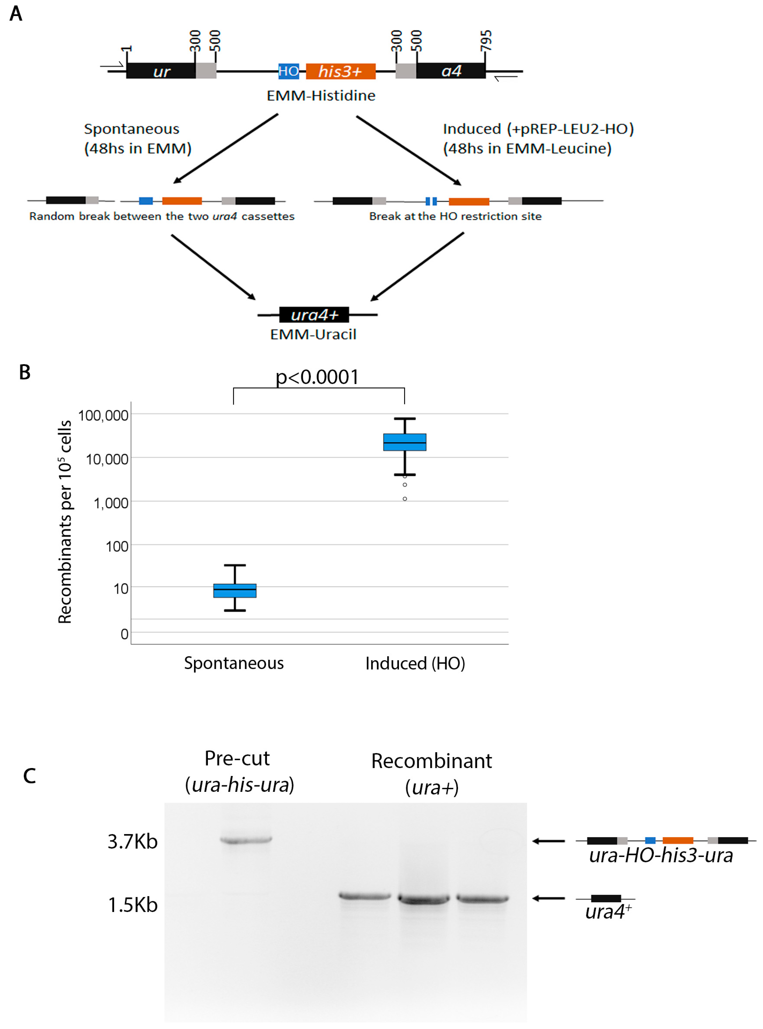
2.3. Characterization of the Assay

Spontaneous ura4+ his3 recombinants arise at an average frequency of approximately 1 in 104 cells (Figure 1B). As expected, when the break is made by the endonuclease, the frequency is much higher (2.5 in 10 colonies) (Figure 1B). This assay does not appear to report conversion (e.g., ura4+ his3+ colonies). PCR analysis of several recombinants with primers flanking the ura4+ ORF showed that the ura-his-ura cassette has been converted to ura4+ (Figure 1C). Sporadically, we did find some colonies that were ura4+ his3+, which appeared at a much lower frequency and only when we induced the break (Supplemental Figure S1A). To understand what these ura4+ his3+ colonies were, we used PCR to check the size of the locus in the HO-induced recombinant colonies (Supplementary Figure S1B). When primers flanking the ura4+ ORF are used, we found that both the ura4+ and the ura4+ his3+ are the same size—indicating that both are deletion outcomes. Next, we checked whether the ura4+ his3+ colonies arose as a result of gene conversion between the his3+ in our assay and the his3-D1 locus [30]. PCR across the his3+ locus showed that the his3-D1 deletion is present in both the ura-his-ura (pre) and the recombinant ura4+ his3+ colonies, suggesting that the his3-D1 allele has not been converted to his3+ (WT) (Supplementary Figure S1C). However, PCR with primers within the ORF his3+ detected the presence of the his3+ ORF in the ura4+ his3+ strains. We concluded from this PCR analysis and the very low frequency of the ura4+ his3+ recombinants that the his3+ must arise due to some spurious integration of the ORF elsewhere in the genome. Thus, this assay can be primarily used to test deletions.

2.4. Strains

The strains used in this study are listed in Supplementary Table S1. The construction of the ura-his-ura recombination cassette has been described previously [27]. This cassette has been slightly modified by cloning the S. cerevisiae HO endonuclease restriction site immediately upstream of the his3+ gene, as described above. The HO restriction site sequence is identical to that described in [28]. Standard yeast genetics have been used to cross the recombination mutants with the ura-his-ura cassette.

2.5. PCR Analysis

The primers for the PCR in Figure 1C were, 5′-agctacaaatcccactggct-3′ and 5′-tgatattgacgaaacttttt-3′. PCR was performed using Phusion® High-Fidelity DNA polymerase (NEB) and GC buffer at 55 °C annealing temperature (34 cycles).

2.6. Data Analysis

For all assays, the data were adjusted for viability and error in plating using the numbers on the YES plates (# colonies EMM-Uracil/(# colonies on YES/1000). The number “1000” represents the colonies intended to be plated on the YES plate. For example, if only 900 colonies appeared on the YES plate, then 900/1000 = 0.9 efficiency of plating. This means that a 10% error was made and the division of EMM-Uracil colonies by 0.9 normalizes the numbers to 100% plating efficiency. Thus, the YES plate serves as a control for both viability and plating errors and normalizes all experiments. This normalization was also important in order to control for systematic errors that might have been introduced as different people did the experiments, and is not unlike normalizations used previously [27,31]. The resulting value was then multiplied by the dilution factor so that the results were normalized to a recombination frequency of 105 for spontaneous breaks and 104 for induced breaks. Descriptive statistics and graphs were generated using SPSS.

3. Genetic Validation of the Assay and Discussion

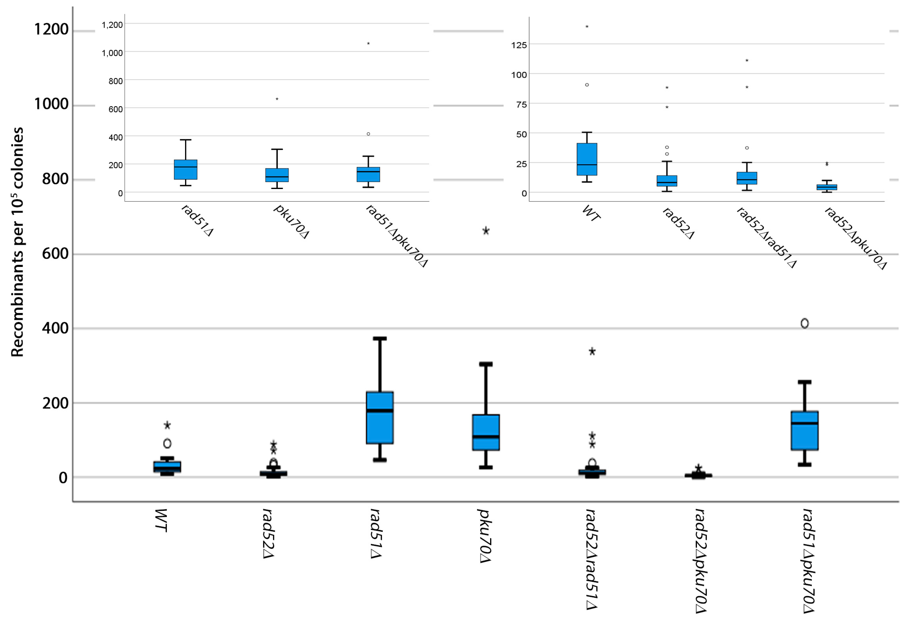
3.1. Analysis of Spontaneous Breaks

We next carried out some preliminary characterization of the genetic requirements for these deletions (Figure 2). We found that rad52+ is required for spontaneous breaks but rad51+ and pku70+ are not (Figure 2, Supplementary Tables S2 and S3). In fact, both rad51+ and pku70+ appeared to inhibit deletion outcomes. These results suggest that spontaneous break repair relies on a pathway that requires both rad51+ and pku70+. rad51+ has been previously shown to suppress chromosomal rearrangements in S. pombe arising from improperly repaired spontaneous breaks [25,32]. rad51+ is also not required for deletion outcomes, but is essential for gene conversion [33]. This indicates that spontaneous break repair is initially channeled through a homologous recombination pathway that requires rad51+, presumably by attempting to initiate a crossover. However, this pathway is not very efficient, most likely because the break occurs between direct repeats—a process which favors repair by single-strand annealing. Mechanisms of repair of spontaneous breaks by single-strand annealing that do not rely on rad51+ have been proposed in S. pombe [34].
The loss of pku70+ also increases recombination outcomes arising from spontaneous breaks, indicating that pku70+ suppresses these deletions as well. Recent evidence in S. pombe shows that pku70+ controls resection at stalled replication forks [35]. The loss of pku70+ leads to an increase in the resection tract but a decrease in Rad51 binding. These two events combined suggest that pku70+ and rad51+ work in the same pathway and in the repair of direct repeats in our assay. This repair pathway most likely occurs through single-strand annealing when either pku70+ or rad51+ is lost.
In higher eukaryotes and fission yeast, rad52+ is not essential for all forms of homologous recombination repair [36,37]. The fact that some repair still occurs in the absence of rad52+ indicates that, at a low percentage, ura4+ may be reconstituted by some other form of repair that does not rely on rad52+. These genetic results validate that this assay reports deletions only.
Sometimes the deletion of rad52+ in S. pombe acquires a suppressor that attenuates the sensitivity of the strains to DNA-damaging drugs. However, these results are not due to the effect of such a suppressor, because all strains used here are still sensitive to methyl–methanosulfonate (MMS) (Supplementary Figure S2).

3.2. Analysis of Induced Breaks

Next, we validated the genetic requirements for induced breaks. Because the HO endonuclease is expressed from a plasmid behind the nmt1 promoter, we used the following controls: 1) a control with thiamine to check whether the promoter is leaky and 2) a vector control which should give a frequency comparable to the spontaneous frequency (Figure 3, Supplementary Table S4). We found that recombinants appeared at about three orders of magnitude higher when the break was induced. The addition of thiamine drastically reduced the recombination frequency—albeit not to the spontaneous levels, suggesting that there is some promoter leakage. The vector control gives spontaneous level recombinants.
Both rad52+ and rad51+ are required for recombinants arising from induced breaks, suggesting that induced breaks are likely repaired through a crossover—either intrachromosomal or unequal sister chromatid exchange. As previously shown [38,39], pku70+ antagonizes recombination and, not unexpectedly, we also show that the deletion of pku70+ increases recombination outcomes. However, the function of pku70+ here is distinct from its function at spontaneous breaks, because it does not have the same phenotype as rad51+. The small increase in the frequency of recombinants in the absence of pku70+ suggests that the cell attempts to repair some of the generated two-ended breaks through NHEJ, which does not result in a ura4+ phenotype. Non-homologous end-joining and pku70+ have been previously shown to compete with recombination [29,38,39]. Because we select for ura4+, these cells will die. The requirement of pku70+ for the induced breaks, but not rad51+, may be explained by the stage of the cell cycle where the breaks are repaired. We presume that spontaneous breaks are generated as a consequence of DNA replication and may be repaired in S-phase. HO-induced breaks cause a cell cycle delay due to activation of the rad3+-dependent checkpoint [29]. It is therefore possible that induced breaks pile up in G2, while spontaneous breaks are repaired in S-phase. These results suggest that the mechanism of repair of induced breaks is distinct from that of spontaneous breaks.

4. Conclusions

Here we describe an assay to study intra-chromosomal deletions arising at regions of non-tandem repeats and provide preliminary data that show that induced breaks have different genetic requirements than spontaneous breaks—in agreement with what has been previously shown. We believe that this assay will be an important tool in the field of DNA damage repair to study deletions.
It is also worth noting that some cell cycle regulators, such as CDKN2A, are inactivated in cancer cells by deletion of the entire gene rather than by point mutations [40,41]. This shows that these intra-chromosomal deletions could introduce enough genetic change in human cells that may cause cancer. Since there is enough conservation in repair genes between yeast and human cells, this assay could be used to leverage the powerful yeast genetics to identify the mechanisms of these intra-chromosomal deletions.

Supplementary Materials

The following are available online at https://www.mdpi.com/2409-9279/2/3/74/s1, Figure S1: Analysis of Ura+His+ recombinants, Figure S2: Sensitivity of strains to MMS, Table S1: Strains used in this study, Table S2: Descriptive statistics for spontaneous breaks when cells were released in Edinburgh Minimal Media and platted on EMM-Uracil with Phloxin B, Table S3: Descriptive statistics for spontaneous breaks when cells were released in Edinburgh Minimal Media and platted on EMM-Uracil without Phloxin B, Table S4: Descriptive statistics for induced breaks when cells were released in Edinburgh Minimal Media.

Author Contributions

Conceptualization, methodology, software, R.C.P.; validation, R.C.P., B.E.L., M.T.M.; formal analysis, R.C.P.; investigation, B.E.L., M.T.M., T.M.H., L.N.W., J.M.K., K.G.H., S.R.F., W.M.D., P.D.R., H.M.H., M.A.B., P.N.M.; resources, R.C.P., J.-Q.W.; data curation, RCP; writing—original draft preparation, RCP; writing—review and editing, R.C.P., J.-Q.W.; visualization, R.C.P.; supervision, R.C.P., J.-Q.W.; project administration, R.C.P.; funding acquisition, R.C.P.

Funding

This work was supported by the National Institutes of Health [grant number RO3 CA223545-01].

Acknowledgments

Bailey E. Lucas is an Ohio State University Cancer Center Pelotonia Undergraduate Fellow. We thank James and Ellen Bazzoli for their monetary sponsorship of our laboratory.

Conflicts of Interest

The authors declare no conflict of interest.

References

  1. Janssen, A.; van der Burg, M.; Szuhai, K.; Kops, G.J.; Medema, R.H. Chromosome segregation errors as a cause of DNA damage and structural chromosome aberrations. Science 2011, 333, 1895–1898. [Google Scholar] [CrossRef] [PubMed]
  2. Lasko, D.; Cavenee, W.; Nordenskjold, M. Loss of constitutional heterozygosity in human cancer. Annu. Rev. Genet 1991, 25, 281–314. [Google Scholar] [CrossRef] [PubMed]
  3. Ortega, V.; Chaubey, A.; Mendiola, C.; Ehman, W., Jr.; Vadlamudi, K.; Dupont, B.; Velagaleti, G. Complex Chromosomal Rearrangements in B-Cell Lymphoma: Evidence of Chromoanagenesis? A Case Report. Neoplasia 2016, 18, 223–228. [Google Scholar] [CrossRef] [Green Version]
  4. Mehta, A.; Haber, J.E. Sources of DNA double-strand breaks and models of recombinational DNA repair. Cold Spring Harb. Perspect. Biol. 2014, 6, a016428. [Google Scholar] [CrossRef] [PubMed]
  5. Nikolov, I.; Taddei, A. Linking replication stress with heterochromatin formation. Chromosoma 2016, 125, 523–533. [Google Scholar] [CrossRef] [PubMed]
  6. Tabancay, A.P., Jr.; Forsburg, S.L. Eukaryotic DNA replication in a chromatin context. Curr. Top Dev. Biol. 2006, 76, 129–184. [Google Scholar]
  7. Prado, F.; Aguilera, A. Impairment of replication fork progression mediates RNA polII transcription-associated recombination. EMBO J. 2005, 24, 1267–1276. [Google Scholar] [CrossRef] [Green Version]
  8. Cavalier-Smith, T. Origins of the machinery of recombination and sex. Heredity (Edinb) 2002, 88, 125–141. [Google Scholar] [CrossRef]
  9. Mortensen, U.H.; Lisby, M.; Rothstein, R. Rad52. Curr. Biol. 2009, 19, R676–R677. [Google Scholar] [CrossRef] [Green Version]
  10. Symington, L.S. Role of RAD52 epistasis group genes in homologous recombination and double-strand break repair. Microbiol. Mol. Biol. Rev. 2002, 66, 630–670. [Google Scholar] [CrossRef]
  11. Chang, H.H.Y.; Pannunzio, N.R.; Adachi, N.; Lieber, M.R. Non-homologous DNA end joining and alternative pathways to double-strand break repair. Nat. Rev. Mol. Cell Biol. 2017, 18, 495–506. [Google Scholar] [CrossRef] [PubMed]
  12. Symington, L.S.; Gautier, J. Double-strand break end resection and repair pathway choice. Annu. Rev. Genet. 2011, 45, 247–271. [Google Scholar] [CrossRef] [PubMed]
  13. Aylon, Y.; Liefshitz, B.; Kupiec, M. The CDK regulates repair of double-strand breaks by homologous recombination during the cell cycle. EMBO J. 2004, 23, 4868–4875. [Google Scholar] [CrossRef] [PubMed] [Green Version]
  14. Barlow, J.H.; Lisby, M.; Rothstein, R. Differential regulation of the cellular response to DNA double-strand breaks in G1. Mol. Cell 2008, 30, 73–85. [Google Scholar] [CrossRef]
  15. Clerici, M.; Mantiero, D.; Guerini, I.; Lucchini, G.; Longhese, M.P. The Yku70-Yku80 complex contributes to regulate double-strand break processing and checkpoint activation during the cell cycle. EMBO Rep. 2008, 9, 810–818. [Google Scholar] [CrossRef] [PubMed]
  16. Ira, G.; Bressan, D.; Wan, L.; Hollingsworth, N.M.; Haber, J.E.; Foiani, M. DNA end resection, homologous recombination and DNA damage checkpoint activation require CDK1. Nature 2004, 431, 1011–1017. [Google Scholar] [CrossRef] [PubMed] [Green Version]
  17. Caspari, T.; Murray, J.M.; Carr, A.M. Cdc2-cyclin B kinase activity links Crb2 and Rqh1-topoisomerase III. Genes Dev. 2002, 16, 1195–1208. [Google Scholar] [CrossRef]
  18. Fortunato, E.A.; Osman, F.; Subramani, S. Analysis of spontaneous and double-strand break-induced recombination in rad mutants of S. pombe. Mutat. Res. 1996, 364, 14–60. [Google Scholar] [CrossRef]
  19. Cullen, J.K.; Prudden, J.; Wee, B.Y.; Davé, A.; Findlay, J.S.; Savory, A.P.; Humphrey, T.C. Break-induced loss of heterozygosity in fission yeast: Dual roles for homologous recombination in promoting translocations and preventing de novo telomere addition. Mol. Cell Biol. 2007, 27, 7745–7757. [Google Scholar] [CrossRef]
  20. Pierce, A.J.; Johnson, R.D.; Thompson, L.H.; Jasin, M. XRCC3 promotes homology-directed repair of DNA damage in mammalian cells. Genes Dev. 1999, 13, 2633–2638. [Google Scholar] [CrossRef] [Green Version]
  21. Stark, J.M.; Pierce, A.J.; Oh, J.; Pastink, A.; Jasin, M. Genetic steps of mammalian homologous repair with distinct mutagenic consequences. Mol. Cell Biol. 2004, 24, 9305–9316. [Google Scholar] [CrossRef]
  22. Sugawara, N.; Haber, J.E. Repair of DNA double strand breaks: In vivo biochemistry. Methods Enzymol. 2006, 408, 416–429. [Google Scholar]
  23. Gonzalez-Barrera, S.; Cortés-Ledesma, F.; Wellinger, R.E.; Aguilera, A. Equal sister chromatid exchange is a major mechanism of double-strand break repair in yeast. Mol. Cell 2003, 11, 1661–1671. [Google Scholar] [CrossRef]
  24. Liddell, L.; Manthey, G.; Pannunzio, N.; Bailis, A. Quantitation and analysis of the formation of HO-endonuclease stimulated chromosomal translocations by single-strand annealing in Saccharomyces cerevisiae. J. Vis. Exp. 2011, 55, 3150. [Google Scholar] [CrossRef]
  25. McDonald, J.P.; Rothstein, R. Unrepaired heteroduplex DNA in Saccharomyces cerevisiae is decreased in RAD1 RAD52-independent recombination. Genetics 1994, 137, 393–405. [Google Scholar]
  26. Osman, F.; Fortunato, E.A.; Subramani, S. Double-strand break-induced mitotic intrachromosomal recombination in the fission yeast Schizosaccharomyces pombe. Genetics 1996, 142, 341–357. [Google Scholar]
  27. Li, P.C.; Petreaca, R.C.; Jensen, A.; Yuan, J.P.; Green, M.D.; Forsburg, S.L. Replication fork stability is essential for the maintenance of centromere integrity in the absence of heterochromatin. Cell Rep. 2013, 3, 638–645. [Google Scholar] [CrossRef]
  28. Du, L.L.; Nakamura, T.M.; Moser, B.A.; Russell, P. Retention but not recruitment of Crb2 at double-strand breaks requires Rad1 and Rad3 complexes. Mol. Cell Biol. 2003, 23, 6150–6158. [Google Scholar] [CrossRef]
  29. Prudden, J.; Evans, J.S.; Hussey, S.P.; Deans, B.; O’Neill, P.; Thacker, J.; Humphrey, T. Pathway utilization in response to a site-specific DNA double-strand break in fission yeast. EMBO J. 2003, 22, 1419–1430. [Google Scholar] [CrossRef] [Green Version]
  30. Burke, J.D.; Gould, K.L. Molecular cloning and characterization of the Schizosaccharomyces pombe his3 gene for use as a selectable marker. Mol. Gen Genet. 1994, 242, 169–176. [Google Scholar] [CrossRef]
  31. Osman, F.; Whitby, M.C. Monitoring homologous recombination following replication fork perturbation in the fission yeast Schizosaccharomyces pombe. Methods Mol. Biol. 2009, 521, 535–552. [Google Scholar]
  32. Nakamura, K.; Shitanda, T.; Kaweeteerawat, C.; Takahashi, T.S.; Itoh, T.; Shirahige, K.; Masukata, H.; Nakagawa, T. Rad51 suppresses gross chromosomal rearrangement at centromere in Schizosaccharomyces pombe. EMBO J. 2008, 27, 3036–3046. [Google Scholar] [CrossRef] [Green Version]
  33. Doe, C.L.; Osman, F.; Dixon, J.; Matthew, C. WhitbyDNA repair by a Rad22-Mus81-dependent pathway that is independent of Rhp51. Nucleic Acids Res. 2004, 32, 5570–5581. [Google Scholar] [CrossRef]
  34. Onaka, A.T. Rad51 and Rad54 promote noncrossover recombination between centromere repeats on the same chromatid to prevent isochromosome formation. Nucleic Acids Res. 2016, 44, 10744–10757. [Google Scholar] [CrossRef] [Green Version]
  35. Teixeira-Silva, A. The end-joining factor Ku acts in the end-resection of double strand break-free arrested replication forks. Nat. Commun. 2017, 8, 1982. [Google Scholar] [CrossRef]
  36. Osman, F.; Adriance, M.; McCready, S. The genetic control of spontaneous and UV-induced mitotic intrachromosomal recombination in the fission yeast Schizosaccharomyces pombe. Curr. Genet. 2000, 38, 113–125. [Google Scholar] [CrossRef]
  37. Yamaguchi-Iwai, Y. Homologous recombination, but not DNA repair, is reduced in vertebrate cells deficient in RAD52. Mol. Cell Biol. 1998, 18, 6430–6435. [Google Scholar] [CrossRef]
  38. Tomita, K.; Matsuura, A.; Caspari, T.; Carr, A.M.; Akamatsu, Y.; Iwasaki, H.; Mizuno, K.; Ohta, K.; Uritani, M.; Ushimaru, T. Competition between the Rad50 complex and the Ku heterodimer reveals a role for Exo1 in processing double-strand breaks but not telomeres. Mol. Cell Biol. 2003, 23, 5186–5197. [Google Scholar] [CrossRef]
  39. Ferreira, M.G.; Cooper, J.P. Two modes of DNA double-strand break repair are reciprocally regulated through the fission yeast cell cycle. Genes Dev. 2004, 18, 2249–2254. [Google Scholar] [CrossRef] [Green Version]
  40. Florl, A.R.; Schulz, W.A. Peculiar structure and location of 9p21 homozygous deletion breakpoints in human cancer cells. Genes Chromosomes Cancer 2003, 37, 141–148. [Google Scholar] [CrossRef]
  41. Frazao, L.; Carmo, M.; Mafra, M. BRAF V600E mutation and 9p21: CDKN2A/B and MTAP co-deletions Markers in the clinical stratification of pediatric gliomas. BMC Cancer 2018, 18, 1259. [Google Scholar] [CrossRef]
Figure 1. An assay to study spontaneous and induced double-strand breaks at regions of non-tandem repeats. (A). The ura-his-ura assay. In this assay, two non-functional ura4 alleles flank a functional his3+ allele. The ura4 alleles have 200 bps of identical overlapping sequences, creating two non-tandem repeats (gray areas). The S. cerevisiae homothallic endonuclease (HO) is cloned just upstream of the his3+ gene. The HO enzyme is on a LEU2 plasmid under the control of the nmt1 promoter which can be repressed with thiamine. Spontaneous ura4+his3 recombinants are assayed by growing cells in EMM+UraHisAdeLeu media for 48 h then plating on selective EMM-Uracil. Induced break recombinants are assayed by growing cells for 48 h in media without thiamine to de-repress the HO endonuclease, while maintaining selection for the plasmid (EMM-Leucine). Cells are then plated on EMM-Uracil. All experiments were performed at 32 °C. (B). Box plot showing the frequency of recombinants for both induced and spontaneous breaks. (C). PCR across the ura-his-ura cassette in both pre- and post-recombination strains. Half arrowheads in (A) show approximate positions of primers.
Mps 02 00074 g001
Figure 2. Genetic requirements for spontaneous breaks. Box plots showing the spontaneous recombination frequency per 105 colonies. Cells were grown on EMM-Uracil plates for 3–5 days at 32 °C. For clarity, insets are shown for strains with similar recombination frequencies.
Mps 02 00074 g002
Figure 3. Genetic requirements for induced breaks. Box plots showing the HO-endonuclease-induced recombination frequency per 104 colonies. Cells were grown on EMM-Uracil plates for 3–5 days at 32 °C. For clarity, insets are shown for strains with similar recombination frequencies.
Mps 02 00074 g003

