Article

Future of Dutch NGS-Based Newborn Screening: Exploring the Technical Possibilities and Assessment of a Variant Classification Strategy

by
Gea Kiewiet
1,*,†,
Dineke Westra
2,†,
Eddy N. de Boer
1,†,
Emma van Berkel
2,
Tom G. J. Hofste
2,
Martine van Zweeden
2,
Ronny C. Derks
2,
Nico F. A. Leijsten
2,
Martina H. A. Ruiterkamp-Versteeg
2,
Bart Charbon
1,
Lennart Johansson
1,
Janneke Bos-Kruizinga
1,
Inge J. Veenstra
1,
Monique G. M. de Sain-van der Velden
3,
Els Voorhoeve
4,
M. Rebecca Heiner-Fokkema
5,
Francjan van Spronsen
6,
Birgit Sikkema-Raddatz
1,‡ and
Marcel Nelen
2,7,‡
1
Department of Genetics, University Medical Center Groningen, University of Groningen, 9713 GZ Groningen, The Netherlands
2
Department of Human Genetics, Radboud University Medical Center, 6500 HB Nijmegen, The Netherlands
3
Section Metabolic Diagnostics, Department of Genetics, University Medical Center Utrecht, 3584 CX Utrecht, The Netherlands
4
Centre for Health Protection, National Institute for Public Health and the Environment, 3720 BA Bilthoven, The Netherlands
5
Department of Laboratory Medicine, University Medical Center Groningen, University of Groningen, 9713 GZ Groningen, The Netherlands
6
Division of Metabolic Diseases, Beatrix Children’s Hospital, University Medical Center Groningen, University of Groningen, 9713 GZ Groningen, The Netherlands
7
Department of Genetics, University Medical Center Utrecht, 3584 CX Utrecht, The Netherlands
*
Author to whom correspondence should be addressed.
These authors contributed equally to this work.
Int. J. Neonatal Screen. 2024, 10(1), 20; https://doi.org/10.3390/ijns10010020
Submission received: 22 January 2024 / Revised: 19 February 2024 / Accepted: 1 March 2024 / Published: 7 March 2024

Abstract

:
In this study, we compare next-generation sequencing (NGS) approaches (targeted panel (tNGS), whole exome sequencing (WES), and whole genome sequencing (WGS)) for application in newborn screening (NBS). DNA was extracted from dried blood spots (DBS) from 50 patients with genetically confirmed inherited metabolic disorders (IMDs) and 50 control samples. One hundred IMD-related genes were analyzed. Two data-filtering strategies were applied: one to detect only (likely) pathogenic ((L)P) variants, and one to detect (L)P variants in combination with variants of unknown significance (VUS). The variants were filtered and interpreted, defining true/false positives (TP/FP) and true/false negatives (TN/FN). The variant filtering strategies were assessed in a background cohort (BC) of 4833 individuals. Reliable results were obtained within 5 days. TP results (47 patient samples) for tNGS, WES, and WGS results were 33, 31, and 30, respectively, using the (L)P filtering, and 40, 40, and 38, respectively, when including VUS. FN results were 11, 13, and 14, respectively, excluding VUS, and 4, 4, and 6, when including VUS. The remaining FN were mainly samples with a homozygous VUS. All controls were TN. Three BC individuals showed a homozygous (L)P variant, all related to a variable, mild phenotype. The use of NGS-based workflows in NBS seems promising, although more knowledge of data handling, automated variant interpretation, and costs is needed before implementation.

1. Introduction

Many countries worldwide use newborn screening (NBS) programs for treatable disorders [1,2]. The Dutch NBS program tests for 27 disorders, including 19 inherited metabolic disorders (IMDs) with a monogenetic origin [2]. Since the introduction of NBS, there has been ongoing demand for its expansion and for a reduction in the proportion of false positives (FP) [3]. However, including new disorders in the NBS program is labor-intensive, because disorder-specific tests need to be developed, while reducing FP remains challenging [4,5,6]. Furthermore, the biochemical testing approach hampers the inclusion of disorders that lack a biochemical footprint. As IMDs are hereditary, genetic testing using next-generation sequencing (NGS) techniques can overcome these shortcomings. NGS-based genetic testing is an all-in-one assay that can easily be modified to include new disorders, and NGS is already widely used in clinical care to diagnose patients with a suspected genetic condition [7,8,9,10,11]. Several recent studies also started to explore the use of NGS-based tests in NBS [12,13].
However, while the advantages of NGS-based genetic testing are widely recognized, many challenges remain [14,15]. One of these is selection of the most suitable NGS approach. In principle, targeted sequencing of a panel of genes (tNGS), whole-exome sequencing (WES), and whole-genome sequencing (WGS) are all reliable methods for detecting genetic disorders in a high-throughput manner, and tNGS and WES have proven their usefulness in the clinical setting. At the same time, tNGS, WES, and WGS differ in their ability to detect different types of variants, their turnaround times, and their costs. Furthermore, high-throughput data analysis, filtering, and automated variant interpretation would be necessary in NBS where fast turnaround times are needed, and the majority of newborns are healthy. In this study we explored three different NGS approaches for use in NBS: tNGS, WES, and WGS performed on DNA obtained from dried blood spots (DBS). The techniques were compared with respect to technical aspects, variant detection, interpretation, and turnaround times. We also assessed the variant classification strategy we used in a background cohort to identify the potential number of FP. The outcomes of this study provide recommendations for the implementation of a large-scale NGS-based approach.

2. Materials and Methods

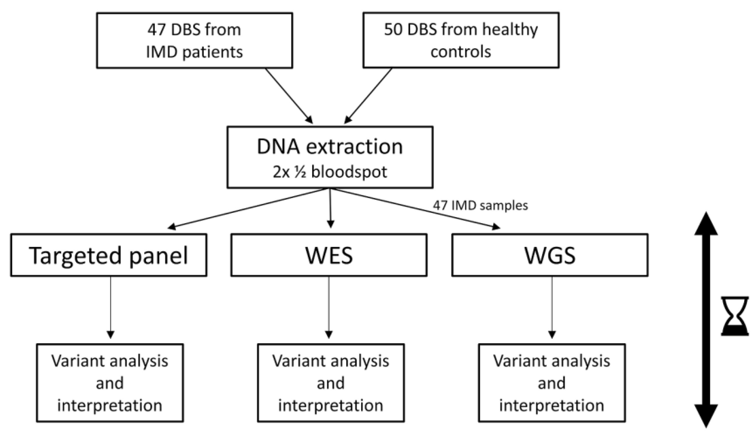
2.1. Experimental Setup

In total, 100 genes associated with 95 metabolic disorders were previously selected as candidates for inclusion in a NGS-based NBS (Supplementary Table S1) (Veldman et al. manuscript in preparation). Disease severity, treatability, and age of onset were taken as the main inclusion criteria. To test the performance of different NGS techniques for use in NBS, we compiled a test cohort of 100 DBS: 50 from patients with a genetically confirmed IMD and 50 from healthy controls. After DNA extraction (using 1 blood spot in total), tNGS and WES were performed on all samples. WGS was performed on the 50 DBS from patients. All analyses were carried out following standard procedures used in diagnostics (Figure 1), with both single nucleotide variants (SNVs) and copy number variants (CNVs) analyzed. After data processing and the application of a virtual panel to the WES and WGS data, variant filtering was performed. Variants were assessed and interpreted blindly, with the outcomes of the NGS approaches and previous genetic results then compared ‘post-blind’. To assess the SNV variant filtering strategy, it was retrospectively applied to WES data from a cohort of 4833 individuals (further referred to as the background cohort (BC)).

2.2. Sample Collection and DNA Extraction

Fifty DBS from patients with an IMD associated with one of the selected one hundred genes were kindly provided by the sample archives of the Laboratory of Metabolic Diseases of the University Medical Center Groningen (UMCG) and the Laboratory of Metabolic Diseases of the University Medical Center Utrecht. Samples were only included when no patient or parental non-consent for use for research purposes was registered in the research registries of the UMCs. The Ethical Review Board (ERB) of the UMCG has declared that this study does not fall under the Medical Research Involving Humans Act, so no further ERB approval was needed (METc 2021/611). In all patients, a genetic diagnosis that fits the aberrant metabolic results had been made in a clinical setting. Fifty DBS from randomly selected anonymous blood samples from individuals without a (known) IMD served as negative controls. Preparation of these DBS and DNA extraction from all DBS are described in Supplementary File S1. An existing WES dataset of 4833 anonymized (METc Radboud university medical center (number 2011-188 and 2020-7142)) individuals was included to compare the SNV results of the DBS in a BC representing a large background population. These BC data were collected previously in a diagnostic setting, coming from presumed healthy individuals who were included in trio-analysis (parents of an affected child). All BC individuals were born before the first IMDs, aside from phenylketonuria, became target diseases in the Dutch NBS in 2007.

2.3. Preparation of Samples, Sequencing, Data Filtering, and Classification

A detailed description of sample preparation and sequencing for all NGS techniques is provided in Supplementary File S1, as are the methods for alignment, coverage calculation and variant calling. The filtering strategies, which were applied to all data, are shown in Figure 2. To start, we applied a strict filtering strategy designed to only detect pathogenic (P) and likely pathogenic (LP) variants, according to recommendations for other genetic screening approaches [16] and in line with other studies [17]. We included filter steps that selected (1) variants conclusively described as P and LP in any of the following variant databases (either a disease-causing mutation in the Human Gene Mutation Database pro v.2022, (L)P in ClinVar23 (last update 6 October 2022), (L)P in the national database of the Dutch Society of Clinical Laboratory Genetics (December 2021 release, https://vkgl.molgeniscloud.org/ accessed on 1 May 2022), or (L)P in local databases), (2) truncating variants not described in databases (nonsense, frameshift, start/stoploss, canonical splice site; unless present downstream of the nonsense-mediated decay boundary) [18], and (3) variants with possible splicing effects (positions ± 3). Filtered variants also needed to have an allele frequency <1% (Figure 2A). The variants that remained after these filtering steps were evaluated by a clinical laboratory geneticist and classified according to the American College of Medical Genetics and Genomics (ACMG) guidelines [19], considering the mode of inheritance (MOI) of the associated disease. Afterwards, we extended the filtering strategy to obtain VUS from genes in which one P or LP variant with a variant allele frequency (VAF) of 40–60% was found in an autosomal recessive (AR) or X-linked recessive (XLR) gene (Figure 2B). All identified variants and combinations thereof were manually reviewed and, if necessary, reclassified.

2.4. Outcome of the Study

To compare the performance of the NGS workflows, we measured the following outcomes:
  • Technical performance of tNGS, WES, and WGS. The number of failed samples, overall coverage, and specific regions not covered >20x were reported.
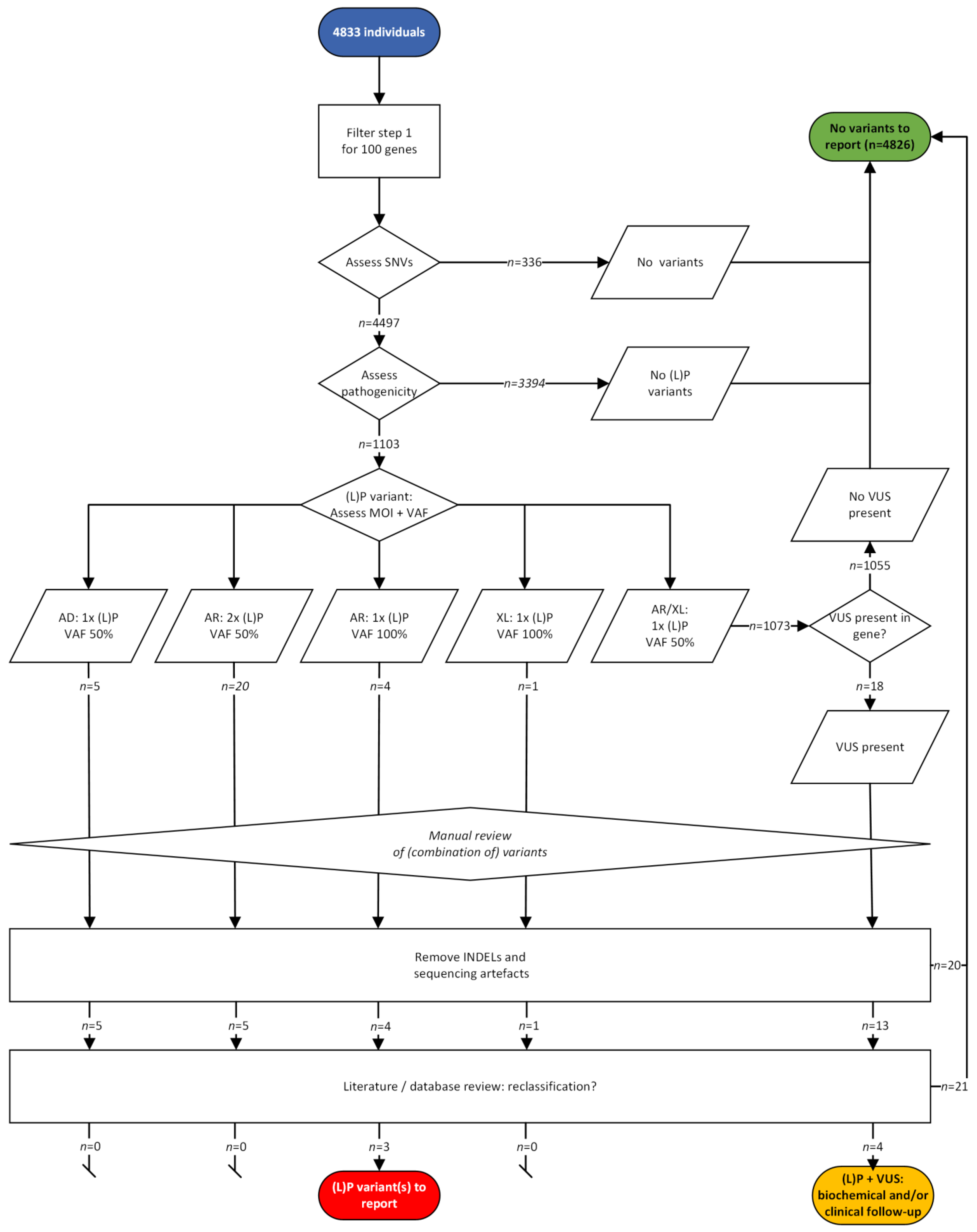
  • Outcomes of the two variant filtering strategies. First, we measured the number of variants using the strict filtering strategy reporting only P and LP variants ((L)P) filter strategy; Figure 2B, left). We then calculated the number of true positives (TP), true negatives (TN), FP and false negatives (FN) based on the definitions described in Supplementary File S1. Second, we tested a filtering strategy with an additional step (Figure 2B, right). When only one P or LP variant was found in an AR or XLR gene, we also reported VUS found in the same gene (extra VUS filter strategy). Here, we used the same definitions for TP/FP/FN/TN, but the samples in which one LP or P variant and a VUS were detected, were now considered positive. Carriership was defined as presence of one LP or P variant detected with a VAF of 50% in an AR gene in any sample.
  • Estimated turnaround time. For a series of 96 samples, we measured the time needed to obtain results. We included the time needed for automated sample preparation, sequencing, data processing, and data analysis. For tNGS data analysis, an automated variant interpretation pipeline was used to obtain relevant variants for each sample (https://github.com/molgenis/vip, accessed on 1 May 2022). For WES and WGS, downstream processing was performed using an automated data analysis pipeline and custom-made annotation [20], with a bioinformatic filter for the selected genes.

3. Results

3.1. Technical Performance

The DNA isolation procedure yielded an average 702.45 ng DNA per sample (range 393.4–3458.4 ng). No data could be obtained due to the failure of library preparation for 3/100 samples in all 3 approaches. All the DNA samples that failed were archival DBS. The overall coverage for tNGS and WES fulfilled local diagnostic standard requirements (>20x coverage for >95% of the target regions). For WGS, >85% (38/44) of samples had >20x coverage. An overview of the genome positions that did not comply with these requirements is shown, per method, in Supplementary File S2. The average read depths were 195 ± 74 for tNGS, 182.5 ± 36.7 for WES, and 31.4 ± 11.6 for WGS.

3.2. Filter Strategy Assessment

We calculated the average number of variants remaining after filtering using the strict (L)P filter strategy. On average, 2.4 variants (range 0–6) were found using tNGS, and 1.3 variants (range 0–3) were found using the WES and WGS workflow in the IMD-positive samples. In the control samples, a mean of 0.89 variants (range 0–4) were left after filtering using tNGS, and this mean was 0.17 (range 0–2) after WES-based analysis.
Using the strict (L)P filtering strategy (Figure 2A), 33 of the IMD samples were TP using the tNGS workflow, as were 31 when using WES and 30 when using WGS (Figure 2C). For WGS, 7/9 samples with low coverage (<20x) were correctly identified as TP. However, 11 (tNGS), 13 (WES), and 14 (WGS) samples were found to be FN. For both tNGS and WES, the 50 control samples were TN.
When we applied the VUS filter strategy (Figure 2B), 40 samples were TP in the tNGS and WES workflows, as were 38 in the WGS workflow (Figure 2C). Comparing these numbers with the TP numbers of the strict filtering strategy shows that the majority of FN samples were resolved using this strategy. Nonetheless, four (tNGS and WES) and six (WGS) samples were found to be FN. Table 1 provides a detailed overview of the variants detected per sample, technique, and filter strategy.
The additional use of the VUS filter strategy identified three samples (#31, #35, and #37) to be FN in all three workflows. The known variant in these samples was registered as a homozygous VUS in the databases used (Table 2). Sample #6 contained a disease-causing intronic variant, c.-149G>A in SLC22A5, that was only seen with tNGS, as non-coding regions (in this case the 5′UTR of the gene) were not included in the bioinformatic analysis for WES and WGS. Manual curation of the data, however, did identify the variant for WES and WGS. Variants in CBS (#3) and one variant in ABCD1 (#31) were not identified in the WGS workflow because of reduced coverage (<20x) and known pseudogenes that complicated the analysis for these genes.
Both of the known CNVs in the IMD-positive samples were detected in all workflows. However, the WES and WGS data analysis regarding CNVs failed in 14 of 47 samples due to reference pool issues.
For the BC (Figure 3), we first looked for the presence of any SNV in the coding and splicing region (MANE transcript [21], if applicable) of the 100 genes, and 336 individuals (7%) did not have any SNV in any of the genes. The pathogenicity of the SNVs present in the remaining 4497 individuals were evaluated via the above-mentioned strict strategy, and no (L)P variant was identified in 3394 individuals (70%). Considering the VAF and the MOI of the genes in which (L)P variants were found, the number or zygosity of the (L)P variants fitted the MOI in 30 individuals (0.6%). After reviewing these variants (excluding deletion–insertions located in cis, but called as separate events, sequencing artefacts, and variants not relevant based on predictions and literature), a homozygous pathogenic variant remained in three individuals in ACADVL, CPT1A, and MCCC2, respectively (Supplementary Table S2). These variants are associated with a variable phenotype that is mostly asymptomatic or mild (Supplementary Table S2) and not with severe disease.
In 1073 individuals (22%), only 1 (L)P with a VAF of 40–60% was seen in an AR or XLR gene. For these individuals, the VUS filter strategy was applied, which resulted in an extra 18 individuals with 1 (L)P variant and a VUS fitting the MOI of the gene. After reviewing these variants, a combination of (L)P and relevant VUS in an AR gene was seen in four individuals (Supplementary Table S3).

3.3. Carriership

In the 100 DBS samples, 28 carrierships were detected (Supplementary Table S4) with a maximum of 1 carriership per sample. In total, 23 carrierships were detected with tNGS, 12 with WES and 12 with WGS. The differences in detection were mostly due to the presence of a (L)P classified variant in the local database. Carriership was detected in 21 different genes, with the most variants (n = 2) detected in the PAH and SI genes. In sample #2, with two known variants in GCDH, the outcome remained inconclusive after the WES and WGS workflows because one GCDH variant was filtered out, while a variant (carriership) in BTD was found.

3.4. Turnaround Time

The workflow and observed turnaround time of the experiments are summarized in Figure 4. The procedure from library preparation through variant interpretation was performed in 5 working days for both tNGS and WES. Automated library preparation took two days, followed by approximately 28 h of sequencing. The following 36 h were devoted to data processing, after which variants were filtered and prioritized to obtain the relevant variants for each sample. On day 5, all variants were manually assessed by an experienced technician and clinical laboratory geneticist. WGS was performed in 4 days, with the main difference being the enrichment process needed for WES and tNGS.

4. Discussion

In this study we demonstrate that using NGS as a first tier in NBS is a promising and realistic approach. We show that all three NGS strategies perform equally well and that results can be reported within 4 to 5 working days. Even though no variants associated with a severe phenotype were present in the BC, the experimental setup clearly demonstrated the need for robust filter and interpretation strategies.
We detected no relevant differences between tNGS and WES regarding technical performance. Only a few samples failed, all of them from archived DBS, in line with data showing that DNA from archived DBS deteriorates over time, increasing the risk of failure [22]. However, implementation of robust quality control parameters will allow early detection, limiting failure rates in sequencing. The NGS procedure can, thus, be repeated with little delay. For WGS, further testing is needed to confirm that an average WGS coverage of ~30x provides the sensitivity required in NBS programs. CNV calling for the tNGS data using the 100 DBS samples as a reference pool was successful for all samples, while CNV calling for WES and WGS was hampered by the use of a reference pool of EDTA blood samples. This shows the importance of using a DBS-specific reference pool. Carriership of one (L)P variant was found in 28% of the samples but was not considered for reporting since this is not within the scope of NBS, which is identifying treatable disorders in newborns.
Our filter strategy assessment showed that only a limited number of variants per sample needed manual interpretation for both the DBS and the BC, using a strict (L)P filter strategy. Although the number of variants that needed to be interpreted was low, this process has to be optimized, as NBS screening procedures are carried out on large scales. One important aspect for reducing the number of variants is the need for a highly NBS-focused, curated, open access variant database. This need is emphasized by the small differences in outcome that we observed between NGS workflows due to differences in curated variants in local databases and to experimental setup (with, e.g., coverage of non-coding regions in tNGS, but not in WES/WGS). The Kingsmore et al. study that evaluated the pathogenicity of almost 30,000 variants in 317 genes is a good starting point for the development of such a database [17].
In current Dutch biochemical NBS, the FP rate is relatively high, at least for a selection of disorders, with an overall positive predictive value of 43% reported for 2021 [23,24,25]. This is because some biomarkers are influenced by, e.g., (maternal) diet, medication, and maternal metabolism [24]. This not only creates unnecessary anxiety in parents, but the current FP rate also leads to costs due to follow-up of newborns, including more healthcare consumption [26]. In our study, we observed a false positive rate of 0.06% (3/4883), when combining the negative DBS and the BC and using our strict filter strategy. The identified variants in ACADVL, CPT1A, and MCCC2 are associated with variable but mostly asymptomatic or mild phenotypes (Supplementary Table S2), and it is questionable whether they should be reported in NBS, as the severe disease form is the target disease. In a pre-implementation research setting, we suggest discussing the relevance of variants like these in an expert panel. Gained knowledge on these kind of variants can later be used to decide whether these variants should still be reported in NBS in the future. An interesting approach to further decreasing the number of FPs is to combine both biochemical and genetic techniques, usually with NGS as second tier, but also in a procedure where both NGS and biomarkers are used in a first-tier setting [27,28,29].
Reducing the number of FNs is a key goal in a genetics-first NBS. In our study, most of the cases contained one LP or P variant, as well as a VUS in the same gene. The percentage decreased to 6–9% when applying the VUS filter strategy, but these numbers are still high compared to the FN rate of current NBS. The key reason for this is the challenge of interpreting NGS data in the absence of any phenotypic information, a problem intrinsic to the screening setting. In the current study, variant classification was carried out according to current ACMG guidelines, i.e., what is known from various variant databases and the literature. This finding again emphasizes the importance of NBS-focused databases of curated variants. In addition, we need a biochemical and/or clinical evaluation of a (suspicious) VUS in a specific gene [30]. For many of the 95 IMD investigated, functional testing is possible (Arar et al., manuscript in preparation). Furthermore, the differences in sensitivity and specificity between biochemical methods and NGS highlight the need for a proper comparison between the yield of the two strategies when used as a first tier. It is possible that a combination of both methods will be optimal, at least for some disorders.
With our experiments, we demonstrated that automated, high-throughput NGS analysis of DBS is feasible. The NGS experiment, from library preparation to data interpretation, was carried out in a maximum of 5 days. In current Dutch NBS, the target is a first-tier result within 10 days after birth, with DBS sampling occurring on day 3 through day 7. With sampling at birth (DBS or buccal swab), NGS results can be obtained within the first week after birth. WGS offers both the fastest workflow and the possibility to use the complete genomic data.
However, before NGS in NBS can be implemented, several additional aspects need to be considered. One is testing capacity. Based on ~170,000 babies born annually in the Netherlands (and assuming almost 100% participation), we calculate a need to analyze >640 samples per day in a 5-day laboratory service. This requires 7 library prep runs per day (in a 96-well plate format). Sequencing would require multiple runs, depending on the strategy of choice. The most demanding sequencing strategy would be WGS (mean coverage 30x, 120 Gb per sample), which would require around ten NovaSeqX+ 25B flowcells (output 8 Tb) per day (which equals ten machines (dual flowcells)), as the runtime is 48 h. Another crucial aspect is the amount of sequencing data generated. Secure data storage capacity, as well as duration of storage and related costs for the different NGS techniques, needs further discussion [31]. Data analysis needs to be fully automated for all negative samples, in order to provide a fast result and to not require too much work from lab technicians and clinical laboratory geneticists. The current voluntary participation rate in NBS in the Netherlands is almost 100% [25]. To secure this for the future, investigation of the opinions of parents and the general population regarding storage and use of genetic data will be key for developing a widely supported plan for the implementation of NGS in NBS [31,32,33]. Thus, we emphasize that such ethical, social, and legal topics need attention in future studies, as well as studies on the cost-effectiveness of a genetics approach for NBS.
In conclusion, we show that all three NGS methods tested can be used to obtain reliable data from DBS samples within 5 days. Based on our results, data analysis strategies need to be optimized to enable a significant reduction in FN results. Assessment of the filter strategy when used in the BC resulted in a low number of FPs. Large cohort studies are needed for better variant classification and to learn more about large-scale data handling, turnaround times and costs involved. Based on this study, we feel that the use of NGS-based screening will benefit NBS, particularly for diseases with a high FP rate in current biochemical NBS. NGS implementation also comes with the flexibility to add new target diseases to the screening program, especially those without a biochemical marker.

Supplementary Materials

The following supporting information can be downloaded at: https://www.mdpi.com/article/10.3390/ijns10010020/s1, Supplementary File S1: Methods: Dried blood spots and DNA extraction, Design of the targeted panel, Preparation of samples and sequencing targeted panel, Preparation of samples and sequencing WES, Preparation of samples and sequencing WGS, Definitions of TP/TN/FP/FN in the (L)P filter strategy. Table S1: Genes and associated disorders included in gene panel. Table S2: Identified homozygous pathogenic variants in three individuals in the background population that would be reported in a NBS setting. Table S3: Identified combinations of a (L)P variant and VUS in four individuals in the background cohort that might need to be reported in a NBS setting. Table S4. List of heterozygous (likely) pathogenic variants in autosomal recessive genes, indicating carriership, detected with one or more NGS techniques. Supplementary File S2: overview of genome positions that did not comply with quality requirements per method.

Author Contributions

Conceptualization, G.K., D.W., E.N.d.B., E.V., M.R.H.-F., F.v.S., B.S.-R. and M.N.; Formal analysis, G.K., D.W., E.N.d.B., E.v.B., M.v.Z., R.C.D., N.F.A.L., M.H.A.R.-V., B.C., L.J., J.B.-K. and I.J.V.; Funding acquisition, F.v.S. and M.N.; Methodology, G.K., D.W., E.N.d.B. and T.G.J.H.; Resources, M.G.M.d.S.-v.d.V. and M.R.H.-F.; Software, B.C. and L.J.; Supervision, G.K., D.W., F.v.S., B.S.-R. and M.N.; Visualization, G.K. and D.W.; Writing—original draft, G.K.; Writing—review and editing, G.K., D.W., E.N.d.B., E.v.B., T.G.J.H., M.v.Z., R.C.D., N.F.A.L., M.H.A.R.-V., B.C., L.J., J.B.-K., I.J.V., M.G.M.d.S.-v.d.V., E.V., M.R.H.-F., F.v.S., B.S.-R. and M.N. All authors have read and agreed to the published version of the manuscript.

Funding

This study was funded by a ZonMW grant, 05430021810015. We also received support in the form of WGS kits from Illumina. The authors confirm their independence from the sponsors; the content of the article has not been influenced by the sponsors.

Institutional Review Board Statement

The Ethical Review Board (ERB) of the UMCG has declared that this study does not fall under the Medical Research Involving Humans Act, so no further ERB approval was needed (METc 2021/611).

Informed Consent Statement

Samples were only included when no patient or parental non-consent for use for research purposes was registered in the research registries of the UMCs, and the samples were completely anonymized.

Data Availability Statement

The data that support the findings of this study are available from the corresponding author, G.K., upon reasonable request. Raw data files are not available due to privacy issues related to genetic data.

Conflicts of Interest

L.J. is involved in the European Union’s Horizon 2020 research and innovation programs EJP-RD (H2020 825575), in collaboration with the Solve-RD (H2020 779257) and CINECA (H2020 825775) projects, as well as the Dutch NWA.1332.20.006 ELSA AI lab Northern Netherlands (ELSA-NN) project. F.v.S. received grants paid to the university from BioMarin, ESPKU, NPKUA, NPKUV, Nutricia, received consultancy fees paid to the university from APR, Axcella, Alexion, BioMarin, and Pluvia Biotech, is involved in the clinical trials PTC, is a member of the Data Safety Monitoring Board of Sanofi, received lecture fees paid to the university from BioMarin, Nutricia, and Vitaflo, and receives shares/financials from NAlex. Furthermore, he is chair and member of the Dutch advisory committee for newborn screening for metabolic diseases, Dutch advisory committee for the RIVM on newborn screening and the working group on research in relation to newborn screening. He is also the chair and a member of the scientific advisory committees of the following patient organizations: European Society for PKU and allied disorders, Dutch PKU society, Tyrosinemia Foundation, and the scientific advisory committee for the National PKU Alliance USA. All the other authors declare no conflicts of interest. The funder had no role in in the design of the study; in the collection, analyses, or interpretation of data; in the writing of the manuscript, or in the decision to publish the results.

References

  1. Therrell, B.L.; Padilla, C.D.; Loeber, J.G.; Kneisser, I.; Saadallah, A.; Borrajo, G.J.C.; Adams, J. Current Status of Newborn Screening Worldwide: 2015. Semin. Perinatol. 2015, 39, 171–187. [Google Scholar] [CrossRef] [PubMed]
  2. Castiñeras, D.E.; Couce, M.-L.; Marín, J.L.; González-Lamuño, D.; Rocha, H. Newborn Screening for Metabolic Disorders in Spain and Worldwide. An. Pediatría 2019, 91, 128e.1–128e.14. [Google Scholar] [CrossRef]
  3. van Dijk, T.; Kater, A.; Jansen, M.; Dondorp, W.J.; Blom, M.; Kemp, S.; Langeveld, M.; Cornel, M.C.; van der Pal, S.M.; Henneman, L. Expanding Neonatal Bloodspot Screening: A Multi-Stakeholder Perspective. Front. Pediatr. 2021, 9, 706394. [Google Scholar] [CrossRef] [PubMed]
  4. Vasquez-Loarte, T.; Thompson, J.D.; Merritt, J.L. Considering Proximal Urea Cycle Disorders in Expanded Newborn Screening. Int. J. Neonatal Screen. 2020, 6, 77. [Google Scholar] [CrossRef] [PubMed]
  5. Stroek, K.; Boelen, A.; Bouva, M.J.; De Sain-van der Velden, M.; Schielen, P.C.J.I.; Maase, R.; Engel, H.; Jakobs, B.; Kluijtmans, L.A.J.; Mulder, M.F.; et al. Evaluation of 11 Years of Newborn Screening for Maple Syrup Urine Disease in the Netherlands and a Systematic Review of the Literature: Strategies for Optimization. JIMD Rep. 2020, 54, 68–78. [Google Scholar] [CrossRef]
  6. Derks, T.G.J.; Boer, T.S.; Assen, A.; Bos, T.; Ruiter, J.; Waterham, H.R.; Niezen-Koning, K.E.; Wanders, R.J.A.; Rondeel, J.M.M.; Loeber, J.G.; et al. Neonatal Screening for Medium-Chain Acyl-CoA Dehydrogenase (MCAD) Deficiency in The Netherlands: The Importance of Enzyme Analysis to Ascertain True MCAD Deficiency. J. Inherit. Metab. Dis. 2008, 31, 88–96. [Google Scholar] [CrossRef]
  7. French, C.E.; Delon, I.; Dolling, H.; Sanchis-Juan, A.; Shamardina, O.; Mégy, K.; Abbs, S.; Austin, T.; Bowdin, S.; Branco, R.G.; et al. Whole Genome Sequencing Reveals That Genetic Conditions Are Frequent in Intensively Ill Children. Intensive Care Med. 2019, 45, 627–636. [Google Scholar] [CrossRef]
  8. Meng, L.; Pammi, M.; Saronwala, A.; Magoulas, P.; Ghazi, A.R.; Vetrini, F.; Zhang, J.; He, W.; Dharmadhikari, A.V.; Qu, C.; et al. Use of Exome Sequencing for Infants in Intensive Care Units: Ascertainment of Severe Single-Gene Disorders and Effect on Medical Management. JAMA Pediatr. 2017, 171, e173438. [Google Scholar] [CrossRef]
  9. de Ligt, J.; Willemsen, M.H.; van Bon, B.W.M.; Kleefstra, T.; Yntema, H.G.; Kroes, T.; Vulto-van Silfhout, A.T.; Koolen, D.A.; de Vries, P.; Gilissen, C.; et al. Diagnostic Exome Sequencing in Persons with Severe Intellectual Disability. N. Engl. J. Med. 2012, 367, 1921–1929. [Google Scholar] [CrossRef]
  10. Neveling, K.; Feenstra, I.; Gilissen, C.; Hoefsloot, L.H.; Kamsteeg, E.J.; Mensenkamp, A.R.; Rodenburg, R.J.T.; Yntema, H.G.; Spruijt, L.; Vermeer, S.; et al. A Post-Hoc Comparison of the Utility of Sanger Sequencing and Exome Sequencing for the Diagnosis of Heterogeneous Diseases. Hum. Mutat. 2013, 34, 1721–1726. [Google Scholar] [CrossRef]
  11. Olde Keizer, R.A.C.M.; Marouane, A.; Kerstjens-Frederikse, W.S.; Deden, A.C.; Lichtenbelt, K.D.; Jonckers, T.; Vervoorn, M.; Vreeburg, M.; Henneman, L.; de Vries, L.S.; et al. Rapid Exome Sequencing as a First-Tier Test in Neonates with Suspected Genetic Disorder: Results of a Prospective Multicenter Clinical Utility Study in the Netherlands. Eur. J. Pediatr. 2023, 182, 2683–2692. [Google Scholar] [CrossRef]
  12. Holm, I.A.; Agrawal, P.B.; Ceyhan-Birsoy, O.; Christensen, K.D.; Fayer, S.; Frankel, L.A.; Genetti, C.A.; Krier, J.B.; LaMay, R.C.; Levy, H.L.; et al. The BabySeq Project: Implementing Genomic Sequencing in Newborns. BMC Pediatr. 2018, 18, 225. [Google Scholar] [CrossRef]
  13. van Campen, J.C.; Sollars, E.S.A.; Thomas, R.C.; Bartlett, C.M.; Milano, A.; Parker, M.D.; Dawe, J.; Winship, P.R.; Peck, G.; Grafham, D.; et al. Next Generation Sequencing in Newborn Screening in the United Kingdom National Health Service. Int. J. Neonatal Screen. 2019, 5, 40. [Google Scholar] [CrossRef]
  14. Burton, B.K.; Veldman, A.; Kiewiet, M.B.G.; Rebecca Heiner-Fokkema, M.; Nelen, M.R.; Sinke, R.J.; Sikkema-Raddatz, B.; Voorhoeve, E.; Westra, D.; Dollé, M.E.T.; et al. Neonatal Screening Towards Next-Generation Sequencing (NGS)-Based Newborn Screening: A Technical Study to Prepare for the Challenges Ahead. Int. J. Neonatal Screen. 2022, 8, 17. [Google Scholar] [CrossRef]
  15. Spiekerkoetter, U.; Bick, D.; Scott, R.; Hopkins, H.; Krones, T.; Gross, E.S.; Bonham, J.R. Genomic Newborn Screening: Are We Entering a New Era of Screening? J. Inherit. Metab. Dis. 2023, 46, 778–795. [Google Scholar] [CrossRef] [PubMed]
  16. Gregg, A.R.; Aarabi, M.; Klugman, S.; Leach, N.T.; Bashford, M.T.; Goldwaser, T.; Chen, E.; Sparks, T.N.; Reddi, H.V.; Rajkovic, A.; et al. Screening for Autosomal Recessive and X-Linked Conditions during Pregnancy and Preconception: A Practice Resource of the American College of Medical Genetics and Genomics (ACMG). Genet. Med. 2021, 23, 1793–1806. [Google Scholar] [CrossRef] [PubMed]
  17. Kingsmore, S.F.; Smith, L.D.; Kunard, C.M.; Bainbridge, M.; Batalov, S.; Benson, W.; Blincow, E.; Caylor, S.; Chambers, C.; del Angel, G.; et al. A Genome Sequencing System for Universal Newborn Screening, Diagnosis, and Precision Medicine for Severe Genetic Diseases. Am. J. Hum. Genet. 2022, 109, 1605–1619. [Google Scholar] [CrossRef] [PubMed]
  18. Nguyen, L.S.; Wilkinson, M.F.; Gecz, J. Nonsense-Mediated MRNA Decay: Inter-Individual Variability and Human Disease. Neurosci. Biobehav. Rev. 2014, 46, 175–186. [Google Scholar] [CrossRef] [PubMed]
  19. Richards, S.; Aziz, N.; Bale, S.; Bick, D.; Das, S.; Gastier-Foster, J.; Grody, W.W.; Hegde, M.; Lyon, E.; Spector, E.; et al. Standards and Guidelines for the Interpretation of Sequence Variants: A Joint Consensus Recommendation of the American College of Medical Genetics and Genomics and the Association for Molecular Pathology. Genet. Med. 2015, 17, 405–424. [Google Scholar] [CrossRef] [PubMed]
  20. Lelieveld, S.H.; Reijnders, M.R.F.; Pfundt, R.; Yntema, H.G.; Kamsteeg, E.J.; De Vries, P.; De Vries, B.B.A.; Willemsen, M.H.; Kleefstra, T.; Löhner, K.; et al. Meta-Analysis of 2104 Trios Provides Support for 10 New Genes for Intellectual Disability. Nat. Neurosci. 2016, 19, 1194–1196. [Google Scholar] [CrossRef]
  21. Morales, J.; Pujar, S.; Loveland, J.E.; Astashyn, A.; Bennett, R.; Berry, A.; Cox, E.; Davidson, C.; Ermolaeva, O.; Farrell, C.M.; et al. A Joint NCBI and EMBL-EBI Transcript Set for Clinical Genomics and Research. Nature 2022, 604, 310–315. [Google Scholar] [CrossRef]
  22. Hwang, J.; Jaroensuk, J.; Leimanis, M.L.; Russell, B.; McGready, R.; Day, N.; Snounou, G.; Nosten, F.; Imwong, M. Long-Term Storage Limits PCR-Based Analyses of Malaria Parasites in Archival Dried Blood Spots. Malar. J. 2012, 11, 339. [Google Scholar] [CrossRef]
  23. Tangeraas, T.; Sæves, I.; Klingenberg, C.; Jørgensen, J.; Kristensen, E.; Gunnarsdottir, G.; Hansen, E.V.; Strand, J.; Lundman, E.; Ferdinandusse, S.; et al. Performance of Expanded Newborn Screening in Norway Supported by Post-Analytical Bioinformatics Tools and Rapid Second-Tier DNA Analyses. Int. J. Neonatal Screen. 2020, 6, 51. [Google Scholar] [CrossRef]
  24. Malvagia, S.; Forni, G.; Ombrone, D.; la Marca, G. Development of Strategies to Decrease False Positive Results in Newborn Screening. Int. J. Neonatal Screen. 2020, 6, 84. [Google Scholar] [CrossRef]
  25. Monitor van de Neonatale Hielprikscreening 2021 | Prenatale En Neonatale Screeningen. Available online: https://www.pns.nl/documenten/monitor-van-neonatale-hielprikscreening-2021 (accessed on 21 May 2023).
  26. Karaceper, M.D.; Chakraborty, P.; Coyle, D.; Wilson, K.; Kronick, J.B.; Hawken, S.; Davies, C.; Brownell, M.; Dodds, L.; Feigenbaum, A.; et al. The Health System Impact of False Positive Newborn Screening Results for Medium-Chain Acyl-CoA Dehydrogenase Deficiency: A Cohort Study. Orphanet J. Rare Dis. 2016, 11, 12. [Google Scholar] [CrossRef]
  27. Lund, A.M.; Wibrand, F.; Skogstrand, K.; Bækvad-Hansen, M.; Gregersen, N.; Andresen, B.S.; Hougaard, D.M.; Dunø, M.; Olsen, R.K.J. Use of Molecular Genetic Analyses in Danish Routine Newborn Screening. Int. J. Neonatal Screen. 2021, 7, 50. [Google Scholar] [CrossRef] [PubMed]
  28. Wojcik, M.H.; Zhang, T.; Ceyhan-Birsoy, O.; Genetti, C.A.; Lebo, M.S.; Yu, T.W.; Parad, R.B.; Holm, I.A.; Rehm, H.L.; Beggs, A.H.; et al. Discordant Results between Conventional Newborn Screening and Genomic Sequencing in the BabySeq Project. Genet. Med. 2021, 23, 1372–1375. [Google Scholar] [CrossRef]
  29. la Marca, G.; Carling, R.S.; Moat, S.J.; Yahyaoui, R.; Ranieri, E.; Bonham, J.R.; Schielen, P.C.J.I. Current State and Innovations in Newborn Screening: Continuing to Do Good and Avoid Harm. Int. J. Neonatal Screen. 2023, 9, 15. [Google Scholar] [CrossRef]
  30. Narravula, A.; Garber, K.B.; Askree, S.H.; Hegde, M.; Hall, P.L. Variants of Uncertain Significance in Newborn Screening Disorders: Implications for Large-Scale Genomic Sequencing. Genet. Med. 2017, 19, 77–82. [Google Scholar] [CrossRef] [PubMed]
  31. Bick, D.; Ahmed, A.; Deen, D.; Ferlini, A.; Garnier, N.; Kasperaviciute, D.; Leblond, M.; Pichini, A.; Rendon, A.; Satija, A.; et al. Newborn Screening by Genomic Sequencing: Opportunities and Challenges. Int. J. Neonatal Screen. 2022, 8, 40. [Google Scholar] [CrossRef] [PubMed]
  32. Armstrong, B.; Christensen, K.D.; Genetti, C.A.; Parad, R.B.; Robinson, J.O.; Blout Zawatsky, C.L.; Zettler, B.; Beggs, A.H.; Holm, I.A.; Green, R.C.; et al. Parental Attitudes toward Standard Newborn Screening and Newborn Genomic Sequencing: Findings from the BabySeq Study. Front. Genet. 2022, 13, 867371. [Google Scholar] [CrossRef] [PubMed]
  33. Goldenberg, A.J.; Lloyd-Puryear, M.; Brosco, J.P.; Therrell, B.; Bush, L.; Berry, S.; Brower, A.; Bonhomme, N.; Bowdish, B.; Chrysler, D.; et al. Including ELSI Research Questions in Newborn Screening Pilot Studies. Genet. Med. 2019, 21, 525–533. [Google Scholar] [CrossRef] [PubMed]
Figure 1. Study design. One hundred dried blood spots were analyzed using three different NGS methods, and the turnaround time of sample analysis and variant interpretation were assessed (indicated with the hourglass). DBS: dried blood spots; IMD: inherited metabolic disorder; WES: whole-exome sequencing; WGS: whole-genome sequencing.
Ijns 10 00020 g001
Figure 2. Variant filtering and outcome in dried blood spots. (A) Filtering steps used in the (likely) pathogenic ((L)P) filtering strategy. (B) (L)P filtering strategy and the additional VUS strategy. (C) Number of TP, TN, and FN results. AD: autosomal dominant; AR: autosomal recessive; CLG: clinical laboratory geneticist; MAF: minor allele frequency; VAF: variant allele frequency; VUS: variant of unknown significance; XL: X-linked recessive.
Ijns 10 00020 g002
Figure 3. Schematic overview and outcome of the (likely) pathogenic variant filtering strategy and the extra VUS filter strategy in the background cohort. Single nucleotide variants (SNVs) present in 100 IMD genes were assessed in 4833 individuals. AD: autosomal dominant; AR: autosomal recessive; INDEL: insertion–deletion; (L)P:(likely) pathogenic; MOI: mode of inheritance; tNGS: targeted NGS; VAF: variant allele frequency; VUS: variant of unknown significance; XL: X-linked recessive.
Ijns 10 00020 g003
Figure 4. Workflows and observed turnaround times of NGS strategies including data analysis and interpretation. CNV: copy number variant; NGS: next-generation sequencing; QC: quality control; PCR: polymerase chain reaction; SNV: single nucleotide variant; tNGS: targeted next-generation sequencing; WES: whole-exome sequencing; WGS: whole-genome sequencing.
Ijns 10 00020 g004
Table 1. Variants detected per method per sample and considered as (L)P (“Yes”) when applying the strict (L)P filtering strategy or VUS if detected additionally using the extra VUS filtering strategy (“No *”). Hom: homozygous; het: heterozygous; hem: hemizygous; tNGS: targeted NGS; WES: whole-exome sequencing; WGS: whole-genome sequencing.
SampleVariantDetected?
tNGSWESWGS
1SLC2A2 Chr3(GRCh37):g.170716187T>C
NM_000340.3:c.1771-2A>G p.?; homozygous
YesYesYes
2GCDH Chr19(GRCh37):g.13002736del
NM_000159.4:c.219del p.(Tyr74fs); heterozygous
YesNo *No *
GCDH Chr19(GRCh37):g.13004444G>A
NM_000159.4:c.482G>A p.(Arg161Gln); heterozygous
YesYesYes
3CBS Chr21(GRCh37):g.44478972C>T
NM_000071.3:c.1330G>A p.(Asp444Asn); heterozygous
YesYesNo
CBS Chr21(GRCh37):g.44484032_4484034del
NM_000071.3:c.805_807del p.(Lys269del); heterozygous
YesYesNo
4PCCB Chr3(GRCh37):g.136035806dup
NM_001178014.2:c.1050dup p.(Glu351*); heterozygous
YesNo dataYes
PCCB Chr3(GRCh37):g.136046016_136046029delinsTAGAGCACAGGA NM_001178014.2:c.1278_1291delinsTAGAGCACAGGA p.(Gly427fs); heterozygousYesNo dataYes
5PAH Chr12(GRCh37):g.103234177C>T
NM_000277.3:c.1315+1G>A p.?; het
YesYesYes
PAH Chr12(GRCh37):g.103288604G>T
NM_000277.3:c.261C>A p.(Ser87Arg); heterozygous
YesYesYes
6SLC22A5 Chr5(GRCh37):g.131705516G>A
NM_003060.4:c.-149G>A p.?; heterozygous
YesNoNo
SLC22A5 Chr5(GRCh37):g.131719951G>A
NM_003060.4:c.610G>A p.(Gly204Ser); heterozygous
No *NoNo
7SLC2A1 Chr1(GRCh37):g.43395707C>G
NM_006516.4:c.517-1G>C p.?; heterozygous
YesYesYes
8PCCA Chr13(GRCh37):g.100888120G>C
NM_000282.4:c.625G>C p.(Ala209Pro); heterozygous
No *No *No *
PCCA Chr13(GRCh37):g.100925458dup
NM_000282.4:c.923dup p.(Leu308fs); heterozygous
YesYesYes
9OAT Chr10(GRCh37):g.126089510C>T
NM_000274.4:c.1058G>A p.(Gly353Asp); heterozygous
YesYesYes
OAT Chr10(GRCh37):g.126086661del
NM_000274.4:c.1171del p.(Trp391fs); heterozygous
YesYesYes
10ACADM Chr1(GRCh37):g.76198409T>C
NM_000016.6:c.199T>C p.(Tyr67His); heterozygous
YesYesYes
ACADM Chr1(GRCh37):g.76226846A>G
NM_000016.6:c.985A>G p.(Lys329Glu); heterozygous
YesYesYes
11SLC52A3 Chr20(GRCh37):g.744576G>C
NM_033409.4:c.639C>G p.(Tyr213*); heterozygous
YesYesYes
SLC52A3 Chr20(GRCh37):g.744542_744544del
NM_033409.4:c.678_680del p.(Leu227del); heterozygous
No *No *No *
12FAH Chr15(GRCh37):g.80464558T>G
NM_000137.4:c.674T>G p.(Ile225Ser); heterozygous
No *No *No *
FAH Chr15(GRCh37):g.80472572G>A
NM_000137.4:c.1062+5G>A p.?; heterozygous
YesYesYes
13ASS1 Chr9(GRCh37):g.133352345_133352352del NM_054012.4:c.685_688+4del p.(fs232*); heterozygousYesYesYes
ASS1 Chr9(GRCh37):g.133355813G>A
NM_054012.4:c.815G>A p.(Arg272His); heterozygous
YesYesYes
14BCKDHB Chr6(GRCh37):g.80982870C>T
NM_183050.4:c.970C>T p.(Arg324*); homozygous
YesYesYes
15GAMT Chr19(GRCh37):g.1398988A>G
NM_000156.6:c.497T>C p.(Leu166Pro); homozygous
YesYesYes
16ETFB Chr19(GRCh37):g.51848627_51848629del
NM_001985.3:c.614_616delAGA p.(Lys205del); homozygous
YesYesYes
17MMACHC Chr1(GRCh37):g.45973222G>T
NM_015506.3:c.276G>T p.(Glu92Asp); homozygous
YesYesYes
18ACAT1 Chr11(GRCh37):g.108010834C>T
NM_000019.4:c.662C>T p.(Arg208*); heterozygous
YesYesYes
ACAT1 Chr1(GRCh37):g.108016927A>C
NM_000019.4:c.1006-2A>C p.?; heterozygous
YesYesYes
19MMUT Chr6(GRCh37):g.49425703G>A
NM_000255.4:c.454C>T p.(Arg152*); heterozygous
YesYesYes
MMUT Chr6(GRCh37):g.49425502T>A
NM_00255.4:c.665A>T p.(Asn219Tyr); heterozygous
YesYesYes
20OTC ChrX(GRCh37):g.38271205C>T
NM_000531.6:c.958C>T p.(Arg320*); homozygous
YesYesNo data
21ACADVL Chr17(GRCh37):g.7123482del
NM_000018.4:c.104del p.(Pro35fs); heterozygous
YesYesYes
ACADVL Chr17(GRCh37):g.7125591T>C
NM_000018.4:c.848T>C p.(Val283Ala); heterozygous
YesYesYes
22SLC52A2 Chr8(GRCh37):g.145583300dup
NM_001363118.2:c.148dup p.(Tyr50fs); heterozygous
YesYesYes
SLC52A2 Chr8(GRCh37):g.145584264T>C
NM_001363118.2:c.1016T>C p.(Leu339Pro); heterozygous
YesYesYes
23MMACHC Chr1(GRCh37):g.45973217dup
NM_015506.3:c.271dup p.(Arg91fs); heterozygous
No dataNo dataYes
MMACHC Chr1(GRCh37):g.45973222G>T
NM_015506.3:c.276G>T p.(Glu92Asp); heterozygous
No dataNo dataYes
24DNAJC12 Chr10(GRCh37):g.69583144del
NM_021800.3:c.85del p.(Gln29fs); heterozygous
YesYesYes
DNAJC12 Chr10(GRCh37):g.69556875C>A
NM_021800.3:c.596G>T p.(*199Leuext*42); heterozygous
YesYesYes
25ALDH7A1 Chr5(GRCh37):g.1288206del
NM_001182.5:c.1513del p.(Ala505fs); homozygous
YesYesYes
26IVD Chr15(GRCh37):g.40699855A>T
NM_002225.5:c.163A>T p.(Lys55*); heterozygous
YesYesYes
IVD Chr15(GRCh37):g.40710350A>G
NM_002225.3:c.1169A>G p.(Asp390Gly); heterozygous
No *No *No *
27HMGCL Chr1(GRCh37):g.24147022C>T
NM_000191.3:c.122G>A p.(Arg41Gln); homozygous
YesYesYes
28Chr17(GRCh37):g.3493545_3564028del; heterozygous
(57 kb deletion including CTNS gene)
YesYesYes
CTNS Chr17(GRCh37):g.3543518_3543521del
NM_004937.3:c.18_21del p.(Thr7fs); hemizygous
YesYesYes
29G6PC Chr17(GRCh37):g.41052972dell
NM_000151.4:c.79del p.(Gln27fs); heterozygous
YesYesYes
G6PC Chr17(GRCh37):g.41063157del
NM_000151.4:c.788del p.(Lys263fs); heterozygous
YesYesYes
30ETFA Chr15(GRCh37):g.76603769C>T
NM_000126.4:c.-40G>A p.?: homozygous
NoNoNo
31ABCD1 ChrX(GRCh37):g.152991164A>G
NM_000033.4:c.443A>G p.(Asn148Ser); hem.
YesYesNo
32AGL Chr1(GRCh37):g.100316614C>T
NM_000642.3:c.16C>T p.(Gln6*); heterozygous
No dataYesYes
AGL Chr1(GRCh37):g.100387137dup
NM_000642.3:c.4529dup p.(Tyr1510*); heterozygous
No dataYesYes
33ASL Chr7(GRCh37):g.65551586T>C
NM_000048.4:c.461T>C p.(Leu154Pro); heterozygous
YesNo dataYes
ASL Chr7(GRCh37):g.65551738G>A
NM_000048.4:c.532G>A p.(Val178Met); heterozygous
YesNo dataYes
34BCKDHA Chr19(GRCh37):g.41916527T>A
NM_000709.4:c.109-15T>A p.?; homozygous
NoNoNo
35BTD Chr3(GRCh37):g.15676984_15676990delinsTCC NM_000060.2:c.98_104delinsTCC p.(Cys33fs); heterozygousYesYesYes
No second variant found in diagnostic setting n.a.
36CAD Chr2(GRCh37):g.27460617C>T
NM_004341.5:c.4595C>T p.(Ala1532Val); homozygous
NoNoNo
37CPT2 Chr1(GRCh37):g.53666438C>G
NM_000098.3:c.200C>G p.(Ala67Gly); heterozygous
YesNo *No *
CPT2 Chr1(GRCh37):g.53676026C>T
NM_000098.3:c.680C>T p.(Pro227Leu); heterozygous
YesYesYes
38CYP27A1 Chr2(GRCh37):g.219677818C>T
NM_000784.4:c.1016C>T p.(Thr339Met); heterozygous
YesYesYes
CYP27A1 Chr2(GRCh37):g.219678909C>T
NM_000784.4:c.1183C>T p.(Arg395Cys); heterozygous
YesYesYes
39FOLR1 Chr11(GRCh37):g.71906952T>C
NM_016729.3:c.505T>C p.(Cys169Arg); homozygous
NoYesYes
40Chr12(GRCh37):g.1955262_22837888del; heterozygous
deletion including GYS2
YesYesYes
GYS2 c.495+1G>T p.?; heterozygousYesYesYes
41HADHA Chr2(GRCh37):g.26418053C>G
NM_000182.5:c.1528G>C p.(Glu510Gln); heterozygous
YesYesYes
HADHA Chr2(GRCh37):g.26414401del
NM_000182.5:c.2099del p.(Gly700fs); heterozygous
YesYesYes
42HMGCS2 Chr1(GRCh37):g.120307008G>A
NM_005518.4:c.346C>T p.(Arg116Cys); heterozygous
No *NoNo data
HMGCS2 Chr1(GRCh37):g.120302538C>T
NM_005518.4:c.634G>A p.(Gly212Arg); heterozygous
YesYesNo data
43MCCC1 Chr3(GRCh37):g.18278896A>T
NM_020166.5:c.639+2T>A p.?; homozygous
YesYesNo data
44MCCC2 Chr5(GRCh37):g.70945074C>T
NM_022132.5:c.1367C>T p.(Ala456Val); heterozygous
YesNo *No *
MCCC2 Chr5(GRCh37):g.70948566A>G
NM_022132.5:c.1559A>G p.(Tyr520Cys); heterozygous
YesYesYes
45OXCT1 Chr5(GRCh37):g.41803250C>T
NM_000436.4:c.971G>A p.(Gly324Glu); homozygous
No dataYesYes
46TH Chr11(GRCh37):g.2189135C>T
NM_199292.3:c.698G>A p.(Arg233His); heterozygous
YesYesYes
TH Chr11(GRCh37):g.2186980G>A
NM_199292.3:c.1211C>T p.(Thr404Met); heterozygous
No *No *No *
47SLC2A1 Chr1(GRCh37):g.43395453T>A
NM_006516.4:c.680-2A>T p.?; heterozygous
YesYesYes
Table 2. Overview of false negative samples. Variants not detected after applying the less strict extra VUS filter strategy are listed. tNGS: targeted NGS; WES: whole-exome sequencing; WGS: whole-genome sequencing; hom.: homozygous; AR: autosomal recessive; XL: recessive X-linked; VUS: variant of unknown significance.
tNGSWESWGS
SampleVariant (s)Reason Variant Missed SampleVariant (s)Reason Variant Missed SampleVariant (s)Reason Variant Missed
6SLC22A5 Chr5(GRCh37):g.131705516G>A NM_003060.4:c.-149G>A p.?; heterozygous3′UTR variant filtered out, but present in raw data/other variant VUS6SLC22A5 Chr5(GRCh37):g.131705516G>A NM_003060.4:c.-149G>A p.?; heterozygous3′UTR variant filtered out, but present in raw data/other variant VUS
30ETFA Chr15(GRCh37):g.76603769C>T NM_000126.4:c.-40G>A p.?: homozygoushom. VUS/present in raw data30ETFA Chr15(GRCh37):g.76603769C>T NM_000126.4:c.-40G>A p.?: homozygoushom. VUS/present in raw data30ETFA Chr15(GRCh37):g.76603769C>T NM_000126.4:c.-40G>A p.?: homozygoushom. VUS/present in raw data
34BCKDHA Chr19(GRCh37):g.41916527T>A NM_000709.4:c.109-15T>A p.?; homozygoushom. VUS/present in raw data34BCKDHA Chr19(GRCh37):g.41916527T>A NM_000709.4:c.109-15T>A p.?; homozygoushom. VUS/present in raw data34BCKDHA Chr19(GRCh37):g.41916527T>A NM_000709.4:c.109-15T>A p.?; homozygoushom. VUS/present in raw data
36CAD Chr2(GRCh37):g.27460617C>T NM_004341.5:c.4595C>T p.(Ala1532Val); homozygoushom. VUS/present in raw data36CAD Chr2(GRCh37):g.27460617C>T NM_004341.5:c.4595C>T p.(Ala1532Val); homozygoushom. VUS/present in raw data36CAD Chr2(GRCh37):g.27460617C>T NM_004341.5:c.4595C>T p.(Ala1532Val); homozygoushom. VUS/present in raw data
39FOLR1 Chr11(GRCh37):g.71906952T>C NM_016729.3:c.505T>C p.(Cys169Arg); homozygoushom. VUS/present in raw data 3CBS Chr21(GRCh37):g.44478972C>T NM_000071.3:c.1330G>A p.(Asp444Asn); heterozygous

CBS Chr21(GRCh37):g.44484032_4484034del NM_000071.3:c.805_807del p.(Lys269del); heterozygous
Low coverage and pseudogene
31ABCD1 ChrX(GRCh37):g.152991164A>G NM_000033.4:c.443A>G p.(Asn148Ser); hem.Low coverage and pseudogene
Disclaimer/Publisher’s Note: The statements, opinions and data contained in all publications are solely those of the individual author(s) and contributor(s) and not of MDPI and/or the editor(s). MDPI and/or the editor(s) disclaim responsibility for any injury to people or property resulting from any ideas, methods, instructions or products referred to in the content.

