Next Article in Journal
The Involvement of Oxidative Stress in Non-Hodgkin’s Lymphomas; A Review of the Literature
Previous Article in Journal
Klebsiella Pneumoniae Cryptogenic Liver Abscess and Endophthalmitis—A Case Report and Review of Literature
 
 
Journal of Mind and Medical Sciences is published by MDPI from Volume 12 Issue 1 (2025). Previous articles were published by another publisher in Open Access under a CC-BY (or CC-BY-NC-ND) licence, and they are hosted by MDPI on mdpi.com as a courtesy and upon agreement with Valparaiso University (ValpoScholar).
Case Report

Congenital Epulis: A Two-Case Report

by
Monica Ivanov
1,*,#,
Bianca Stroe
1,#,
Valeriu Ardeleanu
2,3,4,5#,
Razvan Hainarosie
6,#,
Vlad Denis Constantin
7,#,
Anca Silvia Dumitriu
8,#,
Stana Paunica
8,# and
Anna Kadar
1,6,#
1
Department of Pediatric Surgery, Marie S. Curie Emergency Clinic Hospital For Children, Bucharest, Romania
2
Faculty of Medicine, Ovidius University, Constanta, Romania
3
Dunărea de Jos University, Galaţi, Romania
4
General Hospital CFR, Galati, Romania
5
Arestetic Clinic, Galati, Romania
6
Faculty of General Medicine, Carol Davila University of Medicine and Pharmacy, Bucharest, Romania
7
Department of General Surgery, Carol Davila University of Medicine and Pharmacy, Bucharest, Romania
8
Faculty of Dental Medicine, Carol Davila University of Medicine and Pharmacy, Bucharest, Romania
*
Author to whom correspondence should be addressed.
#
All authors contributed equally to this work and thus share first authorship.
J. Mind Med. Sci. 2021, 8(2), 336-340; https://doi.org/10.22543/7674.82.P336340
Submission received: 10 March 2021 / Revised: 11 March 2021 / Accepted: 29 April 2021 / Published: 10 October 2021

Abstract

:
Congenital epulis is a rare benign tumor of the newborn that could be detected in the prenatal period. Females are more often affected than males and the premaxillary region is usually the predilection site for this oral mass. Excision is the treatment of choice and no recurrences have been reported so far. We present our experience with two cases of congenital epulis, detected in the second trimester of gestation and treated shortly after birth with no further complications. Histopathology should differentiate between congenital epulis and other congenital oral tumors even if its clinical appearance is usually enough to make a tentative diagnosis. A multidisciplinary assessment in the management of this tumor can avoid unwanted complications such as airway obstruction or bleeding.

Introduction

Congenital epulis (CE, from the Greek word—epulis/ epulides, meaning “swelling of the gingiva”) is a rare benign tumor, usually solitary and pedunculated, most frequently noticed at the level of the jaw, on the gingival mucosa of the anterior alveolar ridge of the maxilla or mandible. This is also known as Neumann’s tumor (as Neumann, E. first described it in 1871) or congenital gingival granular cell tumor [1]. The etiology remains unknown, but because CE has a predilection for female newborns (10:1 females: males), some researchers presumed a possible endogenous hormonal intrauterine influence [2,3].
The tumor size varies widely with a predilection for the upper jaw, in the incisive or canine alveolar ridge region (3:1 maxilla: mandible) [2,3]. The tumor is usually firm, and it has normal-appearing overlaying mucosa. Large lesions may interfere with respiration, feeding or adequate closure of the mouth [1,2]. Esthetically, congenital epulides can be alarming, especially to the child’s parents due to their large size [3]. However, multiple simultaneous intraoral locations of the CE have also been reported [4]. Congenital oral tumors of the newborn are rare, and they are usually recognized after birth.
Prenatal diagnostic technique progress makes their identification more precocious [5]. MR imaging is useful to differentiate congenital epulis from other masses of the oral cavity, thereby providing important information for surgical planning [4]. The histopathological examination helps making the final diagnosis, differentiating CE from other similar congenital oral tumors, such as epignathus, hemangioma, fibroma, granuloma, rhabdomyoma, rhabdomyosarcoma, lymphangioma, Epstein pearls or exceptional cases such as infantile hemangiopericytoma [6,7,8].

Case Presentation

We present our experience with congenital epulides by presenting the 2 cases we managed in our clinic. Antenatal diagnosis, clinical presentation, treatment, follow-up and illustrative clinical pictures are presented. As the admissions and surgical treatment were performed during the COVID-19 pandemic, all the general measures were taken in order to prevent the nosocomial transmission of the novel coronavirus infection: PCR testing 24 hours before admission, social distancing, frequent washing and disinfection, mask wearing by the medical personnel and patients older than 6 years, according to the established regulations of the country. As the preoperative testing of surgical patients with RT-PCR for SARS-CoV-2 is associated with a false negative rate of 10% up to 30%, wearing complete PPE, limiting elective hospitalizations, spacing surgeries with keeping 30 min–1 h between them is recommended [9].
In both cases, the informed consent was obtained from the parents’ patients upon admission to our center, after reasonable disclosure presenting the most relevant information and risks associated with the procedure [10].

Case no. 1

A female newborn, delivered by C-section at 40 weeks (Apgar score 9), weighing 2,850 g, referred to our clinic few hours after birth for a large tumor extruding from the anterior part of the mandible. The tumor did not interfere with breathing or swallowing. The intraoral mass was diagnosed prenatally by ultrasound in the 26th week of pregnancy.
The clinical examination revealed an extra-oral, pedunculated, firm, reddish, 40/40 mm tumor of the lower jaw. The tumor was raising from a narrow stalk at the level of lower left incisor (Figure 1). Feeding was difficult and spontaneous rupture was taken into account.
We performed the complete excision of the tumor and no further complications have been reported one year later. The histopathological examination indicated CE.

Case no. 2.

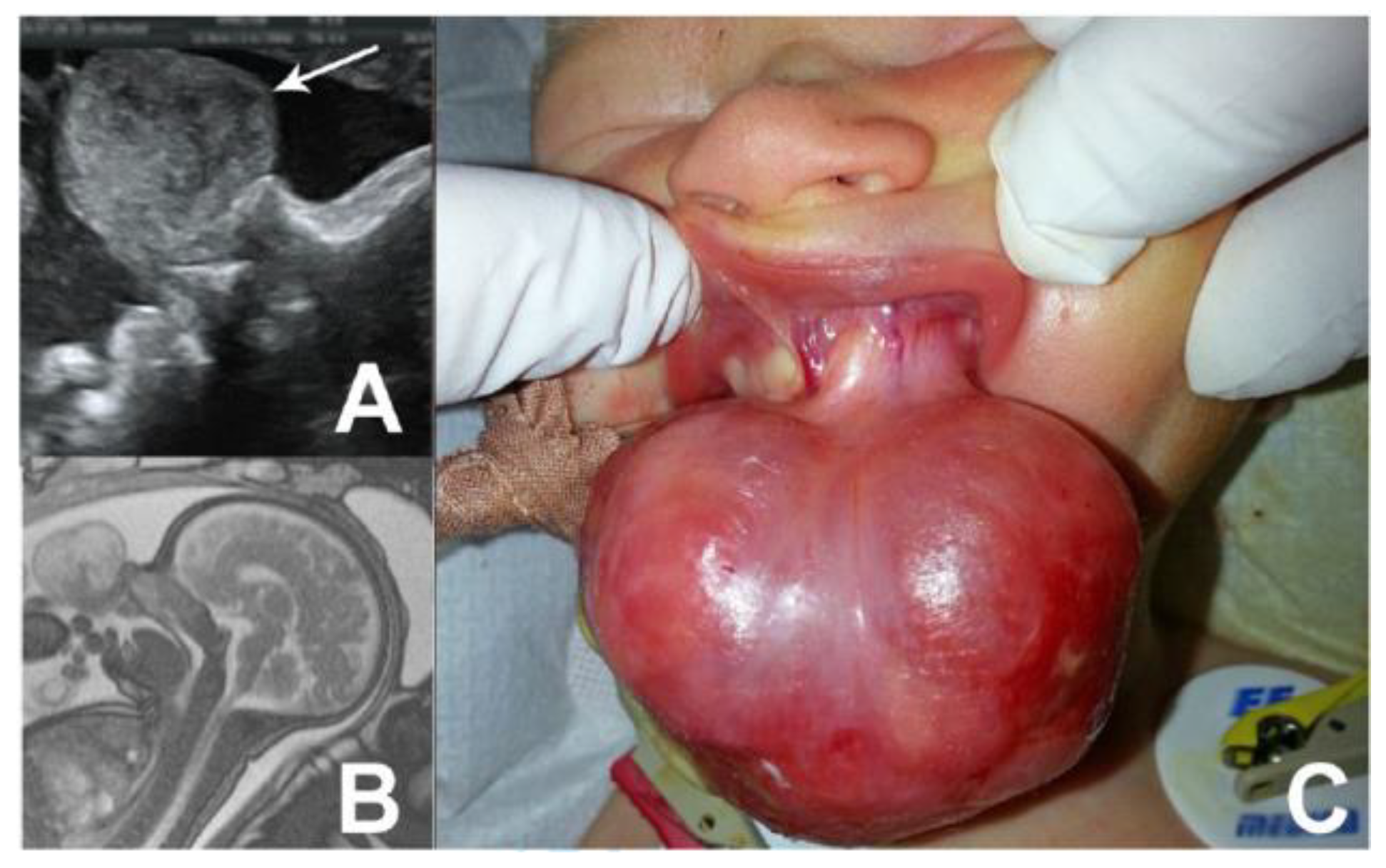
A 31-year-old pregnant woman (20 weeks of gestation), referred to our clinic for counseling over an oral fetal mass detected by ultrasound (Figure 2-A). A subsequent MRI, performed at 21 weeks of gestation, described an apparently pedunculated large tumor of the upper jaw (Figure 2-B). Considering the possibility of spontaneous rupture or asphyxiation, a decision of C-section in the presence of the pediatric surgical team was taken. A female baby was born at 37 weeks of gestation. We noted a 5/4.5 cm reddish, rounded, pedunculated tumor arising from the upper left incisive region (Figure 2-C). No breathing, feeding or bleeding problems were noted. The next day, the tumor was resected under general anesthesia. The HP exam revealed CE. No complications were noted 6 months later.

Discussions

Epulides are benign solid tumors, often pedunculated. They are usually flesh-pink colored and in 10% of the cases, multiple lesions have been noticed. Malignant transformation or recurrence after resection has not been reported so far [1]. Spontaneous regression of epulis is suggested and may be considered in newborns with small lesions which do not interfere with breathing, respiration and with compliant families [11,12].
Dental anomalies have not been reported in association with CE [10], but isolated cases report CE together with polydactyly, goiter, triple X syndrome, polyhydramnios maxillary hypoplasia or neurofibromatosis [13]. Mirror coexistent lesions on both upper and lower alveolar ridges on the same side have also been reported [14]. CE lesion arising from the tongue has also been reported in isolated cases, mainly on its anterior surface [15,16].
The diagnosis of CE is usually clinically suggestive, but histopathological examinations should be performed systematically for differential diagnosis. This reveals large rounded, polyhedral, histiocytic-like cells having small, dark, oval nuclei and excessive eosinophilic granular cytoplasm. The tumor’s cells are spindle-shaped, with a vascular network between granular cells along with a minimal fibrous stroma. The tumor cells extend to the adjacent atrophic epithelium [17]. On the other hand, major immunohistochemical profile differences and minor microscopic differences have been highlighted between the epulis of the newborn (congenital granular cell epulis) and the one found in adulthood (granular cell tumor), so it was suggested to separate them into two different entities [18]. MRI or US can presume the diagnosis of epulis prenatally. Jaw localization, tumor homogeneity and the pedunculated aspect can reduce the differential diagnosis [5]. CE usually presents in maximum growth at the end of the last trimester—maybe under hormonal influences. By using 3D ultrasound airway patency and fetal swallowing may be easily assessed, therefore a multidisciplinary team may be required to assist the child’s delivery. Choosing the right delivery method—vaginal or Cesarean—is essential since vaginal delivery may be dangerous in case of large lesions [17]. Ex utero intra-partum treatment (EXIT) may be considered in obstructive lesions, when a significant airway block is anticipated [7,10], but one must take into account the legal aspect [19] of the medical decision and possible medical errors [20,21].
The main differential diagnosis is myoblastoma [22]. Other several differential diagnoses of epulis may be considered: hemangiomas, lymphatic malformations, schwannomas, fibromas, granulomas, osteogenic sarcomas, rhabdomyosarcomas, chondrogenic sarcomas or heterotopic gastrointestinal cysts [13]. Choristomas or hamartomas of the tongue have similar aspects to CE, but they have a predilection for the dorsum of the tongue, while leyomatous hamartomas are mainly reported on the tip of the tongue or on the midline of the alveolar ridge [15]. CE is very similar to gingival granular cell tumor histologically, but it differs from it epidemiologically as well as clinically. It is seen exclusively on neonatal gingivae, presenting at birth, having a marked predilection for females, while gingival granular cell tumor is rare in the first decade of life, being most frequently diagnosed between the third and sixth decades of life, and affecting a wide variety of visceral and cutaneous sites, also having a predilection for the female gender [22]. While gingival granular cell tumor usually demonstrates strong staining for S-100, CE is negative, but exceptions have been reported [18,22,23]. As demonstrated by Karamchandani et al. [22], SOX10 represents a reliable marker in soft tissue tumor diagnosis and it should be used along with, or even instead of S100.
The cases reported in adults, the term epulis is purely clinical referring to the local reactive hyperplasia of the gum and does not reflect the histopathological diversity of the lesions: peripheral ossifying fibroma, fibroma/ fibrosis, giant cell lesion, granuloma pyogenicum, hyperplastic squamous epithelium, peripheral odontogenic fibromas.
The occurrence of this type of lesion was associated with gingivitis, occlusal trauma, periodontitis and during pregnancy [24,25,26,27].
Different congenital anomalies with potentially obstructive behavior may require immediate correction or can be performed when the biological condition of the patient is appropriate [28,29]. The surgical resection of CE is the treatment of choice when the lesion is obstructive for the feeding and breathing of the newborn. General anesthesia may be considered along with local anesthesia, when intubation cannot be done or when the lesions are small [30,31]. Wide radical excisions should be avoided in order to prevent dentition growth defects [1]. Simple resections, with no need for any mechanical stapler, are desirable [32]. CE excision using different laser methods are documented in the literature [31]. A wait-and-see regimen for tumors smaller than 2 cm that do not interfere with feeding or breathing was indicated, as long as postpartum spontaneous regression has been reported in several cases [7]. The local excision did not report recurrences, malocclusion of the jaws or damage to future dentition so far [5]. In some cases, the CE mass may leave a notch in the alveolus that may appear as an incomplete dental arch, and thus, some authors suggest the possibility of gingivoperiosteoplasty to promote a correct development of the alveolar arch. If this is done early, it restores the continuity of the premaxilla by allowing osteogenic hematoma formation in the cleft palate [22]. Surgical intervention can be delayed to an auspicious moment for the baby, considering the risk of spontaneous rupture with consecutive hemorrhage all this time.

Highlights

Congenital epulis is a rare oral tumor of the newborn.
The size and position of congenital epulis make the tumor at risk for airway obstruction of the newborn or bleeding during delivery or shortly after.
Prenatal diagnosis of oral tumors in the newborn is essential to consider to build-up perinatal multidisciplinary management plan.

Conclusions

Prenatal ultrasound or MRI can suspect CE. Taking into account the high-risk birth (rupture and bleeding, airway obstruction), the obstetrician should perform a C-section. The presence of the pediatric surgical team in the delivery room can prevent further complications. Clinical aspects of CE are usually suggestive, but histopathology is the diagnostic tool. Full oral inspection immediately after birth is indicated in order to prevent misdiagnosing multiple epulides. Surgical excision is the treatment of choice, but a conservative attitude should be considered as long as the tumor is small and uncomplicated.

Conflicts of Interest disclosure

There are no known conflicts of interest in the publication of this article. The manuscript was read and approved by all authors.

Compliance with ethical standards

Any aspect of the work covered in this manuscript has been conducted with the ethical approval of all relevant bodies and that such approvals are acknowledged within the manuscript.

References

  1. Küpers, A.M.; Andriessen, P.; van Kempen, M.J.; van der Tol, I.G.; Baart, J.A.; Dumans, A.G.; van der Waal, I. Congenital epulis of the jaw: a series of five cases and review of literature. Pediatr Surg Int. 2009, 25, 207–210. [Google Scholar] [CrossRef] [PubMed]
  2. Evans, D.A. Congenital epulis. Otolaryngol Head Neck Surg. 2001, 125, 283–284. [Google Scholar] [CrossRef] [PubMed]
  3. Merrett, S.J.; Crawford, P.J. Congenital epulis of the newborn: a case report. Int J Paediatr Dent. 2003, 13, 127–129. [Google Scholar] [CrossRef]
  4. Lee, J.M.; Kim, U.K.; Shin, S.H. Multiple congenital epulis of the newborn: a case report and literature review. J Ped Surg Case Reports. 2013, 1, 32–33. [Google Scholar] [CrossRef]
  5. Roy, S.; Sinsky, A.; Williams, B.; Desilets, V.; Patenaude, Y.G. Congenital epulis: prenatal imaging with MRI and ultrasound. Pediatr Radiol. 2003, 33, 800–803. [Google Scholar] [CrossRef]
  6. Kumar, B.; Sharma, S.B. Neonatal oral tumors: congenital epulis and epignathus. J Pediatr Surg. 2008, 43, e9–e11. [Google Scholar] [CrossRef]
  7. Suciu, N.; Serban, A.; Toader, O.; Oprescu, D.; Spataru, R.I. Case report of fetal lingual tumor—perinatal care and neonatal surgical intervention. J Matern Fetal Neonatal Med. 2014, 27, 314–319. [Google Scholar] [CrossRef]
  8. Kumar, P.; Kim, H.H.; Zahtz, G.D.; Valderrama, E.; Steele, A.M. Obstructive congenital epulis: prenatal diagnosis and perinatal management. Laryngoscope. 2002, 112, 1935–1939. [Google Scholar] [CrossRef]
  9. Serban, D.; Smarandache, C.G.; Tudor, C.; Duta, L.N.; Dascalu, A.M.; Aliuș, C. Laparoscopic Surgery in COVID-19 Era- Safety and Ethical Issues. Diagnostics (Basel). 2020, 10, 673. [Google Scholar] [CrossRef]
  10. Serban, D.; Spataru, R.I.; Vancea, G.; Balasescu, S.A.; Socea, B.; Tudor, C.; Dascalu, A.M. Informed consent in all surgical specialties: from legal obligation to patient satisfaction. Rom J Leg Med. 2020, 28, 317–321. [Google Scholar] [CrossRef]
  11. Ritwik, P.; Brannon, R.B.; Musselman, R.J. Spontaneous regression of congenital epulis: a case report and review of the literature. J Med Case Rep. 2010, 4, 331. [Google Scholar] [CrossRef] [PubMed]
  12. Popescu, B.; Oașă, I.D.; Curcă, C.; et al. First impressions on TOUSS approach of the oral cavity and pharyngeal malignant tumors. J Clin Invest Surg. 2019, 4, 72–76. [Google Scholar] [CrossRef]
  13. Jain, N.; Sinha, P.; Singh, L. Large Congenital Epulis in a Newborn: Diagnosis and Management. Ear Nose Throat, J. 2020, 99, NP79–NP81. [Google Scholar] [CrossRef]
  14. Sharma, D.; Murki, S.; Pratap, T.; Leelakumar, V. Twin congenital epulis in the alveolar ridge of the maxilla and mandible in a newborn: a rare and interesting case. BMJ Case Rep. 2014, 2014, bcr2014206490. [Google Scholar] [CrossRef]
  15. Kayıran, S.M.; Buyukunal, C.; Ince, U.; Gürakan, B. Congenital epulis of the tongue: A case report and review of the literature. JRSM Short Rep. 2011, 2, 62. [Google Scholar] [CrossRef]
  16. Rafique, A.; Habib, H.; Rehman, F.A.; Arshi, S. Impact of rational and experiential thinking styles on interpersonal conflict resolution among young adults. J Mind Med Sci. 2020, 7, 69–78. [Google Scholar] [CrossRef]
  17. Bang, K.O.; Bodhade, A.S.; Dive, A.M. Congenital granular cell epulis of a newborn. Dent Res J (Isfahan). 2012, 9 (Suppl 1), S136–S138. [Google Scholar]
  18. Vered, M.; Dobriyan, A.; Buchner, A. Congenital granular cell epulis presents an immunohistochemical profile that distinguishes it from the granular cell tumor of the adult. Virchows Arch. 2009, 454, 303–310. [Google Scholar] [CrossRef]
  19. Spataru, R.I.; Sirbu, A.; Sirbu, D. Forensic ramifications in diagnosing and treating high forms of the Hirschsprung’s disease. Rom J Leg Med. 2013, 21, 105–110. [Google Scholar]
  20. Serban, D.; Smarandache, A.M.; Cristian, D.; Tudor, C.; Duta, L.; Dascalu, A.M. Medical errors and patient safety culture—shifting the healthcare paradigm in Romanian hospitals. Rom J Leg Med. 2020, 28, 195–201. [Google Scholar] [CrossRef]
  21. Popescu, B.; Oașă, I.D.; Bertesteanu, S.V.; Balalau, C.; Manole, F.; Domuta, M.; Oancea, A.L. Strategies to improve activity and results of the head and neck tumor board. J Clin Invest Surg. 2020, 5, 9–12. [Google Scholar] [CrossRef]
  22. Narasimhan, K.; Arneja, J.S.; Rabah, R. Treatment of congenital epulis (granular cell tumour) with excision and gingivoperiosteoplasty. Can J Plast Surg. 2007, 15, 215–218. [Google Scholar] [CrossRef] [PubMed]
  23. Adeyemi, B.F.; Oluwasola, A.O.; Adisa, A.O. Congenital epulis. Indian J Dent Res. 2010, 21, 292–294. [Google Scholar] [CrossRef]
  24. Kaiserling, E.; Ruck, P.; Xiao, J.C. Congenital epulis and granular cell tumor: a histologic and immunohistochemical study. Oral Surg Oral Med Oral Pathol Oral Radiol Endod. 1995, 80, 687–697. [Google Scholar] [CrossRef]
  25. Karamchandani, J.R.; Nielsen, T.O.; van de Rijn, M.; West, R.B. Sox10 and S100 in the diagnosis of soft-tissue neoplasms. Appl Immunohistochem Mol Morphol. 2012, 20, 445–450. [Google Scholar] [CrossRef]
  26. Truschnegg, A.; Acham, S.; Kiefer, B.A.; Jakse, N.; Beham, A. Epulis: a study of 92 cases with special emphasis on histopathological diagnosis and associated clinical data. Clin Oral Investig. 2016, 20, 1757–1764. [Google Scholar] [CrossRef]
  27. Orosz, M.; Szende, B.; Gábris, K. The clinical and pathological symptoms of pregnancy epulis. Fogorv Sz. 2007, 100, 237–241. [Google Scholar]
  28. Spataru, R.I.; Popoiu, M.C.; Ivanov, M. Foregut duplication cyst associated with esophageal atresia-one-stage neonatal surgical repair. Indian J Surg. 2015, 77 (Suppl 1), 52–55. [Google Scholar] [CrossRef]
  29. Spătaru, R.I.; Martius, E.; Ivan, L.E.; Sirbu, D.; Hostiuc, S. Pseudomembranous colitis complicating the natural course of Crohn’s disease in a pediatric patient. Rom J Legal Med. 2014, 22, 161–166. [Google Scholar] [CrossRef]
  30. Aparna, H.G.; Jayanth, B.S.; Shashidara, R.; Jaishankar, P. Congenital epulis in a newborn: a case report, immunoprofiling and review of literature. Ethiop J Health Sci. 2014, 24, 359–362. [Google Scholar] [CrossRef]
  31. Miulescu, R.E.; Guja, L.; Socea, B.; Dumitriu, A.; Paunica, S.; Ștefănescu, E. Oral pathology induced by excess or deficiency of glucocorticoids in adults. J Mind Med Sci. 2020, 7, 141–147. [Google Scholar] [CrossRef]
  32. Spataru, R. The use of mechanical suture in the treatment of Hirschsprung’s disease: experience of 17 cases. Chirurgia (Bucur). 2014, 109, 208–212. [Google Scholar]
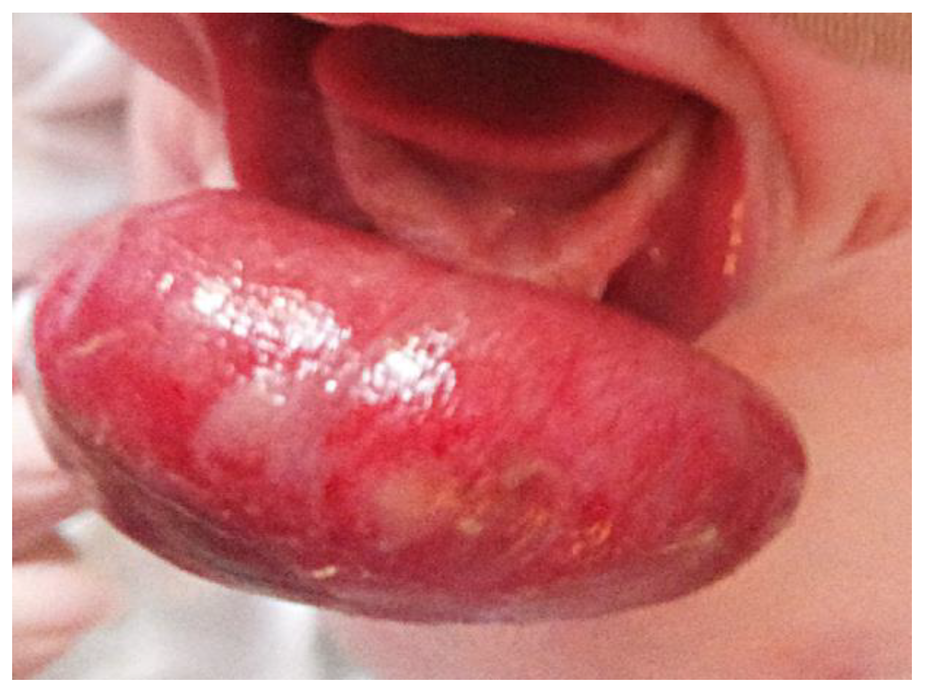
Figure 1. Postnatal clinical picture of CE of the lower jaw.
Jmms 08 00045 g001
Figure 2. Prenatal Imaging (A-ultrasonography and B- fetal MRI) and postnatal aspects (C) of upper jaw CE.
Jmms 08 00045 g002