Klebsiella Pneumoniae Cryptogenic Liver Abscess and Endophthalmitis—A Case Report and Review of Literature
Abstract
:Introduction
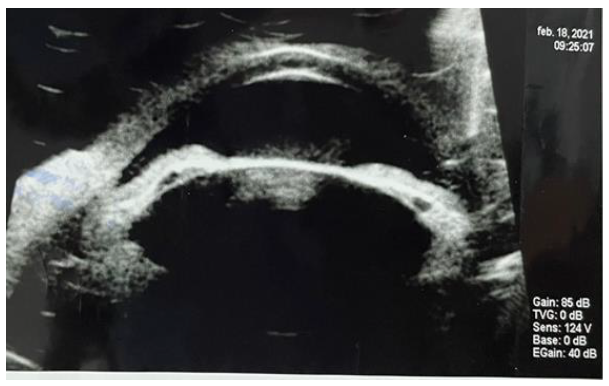
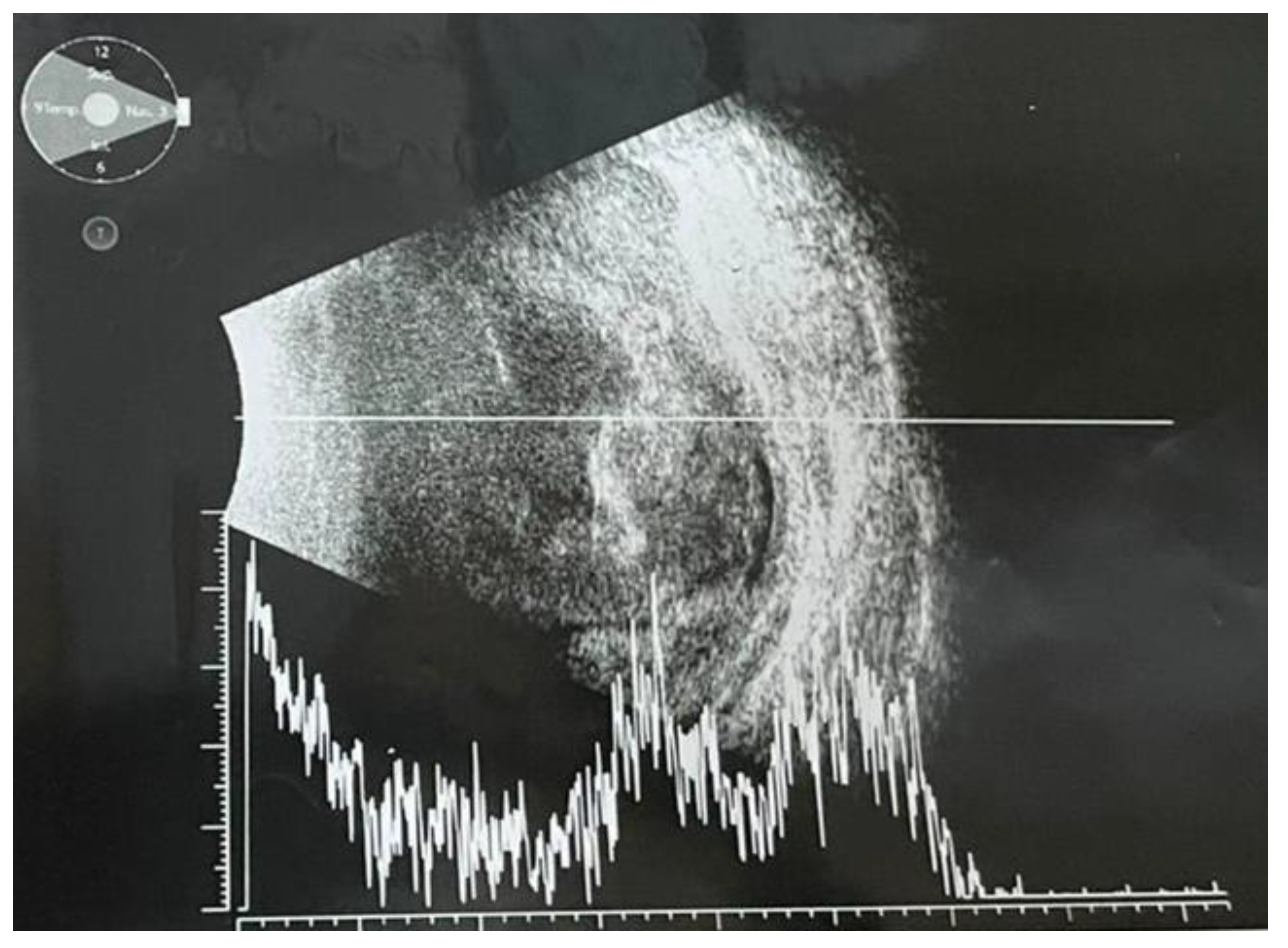
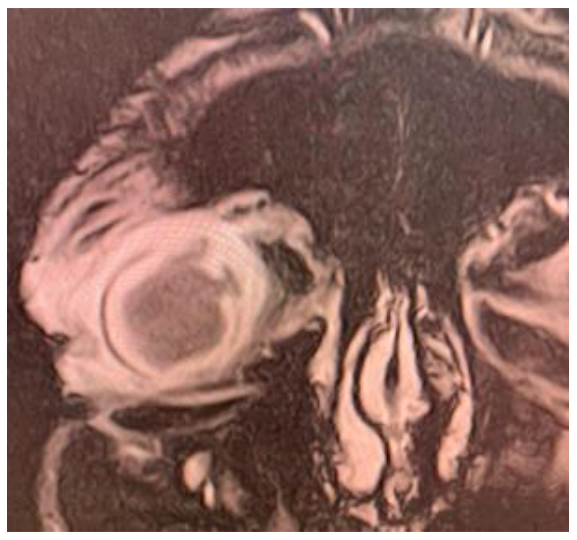
Case Presentation
Discussions
Highlights
- ✓
- Hypervirulent Klebsiella pneumoniae may cause cryptogenic hepatic abscess in previously healthy adults, with metastatic septic determinations, such as endophthalmitis.
- ✓
- Ophthalmic signs may precede hepatic ones and searching for the foculs of the iniatial infection may be challenging.
- ✓
- The therapeutic management is based on necessary interdisciplinary collaboration between several specialists such as ophthalmologist, infectious diseases specialist, surgeon and radiologist.
Conclusions
Abbreviations
Conflicts of Interest disclosure
Compliance with ethical standards
References
- Tsai, F.C.; Huang, Y.T.; Chang, L.Y.; Wang, J.T. Pyogenic liver abscess as endemic disease, Taiwan. Emerg Infect Dis. 2008, 14, 1592–1600. [Google Scholar] [CrossRef] [PubMed]
- Jun, J.B. Klebsiella pneumoniae Liver Abscess. Infect Chemother. 2018, 50, 210–218. [Google Scholar] [CrossRef] [PubMed]
- Han, S.H. Review of hepatic abscess from Klebsiella pneumoniae. An association with diabetes mellitus and septic endophthalmitis. West J Med. 1995, 162, 220–224. [Google Scholar]
- Maggi, U.; Formiga, A.; Lauro, R. Hepatic abscess as a complication of duodenal-jejunal bypass sleeve system and review of the literature. Surg Obes Relat Dis. 2016, 12, e47–e50. [Google Scholar] [CrossRef]
- Qian, Y.; Wong, C.C.; Lai, S.C.; Lin, Z.H.; Zheng, W.L.; Zhao, H.; Pan, K.H.; Chen, S.J.; Si, J.M. Klebsiella pneumoniae invasive liver abscess syndrome with purulent meningitis and septic shock: A case from mainland China. World J Gastroenterol. 2016, 22, 2861–2866. [Google Scholar] [CrossRef]
- Dascalu, A.M.; Tudosie, M.S.; Smarandache, G.C.; Serban, D. Impact of COVID-19 pandemic upon ophthalmological clinical practice. Rom J Leg Med. 2020, 28, 96–100. [Google Scholar] [CrossRef]
- Popescu, B.; Doinița, O.I.; Bălălău, C.; Scăunașu, R.; Manole, F.; Domuța, M.; Oancea, A.L. Fibroscopic examination on ENT patients in COVID-19 era. J Clin Invest Surg. 2020, 5, 63–65. [Google Scholar] [CrossRef]
- Marina, C.N.; Gheoca-Mutu, D.E.; Răducu, L.; Avino, A.; Brîndușe, L.A.; Stefan, C.M.; Scaunasu, R.V.; Jecan, C.R. COVID-19 outbreak impact on plastic surgery residents from Romania. J Mind Med Sci. 2020, 7, 212–216. [Google Scholar] [CrossRef]
- Pichler, C.; Büchsel, M.; Rossen, J.W.; Vavra, M.; Reuter, S.; Kern, W.V.; Thimme, R.; Mischnik, A. First report of invasive liver abscess syndrome with endophthalmitis caused by a K2 serotype ST2398 hypervirulent Klebsiella pneumoniae in Germany, 2016. New Microbes and New Infections. 2017, 17, 77–80. [Google Scholar] [CrossRef]
- Oikonomou, K.G.; Aye, M. Klebsiella Pneumoniae Liver Abscess: A Case Series of Six Asian Patients. Am J Case Rep 2017, 18, 1028–1033. [Google Scholar] [CrossRef]
- Kamal, F.; Williams, G.; Akbar, H.; Khan, M.A.; Kadaria, D. Klebsiella Pneumoniae Liver Abscess: a Case Report and Review of Literature. Cureus. 2017, 9, e970. [Google Scholar] [CrossRef] [PubMed]
- Stefan, D.S.; Constantinescu, R.R.; Meghea, A.; Anghel, R.; Stefan, M.; Tudosie, M.S. Obtaining of Biofertilisers Using Pelt Skin Wastes. Rev. Chim. (Bucharest). 2016, 67, 1401–1405. [Google Scholar]
- Chiu, C.T.; Lin, D.Y.; Liaw, Y.F. Metastatic Septic Endophthalmitis in Pyogenic Liver Abscess. Journal of Clinical Gastroenterology. 1988, 10, 524–527. [Google Scholar] [CrossRef] [PubMed]
- Wang, H.J.; Liu, Y.C.; Lee, S.S.J.; Yen, M.Y.; Chen, Y.S.; Wang, J.H.; Wann, S.R.; Lin, H.H. Primary Liver Abscess Due to Klebsiella pneumoniae in Taiwan. Clinical Infectious Diseases. 1998, 26, 1434–1438. [Google Scholar] [CrossRef]
- Kashani, A.H.; Eliott, D. The emergence of Klebsiella pneumoniae endogenous endophthalmitis in the USA: basic and clinical advances. J Ophthal Inflamm Infect. 2013, 3, 28. [Google Scholar] [CrossRef]
- Jun, J.B. Klebsiella pneumoniae Liver Abscess. Infect Chemother. 2018, 50, 210–218. [Google Scholar] [CrossRef]
- Cheng, D.L.; Liu, Y.C.; Yen, M.; Liu, Y.C.; Wang, R.S. Septic metastatic lesions of pyogenic liver abscess. Their association with Klebsiella pneumoniae bacteremia in diabetic patients. Arch Intern Med 1991, 151, 1557–1559. [Google Scholar] [CrossRef]
- Tudorachi, N.-B.; Eva, I.; Dascalu, C.G.; AL-Hiary, R.; Barbieru, B.; Paunica, M.; Motofei, C.; Moraru, A.-C. The influence of serum calcium and magnesium levels in the radiological evolution of knee osteoarthritis. J Mind Med Sci. 2020, 7, 217–226. [Google Scholar] [CrossRef]
- Braiteh, F.; Golden, M.P. Cryptogenic invasive Klebsiella pneumoniae liver abscess syndrome. Int J Infect Dis. 2007, 11, 16–22. [Google Scholar] [CrossRef]
- García-Cruz, E.; Otero, J.R.; Ineva, P.A.; Pérez, L.M.M.; Elías, L.P.; Asensio, A.A. Robot-assisted aquablation for resection of benign prostatic hyperplasia: A series of cases. J Clin Invest Surg. 2020, 5, 18–23. [Google Scholar] [CrossRef]
- Serban, D.; Papanas, N.; Dascalu, A.M.; et al. Diabetic Retinopathy in Patients with Diabetic Foot Ulcer: A Systematic Review. The International Journal of Lower Extremity Wounds. 2021, 20, 98–103. [Google Scholar] [CrossRef] [PubMed]
- Suceveanu, A.I.; Stoian, A.P.; Parepa, I.; Voinea, C.; Hainarosie, R.; Manuc, D.; Nitipir, C.; Mazilu, L.; Suceveanu, A.P. Gut Microbiota Patterns in Obese and Type 2 Diabetes (T2D) Patients from Romanian Black Sea Coast Region. Rev Chim. 2018, 69, 2260–2267. [Google Scholar] [CrossRef]
- Moraru, D.; Dumitru, A.; Micu, S.I.; Musat, M.; Preda, G.; Popoiag, R.E. The burden of clostridium difficile infection in patients with liver cirrhosis. J Mind Med Sci. 2019, 6, 237–242. [Google Scholar] [CrossRef]
- Yang, C.S.; Tsai, H.Y.; Sung, C.S.; Lin, K.H.; Lee, F.L.; Hsu, W.M. Endogenous Klebsiella Endophthalmitis Associated with Pyogenic Liver Abscess. Ophthalmology. 2007, 114, 876–880. [Google Scholar] [CrossRef]
- Pastagia, M.; Arumugam, V. Klebsiella pneumoniae liver abscesses in a public hospital in Queens, New York. Travel Med Infect Dis. 2008, 6, 228–233. [Google Scholar] [CrossRef]
- Wang, H.; Ren, Y.; Chang, Z.; Liu, Z. The increased recurrence rate of liver abscess caused by extended-spectrum β-lactamase-producing Klebsiella pneumoniae. Eur J Clin Microbiol Infect Dis. 2020, 39, 1315–1320. [Google Scholar] [CrossRef]
- Serban, D.; Socea, B.; Balasescu, S.A.; Badiu, C.D.; Tudor, C.; Dascalu, A.M.; Vancea, G.; Spataru, R.I.; Sabau, A.D.; Sabau, D.; Tanasescu, C. Safety of Laparoscopic Cholecystectomy for Acute Cholecystitis in the Elderly: A Multivariate Analysis of Risk Factors for Intra and Postoperative Complications. Medicina. 2021, 57, 230. [Google Scholar] [CrossRef]
- Park, I.H.; Jun, C.H.; Wi, J.W.; Park, S.Y.; Lee, W.S.; Jung, S.I.; Park, C.H.; Joo, Y.E.; Kim, H.S.; Choi, S.K.; Rew, J.S. Prevalence of and risk factors for endogenous endophthalmitis in patients with pyogenic liver abscesses. Korean J Intern Med. 2015, 30, 453–459. [Google Scholar] [CrossRef]
- Lee, J.Y.; Kim, K.H. Endogenous Endophthalmitis Complicated by Pyogenic Liver Abscess: A Review of 17 Years' Experience at a Single Center. Digestion. 2014, 90, 116–121. [Google Scholar] [CrossRef]
- Lin, J.C.; Siu, L.K.; Fung, C.P.; Tsou, H.H.; Wang, J.J.; Chen, C.T.; Wang, S.C.; Chang, F.Y. Impaired phagocytosis of capsular serotypes K1 or K2 Klebsiella pneumoniae in type 2 diabetes mellitus patients with poor glycemic control. J Clin Endocrinol Metab. 2006, 91, 3084–3087. [Google Scholar] [CrossRef]
- Gheorghe, G.; Stoian, A.P.; Gaman, M.; Socea, B.; Neagu, T.P.; Stanescu, A.M.A.; Bratu, O.G.; Mischianu, D.L.D.; Suceveanu, A.I.; Diaconu, C.C. The Benefits and Risks of Antioxidant Treatment in Liver Diseases. Rev Chim. 2019, 70, 651–655. [Google Scholar] [CrossRef]
- Radu, N.; Voicescu, M.; Radu, E.; Tanasescu, C. Biomaterial with antioxidant and antifungal activities, obtained from romanian indigenous plants. Molecular Crystals and Liquid Crystals. 2017, 655, 243–249. [Google Scholar] [CrossRef]
- Șerban, D.; Brănescu, C.M.; Smarandache, G.C.; Tudor, C.; Tănăsescu, C.; Tudosie, M.S.; Stana, D.; Costea, D.O.; Dascălu, A.M.; Spătaru, R.I. Safe surgery in day care centers: focus on preventing medical legal issues. Rom J Leg Med. 2021, 29, 60–64. [Google Scholar] [CrossRef]
- Fung, C.P.; Chang, F.Y.; Lee, S.C.; Hu, B.S.; Kuo, B.I.; Liu, C.Y.; Ho, M.; Siu, L.K. A global emerging disease of Klebsiella pneumoniae liver abscess: is serotype K1 an important factor for complicated endophthalmitis? Gut. 2002, 50, 420–424. [Google Scholar] [CrossRef]
- Cheng, D.L.; Liu, Y.C.; Yen, M.; Liu, Y.C.; Wang, R.S. Septic metastatic lesions of pyogenic liver abscess. Their association with Klebsiella pneumoniae bacteremia in diabetic patients. Arch Intern Med. 1991, 151, 1557–1559. [Google Scholar]
- Fang, C.T.; Lai, S.Y.; Yi, W.C.; Hsueh, P.R.; Liu, K.L.; Chang, S.C. Klebsiella pneumoniae genotype K1: an emerging pathogen that causes septic ocular or central nervous system complications from pyogenic liver abscess. Clin Infect Dis. 2007, 45, 284–293. [Google Scholar] [CrossRef]
- Chung, C.Y.; Wong, E.S.; Liu, C.C.H.; Wong, M.O.M.; Li, K.K.W. Clinical features and prognostic factors of Klebsiella endophthalmitis-10-year experience in an endemic region. Eye 2017, 31, 1569–1575. [Google Scholar] [CrossRef]
- Wells, J.T.; Lewis, C.R.; Danner, O.K.; Wilson, K.L.; Matthews, L.R. Klebsiella pneumoniae Liver Abscess and Metastatic Endophthalmitis. J Investig Med High Impact Case Rep. 2015, 4, 2324709615624125. [Google Scholar] [CrossRef]
- Liu, Y.C.; Cheng, D.L.; Lin, C.L. Klebsiella pneumoniae liver abscess associated with septic endophthalmitis. Arch Intern Med. 1986, 146, 1913–1916. [Google Scholar]
- Sheu, S.J. Endophthalmitis. Korean J Ophthalmol. 2017, 31, 283–289. [Google Scholar] [CrossRef]

Share and Cite
Grecescu, M.L.; Grecescu, M.; Maria, A.S.; Branescu, C.M.; Anghelache, A.; Dumitriu, A.S.; Paunica, S.; Costea, A.C. Klebsiella Pneumoniae Cryptogenic Liver Abscess and Endophthalmitis—A Case Report and Review of Literature. J. Mind Med. Sci. 2021, 8, 330-335. https://doi.org/10.22543/7674.82.P330335
Grecescu ML, Grecescu M, Maria AS, Branescu CM, Anghelache A, Dumitriu AS, Paunica S, Costea AC. Klebsiella Pneumoniae Cryptogenic Liver Abscess and Endophthalmitis—A Case Report and Review of Literature. Journal of Mind and Medical Sciences. 2021; 8(2):330-335. https://doi.org/10.22543/7674.82.P330335
Chicago/Turabian StyleGrecescu, Mihai Leonard, Monica Grecescu, Andreea Smarandache Maria, Cristian Mihai Branescu, Anca Anghelache, Anca Silvia Dumitriu, Stana Paunica, and Andreea Cristina Costea. 2021. "Klebsiella Pneumoniae Cryptogenic Liver Abscess and Endophthalmitis—A Case Report and Review of Literature" Journal of Mind and Medical Sciences 8, no. 2: 330-335. https://doi.org/10.22543/7674.82.P330335
APA StyleGrecescu, M. L., Grecescu, M., Maria, A. S., Branescu, C. M., Anghelache, A., Dumitriu, A. S., Paunica, S., & Costea, A. C. (2021). Klebsiella Pneumoniae Cryptogenic Liver Abscess and Endophthalmitis—A Case Report and Review of Literature. Journal of Mind and Medical Sciences, 8(2), 330-335. https://doi.org/10.22543/7674.82.P330335