Next Article in Journal
Therapeutic Efficacy of Mesenchymal Stem Cells for Cardiovascular Diseases
Previous Article in Journal
A Pedunculated Esophageal Chondromatous Hamartoma in a Child
 
 
Journal of Mind and Medical Sciences is published by MDPI from Volume 12 Issue 1 (2025). Previous articles were published by another publisher in Open Access under a CC-BY (or CC-BY-NC-ND) licence, and they are hosted by MDPI on mdpi.com as a courtesy and upon agreement with Valparaiso University (ValpoScholar).
Review

The Molecular Mechanisms Linking Metabolic Syndrome to Endometrial and Breast Cancers

by
Andrada-Luciana Lazar
1,#,
Romana Vulturar
2,#,
Adriana Fodor
3,#,
Olga Hilda Orasan
4,*,
Camil-Horia Eusebiu Crișan
5,
Cezar Login
6,
Ioana Para
4,
Vasile Negrean
4,
Brandusa Tiperciuc
7 and
Angela Cozma
4
1
Department of Dermatology, Iuliu Haţieganu University of Medicine and Pharmacy, Cluj-Napoca, Romania
2
Department of Molecular Sciences, Iuliu Haţieganu University of Medicine and Pharmacy, Cluj-Napoca, Romania
3
Clinical Center of Diabetes, Iuliu Haţieganu University of Medicine and Pharmacy, Nutrition, Metabolic Diseases, Cluj-Napoca, Romania
4
4th Medical Clinic, Department of Internal Medicine, Iuliu Haţieganu University of Medicine and Pharmacy, Republicii Street No. 18, Cluj-Napoca, Romania
5
Department of Internal Medicine, Iuliu Haţieganu University of Medicine and Pharmacy, Cluj-Napoca, Romania
6
Department of Physiology, Iuliu Haţieganu University of Medicine and Pharmacy, Cluj-Napoca, Romania
7
Department of Pharmaceutical Chemistry, Iuliu Haţieganu University of Medicine and Pharmacy, Cluj-Napoca, Romania
*
Author to whom correspondence should be addressed.
#
Authors with equal contributions.
J. Mind Med. Sci. 2021, 8(2), 167-178; https://doi.org/10.22543/7674.82.P167178
Submission received: 11 March 2021 / Revised: 12 March 2021 / Accepted: 18 May 2021 / Published: 10 October 2021

Abstract

:
The metabolic syndrome represents a plethora of cardio-metabolic risk factors including obesity, arterial hypertension, atherogenic dyslipidemia, hyperglycemia, accompanied by pro-inflammatory and pro-thrombotic state. The metabolic syndrome is one of the key risk factors for certain types of cancer. Among these malignancies, breast cancer and endometrial neoplasms require special attention. Incriminated major causes for the development of breast and endometrial cancer in metabolic syndrome patients are: the pro-inflammatory status and related cytokines, adipokine imbalances, hyperestrogenism, growth factors, disturbances in cancer microenvironment, insulin resistance and hyperinsulinemia. The metabolic syndrome consists of molecular dysregulations that create a pro-oncogenic status. Our review aims at providing a better understanding of the mechanisms underlying the link between the metabolic syndrome and endometrial and breast cancer.

Introduction

Breast cancer (BC) ranks the first among solid cancers in women, representing a major public health problem globally [1], being a major cause of mortality in female patients. With this in mind, the identification of modifiable risk factors together with the implementation of primary prevention methods should be considered a priority [1]. There are several studies that investigated the link between the metabolic syndrome (MS) and BC with mixed results regarding the implication of obesity, high blood pressure and dyslipidemia. On the other hand, the association of the aforementioned factors proved to increase the risk of developing breast neoplasia [2]. Furthermore, endometrial cancer (EC) is the most common form of gynecological cancer in developed countries [3] and its association with obesity, diabetes and hypertension (the endometrial cancer triad) is irrefutable. Epidemiological studies have shown a 2.45-fold higher risk of developing EC in overweight and obese patients and 2.12-fold higher in diabetic patients. In patients with obesity and hypertension, the risk of developing EC is also increased [4].

Discussions

Metabolic syndrome

The metabolic syndrome represents a group of cardio-metabolic risk factors including: obesity, arterial hypertension, atherogenic dyslipidemia, hyperglycemia together with pro-thrombotic and pro-inflammatory states [5].
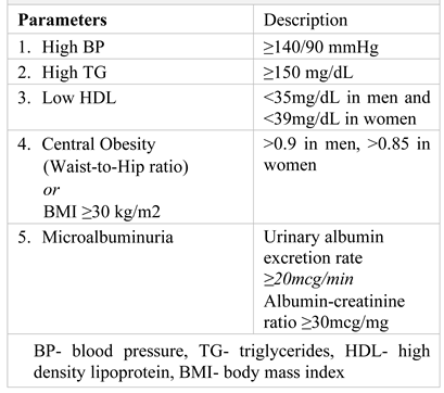
The World Health Organization’s (WHO) definition of MS underwent several adjustments in time. According to WHO, MS is characterized by the presence of: glucose intolerance, impaired glucose tolerance, or diabetes mellitus and/or insulin resistance together with two or more of the parameters listed in Table 1 [5].
Figure 1. Illustration of the metabolic syndrome according to WHO (1).
Jmms 08 00022 g001

Endometrial cancer

Epidemiology

Endometrial cancer (EC) represents the most common gynecological cancer in developed countries, with an average incidence of 14.7/ 100,000 among women [3].

Risk factors

The risk factors associated with EC are body mass index (BMI) ≥ 25, early menarche (before the age 12), nulliparity, nulligravidity, the use of oral contraceptives, infertility, positive family history of endometrial cancer, low level of education. Additionally, there are also studies that suggest an association between infertility treatments and EC [6]. Regarding the involvement of smoking in the development of EC, the results of the studies are contradictory. While there are studies that have identified an association between cigarette consumption and EC [7], there are also studies that support the anti-estrogenic effect of smoking [8,9] or studies that have not identified any association between smoking and endometrial neoplasia [6]. An increased body mass index (BMI) along with increased waist circumference values are associated with premenopausal uterine neoplasia [10]. High blood pressure (HBP) appears to play a role in the development of EC, but the underlying mechanism has not been elucidated to this day [11,12]. Additionally, diabetes mellitus (DM) and hyperinsulinemia have also been identified as risk factors for EC [13,14].

The metabolic syndrome

As mentioned above, EC is a type of neoplasm commonly associated with obesity, diabetes and hypertension (the endometrial cancer triad) [4]. MS is involved in the development of EC through the disruption of blood glucose homeostasis, serum triglycerides levels, insulinemia and insulin-like growth factor (IGF) system [14,15].

Obesity

The increased risk for developing EC among women suffering from obesity is based on the associated hyperestrogenism, hyper-insulinemia and insulin resistance (IR), lipid metabolism disorders, hyperglycemia, chronic inflammation [16].

Hyperestrogenism

A prolonged exposure of the endometrium to the endogenous or exogenous estrogens, in the absence of the antagonist effect of progesterone, represents the main cause of EC. In menopausal women, the adipose tissue, through the enzyme aromatase, which converts androstenedione into estradiol, is the main source of endogenous estrogen [17]. Thus, the serum estradiol resulting from this process interacts with endometrial estrogen receptors [17].
Hyperestrogenemia may also be the result of hyperinsulinemia, common in obese patients [18]. Elevated insulin levels lead to a decrease in sex hormone binding protein (SHBP) synthesis and an increased bioavailability of insulin-like growth factor 1 (IGF-1) [18]. Insulin resistance, in the context of elevated serum levels of adipokines (leptin and visfatin) and pro-inflammatory cytokines (TNF-α, IL-1β, IL-6, MCP-1), underlies hyperinsulinemia, characterized by low serum IGF-1 binding protein resulting in elevated levels of IGF-1 [19]. High levels of insulin and IGF-1 stimulate endometrial cancer cell proliferation by binding to insulin receptors (IR) and IGF-1 receptors (IGF-1R) [20]. There are studies that have demonstrated the synergic effect of insulin and estrogen in the stimulation of endometrial tumor cell proliferation [21].

Chronic inflammation

Obesity is characterized by a chronic pro-inflammatory status characterized by the infiltration of the adipose tissue with macrophages and an increased expression of inflammatory cytokines, which represent an important factor involved in IR [19]. Studies on human and animal subjects with obesity and IR revealed elevated plasma levels of C-reactive protein (CRP) and interleukin-6 (IL-6) [22].

Growth factors

The activity of (VEGF-mTOR)– (the vascular endothelial growth factor - mammalian target of rapamycin) factor is highly increased in obese women with endometrial neoplasm. There are studies proposing VEGF-mTOR derived from adipocytes as a possible therapeutic target in obese women diagnosed with EC [23].

Cancer microenvironment

The adipose tissue can disturb the tumor microenvironment by inducing disturbances in the extracellular matrix, thus dysregulating the homeostasis of the surrounding tissues and creating a favorable status for the development of cancer. In addition to the indirect effects, adipose tissue also acts directly on neoplastic cells through a paracrine mechanism [24]. In addition, there are studies that have reported the possibility of a fusion capacity between endometrial malignant cells and adipose-derived stem cells (ASC), with the subsequent expression of a fibroblast-like phenotype [25]. All the above are associated with a phenomenon of downregulation of E-cadherin expression and upregulation of Vimentin expression [25].

Pro-inflammatory cytokines and adipokines
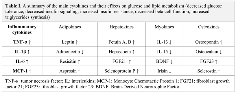
The roles of cytokines in the regulation of metabolism and inter-organ signaling outline the relationship with pathophysiology in metabolic disease. Over nutrition along with low physical activity (a sedentary lifestyle) has led to an increase in metabolic diseases. Several organs and tissues (e.g., adipocytes, hepatocytes, muscles, skeleton cells) secrete specific cytokines for inter-organ communication, and the secretion of these cytokines is modified during nutritional stress and physical activity [26,27].
Jmms 08 00022 i002
Adiponectin, visfatin and leptin are adipokines involved in the development and progression of EC [16]. Obesity, hypertension, insulin resistance and hyperinsulinemia are associated with low serum adiponectin [28], which is further correlated with the risk of developing EC in postmenopausal women who have not received hormone replacement therapy (HRT) [28]. Adiponectin is a cytokine secreted by adipocytes and with low circulating levels in patients diagnosed with EC [29].
Adiponectin exerts its effects through its receptors: the adiponectin receptor-1 (AdipoR1) and the adiponectin receptor-2 (AdipoR2) [30]. An increase in AdipoR1 expression compared to the expression of AdipoR2 was identified in EC. AdipoR1 plays a role in the inhibition of neoplastic cell proliferation, intercellular adhesion and the invasiveness of EC [31] and increases cancer cell insulin sensitivity [32]. Moreover, adiponectin also has an effect on the tumor microenvironment [33]. Thus, the low serum level of adiponectin, found among obese people, is closely related to the risk of developing endometrial neoplasm [34].
Visfatin (Visf) is an insulin-like adipokine. Elevated serum levels of Visf were found in patients diagnosed with EC [35], and its serum level increases with the patients’ BMI [36,37]. Given that the ratio of Visf:Adipo is elevated in EC, studies have shown that a high serum level of Visf represents an independent risk factor for EC [38]. Moreover, the Visf level is associated with the risk of myometrial invasion, lymph node metastases and a poor prognosis for patients diagnosed with EC [36,37].
Leptin (Lept) represents another adipokine which is involved not only in the development of endometrial, but also in breast and colon neoplasm pathogenesis. By interacting with the leptin receptor (ObR), it plays an important role in regulating food intake, energy consumption and cell growth [39,40]. Zhou et al. demonstrated that the serum level of Lept, together with the expression ObR, is strongly correlated with the degree of EC differentiation.
Thus, ObRs are intensely expressed in poorly differentiated tumors [41]. Additionally, the serum level of Lept is an independent risk factor for EC [42]. The cells’ malignant transformation, their proliferation and invasion capacities are based on the Lept over-activation of the PI3K / Akt [42], JNK, STAT3, ERK1 / 2 signaling pathways [43,44].
Inflammation plays a crucial role in tumorigenesis. In patients suffering from obesity, the adipose tissue represents the site of a marked inflammatory process responsible for systemic metabolic alterations and tumor microenvironment disturbances [16].
Interleukin-6 (IL-6), tumor necrosis factor alpha (TNF-α) and plasminogen activator inhibitor-1 (PAI-1) are pro-inflammatory cytokines involved in tumorigenesis in obese people [16].
IL-6 is an inflammatory cytokine, associated with an increased risk of mortality in overweight/obese people [45]. Elevated IL-6 levels represent a predictive factor for poor prognosis in cancer patients [46]. Moreover, estrogen hormones promote cellular IL-6 expression in EC [47].
In turn, IL-6 supports the synthesis of aromatase, thus accelerating the synthesis of estrogen and creating a positive feedback mechanism [48]. The IL-6 / JAK / STAT3 signaling pathway is also involved in the development of neoplasms. IL-6 causes the hyper-activation of the aforementioned pathway, which is associated with an unfavorable prognosis in cancer patients [49]. Additionally, there are studies that confirmed that by blocking the IL-6 inflammatory signal, the spread of neoplastic cells to the liver may be inhibited [49,50].
TNF-α is an endogenous tumor-promoting factor secreted by macrophages and adipocytes, involved in the proliferation, invasion and metastasis of tumor cells [51]. Adipocytes can stimulate endometrial cell proliferation through a paracrine mechanism that appears to be mediated by TNF-α [51]. By activating the NF-Κb signaling pathway, TNF-α inhibits the apoptosis of the neoplastic cells [16].
PAI-1 is a protease inhibitor produced in endothelial, stromal and adipocyte cells [16]. It plays an important role in the invasion and metastasis of tumors associated with obesity [52]. The increased expression of PAI-1 in endometrial neoplastic cells is associated with a poor prognosis, and an increased risk of disease recurrence [53].

Diabetes mellitus

Diabetes mellitus (DM) is a well-known risk factor for EC [54], and it is associated with increased mortality among EC patients [55].

Hyperglycemia

Hyperglycemia acts directly on the signaling pathways involved in the rapid proliferation of tumor cells [56]. In addition to a rapid source of energy, glycolysis is also responsible for the production of a plethora of metabolic intermediates, used for the synthesis of macromolecules, such as: nucleic acids, fatty acids and proteins, essential in supporting the rapid growth of tumors [16].
Hyperglycemia supports the proliferation and invasion of endometrial neoplastic cells through the promotion of VEGF expression, its receptor (VEGFR), and the epithelial-mesenchymal transition (EMT) process by regulating Erα/GLUT4 (glucose transporter) expression [57]. In addition, hyperglycemia supports the proliferation of endometrial neoplastic cells by activating STAT3 expression [58].
An enzyme involved in glycolysis is isoenzyme M2 of pyruvate kinase (PKM2), which not only plays an important role in tumorigenesis, but also in the prognosis of cancer patients [59].
Lactic acid (LA) is a key metabolite in the neoplastic cell glycolysis. It supports the transformation of normal cells into tumor cells, it has an immunosuppressive effect and promotes angiogenesis [60]. Furthermore, LA promotes the transition of M1 anti-tumor macrophages into M2 macrophages, which represent the predominant population of macrophages associated with EC [61]. The uptake of pyruvate and LA by cells is achieved through monocarboxylate transporter 1 (MCT1), which is an independent marker for EC prognosis [62].

Hyperinsulinemia and Insulin resistance

Insulin resistance and hyperinsulinemia play an important role in the development of EC. Moreover, hyperinsulinemia is an independent risk factor for EC [63]. Elevated serum insulin levels together with IGF-1/2 accelerate the conversion of androstenedione into estrogen via aromatase, and also inhibit SHBG synthesis in patients with diabetes [16]. Prolonged exposure to estrogen without the antagonistic effect of progesterone is a well-known risk factor for endometrial dysplasia and malignant transformation [64]. Additionally, IR / IGF-1R are prognostic factors associated with lymph node invasion [65].
Given the involvement of hyperinsulinemia in the development of EC, there are studies that have investigated the potential anti-neoplastic effect of metformin. There are data in the literature suggesting that the combination between metformin with progesterone therapy may have a beneficial effect in patients with an unsatisfactory response to progesterone therapy [66]. Conventional doses of metformin proved to be effective for hyperglycemia and elevated IGF-1 level management in patients with EC [67]. A meta-analysis conducted in 2018 failed to prove the protective effect of metformin against the development of EC, but showed its beneficial effect in terms of survival and risk of recurrence [68]. However, there are also studies that do not support the beneficial effects of metformin in EC management [69].

Dyslipidemia

The association between hyperglycemia, hyperlipemia and HBP doubles the risk of developing EC [70]. A positive correlation has been identified between the EC patients’ BMI and the serum level of palmitic, oleic and stearic acid [71]. An elevated plasma level of eicosapentaenoic acid is also associated with EC recurrence [72]. Noteworthy is that there are studies that have proven the antitumor effect of fatostatin, which has inhibitory effects on the proliferation of neoplastic endometrial cells and also induces the apoptosis of neoplastic cells [73].

Weight loss

Bariatric surgery or weight loss may reduce the risk of developing endometrial hyperplasia or EC [74]. Weight loss in patients with EC decreased C-peptide, insulin, CRP, leptin, IL-1Rα and IL-6 levels and increased SHBG, IGF-BP1 and adiponectin levels [75]. Therefore, bariatric surgery can reduce the risk of developing EC by reducing obesity-associated inflammation [16].

Breast cancer

As mentioned above, breast cancer (BC) ranks as the first type of cancer among women, representing a major public health problem globally. There are several studies that have investigated the link between MS and BC. Numerous epidemiological studies analyzed the causal relationship between the metabolic syndrome or its individual components and BC (1).

Risk factors

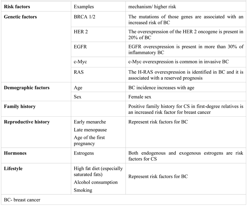
The risk factors for breast cancer are presented in the Table 2.

The metabolic syndrome and breast cancer

Numerous epidemiological studies analyzed the causal relationship between the metabolic syndrome or its individual components and BC. Therefore, several studies have revealed a correlation between diabetes and breast cancer, while there are inconclusive results regarding the role of obesity, high blood pressure and dyslipidemia. Although there is a rather weak association between the individual components of the metabolic syndrome and the development of breast cancer, their combination could increase the risk of developing breast neoplasia [2].
The relationship between MS and breast cancer was investigated in a large population study that included 94,000 women, which was conducted over a period of 14 years; during this time, 5.7% of the women developed breast cancer [77]. The risk of breast cancer increased with the number of MS components, being 45% higher in women who had 4 MS components than in those without MS [77].

Mechanisms

The metabolic syndrome consists in certain endocrine, metabolic and physiological immunological disturbances that can disrupt the molecular pathways involved in the pathogenesis of BC.

Aromatase

An increased aromatase activity along with an increased level of inflammation have been identified in the adipose breast tissue of overweight and obese women [78]. Aromatase activity is responsible for hyperestrogenemia in obese patients (79). Low plasma SHBG levels are associated with IR and other components of the metabolic syndrome [79]. It is well known that estrogen plays an important role in hormone-dependent breast cancer.

Adipokines

The disturbances in the adipokines secretion are also involved in the pathogenesis of BC. Increased levels of leptin and decreased levels of adiponectin were found in obese patients. Leptin stimulates breast cell proliferation while adiponectin has a protective effect by inhibiting uncontrolled cell growth [2].

Insulin resistance and diabetes mellitus

IR has an important pathogenetic role in BC. Hyperinsulinism represents a compensatory mechanism for IR, with the consequent binding of insulin to specific receptors in breast epithelial cells, leading to the activation of the IGF 1 signaling pathway, a pathway known to be involved in carcinogenesis. In addition, hyperinsulinism may contribute to the development of breast cancer by stimulating hepatic IGF-1 production [80].
Agnoli et al. demonstrated the link between MS and BC in a multicenter study and identified the presence of altered fasting blood glucose in patients with MS and BC [81].

Chronic inflammation

Another important feature of MS with implications in tumorigenesis is the chronic inflammation present in these patients, expressed through the secretion of cytokines such as interleukin-6 and tumor necrosis factor alpha that support EC progression [82].

The metabolic syndrome and various types of breast cancer

Four subtypes of BC have been described, each one of them being different in terms of prognosis, treatment options, the presence of estrogen and progesterone hormone receptors, HER2 expression and ki-67 proliferation index [77]. There are several studies in the literature that took into consideration the aforementioned subtypes of BC and showed that the association between breast cancer and the metabolic syndrome is significant only for ER positive BCs [77]. Given the pathogenetic mechanisms described above, in particular the role of estrogen, the risk is expected to be higher for hormone-dependent tumors.
Another study investigated the link between MS and triple negative BC [83]. The authors reported a higher prevalence of MS in triple negative cancer patients (58% compared to 37% for other cancers), but without a well-established causal relationship [83].

The metabolic syndrome and the therapeutic response in breast cancer

The potential implications of MS in the treatment outcomes of BC patients were also investigated. Noteworthy is the association of MS with a high cardiovascular risk and the cardiotoxicity of certain oncological treatments for breast cancer (especially anthracycline-based chemotherapy and anti-HER2 monoclonal antibodies). A higher risk of cardiotoxicity was demonstrated in overweight and obese patients [84]. A low adiponectin production in obese people might represent the explanation for the adverse cardiovascular effects of oncological therapy in obese patients. Animal studies have shown an exacerbation of myocardial dysfunction after the injection of doxorubicin in mice with adiponectin depletion and an improvement in cardiac dysfunction induced by doxorubicin administration after the exogenous injection of adiponectin [85].

Preliminary data

The link between MS and cancer is clearly established. There are several meta-analyses in the scientific literature that showed a significant increase in the incidence of BC in women suffering from MS. At the same time, the authors noted a higher risk in postmenopausal women, thus suggesting the different pathophysiology behind the genesis of pre- and post-menopausal BC [2,86].
Recent data, from a recent meta-analysis of 17 cohort observational studies, confirm a strong association between the metabolic syndrome and breast cancer (RR = 1.15, CI: 1.05-1.26, p = 0.003) [82]. An increased risk was also observed among Caucasian women [82].
The prognostic role of MS in patients with breast cancer was also studied in a recent meta-analysis [87] that included 9 cohort studies with over 17,000 patients diagnosed with BC. The aforementioned study showed that the presence of MS was associated with an increased risk of breast cancer recurrence ([RR] = 1.52, 95%, p = 0.02) and overall mortality [87]. The conclusion of this meta-analysis was that MS is an independent predictive factor for negative prognosis in women with BC [87]. The above findings are consistent with the results of another study which showed that the risk of developing distant metastases is more than double in women with BC and MS than in those with breast cancer without MS [88].
All the above demonstrate that the association between cancer and MS represents an intensely debated subject with a complicated pathophysiology based on multiple, intricate mechanisms. Our paper attempts to shed some light over the underlying mechanisms that link MS to BC and EC.
As mentioned above, the main mechanism underlying hyperestrogenism in overweight and obese patients is the conversion of androgens into estrogens through the action of aromatase present in the adipose tissue, thus, increasing the circulating estradiol [79], all together with the reduced levels of plasma concentration of sex hormone transport protein (SHBG) [78]. The activation of the signaling pathway is accomplished by binding estrogens to ERα receptors, with their consequent dimerization, translocation in the nucleus, followed by gene transcription and protein synthesis with a role in cell proliferation [89].
The disruption of adipokine levels cancels the protective effect of adiponectin against uncontrolled cell growth, while the increased levels of leptin stimulate breast cancer cell proliferation [2].
Insulin resistance and the consequent hyperinsulinism lead to the IGF-1 signaling pathway activation. Furthermore, in the context of hyperinsulinemia, the liver IGF-1 production is stimulated [81], this being another important mechanism involved in BC pathogenesis.
In addition to the aforementioned pathways involved in BC pathophysiology, the chronic inflammatory state that defines MS and is characterized by increased levels of pro-inflammatory cytokines (IL-6 and TNF-alpha) also plays a role in breast tumorigenesis [82]. The mechanisms that mediate the crosstalk between MS and BC are presented in Figure 2.
Obesity, diabetes and hypertension represent the metabolic triad of EC. Insulin resistance, leptin, lactate along with low serum adiponectin and obesity-associated chronic inflammation are important factors involved in EC occurrence [4].
As mentioned above, increased estrogen levels represent a risk factor for EC. In overweight and obese patients, increased adipose tissue aromatase activity is responsible for peripheral estrogen synthesis. Hyperestrogenemia may be the result of hyperinsulinemia by reducing the synthesis of SHBP [18]. As in the case of BC, the chronic inflammation characterized by high levels of pro-inflammatory cytokines (IL-6, TNF-alpha, PAI-1) supports tumorigenesis in obese female patients [16,22]. The adipokines homeostasis is deregulated in MS patients, thus, the dysregulation of Visf:Adipo balance, with high Visf levels, is associated with EC aggressiveness and a poor prognosis [36,37].
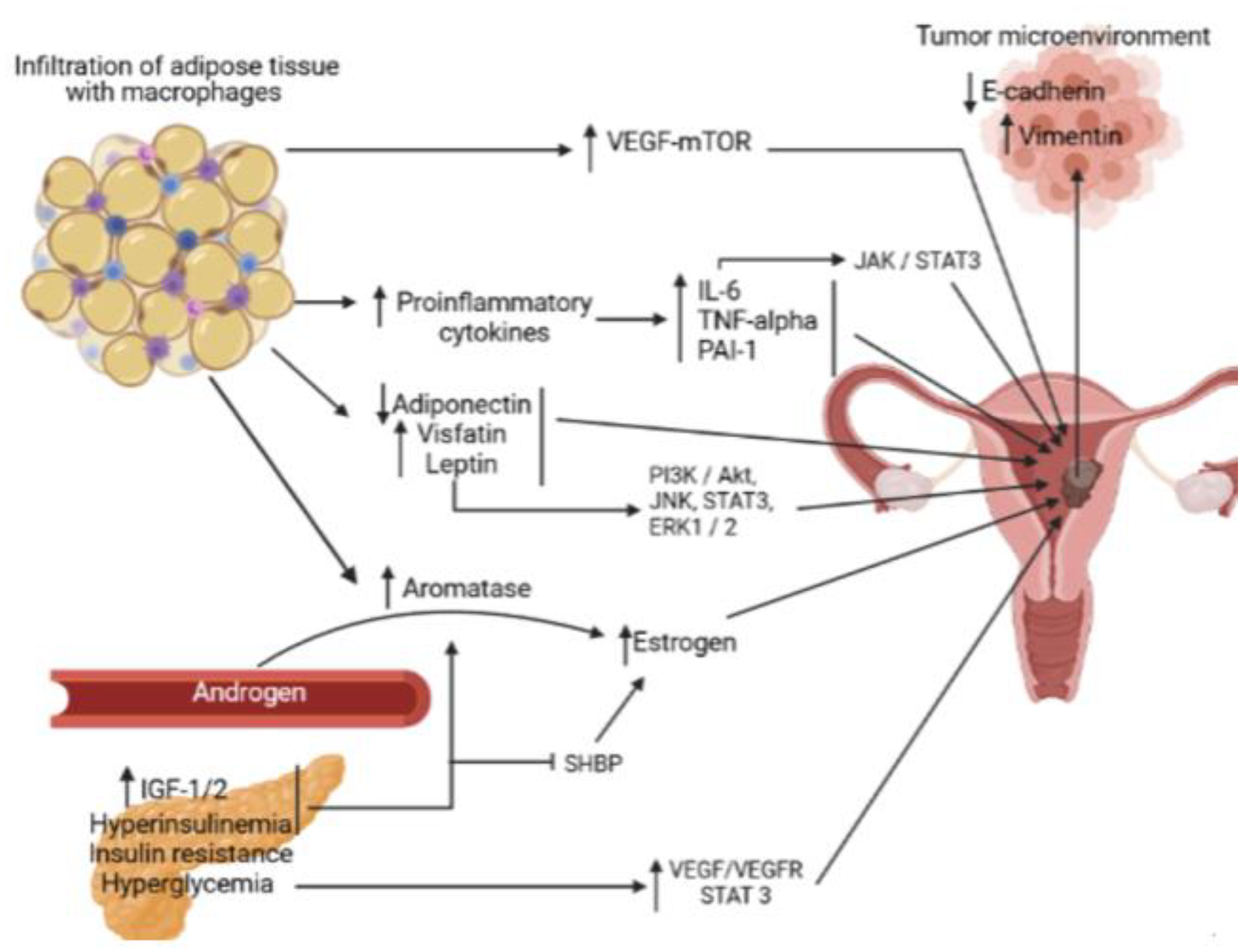
Hyperglycemia not only disrupts the signaling pathways responsible for the rapid cell proliferation in cancer (STAT3, PKM2) but it also favors the EMT via Erα/GLUT4 [57,58,59]. Figure 3 exemplifies the underlying mechanisms that link MS to EC.
In overweight/obese patients, the active adipose tissue produces high levels of leptin and visfatin, while the adiponectin levels are decreased creating a pro-tumorigenic state. Leptin hyperactivates the PI3K/Akt, JNK, STAT3, ERK1/2 signaling pathways, thus mediating the malignant transformation of endometrial cells and their proliferation. Additionally, there is a chronic pro-inflammatory status, characterized by the secretion of inflammatory cytokines: IL-6, TNF-alpha and PAI-1. The hyperactivation of IL-6/JAK/STAT3 signaling pathway regulates tumorigenesis, being associated with poor prognosis. The increased adipose tissue aromatase activity is responsible for the high circulating estrogens that mediate epithelial cell proliferation by interacting with estrogen receptors. The aromatase activity is accelerated by IGF-1/2, which also inhibits the synthesis of SHBP in patients with diabetes mellitus. The invasive phenotype of endometrial cancer cells is supported through the activation of VEGF-mTOR pathway. Additionally, hyperinsulinism stimulates the liver production of IGF-1, its high circulating levels increasing the risk of developing breast cancer. Hyperglycemia is responsible for cancer cell proliferation through the overexpression of VEGF/VEGFR and STAT3. Regarding the tumor microenvironment, the E-cadherin downregulation together with vimentin upregulation are associated with the epithelial–mesenchymal transition.

Highlights

A comprehensive summary of the mechanisms underlying the link between the metabolic syndrome and endometrial and breast cancer
The implication of inflammatory changes associated with the metabolic syndrome in oncogenesis

Conclusions

In conclusion, the metabolic syndrome is a complex condition characterized by insulin resistance, hyperinsulinemia, impaired glucose tolerance, type II diabetes, dyslipidemia and visceral obesity. Most of the molecular disturbances induced by MS are involved in the development, progression and prognosis of BC and EC. More importantly, although the studies of the association between the metabolic syndrome and cancer have shown contributions of genetic, biological factors (age, gender, ethnicity), MS represents an association of modifiable factors and this not only highlights the importance of improving lifestyle, balanced diet and regular physical activity in cancer prevention, but also the importance of combining these measures in the management of patients diagnosed with this disease, thus, in preventing morbidity and mortality in tumors associated with the metabolic syndrome.

Conflict of interest disclosure

There are no known conflicts of interest in the publication of this article. The manuscript was read and approved by all authors.

Compliance with ethical standards

Any aspect of the work covered in this manuscript has been conducted with the ethical approval of all relevant bodies and that such approvals are acknowledged within the manuscript.

References

  1. World Health Organization. GLOBOCAN 2018- Romania. Available online: https://gco.iarc.fr/today/data/factsheets/populations/642-romania-fact-sheets.pdf.
  2. Bhandari, R.; Kelley, G.A.; Hartley, T.A.; et al. Metabolic syndrome is associated with increased breast cancer risk: a systematic review with meta-analysis. Int J Breast Cancer. 2014, 2014, 189384. [Google Scholar] [CrossRef] [PubMed]
  3. Bohîlțea, R.E.; Furtunescu, F.; Dosius, M.; et al. Evaluation of endometrial cancer epidemiology in Romania. J Med Life. 2015, 8, 218–225. [Google Scholar] [PubMed]
  4. Crosbie, E.J.; Zwahlen, A.; Creutzberg, C.; et al. Endometrial cancer. Lancet. 2016, 387, 1094–1108. [Google Scholar] [CrossRef] [PubMed]
  5. Parikh, R.M.; Mohan, V. Changing definitions of metabolic syndrome. Indian J Endocrinol Metab. 2012, 16, 7–12. [Google Scholar] [CrossRef]
  6. Ghanbari Andarieh, M.; Agajani Delavar, M.; Moslemi, D.; Esmaeilzadeh, S. Risk Factors for Endometrial Cancer: Results from a Hospital-Based Case-Control Study. Asian Pac J Cancer Prev. 2016, 17, 4791–4796. [Google Scholar] [CrossRef]
  7. Al-Zoughool, M.; Dossus, L.; Kaaks, R.; et al. Risk of endometrial cancer in relationship to cigarette smoking: results from the EPIC study. Int J Cancer. 2007, 121, 2741–2747. [Google Scholar] [CrossRef]
  8. Viswanathan, A.N.; Feskanich, D.; De Vivo, I.; Hunter, D.J.; Barbieri, R.L.; Rosner, B.; Colditz, G.A.; Hankinson, S.E. Smoking and the risk of endometrial cancer: results from the Nurses' Health Study. Int J Cancer. 2005, 114, 996–1001. [Google Scholar] [CrossRef]
  9. Motofei, I.G. Biology of Cancer; From Cellular Cancerogenesis to Supracellular Evolution of Malignant Phenotype. Cancer Invest. 2018, 36, 309–317. [Google Scholar] [CrossRef]
  10. Raglan, O.; Kalliala, I.; Markozannes, G.; Cividini, S.; Gunter, M.J.; Nautiyal, J.; Gabra, H.; Paraskevaidis, E.; Martin-Hirsch, P.; Tsilidis, K.K.; Kyrgiou, M. Risk factors for endometrial cancer: An umbrella review of the literature. Int J Cancer. 2019, 145, 1719–1730. [Google Scholar] [CrossRef]
  11. Hamet, P. Cancer and hypertension: a potential for crosstalk? J Hypertens. 1997, 15 12 Pt 2, 1573–1577. [Google Scholar] [CrossRef]
  12. Soler, M.; Chatenoud, L.; Negri, E.; Parazzini, F.; Franceschi, S.; la Vecchia, C. Hypertension and hormone-related neoplasms in women. Hypertension. 1999, 34, 320–325. [Google Scholar] [CrossRef] [PubMed]
  13. Giovannucci, E.; Harlan, D.M.; Archer, M.C.; Bergenstal, R.M.; Gapstur, S.M.; Habel, L.A.; Pollak, M.; Regensteiner, J.G.; Yee, D. Diabetes and cancer: a consensus report. CA Cancer J Clin. 2010, 60, 207–221. [Google Scholar] [CrossRef] [PubMed]
  14. Bjørge, T.; Stocks, T.; Lukanova, A.; Tretli, S.; Selmer, R.; Manjer, J.; Rapp, K.; Ulmer, H.; Almquist, M.; Concin, H.; Hallmans, G.; Jonsson, H.; Stattin, P.; Engeland, A. Metabolic syndrome and endometrial carcinoma. Am J Epidemiol. 2010, 171, 892–902. [Google Scholar] [CrossRef]
  15. Trabert, B.; Wentzensen, N.; Felix, A.S.; Yang, H.P.; Sherman, M.E.; Brinton, L.A. Metabolic syndrome and risk of endometrial cancer in the United States: a study in the SEER-medicare linked database. Cancer Epidemiol Biomarkers Prev. 2015, 24, 261–267. [Google Scholar] [CrossRef]
  16. Yang, X.; Wang, J. The Role of Metabolic Syndrome in Endometrial Cancer: A Review. Front Oncol. 2019, 9, 744. [Google Scholar] [CrossRef]
  17. O'Connor, K.A.; Ferrell, R.J.; Brindle, E.; Shofer, J.; Holman, D.J.; Miller, R.C.; Schechter, D.E.; Singer, B.; Weinstein, M. Total and unopposed estrogen exposure across stages of the transition to menopause. Cancer Epidemiol Biomarkers Prev. 2009, 18, 828–836. [Google Scholar] [CrossRef] [PubMed]
  18. Crosbie, E.J.; Zwahlen, M.; Kitchener, H.C.; Egger, M.; Renehan, A.G. Body mass index, hormone replacement therapy, and endometrial cancer risk: a meta-analysis. Cancer Epidemiol Biomarkers Prev. 2010, 19, 3119–3130. [Google Scholar] [CrossRef]
  19. Pintaudi, B.; Di Vieste, G.; Nicolucci, A.; et al. The impact of clinical and psychological characteristics on alexithymia in type 1 diabetes. Mediterranean Journal of Clinical Psychology. 2021, 9. [Google Scholar] [CrossRef]
  20. Bruchim, I.; Sarfstein, R.; Werner, H. The IGF Hormonal Network in Endometrial Cancer: Functions, Regulation, and Targeting Approaches. Front Endocrinol (Lausanne). 2014, 5, 76. [Google Scholar] [CrossRef]
  21. Tian, W.; Teng, F.; Gao, J.; Gao, C.; Liu, G.; Zhang, Y.; Yu, S.; Zhang, W.; Wang, Y.; Xue, F. Estrogen and insulin synergistically promote endometrial cancer progression via crosstalk between their receptor signaling pathways. Cancer Biol Med. 2019, 16, 55–70. [Google Scholar] [CrossRef]
  22. de Luca, C.; Olefsky, J.M. Inflammation and insulin resistance. FEBS Lett. 2008, 582, 97–105. [Google Scholar] [CrossRef]
  23. Sahoo, S.S.; Tanwar, P.S. VEGF-mTOR signaling links obesity and endometrial cancer. Oncoscience. 2018, 5, 150–151. [Google Scholar] [CrossRef] [PubMed]
  24. Quail, D.F.; Dannenberg, A.J. The obese adipose tissue microenvironment in cancer development and progression. Nat Rev Endocrinol. 2019, 15, 139–154. [Google Scholar] [CrossRef]
  25. Li, M.; Li, X.; Zhao, L.; Zhou, J.; Cheng, Y.; Xu, B.; Wang, J.; Wei, L. Spontaneous formation of tumorigenic hybrids between human omental adipose-derived stromal cells and endometrial cancer cells increased motility and heterogeneity of cancer cells. Cell Cycle. 2019, 18, 320–332. [Google Scholar] [CrossRef]
  26. Shi, J.; Fan, J.; Su, Q.; Yang, Z. Cytokines and Abnormal Glucose and Lipid Metabolism. Front Endocrinol (Lausanne). 2019, 10, 703. [Google Scholar] [CrossRef] [PubMed]
  27. Cao, H. Adipocytokines in obesity and metabolic disease. J Endocrinol. 2014, 220, T47–T59. [Google Scholar] [CrossRef] [PubMed]
  28. Zeng, F.; Shi, J.; Long, Y.; Tian, H.; Li, X.; Zhao, A.Z.; Li, R.F.; Chen, T. Adiponectin and Endometrial Cancer: A Systematic Review and Meta-Analysis. Cell Physiol Biochem. 2015, 36, 1670–1678. [Google Scholar] [CrossRef]
  29. Gelsomino, L.; Naimo, G.D.; Catalano, S.; Mauro, L.; Andò, S. The Emerging Role of Adiponectin in Female Malignancies. Int J Mol Sci. 2019, 20, 2127. [Google Scholar] [CrossRef]
  30. Kadowaki, T.; Yamauchi, T. Adiponectin and adiponectin receptors. Endocr Rev. 2005, 26, 439–451. [Google Scholar] [CrossRef]
  31. Yabushita, H.; Iwasaki, K.; Obayashi, Y.; Wakatsuki, A. Clinicopathological roles of adiponectin and leptin receptors in endometrial carcinoma. Oncol Lett. 2014, 7, 1109–1117. [Google Scholar] [CrossRef]
  32. Peng, J.; Tsang, J.Y.; Ho, D.H.; Zhang, R.; Xiao, H.; Li, D.; Zhu, J.; Wang, F.; Bian, Z.; Lui, V.C.; Xu, A.; Tam, P.K.; Lamb, J.R.; Xia, H.; Chen, Y. Modulatory effects of adiponectin on the polarization of tumor-associated macrophages. Int J Cancer. 2015, 137, 848–858. [Google Scholar] [CrossRef]
  33. Cai, Z.F.; Deng, L.; Wang, M.M.; Zhang, J.Q.; Li, L. Effect of AMPK/mTOR/S6K1 pathways and the insulin- sensitizing effect for adiponectin in endometrial cancer cells. Zhonghua Fu Chan Ke Za Zhi. 2018, 53, 554–560. [Google Scholar] [CrossRef] [PubMed]
  34. Zheng, Q.; Wu, H.; Cao, J. Circulating adiponectin and risk of endometrial cancer. PLoS One. 2015, 10, e0129824. [Google Scholar] [CrossRef] [PubMed]
  35. Mohammadi, M.; Mianabadi, F.; Mehrad-Majd, H. Circulating visfatin levels and cancers risk: A systematic review and meta-analysis. J Cell Physiol. 2019, 234, 5011–5022. [Google Scholar] [CrossRef]
  36. Cymbaluk-Płoska, A.; Chudecka-Głaz, A.; Pius- Sadowska, E.; Sompolska-Rzechuła, A.; Machaliński, B.; Menkiszak, J. Circulating Serum Level of Visfatin in Patients with Endometrial Cancer. Biomed Res Int. 2018, 2018, 8576179. [Google Scholar] [CrossRef]
  37. Ciuhu, A.N.; Pantea-Stoian, A.M.; Nitipir, C.; et al. Assessment of cachexia in cancer patients with advanced disease. International Conference on Interdisciplinary Management of Diabetes Mellitus and its Complications (INTERDIAB) Interdiab 2017: Diabetes mellitus in internal medicine. 2017, 139–147. ISSN: 2393-3488.
  38. Wang, Z.; Gao, S.; Sun, C.; Li, J.; Gao, W.; Yu, L. Clinical significance of serum adiponectin and visfatin levels in endometrial cancer. Int J Gynaecol Obstet. 2019, 145, 34–39. [Google Scholar] [CrossRef]
  39. Zahid, H.; Subbaramaiah, K.; Iyengar, N.M.; Zhou, X.K.; Chen, I.C.; Bhardwaj, P.; Gucalp, A.; Morrow, M.; Hudis, C.A.; Dannenberg, A.J.; Brown, K.A. Leptin regulation of the p53-HIF1α/PKM2-aromatase axis in breast adipose stromal cells: a novel mechanism for the obesity-breast cancer link. Int J Obes (Lond). 2018, 42, 711–720. [Google Scholar] [CrossRef]
  40. Koushiou, M.; Kapatais, A.; Iasonidou, E.; Adonis, M.; Ferreira, N. The moderating role of body image inflexibility in the relation between weight concerns and symptoms of eating disorders in Cypriot University students. Mediterranean Journal of Clinical Psychology. 2021, 9. [Google Scholar] [CrossRef]
  41. Zhou, X.; Li, H.; Chai, Y.; Liu, Z. Leptin Inhibits the Apoptosis of Endometrial Carcinoma Cells Through Activation of the Nuclear Factor κB-inducing Kinase/IκB Kinase Pathway. Int J Gynecol Cancer. 2015, 25, 770–778. [Google Scholar] [CrossRef]
  42. Petridou, E.; Belechri, M.; Dessypris, N.; Koukoulomatis, P.; Diakomanolis, E.; Spanos, E.; Trichopoulos, D. Leptin and body mass index in relation to endometrial cancer risk. Ann Nutr Metab. 2002, 46, 147–51. [Google Scholar] [CrossRef]
  43. Gao, J.; Tian, J.; Lv, Y.; Shi, F.; Kong, F.; Shi, H.; Zhao, L. Leptin induces functional activation of cyclooxygenase-2 through JAK2/STAT3, MAPK/ERK, and PI3K/AKT pathways in human endometrial cancer cells. Cancer Sci. 2009, 100, 389–395. [Google Scholar] [CrossRef] [PubMed]
  44. Liu, Y.; Lv, L.; Xiao, W.; Gong, C.; Yin, J.; Wang, D.; Sheng, H. Leptin activates STAT3 and ERK1/2 pathways and induces endometrial cancer cell proliferation. J Huazhong Univ Sci Technolog Med Sci. 2011, 31, 365. [Google Scholar] [CrossRef]
  45. Ekremoğlu, M.; Severcan, Ç.; Pasaoğlu, Ö.T.; Şen, B.; Pasaoğlu, H. An investigation of acute effects at various doses of malathion on glucose homeostasis and insulin resistance in rat liver, pancreas and serum. J Mind Med Sci. 2020, 7, 85–93. [Google Scholar] [CrossRef]
  46. Lippitz, B.E.; Harris, R.A. Cytokine patterns in cancer patients: A review of the correlation between interleukin 6 and prognosis. Oncoimmunology. 2016, 5, e1093722. [Google Scholar] [CrossRef]
  47. He, Y.Y.; Cai, B.; Yang, Y.X.; Liu, X.L.; Wan, X.P. Estrogenic G protein-coupled receptor 30 signaling is involved in regulation of endometrial carcinoma by promoting proliferation, invasion potential, and interleukin-6 secretion via the MEK/ERK mitogen-activated protein kinase pathway. Cancer Sci. 2009, 100, 1051–1061. [Google Scholar] [CrossRef]
  48. Che, Q.; Liu, B.Y.; Liao, Y.; et al. Activation of a positive feedback loop involving IL-6 and aromatase promotes intratumoral 17β-estradiol biosynthesis in endometrial carcinoma microenvironment. Int J Cancer. 2014, 135, 282–94. [Google Scholar] [CrossRef]
  49. Johnson, D.E.; O'Keefe, R.A.; Grandis, J.R. Targeting the IL-6/JAK/STAT3 signalling axis in cancer. Nat Rev Clin Oncol. 2018, 15, 234–248. [Google Scholar] [CrossRef]
  50. Rahnea-Nita, G.; Badiu, D.C.; Popescu, C.G.; Grigorean, V.T.; Serban, D.; Smarandache, C.G.; Rahnea-Nita, R.A.; Ciuhu, A.N.; Mandu, M.; Andronache, L.F.; Stoian, A.R. The impact of Coronavirus disease 2019 pandemic on the treatment of cancer patients: the first steps in this fight. Mediterranean Journal of Clinical Psychology. 2021, 9. [Google Scholar] [CrossRef]
  51. Nair, S.; Nguyen, H.; Salama, S.; Al-Hendy, A. Obesity and the Endometrium: Adipocyte-Secreted Proinflammatory TNF α Cytokine Enhances the Proliferation of Human Endometrial Glandular Cells. Obstet Gynecol Int. 2013, 2013, 368543. [Google Scholar] [CrossRef]
  52. Vousden, K.A.; Lundqvist, T.; Popovic, B.; et al. Discovery and characterisation of an antibody that selectively modulates the inhibitory activity of plasminogen activator inhibitor-1. Sci Rep. 2019, 9, 1605. [Google Scholar] [CrossRef]
  53. Abbink, K.; Zusterzeel, P.L.M.; Geurts-Moespot, A.; van der Steen, R.; Span, P.N.; Sweep, F.C.G.J. Prognostic significance of VEGF and components of the plasminogen activator system in endometrial cancer. J Cancer Res Clin Oncol. 2020, 146, 1725–1735. [Google Scholar] [CrossRef] [PubMed]
  54. Suceveanu, A.I.; Stoian, A.P.; Parepa, I.; et al. Gut Microbiota Patterns in Obese and Type 2 Diabetes (T2D) Patients from Romanian Black Sea Coast Region. Revista de Chimie. 2018, 69, 2260–2267. [Google Scholar] [CrossRef]
  55. Folsom, A.R.; Anderson, K.E.; Sweeney, C.; Jacobs, D.R., Jr. Diabetes as a risk factor for death following endometrial cancer. Gynecol Oncol. 2004, 94, 740–5. [Google Scholar] [CrossRef] [PubMed]
  56. Byrne, F.L.; Martin, A.R.; Kosasih, M.; Caruana, B.T.; Farrell, R. The Role of Hyperglycemia in Endometrial Cancer Pathogenesis. Cancers (Basel). 2020, 12, 1191. [Google Scholar] [CrossRef]
  57. Gu, C.J.; Xie, F.; Zhang, B.; Yang, H.L.; Cheng, J.; He, Y.Y.; Zhu, X.Y.; Li, D.J.; Li, M.Q. High Glucose Promotes Epithelial-Mesenchymal Transition of Uterus Endometrial Cancer Cells by Increasing ER/GLUT4- Mediated VEGF Secretion. Cell Physiol Biochem. 2018, 50, 706–720. [Google Scholar] [CrossRef]
  58. Shah, H.; Shah, R.; Sanghani, H.; Lakhani, N. Health related quality of life (HRQoL) and its associated surgical factors in diabetes foot ulcer patients. J Clin Invest Surg. 2020, 5, 83–90. [Google Scholar] [CrossRef]
  59. Hsu, M.C.; Hung, W.C. Pyruvate kinase M2 fuels multiple aspects of cancer cells: from cellular metabolism, transcriptional regulation to extracellular signaling. Mol Cancer. 2018, 17, 35. [Google Scholar] [CrossRef]
  60. Choi, S.Y.; Collins, C.C.; Gout, P.W.; Wang, Y. Cancer- generated lactic acid: a regulatory, immunosuppressive metabolite? J Pathol. 2013, 230, 350–355. [Google Scholar] [CrossRef]
  61. Colegio, O.R. Lactic acid polarizes macrophages to a tumor-promoting state. Oncoimmunology. 2015, 5, e1014774. [Google Scholar] [CrossRef]
  62. Latif, A.; Chadwick, A.L.; Kitson, S.J.; Gregson, H.J.; Sivalingam, V.N.; Bolton, J.; McVey, R.J.; Roberts, S.A.; Marshall, K.M.; Williams, K.J.; Stratford, I.J.; Crosbie, E.J. Monocarboxylate Transporter 1 (MCT1) is an independent prognostic biomarker in endometrial cancer. BMC Clin Pathol. 2017, 17, 27. [Google Scholar] [CrossRef]
  63. Hernandez, A.V.; Pasupuleti, V.; Benites-Zapata, V.A.; Thota, P.; Deshpande, A.; Perez-Lopez, F.R. Insulin resistance and endometrial cancer risk: A systematic review and meta-analysis. Eur J Cancer. 2015, 51, 2747–2758. [Google Scholar] [CrossRef]
  64. Rodriguez, A.C.; Blanchard, Z.; Maurer, K.A.; Gertz, J. Estrogen Signaling in Endometrial Cancer: a Key Oncogenic Pathway with Several Open Questions. Horm Cancer. 2019, 10, 51–63. [Google Scholar] [CrossRef]
  65. Joehlin-Price, A.S.; Stephens, J.A.; Zhang, J.; Backes, F.J.; Cohn, D.E.; Suarez, A.A. Endometrial Cancer Insulin- Like Growth Factor 1 Receptor (IGF1R) Expression Increases with Body Mass Index and Is Associated with Pathologic Extent and Prognosis. Cancer Epidemiol Biomarkers Prev. 2016, 25, 438–445. [Google Scholar] [CrossRef] [PubMed]
  66. Bacalbasa, N.; Balescu, I.; Dimitriu, M.; Balalau, C.; Vilcu, M.; Brezean, I. Does sentinel lymph node detection play a role in patients with vaginal cancer? J Clin Invest Surg. 2019, 4, 1–4. [Google Scholar] [CrossRef]
  67. Mu, N.; Xu, T.; Gao, M.; Dong, M.; Tang, Q.; Hao, L.; Wang, G.; Li, Z.; Wang, W.; Yang, Y.; Hou, J. Therapeutic effect of metformin in the treatment of endometrial cancer. Oncol Lett. 2020, 20, 156. [Google Scholar] [CrossRef] [PubMed]
  68. Chu, D.; Wu, J.; Wang, K.; Zhao, M.; Wang, C.; Li, L.; Guo, R. Effect of metformin use on the risk and prognosis of endometrial cancer: a systematic review and meta- analysis. BMC Cancer. 2018, 18, 438. [Google Scholar] [CrossRef]
  69. Nevadunsky, N.S.; Van Arsdale, A.; Strickler, H.D.; Moadel, A.; Kaur, G.; Frimer, M.; Conroy, E.; Goldberg, G.L.; Einstein, M.H. Metformin use and endometrial cancer survival. Gynecol Oncol. 2014, 132, 236–240. [Google Scholar] [CrossRef]
  70. Arthur, R.S.; Kabat, G.C.; Kim, M.Y.; Wild, R.A.; Shadyab, A.H.; Wactawski-Wende, J.; Ho, G.Y.F.; Reeves, K.W.; Kuller, L.H.; Luo, J.; Beebe-Dimmer, J.; Simon, M.S.; Strickler, H.; Wassertheil-Smoller, S.; Rohan, T.E. Metabolic syndrome and risk of endometrial cancer in postmenopausal women: a prospective study. Cancer Causes Control. 2019, 30, 355–363. [Google Scholar] [CrossRef]
  71. Bruning, P.F.; Bonfrèr, J.M. Free fatty acid concentrations correlated with the available fraction of estradiol in human plasma. Cancer Res. 1986, 46, 2606–2609. [Google Scholar]
  72. Li, P.; Shan, B.; Jia, K.; Hu, F.; Xiao, Y.; Zheng, J.; Gao, Y.T.; Wang, H.; Gao, Y. Plasma omega-3 polyunsaturated fatty acids and recurrence of endometrial cancer. BMC Cancer. 2020, 20, 576. [Google Scholar] [CrossRef]
  73. Yao, L.; Chen, S.; Li, W. Fatostatin inhibits the development of endometrial carcinoma in endometrial carcinoma cells and a xenograft model by targeting lipid metabolism. Arch Biochem Biophys. 2020, 684, 108327. [Google Scholar] [CrossRef] [PubMed]
  74. Mackintosh, M.L.; Crosbie, E.J. Obesity-driven endometrial cancer: is weight loss the answer? BJOG. 2013, 120, 791–794. [Google Scholar] [CrossRef]
  75. Linkov, F.; Goughnour, S.L.; Ma, T.; Xu, Z.; Edwards, R.P.; Lokshin, A.E.; Ramanathan, R.C.; Hamad, G.G.; McCloskey, C.; Bovbjerg, D.H. Changes in inflammatory endometrial cancer risk biomarkers in individuals undergoing surgical weight loss. Gynecol Oncol. 2017, 147, 133–138. [Google Scholar] [CrossRef]
  76. Sun, Y.S.; Zhao, Z.; Yang, Z.N.; Xu, F.; Lu, H.J.; Zhu, Z.Y.; Shi, W.; Jiang, J.; Yao, P.P.; Zhu, H.P. Risk Factors and Preventions of Breast Cancer. Int J Biol Sci. 2017, 13, 1387–1397. [Google Scholar] [CrossRef] [PubMed]
  77. Dibaba, D.T.; Braithwaite, D.; Akinyemiju, T. Metabolic Syndrome and the Risk of Breast Cancer and Subtypes by Race, Menopause and BMI. Cancers (Basel). 2018, 10, 299. [Google Scholar] [CrossRef]
  78. van Oers, H.; Schlebusch, L. Indicators of psychological distress and body image disorders in female patients with breast cancer. J Mind Med Sci. 2020, 7, 179–187. [Google Scholar] [CrossRef]
  79. Chen, Y.; Wen, Y.Y.; Li, Z.R.; Luo, D.L.; Zhang, X.H. The molecular mechanisms between metabolic syndrome and breast cancer. Biochem Biophys Res Commun. 2016, 471, 391–395. [Google Scholar] [CrossRef]
  80. Belardi, V.; Gallagher, E.J.; Novosyadlyy, R.; LeRoith, D. Insulin and IGFs in obesity-related breast cancer. J Mammary Gland Biol Neoplasia. 2013, 18, 277–89. [Google Scholar] [CrossRef] [PubMed]
  81. Agnoli, C.; Grioni, S.; Sieri, S.; Sacerdote, C.; Ricceri, F.; Tumino, R.; Frasca, G.; Pala, V.; Mattiello, A.; Chiodini, P.; Iacoviello, L.; De Curtis, A.; Panico, S.; Krogh, V. Metabolic syndrome and breast cancer risk: a case- cohort study nested in a multicentre italian cohort. PLoS One. 2015, 10, e0128891. [Google Scholar] [CrossRef]
  82. Guo, M.; Liu, T.; Li, P.; Wang, T.; Zeng, C.; Yang, M.; Li, G.; Han, J.; Wu, W.; Zhang, R. Association Between Metabolic Syndrome and Breast Cancer Risk: An Updated Meta-Analysis of Follow-Up Studies. Front Oncol. 2019, 9, 1290. [Google Scholar] [CrossRef]
  83. Maiti, B.; Kundranda, M.N.; Spiro, T.P.; et al. The association of metabolic syndrome with triple-negative breast cancer. Breast Cancer Res Treat. 2010, 121, 479–483. [Google Scholar] [CrossRef] [PubMed]
  84. Guenancia, C.; Lefebvre, A.; Cardinale, D.; et al. Obesity as a Risk Factor for Anthracyclines and Trastuzumab Cardiotoxicity in Breast Cancer: A Systematic Review and Meta-Analysis. J Clin Oncol. 2016, 34, 3157–3165. [Google Scholar] [CrossRef] [PubMed]
  85. Maruyama, S.; Shibata, R.; Ohashi, K.; Ohashi, T.; Daida, H.; Walsh, K.; Murohara, T.; Ouchi, N. Adiponectin ameliorates doxorubicin-induced cardiotoxicity through Akt protein-dependent mechanism. J Biol Chem. 2011, 286, 32790–32800. [Google Scholar] [CrossRef]
  86. Esposito, K.; Chiodini, P.; Capuano, A.; Bellastella, G.; Maiorino, M.I.; Rafaniello, C.; Giugliano, D. Metabolic syndrome and postmenopausal breast cancer: systematic review and meta-analysis. Menopause. 2013, 20, 1301–1309. [Google Scholar] [CrossRef] [PubMed]
  87. Li, P.; Wang, T.; Zeng, C.; et al. Association between metabolic syndrome and prognosis of breast cancer: a meta-analysis of follow-up studies. Diabetol Metab Syndr. 2020, 12, 10. [Google Scholar] [CrossRef]
  88. Berrino, F.; Villarini, A.; Traina, A.; Bonanni, B.; Panico, S.; Mano, M.P.; Mercandino, A.; Galasso, R.; Barbero, M.; Simeoni, M.; Bassi, M.C.; Consolaro, E.; Johansson, H.; Zarcone, M.; Bruno, E.; Gargano, G.; Venturelli, E.; Pasanisi, P. Metabolic syndrome and breast cancer prognosis. Breast Cancer Res Treat. 2014, 147, 159–165. [Google Scholar] [CrossRef]
  89. Tecalco-Cruz, A.C.; Pérez-Alvarado, I.A.; Ramírez- Jarquín, J.O.; Rocha-Zavaleta, L. Nucleo-cytoplasmic transport of estrogen receptor alpha in breast cancer cells. Cell Signal. 2017, 34, 121–132. [Google Scholar] [CrossRef]
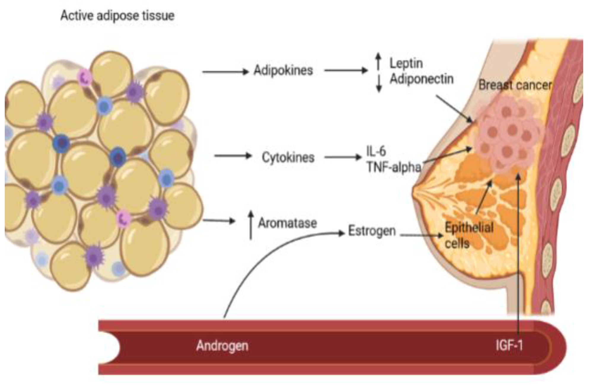
Figure 2. The major mechanisms linking MS to BC pathogenesis. The active adipose tissue disrupts the adipokine homeostasis by producing high levels of leptin, while the adiponectin levels are decreased, thus creating a pro-tumorigenic state. Additionally, the chronic pro-inflammatory status, characterized by the secretion of inflammatory cytokines (IL-6, TNF-alpha), regulates several processes in tumorigenesis. The increased adipose tissue aromatase activity is responsible for the high circulating estrogens that mediate epithelial cell proliferation by interacting with the estrogen receptors. Additionally, hyperinsulinism stimulates the liver production of IGF-1, its high circulating levels increasing the risk of developing breast cancer.
Jmms 08 00022 g002
Figure 3. The underlying mechanisms that link MS to EC.
Jmms 08 00022 g003
Table 1. WHO definition of MS (5).
Jmms 08 00022 i001
Table 2. Risk factors for breast cancer [76].
Jmms 08 00022 i003