Next Article in Journal
Early Maladjustment Schemas in Individuals With and Without Type 2 Diabetes Mellitus
Previous Article in Journal
A Perfusion Decellularization Heart Model—An Interesting Tool for Cell-Matrix Interaction Studies
 
 
Journal of Mind and Medical Sciences is published by MDPI from Volume 12 Issue 1 (2025). Previous articles were published by another publisher in Open Access under a CC-BY (or CC-BY-NC-ND) licence, and they are hosted by MDPI on mdpi.com as a courtesy and upon agreement with Valparaiso University (ValpoScholar).
Article

The Correlation Between Histopathological and Ultrasound Findings Regarding Cesarean Section Scars—A Three-Year Survey Study

by
Oana D. Bălălău
1,2,
Nicolae Bacalbașa
1,
Cristian Bălălău
1,*,
Carolina Negrei
1,
Bianca Gălățeanu
3,
Octav Ghinghină
1,4,
Cristina Răduță
2,
Liana Pleș
1,2,
Anca D. Stănescu
1,2 and
Vasile A. Dumitru
1,5
1
Carol Davila University of Medicine and Pharmacy, Bucharest, Romania
2
Bucur Maternity, St. Ioan Emergency Hospital, Bucharest, Romania
3
Department of Biochemistry and Molecular Biology, The University of Bucharest, Bucharest, Romania
4
Department of Surgery, St. Ioan Clinical Emergency Hospital, Bucharest, Romania
5
Department of Pathology, The Emergency University Hospital Bucharest, Bucharest, Romania
*
Author to whom correspondence should be addressed.
J. Mind Med. Sci. 2019, 6(1), 143-149; https://doi.org/10.22543/7674.61.P143149
Submission received: 2 March 2018 / Revised: 20 April 2018 / Accepted: 6 July 2018 / Published: 27 April 2019

Highlights

  • The predictive ultrasound parameters for the risk of rupture/dehiscence of the uterine scar showed varying cut-off values, ranging between 2.0 and 3.5 mm for the lower segment and up to 0.97 mm for the myometrium.
  • This observation of inverse proportionality between the uterine thickness and the risk of rupture/dehiscence of the scar seems to be correlated with the histopathological features of the cesarean section scar.

Abstract

:

Highlights

  • The predictive ultrasound parameters for the risk of rupture/dehiscence of the uterine scar showed varying cut-off values, ranging between 2.0 and 3.5 mm for the lower segment and up to 0.97 mm for the myometrium.
  • This observation of inverse proportionality between the uterine thickness and the risk of rupture/dehiscence of the scar seems to be correlated with the histopathological features of the cesarean section scar.

Abstract

The cesarean operation represents a major surgery, with a higher risk of postoperative complications and longer postoperative recovery than vaginal delivery. Due to the increasing frequency of cesarean sections, the ultrasound imaging of the uterine scar has become a particularly useful tool in identifying its potential long-term complications. This should be done pre-conceptively and quarterly or whenever necessary during pregnancy. Currently, there are only few histopathological studies on the uterine scar, trying to assess the myometrial repair and certain factors that influence the quality of the scar. The study was performed on a batch of 123 patients with previous C-sections, with multiple ultrasound exams during pregnancy and post-operative pathologic evaluation of the uterine scar in order to assess the possibility of a new prognostic score by correlating these two factors. Our study found solid evidence related to possible correlations between histopathological and ultrasound data on the cesarean section scar, which could lead to a possible predictive algorithm with implications for both prognostic and therapeutic fields.

Introduction

The uterine scar is the result of numerous events responsible for the tissular healing process, the most common manifestations of which are: connective tissue with increased amount of immature collagen, partially acellular in the subserosa, hemorrhagic extravasation between the myometrium and the subserosa and microhematomata between the myometrium and scar tissue. In some cases, the architecture of the scar may be significantly altered with the emergence of rigid structures due to muscle fiber merging that are subsequently replaced by connective tissue rich in fibroblasts or adult acellular connective tissue. Sometimes, persistent inflammatory processes are seen for many years after the last cesarean section or areas of endometriosis that extend from the lumen to the serosa. The thickness of the scar can be significantly diminished, and an atrophic and very thin myometrium can be observed, covered by well vascularized visceral peritoneum. Granulomata, sclerohialin areas with microcalcifications and isolated pseudomixomatous lesions have also been described [1,2].
Along with the increasing frequency of cesarean sections, the ultrasound imaging of uterine scar has become a particularly useful tool in identifying its potential long-term complications.
Attempts to visualize scar defects began in 1961 through hysterosalpingography [1,3], later followed by the transabdominal ultrasound approach and transvaginal ultrasound starting with 1982 [2] and 1990 [3] respectively, and more recently by sonohysterography with saline or gel instillation (SHG) or hysteroscopy [4,5,6].
Uterine scar defects, described as echogenic areas at previous C-section levels, were named niche by Monteagudo et al. in 2001 [7], but also diverticulum, isthmocele or dehiscence [8].
The percentage of uterine scar defects varied throughout the studies according to the method of assessment used, the criteria to define the niche and the study group [9]. In 2011, Bij de Vaate et al. conducted a study and found a 24% prevalence of niche in patients evaluated through transvaginal ultrasound and a 56% prevalence for SHG 6-12 months after the C-section [5]. In 2014, Van der Voet et al. identified higher percentages on a similar group of women, 49.6% through transvaginal ultrasound assessment and 64.5% through SHG assessment [6].

Materials and Methods

This longitudinal prospective study was conducted at “Bucur” Maternity for a period of 4 years, and its aim was to correlate the dynamic evolution of the uterine scar (monitored by means of ultrasound) with the pathological results obtained after the biopsy. For a 4-year survey study, a group of 123 patients with scar tissue after the cesarean surgery had undergone two measurements of the uterine scar (in the 2nd and 3rd trimester of pregnancy) through abdominal and pelvic ultrasound.
To avoid autolysis and to preserve the integrity of the biopsy items, the biological products were fixed using a buffered formaldehyde solution with a concentration of 10%. The average duration of the fastening process was 12 hours.
After this time, the biopsy pieces were processed using the specific histopathological procedures. The images were obtained using Leica DM750 microscope, after van Giemson, Orceine, Hematoxiline-eozine, Congo red and Masson coloration.
Patients with monofetal pregnancies with segmento-transversal hysterotomy (not “T”-shaped) were included in the study and not those with uterine scars after myomectomy or other interventions on the uterus.

Results

All patients had a history of one or more C-sections, multiparous with both vaginal birth history and cesarean delivery being excluded from the study. The patients’ age ranged between 24 and 41 years, with 68% of them aged between 30 and 40 years (Figure 1).
The distribution of multiparity among the study group was 78% with one previous C-section, and in 76% of cases, the gestational age at the time of delivery ranged between 38-39 weeks (Figure 2).
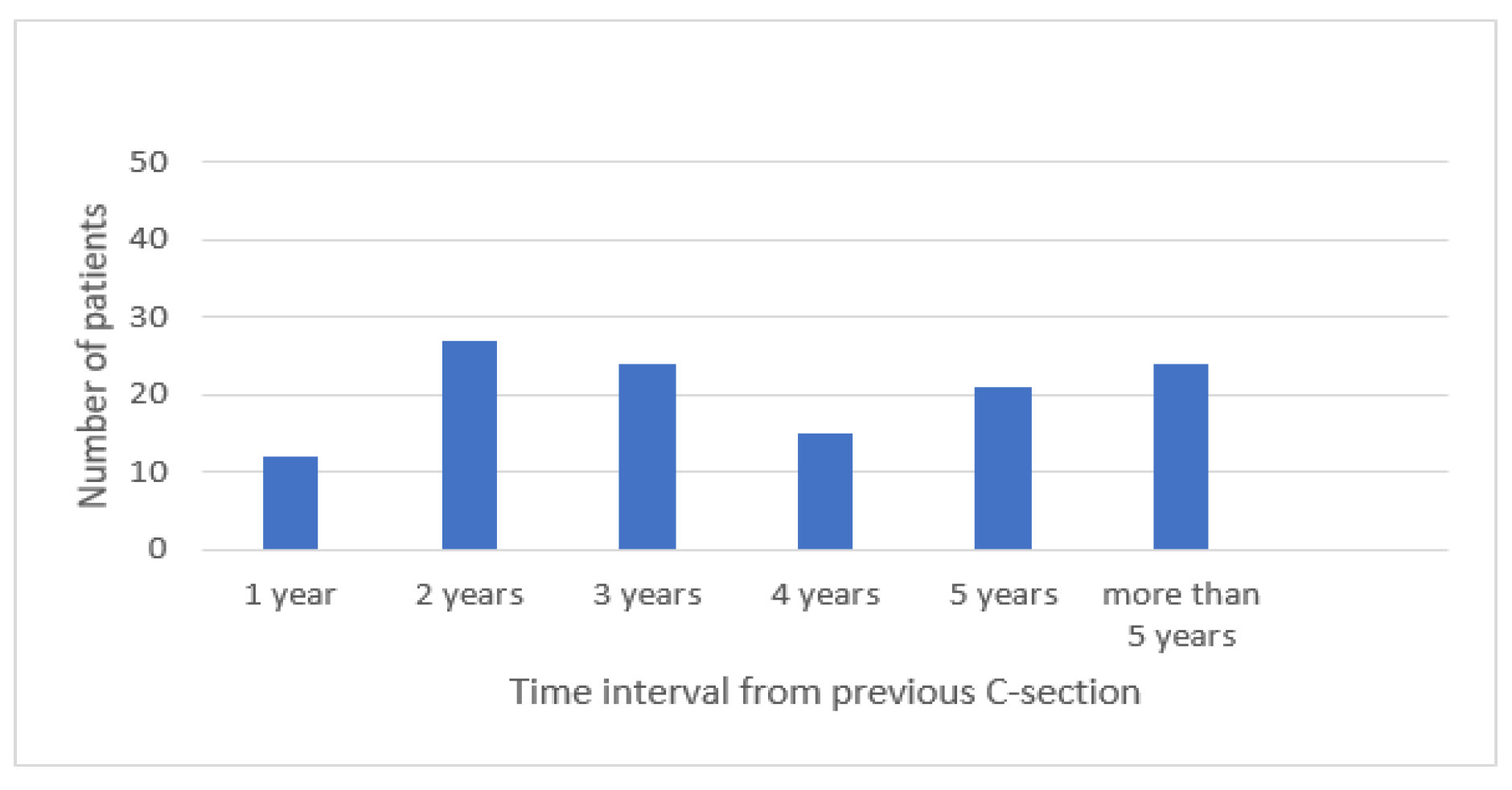
More than 80% of the patients had a previous C-section in the last 5 years, with 52% in the last 3 years (Figure 3).
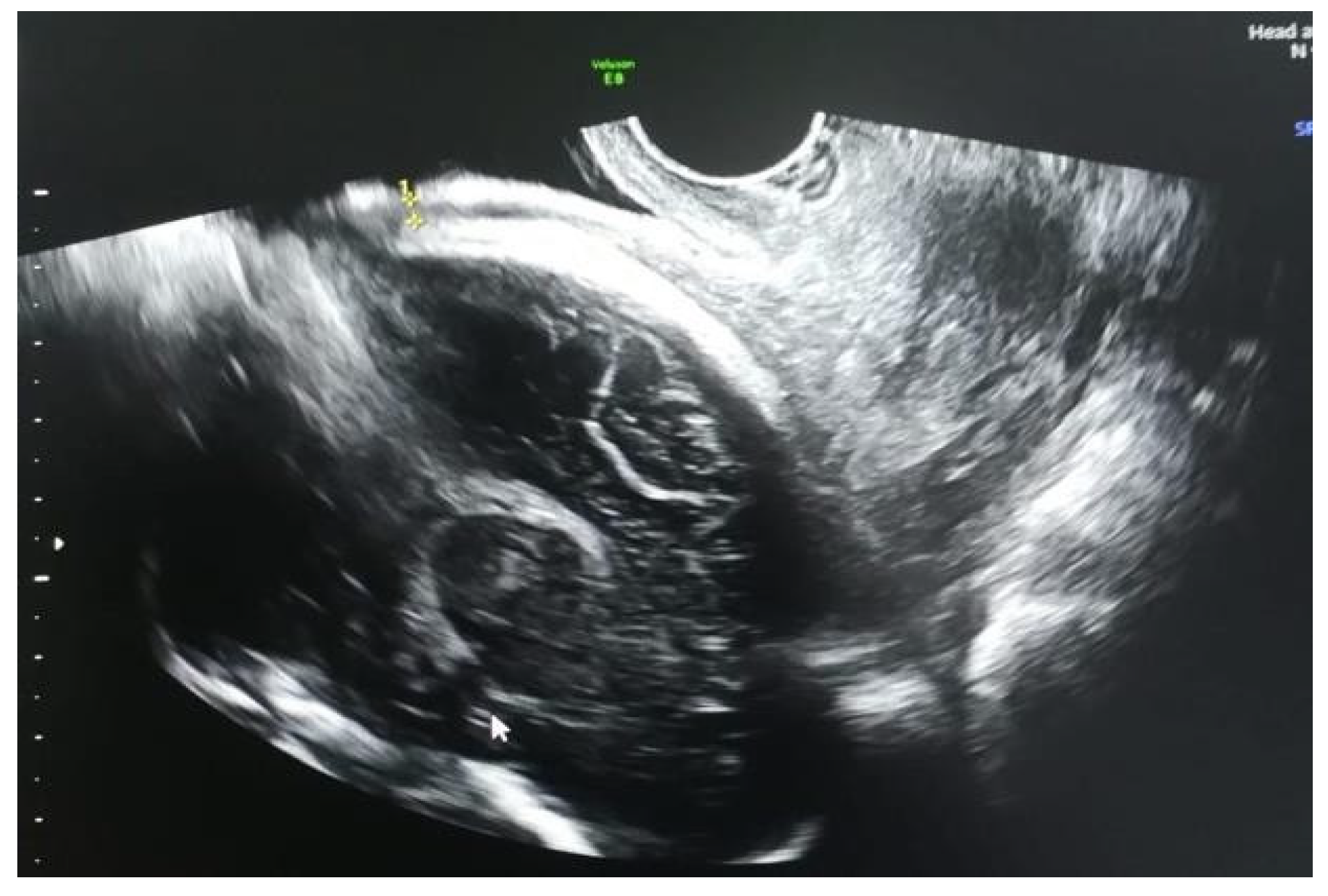
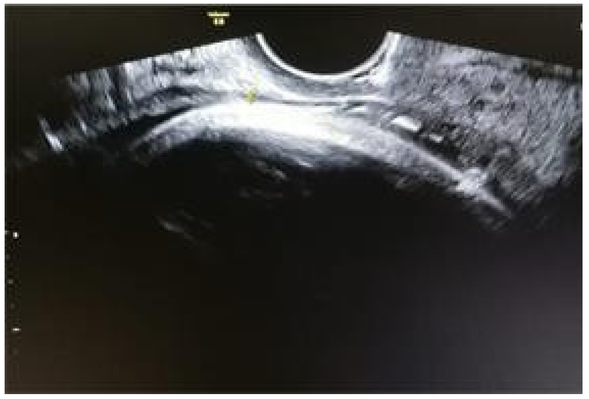
Patients underwent two measurements of the uterine scar, during the 2nd and 3rd trimester of pregnancy through abdominal and pelvic ultrasound (Figure 4 and Figure 5).
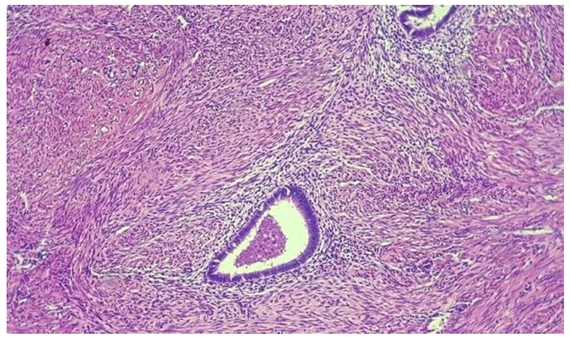
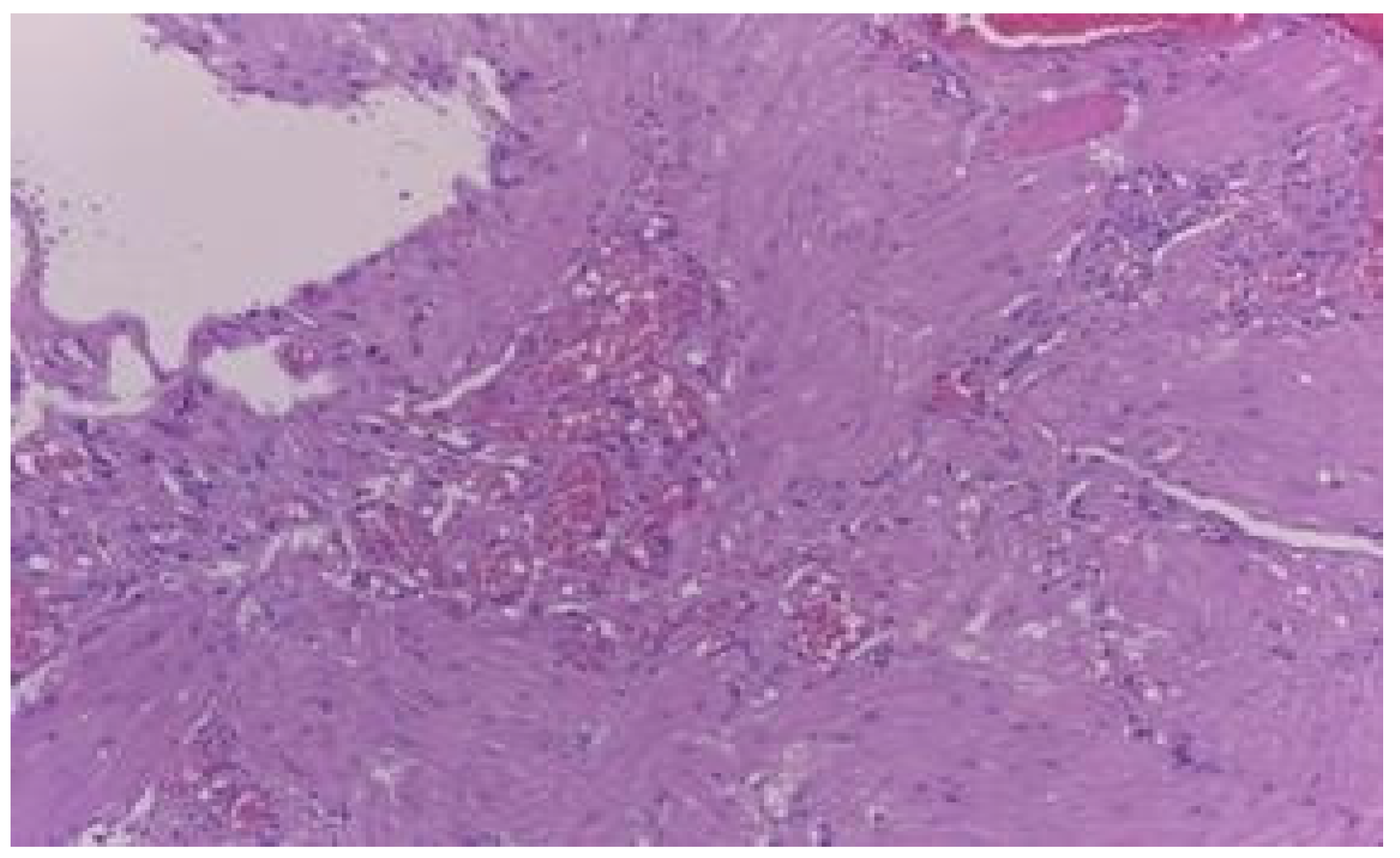
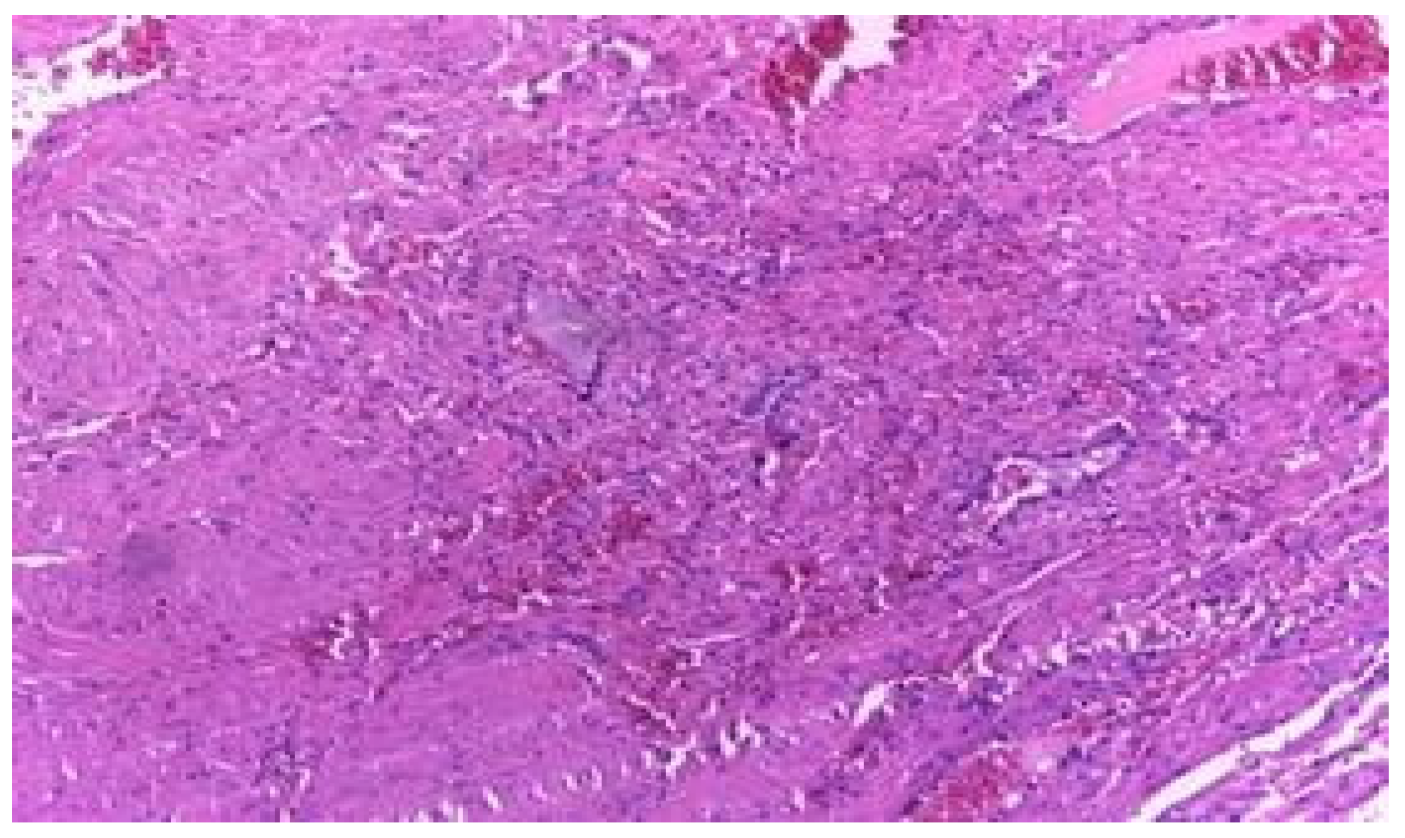
The biopsies taken from the uterine scar at the time of delivery by C-section revealed adenomyosis, inflammatory infiltration, granulation tissue and vascular neoformation, elastin and collagen disposition (Figure 6, Figure 7, Figure 8 and Figure 9).
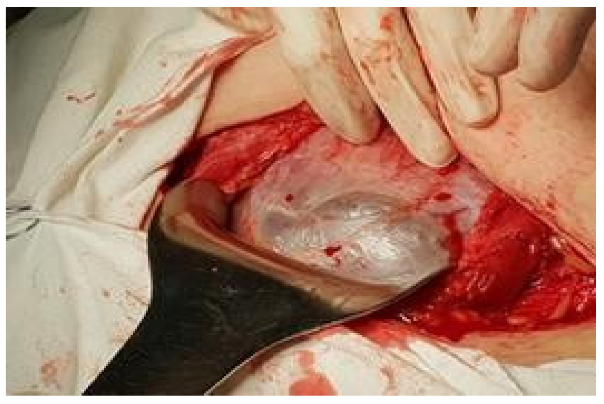
Forty-two patients (34%) presented a scar dehiscence at the moment of the C-section (Figure 10).

Discussions

Most imaging studies of scar defects have been performed on a non-pregnant uterus, making it more difficult to interpret them in the context of a subsequent pregnancy. Osser et al. suggested a possible relationship between the morphology of the scar on the non-pregnant woman and its behavior in a later pregnancy [4].
The enlarged niche, which is less frequent, has been considered in several studies as a defect penetrating up to at least 50% of the thickness of the anterior myometrium or a defect revealed by the thickness of the residual micrometer ≤ 2.2 mm and ≤ 2.5 mm when evaluated by ultrasound, and by SHG respectively [8,9].
Bij de Vaate et al. [9] have systematized several studies that have referred to the risk factors for the occurrence of scarring defects in 4 categories:
  • o factors regarding hysterorrhaphy
  • o factors related to lower segment formation and scar location
  • o factors with possible negative impact on scar healing
  • o other factors: maternal age, multiple pregnancies, vaginal births, placenta praevia, etc.
The suture of the entire thickness of the myometrium, including the endometrial layer as well as the suture within the two myometrial layers or in one myometrial layer but including the endometrium were associated with a lower frequency of the niche [10,11,12].
From the point of view of the factors related to the lower segment and the location of the scar, some of the studies were contradictory. According to Vikhareva Osser et al., the presentation of the fetal mobile below the pelvic inlet, the cervical dilation ≥5 cm or the duration of labor ≥5 hours would constitute risk factors [10], while Yazicioglu et al. find the possibility of a higher niche incidence in association with a lower cervical dilation [11]. Hayakawa et al. have reported premature membrane rupture and high gestational age at birth as risk factors [10], while Yazicioglu et al. did not find any correlation between the gestational age and the occurrence of the uterine defect. Emergency C-section and the presence of labor have not been identified as risk factors [10,11].
Potential factors that have a negative influence such as retroverted uterus, preeclampsia and the number of cesarean operations have been identified [10,11,13].
The lower segment is ultrasound visualized as a two-layer structure: a hyperechogenic one, representing the urinary bladder wall and another, with a lower echogenicity, representing the myometrium. To identify scar defects, most authors suggest longitudinal and transverse sections and at least three measurements, taking into account the smallest value.
A high quality uterine scar in a more than 35-week pregnancy should have the following characters: a minimum thickness of 3.5 mm, a well-shaped and homogeneous scar of triangular shape with a volume up to 10 cubic cc and appropriate perfusion around the scar [8].
The predictive value of the lower segment thickness for the risk of rupture/dehiscence of the uterine scar has been assessed by means of ultrasound in several studies performed during pregnancy, and suggested cut-off values ranging between 2.0 and 3.5 mm [14,15,16]. The conclusions of these studies established a relationship of inverse proportionality between the lower segment thickness and the risk of rupture/dehiscence of the scar, subsequently confirmed by the meta-analysis performed by Kok N et al. in 2014, which synthesized the results of 12 studies [17,18,19]. In 2016, Tadatsugu Kinjo et al. published a paper on the same topic, with similar conclusions, with values higher than 0.97 mm for the myometrium and 3.13 mm for the lower segment which were considered risk factors [20]. The lower segment thickness of less than 2.3 mm is associated with a higher risk of complete uterine rupture. However, no clinically applicable cut-off values have been established so far [21,22].
The rupture of the uterine scar prior to C-section, along with other potential life-threatening co-morbidities like cervical cancer [23], could lead to an emergency hysterectomy, with physical and mental consequences [24,25,26].
The dimensions of the uterine scar are changing during pregnancy, as Naji et al showed in a prospective study conducted in 2013 that monitored the development of uterine scar throughout pregnancy on a group of 320 women. The ultrasound was performed at 11-13, 19-21, 32-34 weeks of gestation. Maternal age was associated with changes in the scar during pregnancy, while, contrary to expectations, the body mass index (BMI), the diabetic history, smoking, postpartum infections and the number of cesarean sections did not. The residual myometrium thickness (RMT) decreased throughout gestation, in complicated cases with uterine rupture reaching a decrease of 2.5-2.7 mm between the first and the second trimester and an average thickness of the residual micrometer of 0.5 mm versus 3.6 mm in the other cases. RMT recorded a lower dynamics of changes during pregnancy and a larger size in women who had vaginal delivery after cesarean surgery than the others, and has been proposed as an indicator of scar integrity. All these ultrasound data could be determined starting with 20 weeks of gestation [27].
The healing mechanism of the uterine scar has been largely debated in time. At the beginning of the 20th century, the hypothesis that healing is the result of a myometrial regeneration that does not result in scar formation was formulated. In 1938 and later in 1952, Schwartz et al. and respectively Siegel showed that a fibroblastic reaction led to the formation of a scar that could then be invaded by muscle cells [28]. In 1970, Wojdecki and Grynsztajn demonstrated the occurrence of grain tissue and fibrosis at the niche level in the first 18 days after cesarean surgery [29].
In 1995, Morris described various changes and the frequency with which they appeared at the niche level, based on the histopathological analysis of 51 hysterectomy specimens from patients with previous cesarean operations: distortion and widening of the isthmus (75%), “overhanging” of congested endometrium above the scar recess (61%), polyps (16%), moderate to important lymphocyte infiltration (65%), residual suture material with giant-cellular foreign body reaction (92%), capillary dilation (65%), recent hemorrhage in the endometrial stroma around niches (59%), fragmentation and breakage of the endometrium (37%), adenomyosis (28%) [30]. Similar histopathological results have been described by Refaat et al. in a study in 2014: lymphocyte infiltration, isthmic distortion, iatrogenic adenomyosis, disorganized muscle fibers in the scar area, congested endometrial fold, changes occurring at as a high frequency as the number of cesarean sections performed [31]. The authors found a higher incidence of ischemic distortion and endometrial folds as far as the distance to the inner cervix was concerned, suggesting that an incision made above is a risk factor for greater pathological changes [31].
The quality of the myometrial healing process has a role in defining the characteristics of the future scar [32]. According to Siegel, increased proliferation of connective interstitial tissue and increased vascularization in pregnancy with the formation of a greater number of fibroblasts would favor the scar of the pregnant uterus compared to the non-pregnant uterus [1].
Larger and deeper myometrial defects are associated with the absence of re-epithelialising of the scar area [32,33].
In 2014, a study on uterine arterial circulation showed that in women with a history of cesarean operations, the resistance in uterine artery is increased and the volume of uterine blood flow distributed to utero-placental circulation is lower compared to patients with spontaneous vaginal births in the past. These data would suggest a possible involvement of the factors described above together with a scarce vascularization of the uterine scar, in the process of re-epithelialising of the scar area [34].

Conclusions

Currently, there are few histopathological studies on uterine scars, and the myometrial repair is somewhat dependent on certain factors, some of which are described above. This requires further detailed analysis for the complete understanding of the pathogenesis of scar defect and its subsequent implications on the non-pregnant uterus, but especially on the pregnant uterus. The latter could lead to complications that may result in the death of the mother.
Ultrasound evaluation of the uterine scar should be performed before conception and especially during pregnancy, when it is necessary to be performed in a dynamic, quarterly manner. Changes in uterine scar, the possibility of opting for spontaneous birth after cesarean surgery or possible birth complications can be identified as early as 20 weeks of pregnancy.
The data provided by the studies conducted so far are insufficient to achieve an algorithm to be introduced into clinical practice.
Our study found solid evidence though correlations between histopathological and ultrasound findings, regarding cesarean section scars, that could lead to a predictive algorithm for both prognostic and therapeutic means.

Conflict of interest disclosure

There are no known conflicts of interest in the publication of this article. The manuscript was read and approved by all authors.

Compliance with ethical standards

Any aspect of the work covered in this manuscript has been conducted with the ethical approval of all relevant bodies and that such approvals are acknowledged within the manuscript.

References

  1. Poidevin, L.O. The value of hysterography in the prediction of cesarean section wound defects. Am J Obstet Gynecol. 1961, 81, 67–71. [Google Scholar] [CrossRef] [PubMed]
  2. Burger, N.F.; Darazs, B.; Boes, E.G. An echographic evaluation during the early puerperium of the uterine wound after caesarean section. J Clin Ultrasound. 1982, 10, 271–274. [Google Scholar] [CrossRef] [PubMed]
  3. Chen, H.Y.; Chen, S.J.; Hsieh, F.J. Observation of cesarean section scar by transvaginal ultrasonography. Ultrasound Med Biol. 1990, 16, 443–447. [Google Scholar] [CrossRef] [PubMed]
  4. Osser, O.V.; Jokubkiene, L.; Valentin, L. Cesarean section scar defects: agreement between transvaginal sonographic findings with and without saline contrast enhancement. Ultrasound Obstet Gynecol. 2010, 35, 75–83. [Google Scholar] [CrossRef]
  5. Bij de Vaate, A.J.; Brölmann, H.A.; van der Voet, L.F.; van der Slikke, J.W.; Veersema, S.; Huirne, J.A. Ultrasound evaluation of the Cesarean scar: relation between a niche and postmenstrual spotting. Ultrasound Obstet Gynecol. 2011, 37, 93–99. [Google Scholar] [CrossRef]
  6. Van der Voet, L.F.; Bij de Vaate, A.M.; Veersema, S.; Brölmann, H.A.; Huirne, J.A. Long-term complications of caesarean section. The niche in the scar: a prospective cohort study on niche prevalence and its relation to abnormal uterine bleeding. BJOG. 2014, 121, 236–244. [Google Scholar] [CrossRef]
  7. Monteagudo, A.; Carreno, C.; Timor-Tritsch, I.E. Saline infusion sonohysterography in nonpregnant women with previous cesarean delivery: the "niche" in the scar. J Ultrasound Med. 2001, 20, 1105–1115. [Google Scholar] [CrossRef]
  8. Basic, E.; Basic-Cetkovic, V.; Kozaric, H.; Rama, A. Ultrasound evaluation of uterine scar after cesarean section. Acta Inform Med. 2012, 20, 149–153. [Google Scholar] [CrossRef]
  9. Bij de Vaate, A.J.; van der Voet, L.F.; Naji, O.; Witmer, M.; Veersema, S.; Brölmann, H.A.; Bourne, T.; Huirne, J.A. Prevalence, potential risk factors for development and symptoms related to the presence of uterine niches following Cesarean section: systematic review. Ultrasound Obstet Gynecol. 2014, 43, 372–382. [Google Scholar] [CrossRef]
  10. Vikhareva Osser, O.; Valentin, L. Risk factors for incomplete healing of the uterine incision after caesarean section. BJOG. 2010, 117, 1119–1126. [Google Scholar] [CrossRef]
  11. Hayakawa, H.; Itakura, A.; Mitsui, T.; Okada, M.; Suzuki, M.; Tamakoshi, K.; Kikkawa, F. Methods for myometrium closure and other factors impacting effects on cesarean section scars of the uterine segment detected by the ultrasonography. Acta Obstet Gynecol Scand. 2006, 85, 429–434. [Google Scholar] [CrossRef] [PubMed]
  12. Yazicioglu, F.; G¨okdogan, A.; Kelekci, S.; Ayg¨un, M.; Savan, K. Incomplete healing of the uterine incision after caesarean section: Is it preventable? Eur J Obstet Gynecol Reprod Biol. 2006, 124, 32–36. [Google Scholar] [CrossRef] [PubMed]
  13. Ofili-Yebovi, D.; Ben-Nagi, J.; Sawyer, E.; Yazbek, J.; Lee, C.; Gonzalez, J.; Jurkovic, D. Deficient lower-segment Cesarean section scars: prevalence and risk factors. Ultrasound Obstet Gynecol. 2008, 31, 72–77. [Google Scholar] [CrossRef]
  14. Mangla, D.; Singh, S.P.; Swasty, *!!! REPLACE !!!*; Chauhan, J. A study to determine scar integrity in pregnant women with previous lower segment caesarean section. Int J Reprod Contracept Obstet Gynecol. 2016, 5, 711–714. [Google Scholar] [CrossRef]
  15. Bujold, E.; Jastrow, N.; Simoneau, J.; Brunet, S.; Gauthier, R.J. Prediction of complete uterine rupture by sonographic evaluation of the lower uterine segment. Am J Obstet Gynecol. 2009, 201, 320.e1–6. [Google Scholar] [CrossRef]
  16. Jastrow, N.; Chaillet, N.; Roberge, S.; Morency, A.M.; Lacasse, Y.; Bujold, E. Sonographic lower uterine segment thickness and risk of uterine scar defect: a systematic review. J Obstet Gynaecol Can. 2010, 32, 321–327. [Google Scholar] [CrossRef]
  17. Rozenberg, P.; Goffinet, F.; Phillippe, H.J.; Nisand, I. Ultrasonographic measurement of lower uterine segment to assess risk of defects of scarred uterus. Lancet 1996, 347, 281–284. [Google Scholar] [CrossRef]
  18. Cheung, V.Y.; Constantinescu, O.C.; Ahluwalia, B.S. Sonographic evaluation of the lower uterine segment in patients with previous cesarean delivery. J Ultrasound Med. 2004, 23, 1441–1447. [Google Scholar] [CrossRef]
  19. Cheung, V.Y. Sonographic measurement of the lower uterine segment thickness in women with previous caesarean section. J Obstet Gynaecol Can. 2005, 27, 674–681. [Google Scholar] [CrossRef]
  20. Sen, S.; Malik, S.; Salhan, S. Ultrasonographic evaluation of lower uterine segment thickness in patients of previous cesarean section. Int J Gynaecol Obstet. 2004, 87, 215–219. [Google Scholar] [CrossRef]
  21. Stanescu, A.D.; Balalau, D.O.; Ples, L.; Paunica, S.; Balalau, C. Postpartum depression: Prevention and multimodal therapy. J Mind Med Sci. 2018, 5, 163–168. [Google Scholar] [CrossRef]
  22. Kok, N.; Wiersma, I.C.; Opmeer, B.C.; de Graaf, I.M.; Mol, B.W.; Pajkrt, E. Sonographic measurement of lower uterine segment thickness to predict uterine rupture during a trial of labor in women with previous Cesarean section: a meta-analysis. Ultrasound Obstet Gynecol. 2014, 42, 132–139. [Google Scholar] [CrossRef]
  23. Kinjo, T.; Masamoto, H.; Mekaru, K.; Taira, Y.; Chinen, Y.; Nitta, H.; Aoki, Y. Measurements of the Lower Uterine Segment at Term in Women with Previous Cesarean Delivery. Open Journal of Obstetrics and Gynecology. 2016, 6, 1–7. [Google Scholar] [CrossRef]
  24. Balalau, D.O.; Sima, R.M.; Bacalbașa, N.; Pleș, L.; Stănescu, A.D. Emergency peripartum hysterectomy, physical and mental consequences: a 6-year study. J Mind Med Sci. 2016, 3, 65–70. [Google Scholar] [CrossRef]
  25. Naji, O.; Daemen, A.; Smith, A.; Abdallah, Y.; Saso, S.; Stalder, C.; Sayasneh, A.; McIndoe, A.; Ghaem-Maghami, S.; Timmerman, D.; Bourne, T. Bourne Changes in Cesarean section scar dimensions during pregnancy: a prospective longitudinal study. Ultrasound Obstet Gynecol. 2013, 41, 556–562. [Google Scholar] [CrossRef]
  26. Bălălău, D.O.; Bacalbașa, N.; Stănescu, A.D. Cesarean scar defects and placental abnormalities – a 3-year survey study. J Mind Med Sci. 2017, 4, 156–162. [Google Scholar] [CrossRef]
  27. Siegel, I. Scars of the pregnant and nonpregnant uterus Histologic comparison of scars two weeks postoperatively. Am J Obstet Gynecol. 1952, 64, 301–308. [Google Scholar] [CrossRef]
  28. Wojdecki, J.; Grynsztajn, A. Scar formation in the uterus after cesarean section. Am J Obstet Gynecol. 1970, 107, 322–324. [Google Scholar] [CrossRef]
  29. Morris, H. Surgical pathology of the lower uterine segment caesarean section scar: is the scar a source of clinical symptoms? Int J Gynecol Pathol. 1995, 14, 16–20. [Google Scholar] [CrossRef]
  30. Balalau, D.O.; Sima, R.M.; Bacalbasa, N.; Banu, P.; Bălălău, C.; Ples, L.; Stanescu, A.D. High-grade cervical dysplasia in pregnancy – psychological and medical challenges. J Mind Med Sci. 2017, 4, 24–30. [Google Scholar] [CrossRef]
  31. Boda, D.; Negrei, C.; Nicolescu, F.; Balalau, C. Assessment of some oxidative stress parameters in methotrexate treated psoriasis patients. Farmacia 2014, 62, 704–710. [Google Scholar]
  32. Pantea-Stoian, A.; Pituru, S.M.; Hainarosie, R.; et al. Testosterone therapy, new opportunities in diabetes mellitus. Farmacia 2018, 66, 1–7. [Google Scholar]
  33. Ben-Nagi, J.; Walker, A.; Jurkovic, D.; Yazbek, J.; Aplin, J.D. Effects of cesarean delivery on the endometrium. Int J Gynaecol Obstet. 2009, 106, 30–34. [Google Scholar] [CrossRef] [PubMed]
  34. Flo, K.; Widnes, C.; Vartun, A.; Acharya, G. Blood flow to the scarred gravid uterus at 22-24 weeks of gestation. BJOC 2014, 121, 210–215. [Google Scholar] [CrossRef]
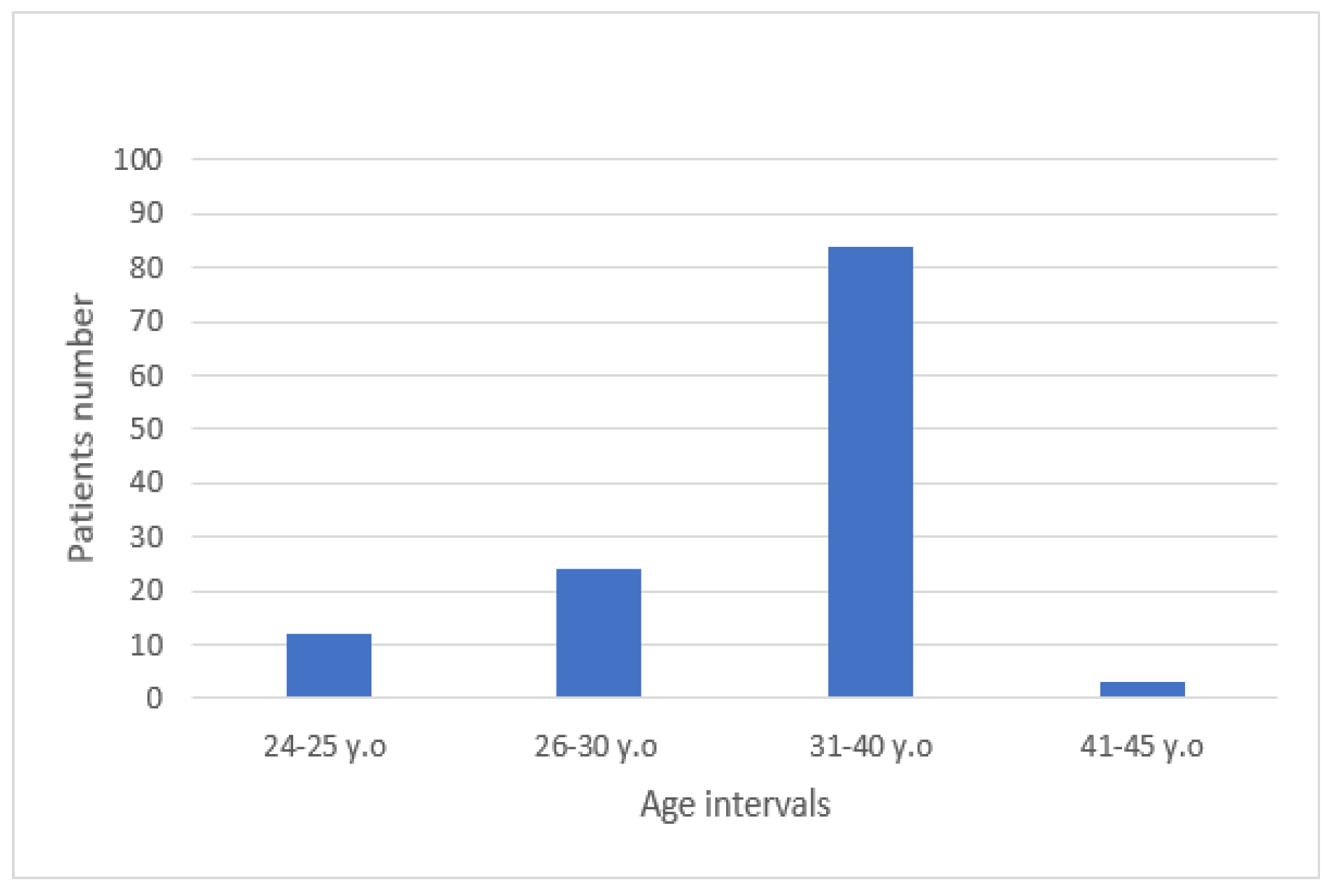
Figure 1. Age intervals
Jmms 06 00021 g001
Figure 2. Age of gestation
Jmms 06 00021 g002
Figure 3. Time interval distribution from the last C-section
Jmms 06 00021 g003
Figure 4. Uterine scar 3 mm (32 weeks)
Jmms 06 00021 g004
Figure 5. Uterine scar 1.1 mm (40 weeks)
Jmms 06 00021 g005
Figure 6. Adenomyosis
Jmms 06 00021 g006
Figure 7. Inflammatory infiltration
Jmms 06 00021 g007
Figure 8. Vascular neoformation
Jmms 06 00021 g008
Figure 9. Granulation tissue
Jmms 06 00021 g009
Figure 10. Uterine scar dehiscence - intraoperative aspect.
Jmms 06 00021 g010