Next Article in Journal
The Correlation Between Histopathological and Ultrasound Findings Regarding Cesarean Section Scars—A Three-Year Survey Study
Previous Article in Journal
Microbiota Signatures in Type-2 Diabetic Patients with Chronic Kidney Disease—A Pilot Study
 
 
Journal of Mind and Medical Sciences is published by MDPI from Volume 12 Issue 1 (2025). Previous articles were published by another publisher in Open Access under a CC-BY (or CC-BY-NC-ND) licence, and they are hosted by MDPI on mdpi.com as a courtesy and upon agreement with Valparaiso University (ValpoScholar).
Article

A Perfusion Decellularization Heart Model—An Interesting Tool for Cell-Matrix Interaction Studies

by
Mihai Meșină
1,
Ion Mîndrilă
2,
Cristian Meșină
3,*,
Cosmin Vasile Obleagă
3 and
Octavian Istrătoaie
4
1
Fundeni Clinical Institute of Bucharest, Department of Cardiovascular Surgery, Bucharest, Romania
2
Department of Thoracic Surgery, Craiova University of Medicine and Pharmacy, Craiova, Romania
3
Department of Surgery, Craiova University of Medicine and Pharmacy, 1 Tabaci Street, 200642 Craiova, Romania
4
Department of Cardiology, Craiova University of Medicine and Pharmacy, Craiova, Romania
*
Author to whom correspondence should be addressed.
J. Mind Med. Sci. 2019, 6(1), 137-142; https://doi.org/10.22543/7674.61.P137142
Submission received: 13 August 2018 / Revised: 20 September 2018 / Accepted: 28 October 2018 / Published: 27 April 2019

Highlights

  • After heart decellularization, there is the possibility of developing research in the field of biomaterials, tissue engineering and cardiac cell cultures.
  • The decellularized extracellular matrix appears to be an attractive substrate for cells grown on these surfaces because they retain their stem cell characteristics for a long time.

Abstract

:

Highlights

  • After heart decellularization, there is the possibility of developing research in the field of biomaterials, tissue engineering and cardiac cell cultures.
  • The decellularized extracellular matrix appears to be an attractive substrate for cells grown on these surfaces because they retain their stem cell characteristics for a long time.

Abstract

Objective. In this paper, we present a technique for whole human, pig, or rat heart decellularization in order to obtain a tool for cell-matrix interaction studies and to obtain the extracellular matrix scaffold with applications to the study of cardiac connective tissue architecture for tissue bioengineering. Material and methods. In order to achieve tissue decellularization we present two separate protocols, the first for large animal hearts (e.g., pig hearts) and human hearts, the second for smaller hearts (e.g., rat hearts). We have performed the cardiac decellularization technique on 20 pig hearts, 5 human hearts, and 20 rat hearts. Results. The decellularization technique on the heart was assessed through histological examination. Conclusion. Although the decellularization technique on the heart is currently under development, this process affords the possibility of developing research in the fields of biomaterials, tissue engineering, and cardiac cell cultures.

Introduction

Collagenous fibers in the heart surround cardiomyocytes and coronary microcirculation [1] and provide support for the macromolecules of electrolytes, oxygen, and metabolic waste diffusion between the cardiomyocytes and the circulatory and lymphatic system [2]. The distribution of myocardial collagen plays an important role in the elastic proprieties of the ventricle [3] and in the kinetics of ventricular contractions [4] by coordinating the transmission of the mechanical forces generated by the contraction of cardio-myocytes. The variety of connective tissue components of the heart muscle reflects the dynamic needs of the heart that undergo various changes throughout the cardiac cycle [5]. The electrophoretic analysis demonstrated that the myocardium of rats [6] and primates [7] contained mainly 2 types of collagen: type I (85%) and type III (11%) collagen. The rest is specific type IV collagenous basal membrane and type V collagen [8], which together with fibronectin, laminin, and elastic fibers form the myocardial connective tissue [9]. The extracellular matrix components are molecularly similar and are well tolerated even as xenografts [10]. By removing the xenogenic or allogenic cellular antigens, the decellularization process can be used to prepare biological scaffolds which have already been used in pre-clinical studies as a means for the development of cardio-myocytes from pluripotent stem cells [11,12]. This technique may prove to be a reliable method for obtaining portions of organs or even neo-organs with a lower rejection rate that can be used in transplant surgery [13].
In this paper, we present a technique for whole human, pig, or rat heart decellularization in order to obtain extracellular matrix scaffolds with applications to the study of cardiac connective tissue architecture for tissue bioengineering.

Materials and Methods

In order to achieve tissue decellularization we present two separate protocols, the first for large animal hearts (pig hearts) and human hearts, the second for smaller hearts (rat hearts). The main difference between the two viscera for consideration is the cellular mass that is much more consistent in the larger hearts (Figure 1). We have performed the cardiac decellularization technique on 20 pig hearts, 5 human hearts, and 20 rat hearts.

Larger hearts (pig and human)

The heart was placed in a 5L container. We cannulated the ascending aorta using an intravenous perfusion infusion set that was connected to a dialysis pump. To prevent the destruction of the tissue through the hyperbaric perfusion trauma, the excess substance was redirected through a separate tubular system connected to the perfusion set that ascended into a reservoir placed above the level of the heart and pump. The excess solution that accumulated in the reservoir could then reflow into the 5L container, where the heart was placed, through a separate tube. We started the process by perfusing the heart with a solution composed of 30g of NaCl and 2L of demineralized water for 20 minutes, and then we washed the solution with 2L of demineralized water for another 20 minutes. We repeated the process 3 times. We then used another solution of 6g of Bromelain and 2L of demineralized water to perfuse the heart at 37°C for about an hour and then resumed only the alternation of 2L demineralized water + 30g NaCl/2L demineralized water for 20 minutes each.
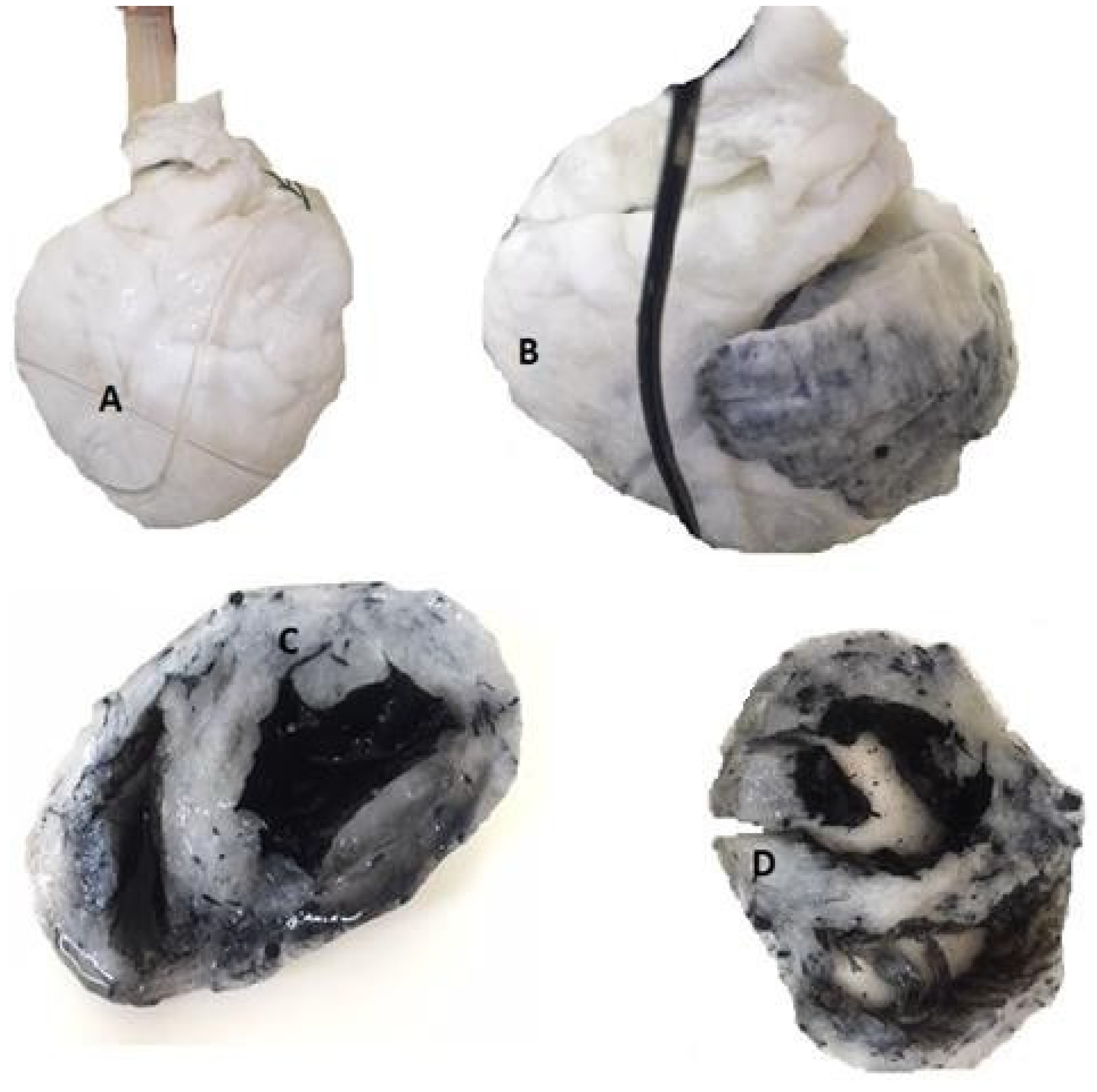
We then perfused the heart with sodium lauryl sulfate (SLS) (10g SLS in 2L of demineralized water) for 24 hours. At the end of the process we reassessed, looking at the aspect of the myocardium and its consistency. We then exchanged the SLS solution with a fresh one and restarted the perfusion process, this time monitoring and reassessing for signs of decellularization every 30 minutes. It took an extra 12 hours for the pig heart to decellularize completely (Figure 2). We then repeated the NaCl + demineralized water/demineralized water alternation for the last time and finally perfused the heart with a hydrogen peroxide 3% and acetic acid solution in a 7/1 ratio (1.4 L hydrogen peroxide + 200 ml acetic acid) for 2 hours.

The rat heart

For smaller hearts, we used a kidney dish instead of the 5L container and only used the 3 alternate perfusions of demineralized water + NaCl/ demineralized water in the same concentrations using the dialysis pump to administer the substances through retrograde coronary perfusion as we did with the larger hearts. After this, we only used SLS (10g SLS in 2L of demineralized water) for a 24h perfusion time and, after that, we reassessed for signs of decellularization. The process took considerably less time and we did not need to wash the heart afterwards with hydrogen peroxide. After the decellularization process, the hearts were preserved by using formaldehyde 10%.

Results

The quality of the decellularization process was assessed through histological microscopic examination. Residual DNA present in decellularized hearts was measured by spectrophotometric quantification and compared to control samples. After decellularization (Figure 1 and Figure 2), the histological examination revealed that all studied hearts (human, pig, and rat) lost intracellular components but maintained the collagen network, proteoglycans, and elastin. Quantitative DNA analysis showed a significant reduction in DNA in decellularized hearts.

Discussions

Decellularization is the process of removing all cellular elements from a tissue, leaving behind the histological skeleton formed exclusively by the extracellular matrix [14]. There is a wide range of methods to achieve this process, either chemical, physical, enzymatic, or a combination of these. The major challenge is not to achieve the decellularization of the tissue or organ in the shortest period of time, but to obtain it in a safe and controlled manner, thus maintaining the integrity of the extracellular matrix. The effectiveness of different agents depends on many factors, including:
-
The quantity of cells
-
Density
-
Lipid content
-
Thickness of tissue.
Depending on the accumulation of these factors, the decellularization process can be chosen and can be customized to each tissue type [15]. Because it is illusory to think that we can completely avoid the alteration of the extracellular matrix structure, this type of customization can help minimize unwanted side effects. The times of the procedure are the same, regardless of the tissue type:
-
The lysis of the cell membrane
-
The separation of cellular components from the extracellular matrix
-
The solubilization of cytosolic and nuclear cellular components
-
The removal of cellular debris from the tissue.
The effectiveness of these steps can be augmented by associating them with mechanical agitation, but the process itself requires a combination of physical, chemical, and enzymatic factors, whose agents differ from one case to another; and their purpose remains the same: the achievement of procedural time [16].
  • Physical factors 
Temperature reduction is a method of lysing the cell membrane by forming intracellular crystals, but the extracellular matrix risks are distorted during this procedure. Increased pressure can break the cells, and agitation can be used to expose cellular debris to chemical and enzymatic decellularization factors, thus leading to their removal.
  • Chemical factors 
Simply alkalinizing and acidifying the solution used to wash the tissue that is being decellularized results in the solubilization of cytoplasmic components and the destruction of nucleic acids, but it also has the secondary effect of removing glycosaminoglycans by modifying the extracellular matrix. Changing osmolality is also a way of achieving cell lysis by osmotic shock. Osmolality can be increased with sodium chloride and decreased by deionization. The lithic effect of osmotic shock is effective but cannot remove the remaining cellular debris. The most commonly used chemicals are non-ionic, ionic, and zwitterion detergents, especially for their lipolytic properties.
-
Non-ionic detergents
Triton X-100 is representative of this class, its properties of denaturalizing lipid-lipid and lipid-protein interactions and leaving intact protein-protein interactions leads to its common use, but results are mixed, depending largely on the tissue.
-
Ionic detergents
Sodium dodecyl sulfate (sodium lauryl sulfate) has the ability to solubilize cell membranes with a tendency of denaturalizing proteins, removing glycosaminoglycans, but because of its proteolytic property it can also destroy native tissue. Sodium doxyclolate is more effective than sodium dodecyl sulfate in association with zwitterion detergents.
-
Zwitterion detergents
These have the combined properties of non-ionic and ionic detergents, the most commonly used representative of the group being CHAPS (3 - [(3-cholamidopropyl) dimethylammonio] -1-propanesulfonate), which has been successfully used in the study of arterial decellularization. Chelating agents such as EDTA and EGTA bind divalent metal ions leading to the disruption of cell adhesion to the extracellular matrix, but the best results are obtained when used in association with enzymatic decellularization methods.
  • Enzymatic factors 
Trypsin is by far the most commonly used enzyme in the decellularization process due to its properties of breaking the peptide bonds of lysine and arginine. The maximum effect of the proteolytic activity of trypsin occurs at 37°C and a pH of 8. Other successfully used enzymes are endonucleases that catalyze the hydrolysis of bonds within the ribonucleotide and de-oxy-ribonucleotide chains, and the exonucleases that catalyze the hydrolysis of terminal bonds from the ribonucleotide and de-oxy-ribonucleotide chains. The adverse effects of enzymatic agents are largely due to prolonged exposure that may affect the structure of the extracellular matrix and remove laminin, fibronectin, and elastin, but also because it is difficult to remove from the tissues and produce an immune response [17].
Biological scaffolds obtained through the decellularization process from various tissues and organs have been successfully used in pre-clinical studies [11]. Although the methods used are different from one case to the next, the goal is a common one, namely the removal of xenogenic or allogenic cellular antigens. These are recognized by the host as foreign, a phenomenon that ultimately leads to an inflammatory response. The extracellular matrix components are molecularly similar and are well tolerated even as xenografts [18].
Biological scaffolds obtained from the extracellular matrix were studied, attempting to re-cultivate them with pluripotent stem cells, most studies being performed on cardiac valves, blood vessels, nerves, skeletal muscles, tendons, ligaments, bladder, and liver [19]. In the field of cardiac tissue engineering, it has been observed that the only cells that can produce cardio-myocytes with viable contractile function are pluripotent stem cells and those obtained through genetic reprogramming [12]. The extracellular matrix obtained through decellularization has been suggested as a means for developing these cells with regenerative potential, attempting to obtain a three-dimensional re-creation of the organ architecture, thus creating portions of organ or even neo-organs with a lower rejection rate that can be used in transplant surgery [13].
Studies in the field of cardiac physiology have demonstrated the essential role of the extracellular matrix in homeostasis and heart development, facilitating not only electrical and mechanical cell signaling functions but also responses to physiological stress and injuries, thus modulating cellular proliferation, migration, adhesion, and change in gene expression during homeostasis and development [20].
Influencing the cell differentiation process is one of the features of the extracellular matrix that gives it an important role as a model for tissue recovery and engineering. This concept stems from the idea that proliferation and differentiation of stem cells into cells specific to each tissue type are influenced by environmental factors acting along with chemical factors. The structure, elasticity, and extracellular matrix components play an important role in this process [21].
Another beneficial effect of the matrix is the presence of exosomes, nanoparticles linked to its structure that can activate the macro-face through their content (DNA fragments, mARN), thus controlling events such as cell proliferation or cell cycle. Exosomes are membranous extracellular vesicles with a diameter of only 30-1000 nm, with a role in intercellular communication due to their properties of RNA transfer, proteins, enzymes, lipids, etc., thus influencing various cellular processes. Their presence in the structure of scaffolds obtained through decelullarization is an additional argument for their use in the field of biological engineering with their pro-angiogenesis, stem cell activation and recruitment, antimicrobial activity, and modulation of the immune response [22].

Conclusions

Although the decellularization technique of the heart is under development, it holds the possibility of developing research in the field of biomaterials, tissue engineering, and cardiac cell cultures.
The decellularized extracellular matrix appears to be an attractive substrate for cells grown on these surfaces because they retain their stem cell characteristics for a long time.

Conflict of interest disclosure

There are no known conflicts of interest in the publication of this article. The manuscript was read and approved by all authors.

Compliance with ethical standards

Any aspect of the work covered in this manuscript has been conducted with the ethical approval of all relevant bodies and that such approvals are acknowledged within the manuscript.

References

  1. Abreu-Velez, A.M.; Howard, M.S. Collagen IV in Normal Skin and in Pathological Processes. N Am J Med Sci. 2012, 4, 1–8. [Google Scholar] [CrossRef] [PubMed]
  2. Wiig, H.; Keskin, D.; Kalluri, R. Interaction between the extracellular matrix and lymphatics - consequences for lymphangiogenesis and lymphatic function. Matrix Biol. 2010, 29, 645–656. [Google Scholar] [CrossRef] [PubMed]
  3. Fomovsky, G.M.; Thomopoulos, S.; Holmes, J.W. Contribution of Extracellular Matrix to the Mechanical Properties of the Heart. J Mol Cell Cardiol. 2010, 48, 490–496. [Google Scholar] [CrossRef] [PubMed]
  4. Kapelko, V.I. Extracellular matrix alterations in cardiomyopathy: The possible crucial role in the dilative form. Exp Clin Cardiol. 2001, 6, 41–49. [Google Scholar] [PubMed]
  5. Eckhouse, S.R.; Spinale, F.G. Changes in the Myocardial Interstitium and Contribution to the Progression of Heart Failure. Heart Fail Clin. 2012, 8, 7–20. [Google Scholar] [CrossRef] [PubMed]
  6. Medugorac, I.; Jacob, R. Characterisation of left ventricular collagen in the rat. Cardiovasc Res. 1983, 17, 15–21. [Google Scholar] [CrossRef] [PubMed]
  7. Weber, K.T.; Janicki, J.S.; Shroff, S.G.; Pick, R.; Chen, R.M.; Bashey, R.I. Collagen remodeling of the pressure-overloaded, hypertrophied nonhuman primate myocardium. Circ Res. 1988, 62, 757–765. [Google Scholar] [CrossRef] [PubMed]
  8. Chapman, D.; Weber, K.T.; Eghbali, M. Regulation of fibrillar collagen types I and III and basement membrane type IV collagen gene expression in pressure overloaded rat myocardium. Circ Res. 1990, 67, 787–794. [Google Scholar] [CrossRef] [PubMed]
  9. Halper, J.; Kjaer, M. Basic Components of Connective Tissues and Extracellular Matrix: Elastin, Fibrillin, Fibulins, Fibrinogen, Fibronectin, Laminin, Tenascins and Thrombospondins. In Progress in Heritable Soft Connective Tissue Diseases. Advances in Experimental Medicine and Biology; Halper, J., Ed.; Springer: Dordrecht, The Netherlands, 2014; Volume 802. [Google Scholar]
  10. Aamodt, J.M.; Grainger, D.W. Extracellular Matrix-based Biomaterial Scaffolds and the Host Response. Biomaterials. 2016, 86, 68–82. [Google Scholar] [CrossRef] [PubMed]
  11. Brown, B.N.; Badylak, S.F. Extracellular matrix as an inductive scaffold for functional tissue reconstruction. Transl Res. 2014, 163, 268–285. [Google Scholar] [CrossRef] [PubMed]
  12. Shiba, Y.; Hauch, K.D.; Laflamme, M.A. Cardiac Applications for Human Pluripotent Stem Cells. Curr Pharm Des. 2009, 15, 2791–2806. [Google Scholar] [CrossRef] [PubMed]
  13. Wang, B.; Wang, G.; To, F.; Butler, J.R.; Claude, A.; McLaughlin, R.M.; Williams, L.N.; de Jongh Curry, A.L.; Liao, J. Myocardial scaffold-based cardiac tissue engineering: application of coordinated mechanical and electrical stimulations. Langmuir. 2013, 29, 11109–11117. [Google Scholar] [CrossRef] [PubMed]
  14. Alberts, B.; Johnson, A.; Lewis, J.; et al. Molecular Biology of the Cell. 4th edition. New York: Garland Science; 2002. Integrins. Available from: https://www.ncbi.nlm.nih.gov/books/NBK26867/.
  15. Gilpin, A.; Yang, Y. Decellularization Strategies for Regenerative Medicine: From Processing Techniques to Applications. Biomed Res Int. 2017, 2017, 9831534. [Google Scholar] [CrossRef] [PubMed]
  16. Gilbert, T.W.; Sellaro, T.L.; Badylak, S.F. Decellularization of tissues and organs. Biomaterials. 2006, 27, 3675–3683. [Google Scholar] [CrossRef] [PubMed]
  17. Crapo, P.M.; Gilbert, T.W.; Badylak, S.F. An overview of tissue and whole organ decellularization processes. Biomaterials. 2011, 32, 3233–3243. [Google Scholar] [CrossRef] [PubMed]
  18. Aamodt, J.M.; Grainger, D.W. Extracellular Matrix-based Biomaterial Scaffolds and the Host Response. Biomaterials. 2016, 86, 68–82. [Google Scholar] [CrossRef] [PubMed]
  19. Yu, Y.; Alkhawaji, A.; Ding, Y.; Mei, J. Decellularized scaffolds in regenerative medicine. Oncotarget 2016, 7, 58671–58683. [Google Scholar] [CrossRef] [PubMed]
  20. Lockhart, M.; Wirrig, E.; Phelps, A.; Wessels, A. Extracellular Matrix and Heart Development. Birth Defects Res A Clin Mol Teratol. 2011, 91, 535–550. [Google Scholar] [CrossRef] [PubMed]
  21. Diaconu, C.C.; Stanescu, A.M.A.; Pantea, S.A.; et al. Hyperkalemia and Cardiovascular Diseases: New Molecules for the Treatment. Revista de Chimie 2018, 69, 1367–1370. [Google Scholar] [CrossRef]
  22. Huleihel, L.; Hussey, G.S.; Naranjo, J.D.; Zang, L.; Dziki, J.L.; Turner, N.J.; Stolz, D.B.; Badylak, S.F. Matrix-bound nanovesicles within ECM bioscaffolds. Sci Adv. 2016, 2, e1600502. [Google Scholar] [CrossRef] [PubMed]
Figure 1. Obtaining the heart decellularization model: A – We cannulated the ascending aorta; B – Anterior view of the cannula inserted into the ascending aorta; C- Posterior view of the cannula inserted into the ascending aorta; D – The excess of the substances was redirected through a separate tubular system connected to the perfusion set; E – At the end of the process we looked at the aspect of the myocardium and its consistency.
Jmms 06 00020 g001
Figure 2. Macroscopic appearance of decellularized heart: A - Posterior view of the decellularized heart; B – Anterior view of the decellularized heart; C - Macroscopic aspect of the decellularized interventricular septum; D- Section at the left atrium of the decellularized heart.
Jmms 06 00020 g002