Next Article in Journal
A Perfusion Decellularization Heart Model—An Interesting Tool for Cell-Matrix Interaction Studies
Previous Article in Journal
Experimental Pharmacological Research Regarding Some New Quinazolin-4-ones Derivatives
 
 
Journal of Mind and Medical Sciences is published by MDPI from Volume 12 Issue 1 (2025). Previous articles were published by another publisher in Open Access under a CC-BY (or CC-BY-NC-ND) licence, and they are hosted by MDPI on mdpi.com as a courtesy and upon agreement with Valparaiso University (ValpoScholar).
Article

Microbiota Signatures in Type-2 Diabetic Patients with Chronic Kidney Disease—A Pilot Study

by
Gratiela P. Gradisteanu
1,
Roxana A. Stoica
2,
Laura Petcu
3,
Ariana Picu
3,
Adrian P. Suceveanu
4,
Teodor Salmen
3,
Diana S. Stefan
2,3,
Cristian Serafinceanu
2,3,
Mariana C. Chifiriuc
1,* and
Anca P. Stoian
2
1
The Research Institute of the University of Bucharest (ICUB), Bucharest, Romania
2
Carol Davila University of Medicine and Pharmacy, 8 Eroii Sanitari Street, 050474 Bucharest, Romania
3
N. C. Paulescu National Institute of Diabetes, Nutrition and Metabolic Diseases, 5-7 Ion Movila Street, Bucharest, Romania
4
The Faculty of Medicine, Ovidius University, Universitatii Street, 900470 Constanta, Romania
*
Author to whom correspondence should be addressed.
J. Mind Med. Sci. 2019, 6(1), 130-136; https://doi.org/10.22543/7674.61.P130136
Submission received: 18 May 2018 / Revised: 20 July 2018 / Accepted: 14 August 2018 / Published: 27 April 2019

Highlights

  • This study investigates the changes present in the fungal microbiota of diabetic patients with diabetic kidney disease, and existing correlations between treatment regimens and microbiota patterns.
  • These preliminary results pave the way for additional studies on larger patient cohorts.

Abstract

:

Highlights

  • This study investigates the changes present in the fungal microbiota of diabetic patients with diabetic kidney disease, and existing correlations between treatment regimens and microbiota patterns.
  • These preliminary results pave the way for additional studies on larger patient cohorts.

Abstract

The human microbiota is paramount for normal host physiology. Altered host-microbiome interactions are part of the pathogenesis of numerous common ailments. Currently, much emphasis is placed on the involvement of the microbiome in the pathogenesis of type-2 diabetes mellitus (T2DM), impaired glucose tolerance, and other metabolic disorders (i.e., obesity). Several studies found highly significant correlations of specific intestinal bacteria with T2DM. A better understanding of the role of the microbiome in diabetes and its complications might provide new insights in the development of new therapeutic principles. Our pilot study investigates the microbiota patterns in Romanian type-2 diabetic patients with diabetic kidney disease. Fecal samples were collected from type 2-diabetic patients and healthy controls and further used for bacterial DNA isolation. Using 16 rDNA qRT-PCR, we analyzed phyla abundance (Bacteroidetes, Firmicutes) as well as the relative abundance of specific bacterial groups (Lactobacillus sp., Enterobacteriaceae, Ruminococus sp., Prevotella sp., Faecalibacterium sp., Clostridium coccoides, Clostridium leptum). Our study also investigates the diabetic fungal microbiome for the first time. Furthermore, we report significant correlations between the treatment regimen and microbiota composition in diabetic nephropathy.

Introduction

Diabetes is a public health problem affecting approximately 10% of adults worldwide [1]. The International Diabetes Federation predicts that there will be 592 million diabetic cases with an additional 175 million undiagnosed diabetic cases by 2035 [2]. In Romania, 11.6% of the population aged between 20-79 suffers from diabetes, thus placing our country on the second place in Europe in terms of diabetic prevalence [3]. This chronic ailment is often complicated by an increased risk of heart disease, stroke and kidney failure [4]. Diabetic kidney disease, classically defined by the presence of proteinuria (macroalbuminuria), is a common complication most likely to occur in patients with poor glycemic control, hypertension, glomerular hyperfiltration or genetic predisposition [5].
Along this line of thinking, there is a growing public and scientific interest in identifying and modulating the key factors that govern the onset of diabetes and its complications. Factors such as rapid environmental changes and modern lifestyle are the major culprits for the alteration of the intestinal microbiota which controls the risk of developing metabolic dysfunction. The gut microbiota is also involved in the development of the immune system and multiple metabolic pathways. Therefore, disturbances in its composition and functionality induced by dietary changes will hinder the gut barrier function. A disrupted gut barrier permits passage of microorganisms, microbial products and foreign antigens into the mucosa subsequently leading to the immune system activation and secretion of inflammatory mediators. This low-grade chronic inflammation induced by a defective gut barrier further modifies host adiposity and insulin resistance [6]. Several recent studies have reported changes in the gut microbiota of individuals at risk of developing diabetes as well as in already diagnosed patients [7]. Even though there has been no clear microbiota pattern linked to diabetes so far, it was shown that diabetic individuals generally exhibit reduced bacterial diversity characterized by a reduction of butyrate-producing bacteria such as Faecalibacterium prausnitzii and Roseburia intestinalis along with an increase in opportunistic pathogens (pathobionts) [8].
Within this context, our study investigates the microbiota signatures in Romanian patients with diabetic nephropathy and compares it with the microbiota patterns identified in other currently available studies. For the first time, our study investigates the fungal microbiota of diabetic patients.

Materials and Methods

Patients

The study population (n=9) was represented by diabetic patients from “N. C. Paulescu” National Institute of Diabetes, Nutrition and Metabolic Diseases in Bucharest, Romania, and healthy volunteers. All participants received and signed the informed consent, and the Ethical Committee approved the study. All the enrolled T2D subjects were diagnosed based on the WHO criteria, namely: polyuria, polydipsia and polyphagia as well as one of the following: a random venous plasma glucose concentration ≥ 11.1 mmol/l or a fasting plasma glucose concentration ≥ 7.0 mmol/l (whole blood ≥ 6.1 mmol/l) or two hour plasma glucose concentration ≥ 11.1 mmol/l two hours after 75g anhydrous glucose in an oral glucose tolerance test [9].

Microbiota analysis

Stool samples were collected at home by the participants following a standardized procedure including antiseptic handling, collection in sterile containers and immediate freezing at −20°C. Fecal DNA was extracted using the AllPrep PowerViral DNA/RNA (Qiagen) according to the manufacturer’s instructions. Genomic DNA concentration was determined at a wavelength of 260 nm using a NanoDrop (Thermo Scientific). For qPCR analysis, DNA samples were diluted in DNAse free water to a concentration of 3 ng/μl. RT-PCR measured the relative abundance of intestinal microorganisms in fecal DNA isolated from T2DM patients and healthy controls on a ViiA7© Fast Real-Time System.
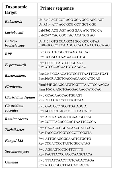
DNA samples were amplified using the bacterial or fungal group-specific primers at their specific annealing temperatures. Eubacteria primers were used to amplify a segment of the 16S rDNA in order to determine the total number of bacteria per sample. The primers used are listed in Table 1. Each PCR reaction included 200 nM of forward and reversed primer, 9 ng of DNA, and 2x SYBR Green Master Mix (Applied Biosystems). Samples without DNA template served as negative controls. Samples were incubated at 95°C for 5 min, and then amplified through 45 cycles of 95°C for 10 s, 60°C for 30 s, and 72°C for 1 s.

The statistical analysis

The data in our study are presented as mean ± SEM and were graphed using the GraphPad Prism 5.0 software. Sample size (n) denotes biological replicates. Differences in microbial relative abundance were assessed using a non-parametric Mann-Whitney test. The *p < 0.05 was considered statistically significant. The statistically significant levels were *, p < 0.05; **, p < 0.01; ***, p < 0.001.

Results

Our pilot study enrolled 9 T2DM patients with diabetic nephropathy and 5 healthy controls. Besides diabetic kidney disease, the enrolled T2DM patients also suffered from neuropathy (7 patients), retinopathy (7 patients), osteoporosis (2 patients), diabetic foot (1 patient), dyslipidemia (8 patients) and steatosis (6 patients) (Figure 1 A).
No statistically significant difference was observed regarding the body mass index (BMI) between the two experimental groups (p=0.0790) (Figure 1 B). In our pilot analysis, we compared the fecal microbiota of patients with type-2 diabetes with that of healthy volunteers using qPCR of the 16S rRNA gene. qPCR analysis was done using SYBR Green primers recognizing different phyla such as Bacteroidetes and Firmicutes, but also bacterial families such as Lactobacillaceae and Enterobacteriaceae. Primers for the Universal Eubacteria 16S were used for normalization.
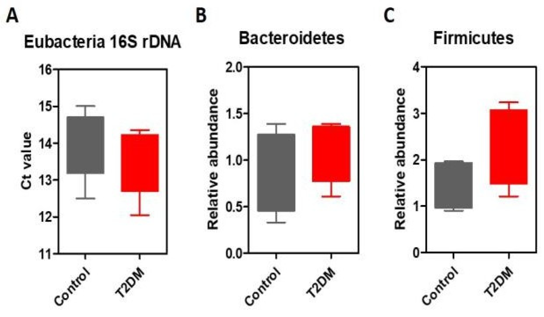
The total amount of bacteria in the stool samples are represented as threshold cycle values (Ct) in Figure 2A. Both healthy controls and T2DM patients with diabetic nephropathy harbored similar levels of Eubacteria (Figure 2 A).
The most common organisms in the human gut microbiota are members of the Gram-positive Firmicutes and the Gram-negative Bacteroidetes phyla, followed by several other phyla, including the Actinobacteria, usobacteria and Verrucomicrobia [10].
The data from animal models and human studies have revealed differences in the two dominant bacterial phyla with a significant increase of Firmicutes and decrease of Bacteroidetes levels in obesity [11]. Regardless their disease status, the subjects enrolled in our study had BMI values ranging from 20 to 38 (Figure 1B), and therefore, there is no surprise that we did not observe significant differences between Bacteroidetes and Firmicutes abundance (Figure 2 B-C), even though T2DM patients exhibited a tendency to harbor more Firmicutes (Figure 2 C).
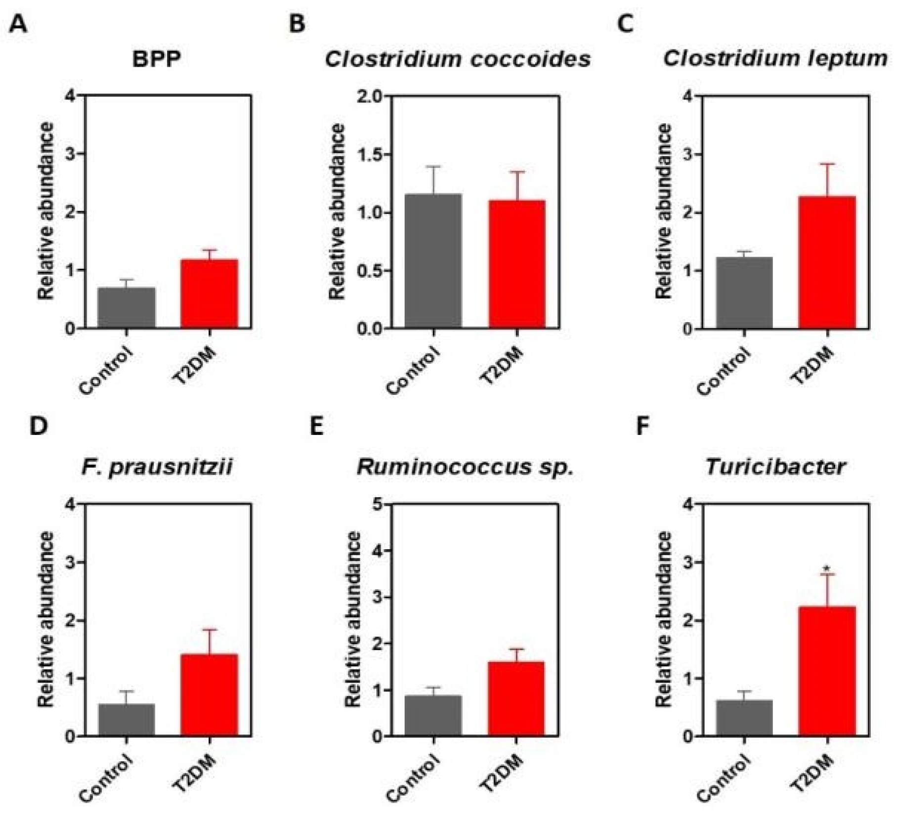
The Bacteroidetes phyla range in relative abundance among individuals, but generally, make up half or more of the gut microbiome. The three predominant Bacteroidetes genera of the human gastrointestinal tract are represented by Bacteroides, Prevotella, and Porphyromonas (BPP). In our analysis, no significant differences were observed in the BPP levels between T2DM patients and healthy controls (Figure 3 A).
The strictly anaerobic Clostridium coccoides group constitutes 25% to 60% of the total microbiota and comprises a large number of species of genera such as Clostridium, Blautia, Anaerostipes, Ruminococcus, Dorea Eubacterium and Roseburia [12,13]. In our analysis, Clostridium coccoides’ relative abundance was similar between healthy controls and T2DM patients with diabetic nephropathy (Figure 3B). The qPCR results indicated that the amounts of Clostridium leptum tended to be higher in T2DM patients, compared to healthy controls (Figure 3 C). The Clostridium leptum group of bacteria (also called Clostridial cluster IV) is a dominant group of fecal bacteria in adult humans, representing around 16-25% of the fecal microbiota and it includes Faecalibacterium prausnitzii and certain species of Eubacterium and Ruminococcus [13].
Following the same pattern, T2DM patients exhibited increased levels of Faecalibacterium prausnitzii and Ruminococcus. No statistically significant differences were observed between the two experimental groups regarding the Clostridium leptum- Ruminococcus-Faecalibacterium prausnitzii levels, highlighting the need to increase the sample size in a subsequent study (Figure 3 D, E). The fecal microbiome of T2DM had significantly higher levels of Turicibacter sp. (*p= 0.0128) (Figure 3 F).
Although the exact role of this butyrate-producing bacteria in the development of metabolic syndrome is not known, one study reported an increased abundance of Turicibacter in the gut of patients with rheumatoid arthritis, an immune-mediated disease [14].
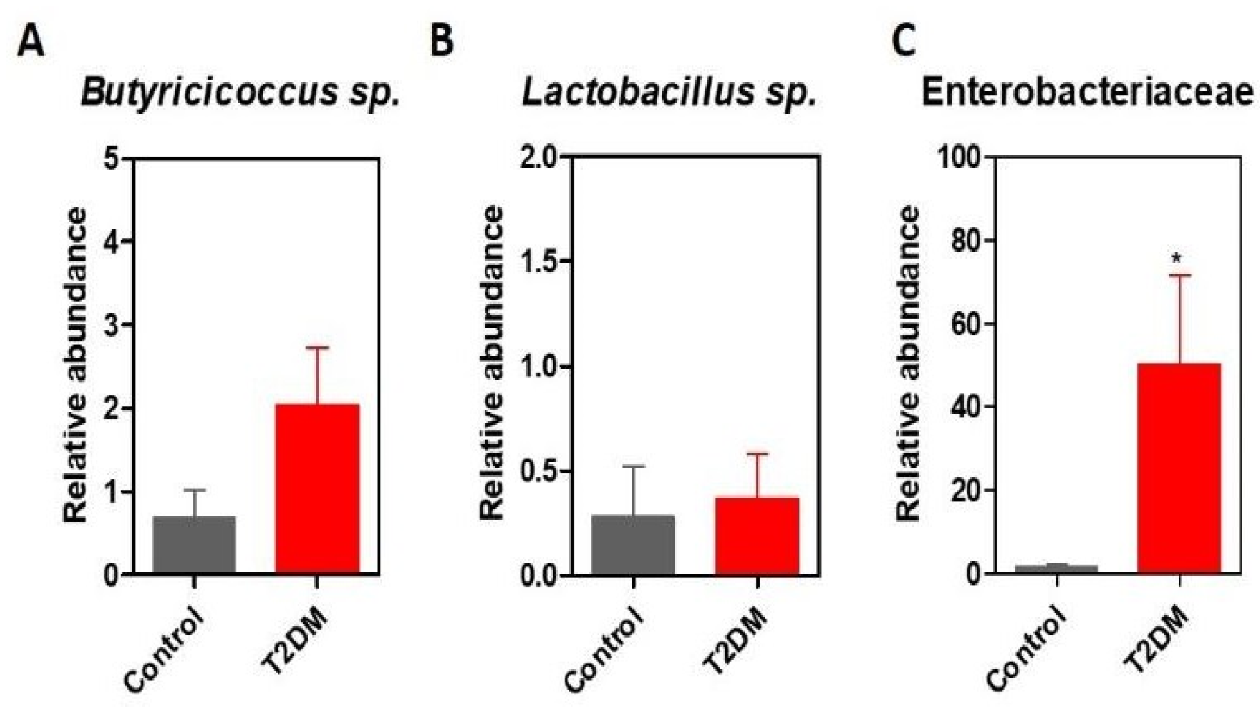
Interestingly though, T2DM patients harbored a microbiota enriched in Butyricicoccus spp. Butyricicoccus members are strictly anaerobic Clostridium cluster IV bacteria that produce high levels of butyrate (a short fatty chain of utmost importance for intestinal homeostasis) and are currently investigated as potential probiotics (Figure 4A). Under such circumstances, lactobacilli have also been widely characterized as potent probiotic strains in numerous studies. Nevertheless, we have tested the relative abundance of lactobacilli in T2DM patients and healthy controls, and we did not observe any differences (Figure 4B).
Oxidative stress and inflammation, especially when accompanied by obesity, mutually potentiate each other and promote the onset of beta-pancreatic cell dysfunction, insulin resistance and T2DM vascular complications. In accordance with this, several studies have linked oxidative stress to dysbiosis [15]. The intestinal tract harbors a radical oxygen gradient and hence microbes that reside in the colonic mucosa exhibit elevated oxygen tolerance and catalase expression compared to luminal or stool-associated bacteria [15]. Moreover, inflammation also promotes an oxidative milieu characterized by the enrichment of aerotolerant phyla such as Proteobacteria and Actinobacteria. Gut inflammation was reported to be responsible for the production of terminal electron acceptors needed by facultative anaerobes such as Enterobacteriaceae further augmenting dysbiosis [16,17]. Specific taxonomic shifts have been linked to intestinal inflammation, including a relative increase in the abundance of Enterobacteriaceae, such as Escherichia coli and Fusobacterium. In our pilot study, the T2DM microbiota was characterized by significantly higher levels of Enterobacteriaceae (*p= 0.0238), hence suggesting a possible link between diabetes and intestinal inflammation (Figure 4 C).
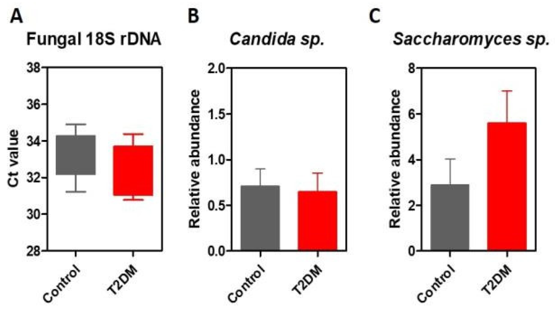
For the first time, this study also investigates the differences in the fungal microbiome of diabetic patients with kidney disease. For this purpose, we quantified the total amount of fungal DNA in stool samples using universal primers for fungal 18S rDNA and the relative abundance of fungal populations such as Candida sp. and Saccharomyces sp. using specific primers (Figure 5 A-C). We observed no statistically significant differences regarding the total amount of fungal DNA sequences or Candida sp. levels. We did, however, find a tendency for higher levels of Saccharomyces sp. in T2DM patients but these findings need to be confirmed among a larger patient cohort.
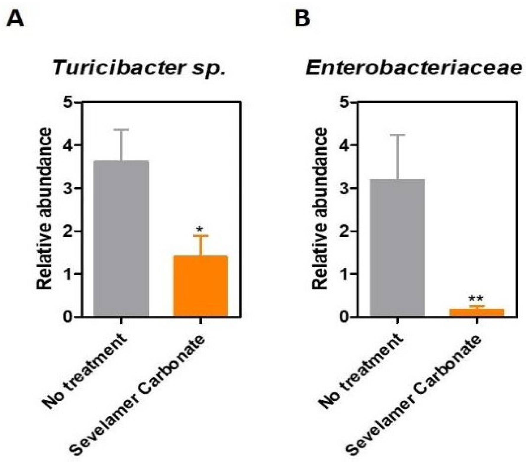
Compelling recent data reveal the important role that medication has in triggering changes in the gut microbiota. For instance, the treatment with metformin alters the microbiome by promoting the growth of Akkermansia muciniphila, Lactobacillus sp., Escherichia spp, and by lowering the levels of Intestinibacter [18]. Bearing this in mind, we tested whether treatment regimens impact microbiota signatures in diabetic nephropathy. Of all the T2DM patients tested, 6 patients (66.6%) received sevelamer carbonate, a phosphate binding drug prescribed to prevent hyperphosphatemia in patients with chronic renal failure. Interestingly, the sevelamer carbonate treatment was significantly correlated with lower levels of both Enterobacteriaceae and Turicibacter (Figure 6 A, B).
Importantly, the sevelamer carbonate treatment decreases levels of potentially pathogenic bacteria (Enterobacteriaceae), and these results need further investigation among a larger patient cohort. The other tested bacterial and fungal populations behaved similarly regardless of the sevelamer administration.

Discussions

Human microbiome profiles depend on diet, ethnic origin, geographic region and age. However, despite these variables, the microbiome of T2DM patients was suggested to have some particular features. The microbiota of T2DM patients has low levels of mucin degrading bacteria such as Akkermansia sp. and Prevotella sp. [19] and also butyrate-producing microorganisms, such as Faecalibacterium prausnitzii, Clostridium sp., Roseburia intestinalis, Eubacterium rectale and Faecalibacterium sp. [20]. The human T2DM intestinal microbiota was shown to be inhabited by opportunistic pathogens including the sulphate-reducing genus Desulfovibrio, Escherichia coli and Bacteroidaceae. A study performed on Chinese T2DM patients revealed an increase in Escherichia coli. Furthermore, a Danish study reported elevated Proteobacteria in T2DM [21]. A study performed on Romanian subjects revealed that the most common aerobic/facultative anaerobic species isolated from the stool cultures of 100 patients with dyslipidemia, diabetes and obesity were Gram-negative bacteria [22]. Our results are partially in accordance with these studies. Similar to the study by Larsen et al., we showed that T2DM microbiota is enriched in Enterobacteriaceae.
In contrast with previously published studies, we observed that T2DM patients harbored a microbiota slightly enriched in butyrate-producing microorganisms, but the difference was not statistically significant. To address this question, we need to perform further studies on a larger patient cohort using next-generation sequencing techniques to ascertain better microbiome features in T2DM associated with nephropathy. Bacterial counts of the L. acidophilus, L. plantarum and L. reuteri subgroups of Lactobacillus sp. were significantly lower among Romanian patients with T2DM and obesity compared to healthy controls [23]. In our study group, no differences were seen in case of lactobacilli abundance in the stool samples harvested from T2DM patients. However, one must consider that the levels of lactobacilli inhabiting the microbiota are highly dependent on the host’s diet.

Conclusions

Herein, we present a pilot study analyzing the microbiome patterns associated with diabetic nephropathy in Romanian patients. For the first time, we investigate the changes present in the fungal microbiota of diabetic patients with diabetic kidney disease. Besides, we describe significant correlations between treatment regimens and microbiota patterns. These preliminary results pave the way for additional studies on larger patient cohorts.

Acknowledgments

All authors had an equal scientific contribution and shared the first authorship.

Conflicts of Interest disclosure

There are no known conflicts of interest in the publication of this article. The manuscript was read and approved by all authors.

Compliance with ethical standards

Any aspect of the work covered in this manuscript has been conducted with the ethical approval of all relevant bodies and that such approvals are acknowledged within the manuscript.

References

  1. Astrup, A.F.N. Redefining type 2 diabetes: ‘diabesity’ or ‘obesity dependent diabetes mellitus’? Obes Rev. 2001, 1, 57–59. [Google Scholar] [CrossRef] [PubMed]
  2. Ayadurai, S.; Hattingh, H.L.; Tee, L.B.; et al. A narrative review of diabetes intervention studies to explore diabetes care opportunities for pharmacists. J Diabetes Res. 2016, 2016, 5897452. [Google Scholar] [CrossRef] [PubMed]
  3. Mota, M.; Popa, S.G.; Mota, E.; Mitrea, A.; Catrinoiu, D.; et al. Prevalence of diabetes mellitus and prediabetes in the adult Romanian population: PREDATORR study. J Diabetes. 2016, 8, 336–344. [Google Scholar] [CrossRef]
  4. Rusu, A.; Bala, C.G.; Craciun, A.E.; et al. HbA1c levels are associated with severity of hypoxemia and not with apnea hypopnea index in patients with type 2 diabetes: Results from a cross-sectional study. Journal of Diabetes. 2017, 9, 555–561. [Google Scholar] [CrossRef]
  5. Bala, C.; Craciun, A.E.; Hancu, N. Updating the concept of metabolically healthy obesity. Acta Endocrinologica-Bucharest. 2016, 12, 197–205. [Google Scholar] [CrossRef]
  6. Musso, G.; Gambino, R.; Cassader, M. Obesity, diabetes, and gut microbiota: The hygiene hypothesis expanded? Diabetes Care. 2010, 33, 2277–2284. [Google Scholar] [CrossRef]
  7. Larsen, N.; Vogensen, F.K.; Van Den Berg, F.W.J.; Nielsen, D.S.; Andreasen, A.S.; Pedersen, B.K.; et al. Gut microbiota in human adults with type 2 diabetes differs from non-diabetic adults. PLoS One. 2010, 5, e9085. [Google Scholar] [CrossRef]
  8. Qin, J.; Li, Y.; Cai, Z.; Li, S.; Zhu, J.; Zhang, F.; Liang, S.; Zhang, W.; Guan, Y.; Shen, D.; et al. A metagenome-wide association study of gut microbiota in type 2 diabetes. Nature. 2012, 490, 55–60. [Google Scholar] [CrossRef]
  9. World Health Organization. Global Report on Diabetes. Isbn. 2016, 978, 88. [Google Scholar]
  10. Eckburg, P.B.; Bik, E.M.; Bernstein, C.N.; Purdom, E.; Dethlefsen, L.; Sargent, M.; et al. Diversity of the human intestinal microbial flora. Science. 2005, 308, 1635–1638. [Google Scholar] [CrossRef]
  11. Ley, R.E.; Bäckhed, F.; Turnbaugh, P.; Lozupone, C.A.; Knight, R.D.; Gordon, J.I. Obesity alters gut microbial ecology. Proc Natl Acad Sci U S A. 2005, 102, 11070–11075. [Google Scholar] [CrossRef] [PubMed]
  12. Hayashi, H.; Sakamoto, M.B.Y. Phylogenetic analysis of the human gut microbiota using 16S rDNA clone libraries and strictly anaerobic culture-based methods. Microbiol Immunol. 2002, 46, 535–548. [Google Scholar] [CrossRef] [PubMed]
  13. Matsuki, T.; Watanabe, K.; Fujimoto, J.; Takada, T.T.R. Use of 16S rRNA gene-targeted group-specific primers for real-time PCR analysis of predominant bacteria in human feces. Appl Env Microbiol. 2004, 70, 7220–7228. [Google Scholar] [CrossRef] [PubMed]
  14. Forbes, J.D.; Van Domselaar, G.; Bernstein, C.N. The Gut Microbiota in Immune-Mediated Inflammatory Diseases. Front Microbiol. 2016, 7, 1081. [Google Scholar] [CrossRef]
  15. Albenberg, L.; Esipova, T.V.; Judge, C.P.; Bittinger, K.; Chen, J.; Laughlin, A.; et al. Correlation between intraluminal oxygen gradient and radial partitioning of intestinal microbiota. Gastroenterology 2014, 147, 1055–1063. [Google Scholar] [CrossRef]
  16. Lopez, C.A.; Miller, B.M.; Rivera-Chavez, F.; et al. Virulence factors enhance Citrobacter rodentium expansion through aerobic respiration. Science. 2016, 353, 1249–1253. [Google Scholar] [CrossRef]
  17. Winter, S.E.; Lopez, C.A.; Bäumler, A.J. The dynamics of gut-associated microbial communities during inflammation. EMBO Rep. 2013, 14, 319–327. [Google Scholar] [CrossRef]
  18. Forslund, K.; Hildebrand, F.; Nielsen, T.; Falony, G.; Le Chatelier, E.; Sunagawa, S.; et al. Disentangling type 2 diabetes and metformin treatment signatures in the human gut microbiota. Nature. 2015, 528, 262–266. [Google Scholar] [CrossRef]
  19. Brown, C.T.; Davis-Richardson, A.G.; Giongo, A.; Gano, K.A.; Crabb, D.B.; Mukherjee, N.; Casella, G.; Drew, J.C.; Ilonen, J.; Knip, M.; et al. Gut microbiome metagenomics analysis suggests a functional model for the development of autoimmunity for type 1 diabetes. PLoS One. 2011, 6, e25792. [Google Scholar] [CrossRef]
  20. Paunica, M.; Gheorghiu, R.; Curaj, A.; Holeab, C. Foresight for restructuring R&D systems. Amfiteatru economic 2009, 11, 201–210. [Google Scholar]
  21. Larsen, N.; Vogensen, F.K.; van den Berg, F.W.; Nielsen, D.S.; Andreasen, A.S.; Pedersen, B.K.; Al-Soud, W.A.; Sørensen, S.J.; Hansen, L.H.J.M. Gut microbiota in human adults with type 2 diabetes differs from non- diabetic adults. PLoS One. 2010, 5, e9085. [Google Scholar] [CrossRef] [PubMed]
  22. Chelariu, M.; Grosu, M.; Gheorghe, I.; Gradisteanu, G.; Picu, A.; Petcu, L.; et al. Host metabolic syndrome can disrupt the intestinal microbiota and promote the acquisition of resistance and virulence genes in Enterobacteriaceae stains. Rom Biotechnol Lett. 2017, 22, 12643–12650. [Google Scholar]
  23. Suceveanu, A.I.; Pantea Stoian, A.; Parepa, R.I.; Voinea, C.; Hainarosie, R.; Manuc, D.; et al. Gut microbiota patterns in obese and type 2 diabetes. (T2D) patients from Romanian black sea coast region. Rev Chim. 2018, 69, 2260–2267. [Google Scholar] [CrossRef]
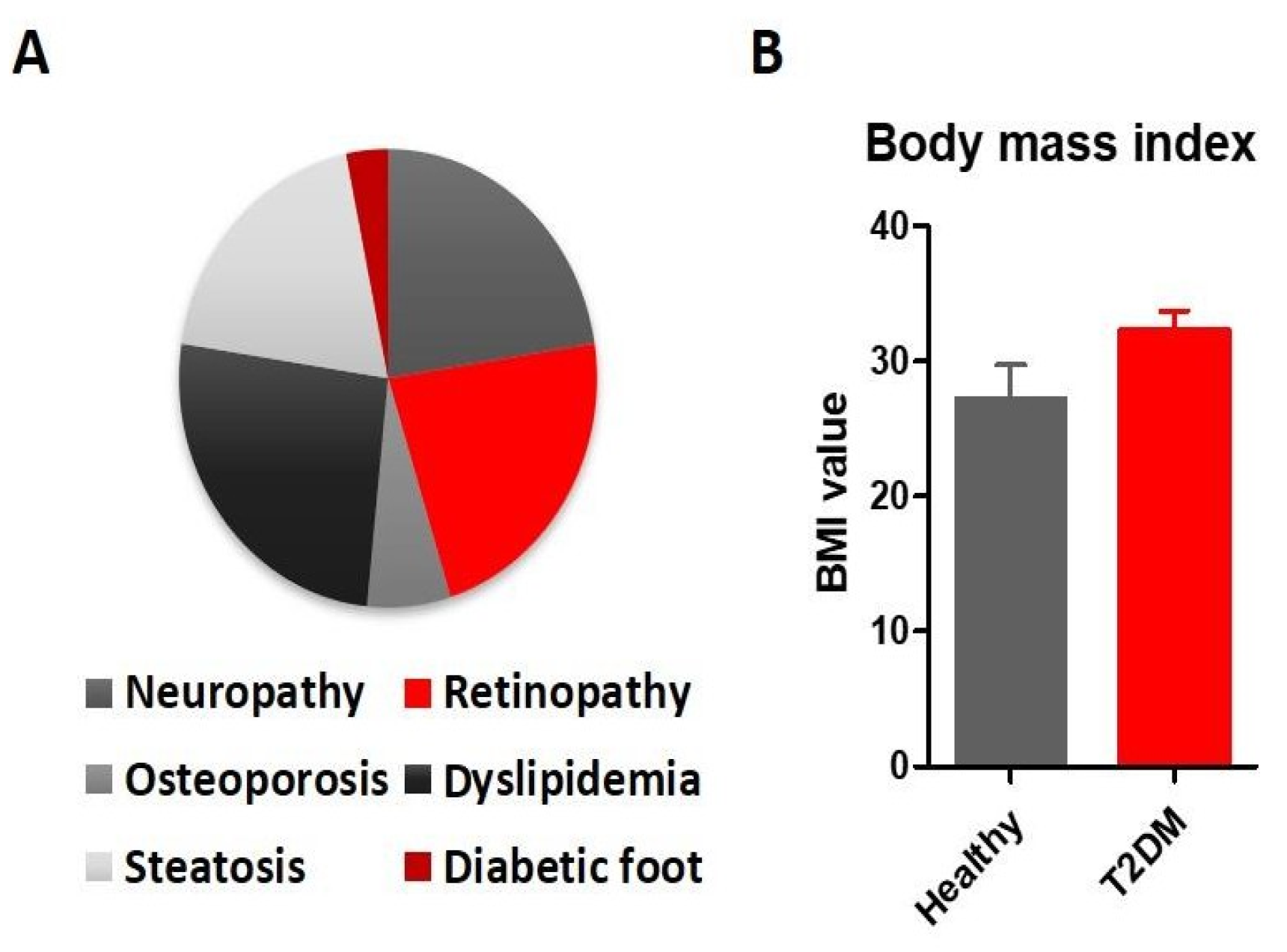
Figure 1. Patient characteristics. A. Associated diabetic complications identified in the enrolled patients. B. Body mass index differences between healthy controls and T2DM patients (Student t-test, p=0.0790).
Jmms 06 00019 g001
Figure 2. Microbiota analysis in patients with T2DM patients with diabetic nephropathy. A. Total bacteria represented as Ct values obtained from qRT-PCR targeting the 16S rDNA of all Eubacteria. The abundance of the Bacteroidetes (B) and Firmicutes (C) phyla in the stool samples harvested from healthy individuals (Control) and patients with diabetic nephropathy (T2DM).
Jmms 06 00019 g002
Figure 3. Microbiota patterns in patients with T2DM patients with diabetic nephropathy. Relative abundance of Bacteroides-Prevotella- Porphyromonas (A), Clostridium coccoides (B), Clostridium leptum (C), Faecalibacterium prausnitzii (D), Ruminococcus (E) and Turicibacter (F) in stool samples harvested from healthy individuals (Control) and patients with diabetic nephropathy (T2DM).
Jmms 06 00019 g003
Figure 4. Relative abundance of Butyricicocus (A), Lactobacillus (B), and Enterobacteriaceae (C) in stool samples harvested from healthy individuals (Control) and patients with diabetic nephropathy (T2DM).
Jmms 06 00019 g004
Figure 5. Fungal microbiome in diabetic nephropathy: total abundance of fungal 18SrDNA (expressed as Ct values), Candida sp. (B) and Saccharomyces sp. (C) abundance in the stool samples harvested from healthy individuals (Control) and patients with diabetic nephropathy (T2DM).
Jmms 06 00019 g005
Figure 6. The sevelamer carbonate treatment triggers changes in microbiota composition: Turicibacter sp. (A) and Enterobacteriaceae (B) are relatively abundant in the stool samples harvested from healthy individuals (Control) and patients with diabetic nephropathy (T2DM).
Jmms 06 00019 g006
Table 1. Primers used in this study
Jmms 06 00019 i001