Article

Natural Alternatives for Pain Relief: A Study on Morus alba, Angelica archangelica, Valeriana officinalis, and Passiflora incarnata

by
Felicia Suciu
1,2,
Oana Cristina Șeremet
2,
Emil Ștefănescu
2,
Ciprian Pușcașu
2,*,
Cristina Isabel Viorica Ghiță
3,
Cerasela Elena Gîrd
2,
Robert Viorel Ancuceanu
2 and
Simona Negreș
2
1
Department of Analysis and Quality Control of Drugs, Faculty of Pharmacy, “Ovidius” University of Constanta, 900470 Constanta, Romania
2
Faculty of Pharmacy, “Carol Davila” University of Medicine and Pharmacy, Traian Vuia 6, 020956 Bucharest, Romania
3
Department of Pharmacology and Pharmacotherapy, “Carol Davila” University of Medicine and Pharmacy, Eroii Sanitari 8, 050471 Bucharest, Romania
*
Author to whom correspondence should be addressed.
J. Mind Med. Sci. 2025, 12(2), 39; https://doi.org/10.3390/jmms12020039
Submission received: 9 May 2025 / Revised: 26 June 2025 / Accepted: 16 July 2025 / Published: 19 July 2025

Abstract

Background: Chronic pain poses a major global health burden, often inadequately managed by conventional analgesics due to limited efficacy and side effects. In this context, plant-based therapies offer a promising alternative. This study aimed to evaluate the antioxidant and analgesic potential of four medicinal plants traditionally used for pain relief: Morus alba, Angelica archangelica, Valeriana officinalis, and Passiflora incarnata. Methods: Phytochemical analyses quantified total phenolic acid, flavonoid, and polyphenolic acid contents in the extracts. Antioxidant activity was assessed using the ABTS radical scavenging assay. Analgesic effects were evaluated in vivo using the hot-plate and tail-flick tests in mice treated for 14 days with plant extracts or paracetamol. Results: Morus alba showed the highest polyphenolic content and strongest antioxidant activity (IC50 = 0.0695 mg/mL). In analgesic tests, Angelica archangelica demonstrated the most significant effect in the hot-plate test (72.2% increase in latency), while Valeriana officinalis had the highest efficacy in the tail-flick test (41.81%), exceeding paracetamol’s performance in that model. Conclusions: While antioxidant activity correlated with polyphenol content, analgesic effects appeared to involve additional mechanisms. These findings support the potential of Angelica archangelica and Valeriana officinalis as effective natural alternatives for pain relief.

1. Introduction

Pain is an uncomfortable sensation. It activates specific nerve fibers that send signals to the brain, where the conscious experience of pain can be influenced by various factors [1]. Although pain serves as a defense mechanism, acting as an alarm signal to protect the body and support human survival, it is also regarded as one of the most significant public health issues worldwide. Chronic pain, in particular, remains a major challenge for modern medicine [2]. The Centers for Disease Control and Prevention reports that around 100 million Americans suffer from common chronic pain conditions, more than those affected by heart disease, diabetes, and cancer combined. Studies indicate that the prevalence of chronic pain ranges from 11% to 40% [3].
The International Association for the Study of Pain (IASP) defines pain as “an unpleasant sensory and emotional experience associated with actual or potential tissue damage” [4]. Acute pain is a sudden, sharp, or throbbing sensation that usually results from tissue injury or trauma. It typically lasts less than six months and subsides as the body heals [5,6,7,8].
Chronic pain persists for over six months and results from nerve damage or faulty signal processing within the nervous system [9,10]. Its causes are often complex and unclear, and it significantly affects quality of life, leading to issues like anxiety, anger, and depression [11,12,13].
Although significant progress has been made in treating chronic pain and inflammatory diseases, these conditions remain a major global health burden due to the limitations and side effects of current therapies. Current anti-inflammatory and analgesic drugs often cause serious side effects, such as gastric damage from NSAIDs (non-steroidal anti-inflammatory drugs) or tolerance and dependence on opioids [14]. Moreover, current pain therapies face two major issues: lack of selectivity and limited scope. Many drugs, like sodium channel blockers or opioids, affect multiple systems due to widespread target distribution, leading to side effects such as euphoria, sedation, or respiratory depression. Additionally, most treatments block only a single pain pathway; for example, COX (cyclooxygenase) inhibitors reduce PGE2 (prostaglandin E2) but not other mediators such IL-1 (interleukin-1) or TNF (tumor necrosis factor), which limits their effectiveness [15]. Therefore, there is an urgent need for alternative therapeutic strategies to manage pain.
According to a World Health Organization report, around 80% of the global population still relies primarily on herbal remedies [1]. In response, researchers are increasingly investigating plant-derived compounds from traditional medicine for their promising bioactivities [16,17,18]. Rooted in centuries of practice, herbal medicine has produced a diverse array of refined treatments. Extensive studies suggest that many of these remedies are effective in managing pain, often with minimal or no side effects [19,20].
The present study aimed to investigate the antioxidant and analgesic potential of four traditionally used medicinal plants, Morus alba (Morus alba cortex (MA)), Angelica archangelica (Angelicae radix (AA)), Valeriana officinalis (Valerianae radix (VO)), and Passiflora incarnata (Passiflora herba (PI)), in mice, to explore alternative approaches to pain management. These plants were selected based on their rich phytochemical profiles and their potential interactions with the γ-aminobutyric acid (GABA) system [21,22,23,24,25]. The originality of this study lies in its integrative approach: rather than focusing on a single extract or isolated compound, it combines in vitro antioxidant assays with in vivo analgesic testing to assess the multi-target therapeutic potential of these natural products, aiming for effective pain relief with reduced toxicity.

2. Materials and Methods

2.1. Plant Materials and Extracts

AA and VO root teas (produced by Stef Mar Ltd., Râmnicu Vâlcea, Romania) and PI aerial parts tea (produced by Fares, Orăștie, Romania) were purchased from retail stores. MA bark was harvested in May 2018 from Buzău (Buzău County (Romania), dried at room temperature, and subsequently placed in an oven at 55 °C for several days, until it became friable and suitable for grinding. A voucher specimen (No. 7/2017) is deposited at the Pharmaceutical Botany department’s herbarium and is available for reference.
The plant materials were ground using an electric mill (Swantech, Gennevilliers, France), and the resulting powder was passed through a V-type sieve (mesh size: 250 µm). The powder was extracted with 70% (v/v) ethyl alcohol (MA bark) or 50% (v/v) ethyl alcohol (AA and VO roots and PI aerial parts) twice by hot reflux (approx. 75 °C). Each extract was filtered through filter paper, and the combined filtrates were concentrated using a rotary evaporator (Büchi, Flawil, Switzerland) at 70 °C. The resulting concentrated product was then lyophilized at −58 °C using a ScanVac CoolSafe freeze dryer (LaboGene, Allerød, Denmark) [26].

2.2. Phytochemical Analyses of Dry Plant Extracts

2.2.1. Determination of Total Phenolic Content (TP)

The TP of the extracts was determined using the Folin–Ciocalteu method [27]. Briefly, phenolic groups reduce molybdenum derivatives Mo6+ (yellow-colored) to molybdenum Mo4+ and Mo5+ (blue-colored), under alkaline conditions provided by sodium carbonate.
The samples were obtained from 0.1 g of soluble extract dissolved in a 100 mL volumetric flask. Volumes of 0.2 mL, 0.3 mL, 0.4 mL, 0.5 mL, and 0.6 mL for MA and 0.5 mL, 0.6 mL, 0.7 mL, 0.8 mL, and 0.9 mL for AA, PI, and VO were transferred to 10 mL volumetric flasks, brought up to 1 mL with distilled water, and then treated with 1 mL of diluted Folin–Ciocalteu reagent (1:1 ratio from the stock solution) (Sigma–Aldrich, Schnelldorf, Germany). The resulting mixture was brought to volume with a sodium carbonate solution of 200 g/L (Sigma–Aldrich, Schnelldorf, Germany). The samples were shaken and incubated in the dark for 40 min at room temperature. Absorbance was measured at λ = 763 nm (the maximum determined for tannic acid), against a blank sample (prepared under the same conditions but without the extract solution), using a Jasco V-530 spectrophotometer (Tokyo, Japan). A standard curve of tannic acid (Sigma–Aldrich, Schnelldorf, Germany) was previously determined, linear in the concentration range of 2.04–9.18 µg/mL (R2 = 0.999). The TP was expressed as mg tannic acid equivalents per gram of sample (mg/g) [28].

2.2.2. Determination of Total Flavonoid Content (TF)

The spectrophotometric determination of TF is based on the formation of stable, yellow-colored complexes between aluminum ions (Al3+) and the hydroxyl groups of flavone structures.
For each extract, 0.25 g was dissolved in 25 mL volumetric flasks using 50% ethanol. Aliquots of 0.3, 0.5, 0.7, 0.9, and 1.1 mL were transferred to 10 mL volumetric flasks for Morus alba (MA), while volumes of 0.9, 1.1, 1.3, 1.5, and 1.7 mL were used for Angelica archangelica (AA), Passiflora incarnata (PI), and Valeriana officinalis (VO). To each sample, 2 mL of sodium acetate solution (100 g/L; Sigma–Aldrich, Schnelldorf, Germany) and 1 mL of aluminum chloride solution (25 g/L; Sigma–Aldrich, Schnelldorf, Germany) were added, followed by dilution to volume with 50% ethanol. After a 45 min incubation, absorbance was measured at 427 nm—the wavelength corresponding to the maximum absorbance for metal ion chelation—using a Jasco V-530 spectrophotometer (Tokyo, Japan). A blank sample was prepared identically, excluding the extract solution. Rutin (Sigma–Aldrich, Schnelldorf, Germany) was used as the reference standard for constructing a calibration curve over the range of 5–35 µg/mL, with an R2 value of 0.9992. Total flavonoid content (TFC) was expressed as milligrams of rutin equivalents per gram of sample (mg/g) [29].

2.2.3. Determination of Total Polyphenol Acids Content (TPA)

The Arnow reagent is a valuable tool in phytochemical analysis due to its high specificity for detecting catechol-type phenolic compounds with ortho-dihydroxy groups, particularly in plant extracts and food samples. The Arnow method relies on the reaction between catechol and the nitrite-molybdate reagent under acidic conditions, initially producing a yellow color that shifts to an intense orange-red upon alkalinization [30]. For the determination of TPA content, 0.25 g of AA, PI, and VO, respectively, was dissolved in 25 mL volumetric flasks with 50% ethanol. For MA, 0.10 g extract was dissolved in a 50 mL volumetric flask with 50% ethanol. Volumes of 0.9 mL, 1.1 mL, 1.3 mL, 1.5 mL, and 1.7 mL were transferred to 10 mL volumetric flasks for MO and VO; for PI and AA, volumes of 1.3 mL, 1.5 mL, 1.7 mL, 1.9 mL, and 2.1 mL were used. To determine the total phenolic acid content, each sample was sequentially treated with 2 mL of 0.5 N hydrochloric acid (Sigma–Aldrich, Germany), followed by 2 mL of Arnow reagent (comprising sodium nitrite and sodium molybdate; Sigma–Aldrich, Schnelldorf, Germany), and then 2 mL of sodium hydroxide solution (85 g/L; Sigma–Aldrich, Schnelldorf, Germany). The final volume was adjusted using distilled water. Absorbance was recorded at 525 nm using a Jasco V-530 spectrophotometer, with a blank sample prepared identically but without the Arnow reagent. Chlorogenic acid (Sigma–Aldrich, Schnelldorf, Germany) served as the reference standard for constructing the calibration curve, which showed linearity in the range of 11–53 µg/mL (R2 = 0.9998). The total phenolic acid content was expressed in milligrams of chlorogenic acid equivalents per gram of sample (mg/g) [31].

2.3. Evaluation of Antioxidant Activity In Vitro

The antioxidant activity of the plant extracts was evaluated using the 2,2′-azino-bis(3-ethylbenzothiazoline-6-sulfonic acid) (ABTS) assay. In this method, the ABTS•+ radical, which exhibits a blue coloration, is decolorized in the presence of antioxidant compounds, resulting in a measurable decrease in absorbance [32].
The ABTS solution was used for evaluating the antioxidant capacity of the samples, due to its sensitivity and reproducibility in both hydrophilic and lipophilic systems. This method has been widely adopted in antioxidant studies [33].
In total, 0.25 g of dry extracts was solubilized in 25 mL of 50% ethanol. Volumes of 50 µL, 60 µL, 70 µL, 80 µL, 90 µL, and 100 µL were transferred to 10 mL volumetric flasks and adjusted to the final volume with 50% ethanol. Then, 0.5 mL of each solution was treated with 3 mL of ethanolic ABTS•+ solution, shaken, and kept in the dark for 4 min. The absorbance of the samples was measured at the maximum absorbance of the ABTS•+ solution (λ = 734 nm), using a Jasco V-530 spectrophotometer, against absolute ethanol as a blank.
The ABTS•+ solution was obtained by mixing 7.4 mM ABTS (Sigma–Aldrich, Schnelldorf, Germany) with 2.6 mM potassium persulfate (Merck, Darmstadt, Germany), followed by incubation at room temperature in the dark for 16 h prior to use. Ascorbic acid (Sigma-Aldrich, Schnelldorf, Germany) was used as a reference for the calibration curve in the range of concentration between 2 and 22 g/mL.
The percentage of inhibition of ABTS•+ was calculated using the formula below [31]:
%   Inhibition   ABTS = A   ( t = 0   min ) A   ( t = 4   min ) A   ( t = 0   min ) × 100
where A (t = 0 min) = absorbance of the blank sample (ABTS•+ sol in the absence of tested compounds: 0.70 ± 0.02);
A (t = 4 min) = absorbance of the vegetal extract (ABTS•+ sol in the presence of tested compounds).
IC50 value, the concentration of sample required to scavenge 50% of the ABTS•+ free radical, was calculated from the plotted graph of radical scavenging activity against the concentration of extracts (IC inhibition). The lower the IC50 value for an extract, the stronger the antioxidant activity [34].

2.4. Evaluation of Pain Sensitivity in Vivo

2.4.1. Animals

All procedures involving experimental animals were conducted in accordance with the bioethical guidelines outlined in Law No. 43/2014 on the protection of animals used for scientific purposes and Directive 2010/63/EU of the European Parliament. The experimental protocol (CFF 08/22.05.2020) was approved by the Bioethics Committee of the Faculty of Pharmacy, “Carol Davila” University of Medicine and Pharmacy, Bucharest, Romania.
Male NMRI mice (4–8 weeks old, weighing 25 ± 2.5 g) were sourced from the Cantacuzino National Institute of Research and Development for Microbiology and Immunology (INCDMI Cantacuzino), Bucharest, Romania. The animals were housed in plexiglass cages with unrestricted access to food (rodent ground chow, INCDMI Cantacuzino) and water. Environmental parameters were continuously monitored using a hygrothermometer and maintained at a temperature of 21–24 °C and a relative humidity of 45–60%. Prior to the start of the experiment, the mice were allowed a one-week acclimatization period in the new environment.
Male NMRI mice were specifically selected to ensure hormonal consistency and reduce biological variability. Female rodents exhibit hormonal fluctuations related to the estrous cycle, which can affect pain perception and treatment responses. The exclusive use of males minimizes this confounding factor, thereby enhancing the reproducibility and comparability of the results [35]. NMRI mice, being an outbred strain, are commonly used in pharmacological research due to their robust physiological responses and the broad translational relevance of experimental outcomes. Their suitability for nociceptive testing is well established. For instance, Vermeirsch et al. utilized NMRI mice to assess morphine-induced analgesia using the hot-plate test [36]. Additionally, a more recent study employed male NMRI mice in both the hot-plate and tail-flick tests to examine the enhancement of morphine-induced analgesia by harmaline, further validating the use of this strain in thermal pain models [37].

2.4.2. Experimental Groups

For this study, we used 60 mice divided into 6 equal groups (n = 10) that received treatments daily, for 14 days by oral gavage, as follows: control group (M)—distilled water 0.1 mL·kg−1; PCT group—paracetamol, 100 mg·kg−1; VO group—Valeriana officinalis extract, 60 mg·kg−1; PI group—Passiflora incarnata extract, 200 mg·kg−1; AA group—Angelica archangelica extract, 200 mg·kg−1; MA group—Morus alba extract, 200 mg·kg−1.
The extracts were administered as solutions or suspensions, prepared by dissolving the dry extract in distilled water. These preparations were stored at 2–8 °C in a refrigerator and used within a maximum of two days.
The 200 mg·kg−1 dose used for AA was selected based on a prior preclinical study, where oral administration of the methanolic extract at this dose produced significant antinociceptive effects in a mouse model of fibromyalgia. These results highlight the plant’s general analgesic potential, including its possible efficacy in conditions characterized by centrally mediated pain [38]. The 200 mg·kg−1 dose of MA bark extract used in this study was selected based on preclinical data demonstrating significant analgesic activity at this dose in mice, including reduced writhing responses and increased latency in thermal pain models [39]. The 200 mg·kg−1 dose of PI extract was chosen based on previous preclinical studies that reported significant central nervous system effects at this dosage, including sedative, depressant, anticonvulsant, and analgesic activities in mice [40]. The dose of 60 mg/kg for VO extract was selected based on preclinical studies indicating that the plant exerts biological effects within a range of 50 to 800 mg/kg [41]. Although our study employed oral administration, the chosen dose of 60 mg/kg represents a deliberately conservative approach—sufficient to investigate potential analgesic effects while minimizing the risk of excessive central nervous system depression. Furthermore, this dose is supported by literature reporting CNS activity, including GABAergic modulation, at doses starting as low as 50 mg/kg in rodent models [42]. A 1% paracetamol solution (Sigma-Aldrich, Schnelldorf, Germany) was also used and stored under the same conditions (2–8 °C) for up to 2 days. The 100 mg·kg−1 oral dose of paracetamol was selected based on its well-documented efficacy in rodent pain models. Mohrland et al. demonstrated that this dose significantly increased tail-flick latency in mice, confirming central analgesic effects in thermal nociceptive assays [43]. Similarly, Bianchi et al. reported that the same dose effectively reduced both central and peripheral hyperalgesia in rats, increasing nociceptive thresholds without affecting paw edema or baseline sensitivity. These findings collectively support 100 mg·kg−1 as a relevant and validated dose for assessing centrally mediated antinociceptive responses in preclinical models [44].

2.4.3. Hot-Plate Test

The hot-plate test evaluates supraspinal nociceptive responses, involving more complex integrative behavior such as paw licking or jumping [45]. It requires only a single exposure to a thermal stimulus, minimizing the risk of tissue damage. Additionally, the method does not require prior habituation and is widely recognized for its high reliability and reproducibility [46]. The hot-plate test was conducted at baseline and on days 7 and 14 after treatment initiation. Mice were placed on a heated surface maintained at 53 °C, and the latency to the first nociceptive response—such as paw licking, jumping, or paw shaking—was recorded as a measure of thermal pain sensitivity [47,48].

2.4.4. Tail-Flick Test

The tail-flick test provides a rapid and objective assessment of spinal nociceptive reflexes, with minimal influence from supraspinal behavioral factors. It is particularly valued for its simplicity, reproducibility, and low variability [45].
The tail-flick test was conducted using an analgesiometer equipped with an infrared radiation source, which delivered focused radiant heat to a single point on the proximal third of the tail. Mice were placed in restraining holders, leaving the tail exposed outside the holder to allow precise targeting of the heat stimulus. The latency to tail withdrawal (flick) was recorded and used as a measure of the nociceptive response [49]. The tests were assessed at baseline, as well as on days 7 and 14 following the initiation of treatment. Both tests were performed on the same day: the hot-plate test was conducted one hour after treatment administration, while the tail-flick test was carried out two hours post-administration. This order was selected to avoid potential interference between supraspinally integrated and spinally mediated nociceptive responses. The hot-plate test evaluates more complex, centrally processed behaviors (paw licking, jumping) and is more susceptible to alterations caused by prior stress or nociceptive input. In contrast, the tail-flick test reflects a simple spinal reflex that is less affected by earlier behavioral activation. Performing the hot-plate test first ensures that supraspinal responses are assessed in a relatively unaltered state, while the subsequent tail-flick test remains valid due to its robustness and minimal behavioral influence [50].

2.4.5. Statistical Analysis

Statistical analysis of the experimental data was performed using GraphPad Prism software, version 5.00 (GraphPad Software, San Diego, CA, USA). The D’Agostino–Pearson test was used to assess the normality of data distribution. For datasets with a normal (parametric) distribution, one-way analysis of variance (ANOVA) followed by Dunnett’s post hoc test were used to compare each treatment group with the control. For non-parametric data, the Kruskal–Wallis test followed by Dunn’s post hoc test were applied. These statistical methods were chosen to ensure an accurate interpretation of group differences according to distribution characteristics, thereby enhancing the robustness and reliability of the analysis. A confidence interval of 95% was used, with the level of statistical significance set at α = 0.05. Results were expressed as mean values ± standard error of the mean (S.E.M.). Percentage variations between experimental groups were calculated using Formula (1) [51]:
Δ% = (Mx − My)/My × 100
where Mx is the mean value for PCT, VO, PI, AA, and MA groups when compared vs. M; My is the mean value for M.
The experimental design, including extract preparation, phytochemical characterization, in vitro antioxidant testing, and in vivo analgesic evaluation, is summarized in Figure 1.

3. Results

3.1. Phytochemical Analyses of Dry Plant Extracts

The results obtained from spectrophotometric determinations on plant extracts are presented in Table 1.
Among the tested extracts, MA exhibited the highest content of all measured components, with a remarkably high TP value (11.50 ± 0.05 g/100 g), followed by significant levels of flavonoids (2.79 ± 0.52 g/100 g) and polyphenolic acids (5.61 ± 0.43 g/100 g). This suggests that MA may possess strong antioxidant potential and could be a valuable source of bioactive compounds.
PI and VO showed moderate levels of total polyphenols, with TP values of 6.36 ± 0.66 g/100 g and 5.95 ± 0.09 g/100 g, respectively. PI contained a higher amount of flavonoids (1.51 ± 0.06 g/100 g) than VO (0.64 ± 0.06 g/100 g), while VO presented slightly more polyphenolic acids (1.29 ± 0.11 g/100 g) than PI (0.85 ± 0.04 g/100 g).
In contrast, AA showed the lowest total polyphenolic content (4.27 ± 1.13 g/100 g), and both flavonoids and polyphenolic acids were not detected in this extract. This could indicate either a low presence of these subclasses of polyphenols or limitations in the sensitivity of the detection methods for this sample.

3.2. Evaluation of Antioxidant Activity in Vitro

Among the tested extracts, MA demonstrated the strongest antioxidant activity with an IC50 of 0.0695 mg/mL, a value close to the reference standard used (ascorbic acid—0.0165 mg/mL). This is consistent with its highest TP (11.50 g/100 g) (Table 2). This strong correlation supports the role of polyphenolic compounds in antioxidant mechanisms.
PI and VO exhibited moderate antioxidant activities, with IC50 values of 0.1044 mg/mL and 0.1272 mg/mL, respectively. These results correlate well with their intermediate levels of total polyphenol (6.36 g/100 g for PI and 5.95 g/100 g for VO). PI, having a higher flavonoid content than VO, also showed slightly better antioxidant performance, which suggests flavonoids may contribute significantly to radical scavenging capacity.
In contrast, AA displayed the weakest antioxidant activity among the extracts, with an IC50 of 0.2599 mg/mL, which aligns with its lowest total polyphenol content (4.27 g/100 g) and the absence of detectable flavonoids and polyphenolic acids.

3.3. Tests for the Evaluation of Pain Sensitivity

3.3.1. Hot-Plate Test

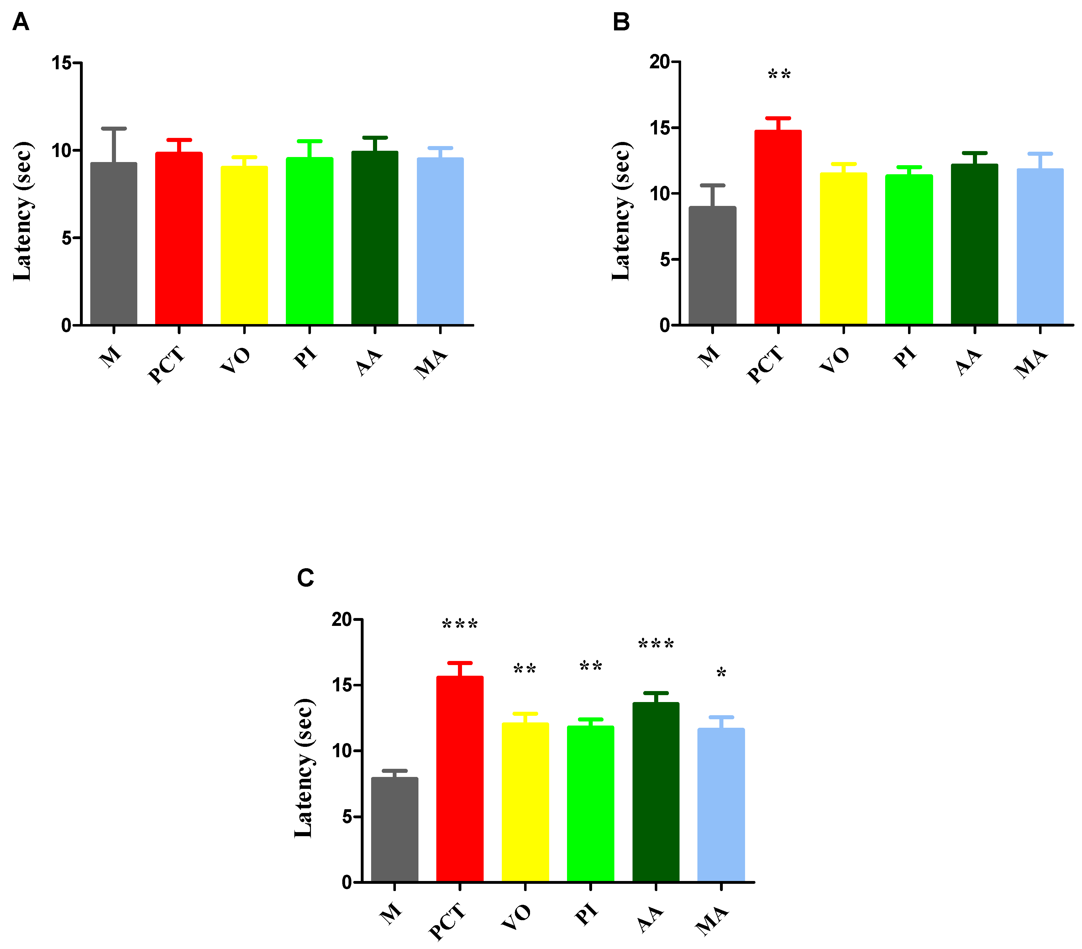
In the hot-plate test, no significant differences were observed between groups prior to treatment administration (Figure 2A). However, after 7 consecutive days of treatment, significant differences in thermal sensitivity emerged (univariate ANOVA, F = 13.78, p = 0.0171; Figure 2B), which became even more pronounced after 14 days (univariate ANOVA, F = 8.918, p < 0.0001; Figure 2C).
All treated groups showed an increase in pain reaction latencies compared to the control group after 7 days of treatment. However, only the group treated with paracetamol demonstrated a statistically significant increase, with a 65.16% improvement.
After 14 days of treatment, all treated groups exhibited significant increases in pain reaction latencies. The paracetamol group recorded the highest increase (97.58%), followed by AA (72.2%), VO (52.28%), PI (49.49%), and MA (47.33%). Among the plant-treated groups, AA produced the most pronounced analgesic effect.

3.3.2. Tail-Flick Test

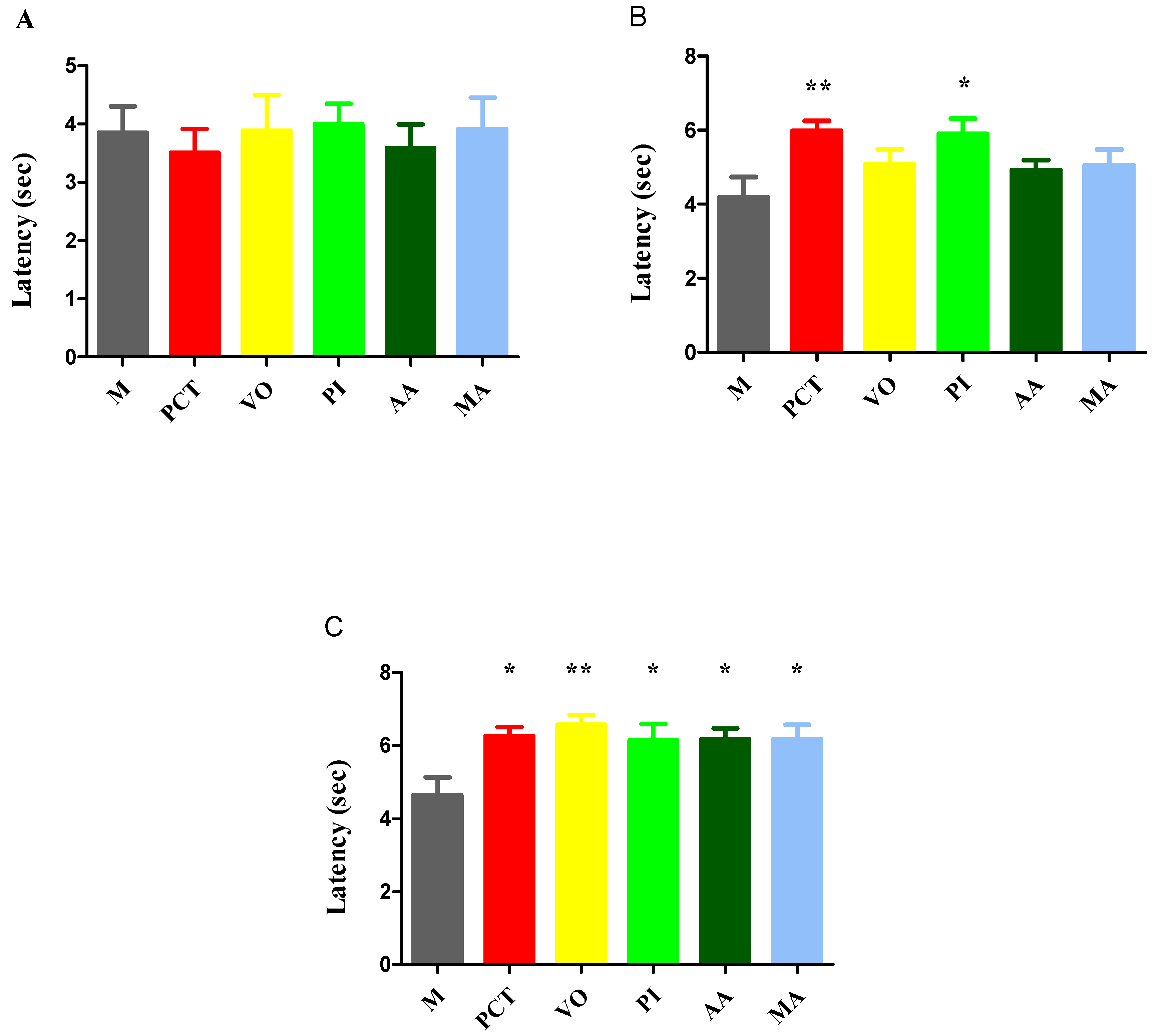
In the tail-flick test, no significant differences were observed between the groups before the initiation of treatment (Figure 3A). Changes in pain reaction latencies were noticed after 7 days (univariate ANOVA, F = 13.63, p = 0.0181, Figure 3B) and after 14 days of treatment (univariate ANOVA, F = 3.534, p < 0.0078, Figure 3C).
After 7 days of treatment, both the paracetamol and Passiflora incarnata (PI) groups exhibited significant increases in pain reaction latency—by 42.72% and 40.81%, respectively—indicating comparable analgesic effects relative to the control group.
After 14 days of treatment, all groups displayed significant analgesic effects. The group treated with VO showed the greatest increase (41.81%), followed by paracetamol (35.12%), with the other extracts showing comparable improvements.

4. Discussion

In recent years, interest in medicinal herbs has grown rapidly due to their natural origin and minimal side effects, appealing to both developed and developing countries. Plants serve as secondary sources for bioactive compounds used in medicines, insecticides, fragrances, dyes, and food additives. Chemical analysis reveals that plants contain a rich mix of bioactive compounds such as phytosterols, flavonoids, alkaloids, glycosides, vitamins, and minerals that contribute to their diverse pharmacological effects [52].
Among the active components of plants, flavonoids play an important role, contributing significantly to their therapeutic effects. Through their antioxidant, anti-inflammatory, anti-cancer, and antimicrobial properties, flavonoids help enhance the healing potential of botanical preparations and support overall health [53]. Some flavonoids exhibit analgesic activities, with several possible mechanisms responsible for this effect, such as the reduction of PGE2 and pro-inflammatory cytokines, blocking of central calcium channels, and the improvement of NO (nitric oxide) content in brain tissue and central serotonin levels [54].
Among the four tested extracts, MA exhibited the highest content of total polyphenols, flavonoids, and polyphenolic acids. These phytochemical parameters correlated with the strongest antioxidant activity, as demonstrated by its lowest IC50 value in the ABTS assay (0.0695 mg/mL). This suggests that the potent radical-scavenging capacity of MA may be largely attributed to its high levels of phenolic compounds, which are known to be effective hydrogen or electron donors. Similarly, PI and VO, which exhibited moderate flavonoid contents, also showed intermediate antioxidant activities. Conversely, AA demonstrated the weakest antioxidant activity (IC50 = 0.2599 mg/mL), correlating with its low total polyphenolic content and the absence of detectable flavonoids and polyphenolic acids.
Also, in our study, we demonstrated the analgesic potential of these four traditionally used medicinal plants, AA, VO, PI, and MA, using two validated pain models in mice: the hot-plate and tail-flick tests. Among these, AA exhibited the strongest analgesic effect in the hot-plate test after 14 days of treatment, showing a 72.2% increase in pain reaction latency, second only to the paracetamol group. In the tail-flick test, VO produced the most pronounced analgesic effect among the plant extracts, with a 41.81% increase in latency, surpassing even the paracetamol group at day 14.
The analgesic effects observed in vivo through the hot-plate and tail-flick tests revealed a more complex relationship between phytochemical content and biological efficacy. Although MA exhibited the highest antioxidant potential, its analgesic performance was comparatively lower than that of AA, which had the lowest total polyphenol content and no detectable flavonoids or polyphenolic acids. In the tail-flick test, VO displayed the strongest analgesic effect after 14 days, despite having moderate levels of polyphenols and flavonoids.
These findings suggest that although flavonoids and polyphenols play a significant role in antioxidant activity, the observed analgesic effects are likely mediated through additional mechanisms beyond polyphenolic content alone. Analgesia involves complex physiological processes, encompassing both central and peripheral pathways. The hot-plate test evaluates supraspinal nociceptive responses integrated into the brain, while the tail-flick test reflects spinal reflex arcs. As such, compounds acting at the level of the central nervous system (e.g., via GABAergic, serotonergic, or endocannabinoid modulation) may exhibit more prominent effects in the hot-plate test, whereas peripheral anti-inflammatory or reflex-inhibitory agents are better detected in the tail-flick test [50].
Therefore, although higher flavonoid content is clearly correlated with increased antioxidant activity, as indicated by lower IC50 values, the in vivo analgesic effects point to the involvement of additional bioactive compounds and complex pharmacodynamic interactions that extend beyond antioxidant mechanisms alone.
These inconsistencies between polyphenolic content and observed analgesic effects suggest that other mechanisms, especially those related to central neurotransmission, may also be involved. The analgesic activity of the four plant extracts investigated in this study appears to be mediated, at least in part, through their interaction with the GABAergic system, according to existing evidence. The selection of MA, AA, VO, and PI was not based on botanical relatedness or plant growth form but rather on a pharmacologically driven strategy. These plants were chosen due to their documented phytochemical composition, especially compounds with potential activity on the GABAergic system, which plays a crucial role in modulating pain. Although these species are not genetically related, they share a functional similarity: the ability to modulate GABA-mediated signaling pathways, which are central to both pain perception and neuronal inhibition. The focus was therefore placed on bioactivity and mechanistic relevance, rather than taxonomy or morphology, to increase the translational potential of the findings.
GABA is a non-protein amino acid synthesized from glutamate via glutamic acid decarboxylase. It is widely present in microorganisms, plants, and vertebrates, with high expression in the brain and spinal cord. As the main inhibitory neurotransmitter in the mammalian nervous system, GABA regulates synaptic transmission, neuronal development, and relaxation and also plays a key role in modulating pain. GABA acts through three receptor types: the ionotropic GABAA and GABAC, which mediate fast transmission, and the metabotropic GABAB, which mediates slower effects. These receptors influence pain perception through distinct signaling pathways, making them promising targets for the development of novel analgesics [55]. The presence of GABA has been quantified in various mulberry species. According to Chen et al., the GABA content was 0.862% in Morus atropurpurea, 0.726% in Morus mongolica, 0.640% in MA, and 0.473% in Morus multicaulis [21]. In PI, the total plant extract has been shown to induce strong currents modulating GABAA receptor activity, attributed to its high GABA content [24]. In AA, several compounds—such as columbianetin, imperatorin, cnidilin, osthol, and columbianedin—were identified and shown to enhance GABA-induced chloride influx through GABAA and GABAB receptors in a concentration-dependent manner [23]. Similarly, Yuan et al. reported that the pharmacological effects of VO extract and valerenic acid are mediated through modulation of GABAA receptor function, suggesting the potential to enhance the activity of anesthetics and other GABAergic drugs [25].
MA, the predominant species within the genus Morus, which comprises over 150 known species [56], is a widely distributed medicinal plant native to China. Due to its adaptability to diverse climatic, topographical, and soil conditions, it is now cultivated across Asia, Europe, the Americas, and even tropical regions of the Southern Hemisphere [52,57]. Medicinally, all parts of the MA plant, leaves, fruits, branches, roots, and the whole plant, have been used. Preliminary analysis shows that it contains a wide range of bioactive compounds, including sitosterol, steroids, tannins, phytosterols, glycosides, carbohydrates, proteins, alkaloids, amino acids, saponins, triterpenes, phenols, flavonoids, benzofuran derivatives, anthocyanins, and anthraquinones [58,59,60].
Active compounds specific to MA bark include the following: morin (found in the highest concentration in the bark—12.3 mcg/g) [61]; albanol A [62]; sanggenon O [63], which has anti-inflammatory properties and acts as a GABA-A receptor modulator [64]; triterpenes such as moruslupenoic acid A and B [65]; arylbenzofurans such as moracin R, C, O, P, mulberrofuran L and Y, kuwanon A, C, E, and T, along with oxyresveratrol and adenosine [66]. MA root bark extract reduced NO production by inhibiting the overexpression of inducible nitric oxide synthase (iNOS). Additionally, it suppressed extracellular signal-regulated kinases 1 and 2 (ERK1/2) activation by promoting inhibitor of kappa B (IκB) degradation and hyperphosphorylation, thereby preventing nuclear factor kappa-light-chain-enhancer of activated B cells (NF-κB) activation through inhibition of p65 nuclear translocation [67]. MA extract also inhibited the production of inflammatory cytokines, including interleukin-6 (IL-6) and TNF, in response to LPS (lipopolysaccharide) stimulation [68]. The traditional use of MA, particularly its leaves and root bark, for pain relief is supported by the presence of these active compounds, which collectively exert analgesic, anti-inflammatory, and GABAergic modulatory effects [52,57].
Regarding the safety profile of MA, available toxicological data indicate favorable tolerance in preclinical models. Oral administration of ethanolic extracts at doses up to 2000 mg/kg did not cause behavioral changes or lethality in mice and was not associated with genotoxic effects [69]. However, intraperitoneal injection induced hematological and histological alterations, emphasizing the relevance of the administration route. Subacute and chronic toxicity studies involving root bark extract administered subcutaneously (50–200 mg/kg/day) also revealed no adverse effects [70]. These findings support the use of MA extract at therapeutic doses and confirm its safety when administered orally, as was the case in the present study.
AA, commonly known as garden Angelica, is an aromatic medicinal species native to northern and eastern Europe, traditionally used in the treatment of digestive and nervous system disorders. The plant thrives in cool, moist climates, especially in Nordic and alpine regions, preferring rich, well-drained soils [71,72]. In traditional and folklore medicine, it is celebrated for its numerous health benefits, earning it the nickname “Angel plant” due to its reputed therapeutic potency. Historically, AA has been used to treat a variety of ailments, including gastrointestinal disturbances, anxiety, sepsis, skin rashes, coughs, fever, headaches, urinary tract infections, and rheumatic disorders. It has also been regarded as a natural blood purifier [73]. Studies have demonstrated that the entire AA plant serves as a potential source of essential oils (ranging from 0.35% to 1%) and coumarins, both of which contribute significantly to its diverse bioactivities [74]. Essential oils from AA contain around 60 compounds, including monoterpenes, sesquiterpenes, alcohols, and macrolides. The plant is also rich in coumarins, particularly linear and angular furanocoumarins, with higher concentrations found in the roots [71]. Preclinical studies have shown that coumarins exhibit a range of pharmacological effects, including analgesic [75], anti-inflammatory [76], anti-anxiety [77], antioxidant [75], and antiseizure activities [78]. While the anti-inflammatory potential of AA is well documented, there is limited literature supporting its analgesic properties [71]. Nevertheless, the presence of furanocoumarins, along with volatile oils and flavonoids, in the root suggests a plausible role in mediating spasmolytic and sedative effects, which may contribute indirectly to pain relief [71]. Acute toxicity data indicate that AA root oil has an oral LD50 (lethal dose, 50%) of 11,000 mg/kg in rats and 2200 mg/kg in mice, suggesting a low level of acute toxicity and a favorable safety profile at therapeutic doses [79].
VO, commonly known as “All-heal” in English, is a well-known medicinal plant cultivated across Europe, Asia, and North America, traditionally valued for its neuroactive and calming properties [80]. VO has a complex phytochemical profile, comprising over 150 identified compounds. Its qualitative and quantitative composition varies depending on environmental factors, climate, plant age, harvest stage, and subspecies. The key constituents include alkaloids (such as chatinine, valerine, valerianine, and actinidine), organic acids and terpenes (including valerenic acid, isovaleric acid, valeric acid, and acetoxyvalerenic acid), iridoids (like valepotriate and isovalepotriate), lignanoids (pinoresinol-4-O-D-glucoside and 8′-hydroxypinoresinol), and flavonoids (such as apigenin, luteolin, and quercetin) [81]. The root of VO has been used since ancient Greece and Rome in traditional medicine and continues to be valued in modern practices for its calming effects and support in sleep promotion. Its primary active compound, the sesquiterpene valerenic acid, exerts these effects by modulating GABA receptors [81]. The European Medicines Agency (EMA) includes various traditional herbal preparations of VO in its monograph, recognizing over 30 years of therapeutic use for relieving mild mental stress and promoting sleep [81]. Additionally, VO is reported to possess other biological activities, including cardiovascular benefits (such as lowering blood pressure and heart rate, antiarrhythmic effects, and lipid regulation), as well as anti-cancer, antimicrobial, and spasmolytic properties [82,83,84].
The medicinal parts used for pain relief are the rhizome and roots, which contain valepotriates, sesquiterpenes, and volatile oils, including valerianic acid and bornyl acetate [81]. However, the analgesic potential of VO remains underexplored in the scientific literature. A recent review by Sánchez et al. highlights that only two preclinical studies have investigated its analgesic effects in animal models [80]. In these studies, alcoholic extracts of VO root, administered at doses of 200 mg/kg and 400 mg/kg, significantly reduced pain scores during the acute phase and lowered pain sensitivity in formalin-induced pain models using Wistar and Sprague Dawley rats [85,86].
The safety profile of VO has been thoroughly documented, with preclinical studies demonstrating low toxicity at therapeutic doses. In animal models, oral administration of VO extract did not induce significant adverse effects on behavior, organ function, or histopathological parameters. Acute toxicity studies reported an oral LD50 greater than 3 g/kg in mice, suggesting a high margin of safety [87]. Additionally, an intraperitoneal LD50 of 3300 mg/kg was reported for the ethanolic root extract in mice, further confirming its low acute toxicity [88]. Although very high doses may lead to enhanced sedative effects or mild hepatic changes, such outcomes are not commonly observed with standard therapeutic use. Overall, the available evidence supports the favorable safety profile of Valeriana officinalis (VO), reinforcing its suitability for oral administration in the management of neuropathic pain [87].
PI is a perennial climbing vine native to the southeastern United States, now cultivated in Europe and other temperate regions worldwide. It grows rapidly and thrives in warm and temperate climates [89]. Currently, researchers believe that only a portion of the active compounds in PI have been identified [90]. The aerial parts of the plant are phytochemically characterized by a range of primary constituents, including flavonoids (notably benzoflavones), maltol, cyanogenic glycosides, and indole alkaloids. The indole alkaloids include harman, harmine, harmaline, harmol, and harmalol [91]. Flavonoids represent approximately 2.5% of the identified constituents in PI, with notable compounds including vitexin, isovitexin, orientin, isoorientin, apigenin, kaempferol, vicenin, lucenin, and saponarin. Additionally, PI contains various organic acids such as phenolic acid, linoleic acid, palmitic acid, oleic acid, myristic acid, formic acid, and butyric acid, as well as coumarins, phytosterols, and essential oils [92]. Preclinical pharmacological studies have highlighted the therapeutic potential of PI, demonstrating anxiolytic, sedative, anticonvulsant, aphrodisiac, antitussive, antiasthmatic, and hypoglycemic properties [91]. In the context of analgesic use, only the aerial parts are considered therapeutically relevant, as they are the source of the neuroactive flavonoids responsible for GABAergic effects [25]. However, its analgesic potential remains poorly documented in the existing literature. Acute toxicity assessments have shown that the ethanolic extract has an oral LD50 of 6200 mg/kg in rats, suggesting minimal acute toxicity and confirming its safety when administered at therapeutic levels [93].
Paracetamol was used as the positive control in our study due to its well-established analgesic efficacy. It is one of the most commonly used analgesics in clinical practice, with treatment guidelines recommending its regular, time-limited use for mild to moderate acute pain and chronic non-malignant pain [94]. Paracetamol produces its analgesic effect primarily by inhibiting prostaglandin synthesis within the central nervous system, particularly in the brain and spinal cord. Although it exhibits weak in vitro inhibition of COX-1 (cyclooxygenase-1) and COX-2 (cyclooxygenase-2), it effectively lowers PGE2 levels in the central nervous system in vivo, suggesting the involvement of a distinct cyclooxygenase isoform, potentially COX-3 (cyclooxygenase-3)—a splice variant of COX-1. While the functional relevance of COX-3 in humans remains controversial, the drug’s ability to cross the blood–brain barrier and exert centrally mediated effects supports a central mechanism of action [95]. Furthermore, recent studies indicate that paracetamol may enhance endocannabinoid signaling and influence descending serotonergic inhibitory pathways, which may also contribute to its analgesic properties [96].
This study presents limitations that should be acknowledged. First, only a single dosage was evaluated for each extract, which precludes analysis of potential dose-dependent effects and may limit the accuracy of the efficacy assessment. Second, the treatment duration was relatively short (14 days), possibly insufficient to observe long-term therapeutic effects or delayed side effects. Furthermore, the study did not include additional biological or mechanistic assays, such as assessments of inflammatory markers, oxidative stress parameters, or analyses of molecular pathways. Although the current in vitro and in vivo methods provide a solid foundation for evaluating antioxidant and analgesic activity, future investigations incorporating these complementary approaches would offer deeper mechanistic insight and strengthen the pharmacological relevance of the findings. Additionally, spectroscopic analyses to confirm and quantify the dominant secondary metabolites in each extract were not performed. This omission limits the chemical specificity of our findings and should be addressed in future work.

5. Conclusions

The growing interest in medicinal plants is driven by their complex phytochemical composition and therapeutic potential with minimal side effects. Among bioactive constituents, flavonoids play a major role due to their antioxidant, anti-inflammatory, and analgesic properties. In this study, we investigated the antioxidant and analgesic activities of four traditionally used medicinal plants: AA, VO, PI, and MA. Although MA exhibited the highest flavonoid and polyphenol content and the strongest antioxidant activity (IC50 = 0.0695 mg/mL), the most potent analgesic effects were observed for AA and VO. After 14 days of treatment, AA showed a 72.2% increase in pain reaction latency in the hot-plate test, second only to paracetamol, while VO demonstrated the greatest analgesic effect in the tail-flick test, surpassing even the reference drug. These findings indicate that although flavonoid and polyphenol levels are closely associated with antioxidant activity, the observed analgesic effects are likely mediated by additional mechanisms beyond antioxidant capacity—potentially involving neurotransmission modulation or other pharmacodynamic pathways. Overall, the results underscore the promising analgesic potential of AA and VO, lending support to their traditional use in pain management.

Author Contributions

Conceptualization, F.S. and S.N.; methodology, C.P., O.C.Ș., E.Ș., C.I.V.G., R.V.A., C.E.G. and S.N.; validation, F.S. and S.N.; formal analysis, F.S., C.P. and S.N.; investigation, C.P., O.C.Ș. and E.Ș.; data curation, F.S.; writing—original draft preparation, F.S. and C.P.; writing—review and editing, O.C.Ș. and C.P.; visualization, S.N.; supervision, S.N. All authors have read and agreed to the published version of the manuscript.

Funding

This research received no external funding.

Institutional Review Board Statement

This study was conducted according to the guidelines of the Declaration of Helsinki and approved by the Bioethics Committee of the Faculty of Pharmacy, Carol Davila University of Medicine and Pharmacy, Bucharest, Romania (CFF 08/22.05.2020).

Informed Consent Statement

Not applicable.

Data Availability Statement

All data generated or analyzed during this study are included in this published article.

Conflicts of Interest

The authors declare no conflicts of interest.

References

  1. Khan, H.; Pervaiz, A.; Intagliata, S.; Das, N.; Nagulapalli Venkata, K.C.; Atanasov, A.G.; Najda, A.; Nabavi, S.M.; Wang, D.; Pittalà, V.; et al. The Analgesic Potential of Glycosides Derived from Medicinal Plants. DARU J. Pharm. Sci. 2020, 28, 387–401. [Google Scholar] [CrossRef]
  2. Silva-Correa, C.R.; Campos-Reyna, J.L.; Villarreal-La Torre, V.E.; Calderon-Pena, A.A.; Gonzalez Blas, M.V.; Aspajo-Villalaz, C.L.; Cruzado-Razco, J.L.; Sagastegui-Guarniz, W.A.; Guerrero-Espino, L.M.; Hilario-Vargas, J. Potential Activity of Medicinal Plants as Pain Modulators: A Review. Pharmacogn. J. 2021, 13, 248–263. [Google Scholar] [CrossRef]
  3. Wang, C.; Meng, Q. Global Research Trends of Herbal Medicine for Pain in Three Decades (1990–2019): A Bibliometric Analysis. J. Pain Res. 2021, 14, 1611–1626. [Google Scholar] [CrossRef] [PubMed]
  4. Pușcașu, C.; Andrei, C.; Olaru, O.T.; Zanfirescu, A. Metabolite-Sensing Receptors: Emerging Targets for Modulating Chronic Pain Pathways. Curr. Issues Mol. Biol. 2025, 47, 63. [Google Scholar] [CrossRef] [PubMed]
  5. Tedore, T.; Weinberg, R.; Witkin, L.; Giambrone, G.P.; Faggiani, S.L.; Fleischut, P.M. Acute Pain Management/Regional Anesthesia. Anesthesiol. Clin. 2015, 33, 739–751. [Google Scholar] [CrossRef]
  6. Argoff, C.E. Recent Management Advances in Acute Postoperative Pain. Pain Pract. 2014, 14, 477–487. [Google Scholar] [CrossRef]
  7. Odoma, S.; Zezi, A.U.; Danjuma, N.M.; Ahmed, A.; Magaji, M.G. Elucidation of the Possible Mechanism of Analgesic Actions of Butanol Leaf Fraction of Olax Subscorpioidea Oliv. J. Ethnopharmacol. 2017, 199, 323–327. [Google Scholar] [CrossRef]
  8. Rauf, A.; Ali, J.; Khan, H.; Mubarak, M.S.; Patel, S. Emerging CAM Ziziphus Nummularia with in Vivo Sedative-Hypnotic, Antipyretic and Analgesic Attributes. 3 Biotech 2016, 6, 11. [Google Scholar] [CrossRef]
  9. Rauf, A.; Khan, R.; Raza, M.; Khan, H.; Pervez, S.; De Feo, V.; Maione, F.; Mascolo, N. Suppression of Inflammatory Response by Chrysin, a Flavone Isolated from Potentilla Evestita Th. Wolf. In Silico Predictive Study on Its Mechanistic Effect. Fitoterapia 2015, 103, 129–135. [Google Scholar] [CrossRef]
  10. Shchegol’kov, E.V.; Shchur, I.V.; Burgart, Y.V.; Saloutin, V.I.; Trefilova, A.N.; Ljushina, G.A.; Solodnikov, S.Y.; Markova, L.N.; Maslova, V.V.; Krasnykh, O.P.; et al. Polyfluorinated Salicylic Acid Derivatives as Analogs of Known Drugs: Synthesis, Molecular Docking and Biological Evaluation. Bioorg. Med. Chem. 2017, 25, 91–99. [Google Scholar] [CrossRef]
  11. Qadir, M.I.; Abbas, K.; Hamayun, R.; Ali, M. Analgesic, Anti-Inflammatory and Anti-Pyretic Activities of Aqueous Ethanolic Extract of Tamarix aphylla L. (Saltcedar) in Mice. Pak. J. Pharm. Sci. 2014, 27, 1985–1988. [Google Scholar]
  12. Ali, M.; Rauf, A.; Hadda, T.; Bawazeer, S.; Abu-Izneid, T.; Khan, H.; Raza, M.; Khan, S.; Shah, S.; Pervez, S.; et al. Mechanisms Underlying Anti-Hyperalgesic Properties of Kaempferol-3,7- Di-O-α-L-Rhamnopyranoside Isolated from Dryopteris Cycadina. Curr. Top. Med. Chem. 2016, 17, 383–390. [Google Scholar] [CrossRef] [PubMed]
  13. Andersen, L.P.H.; Gögenur, I.; Fenger, A.Q.; Petersen, M.C.; Rosenberg, J.; Werner, M.U. Analgesic and Antihyperalgesic Effects of Melatonin in a Human Inflammatory Pain Model. Pain 2015, 156, 2286–2294. [Google Scholar] [CrossRef] [PubMed]
  14. Reis-Pina, P.; Lawlor, P.G.; Barbosa, A. Cancer-Related Pain Management and the Optimal Use of Opioids. Acta Med. Port. 2015, 28, 376–381. [Google Scholar] [CrossRef] [PubMed]
  15. Jayakar, S.; Shim, J.; Jo, S.; Bean, B.P.; Singeç, I.; Woolf, C.J. Developing Nociceptor-Selective Treatments for Acute and Chronic Pain. Sci. Transl. Med. 2021, 13, eabj9837. [Google Scholar] [CrossRef]
  16. Khan, H.; Rengasamy, K.R.R.; Pervaiz, A.; Nabavi, S.M.; Atanasov, A.G.; Kamal, M.A. Plant-Derived MPGES-1 Inhibitors or Suppressors: A New Emerging Trend in the Search for Small Molecules to Combat Inflammation. Eur. J. Med. Chem. 2018, 153, 2–28. [Google Scholar] [CrossRef]
  17. Khan, H.; Nabavi, S.M.; Sureda, A.; Mehterov, N.; Gulei, D.; Berindan-Neagoe, I.; Taniguchi, H.; Atanasov, A.G. Therapeutic Potential of Songorine, a Diterpenoid Alkaloid of the Genus Aconitum. Eur. J. Med. Chem. 2018, 153, 29–33. [Google Scholar] [CrossRef]
  18. Khan, H.; Amin, S.; Patel, S. Targeting BDNF Modulation by Plant Glycosides as a Novel Therapeutic Strategy in the Treatment of Depression. Life Sci. 2018, 196, 18–27. [Google Scholar] [CrossRef]
  19. Arai, Y.-C.; Makino, I.; Ikemoto, T.; Saisu, H.; Terajima, Y.; Owari, K. Kampo for the Treatment of Pain in Japan: A Review. Pain Ther. 2020, 9, 161–170. [Google Scholar] [CrossRef]
  20. Luo, Y.; Wang, C.-Z.; Sawadogo, R.; Tan, T.; Yuan, C.-S. Effects of Herbal Medicines on Pain Management. Am. J. Chin. Med. 2020, 48, 1–16. [Google Scholar] [CrossRef]
  21. Chen, H.; He, X.; Liu, Y.; Li, J.; He, Q.; Zhang, C.; Wei, B.; Zhang, Y.; Wang, J. Extraction, Purification and Anti-Fatigue Activity of γ-Aminobutyric Acid from Mulberry (Morus alba L.) Leaves. Sci. Rep. 2016, 6, 18933. [Google Scholar] [CrossRef] [PubMed]
  22. Gupta, G.; Dua, K.; Kazmi, I.; Anwar, F. Anticonvulsant Activity of Morusin Isolated from Morus alba: Modulation of GABA Receptor. Biomed. Aging Pathol. 2014, 4, 29–32. [Google Scholar] [CrossRef]
  23. Zaugg, J.; Eickmeier, E.; Rueda, D.C.; Hering, S.; Hamburger, M. HPLC-Based Activity Profiling of Angelica Pubescens Roots for New Positive GABAA Receptor Modulators in Xenopus Oocytes. Fitoterapia 2011, 82, 434–440. [Google Scholar] [CrossRef] [PubMed]
  24. Elsas, S.-M.; Rossi, D.J.; Raber, J.; White, G.; Seeley, C.-A.; Gregory, W.L.; Mohr, C.; Pfankuch, T.; Soumyanath, A. Passiflora incarnata L. (Passionflower) Extracts Elicit GABA Currents in Hippocampal Neurons in Vitro, and Show Anxiogenic and Anticonvulsant Effects in Vivo, Varying with Extraction Method. Phytomedicine 2010, 17, 940–949. [Google Scholar] [CrossRef]
  25. Yuan, C.-S.; Mehendale, S.; Xiao, Y.; Aung, H.H.; Xie, J.-T.; Ang-Lee, M.K. The Gamma-Aminobutyric Acidergic Effects of Valerian and Valerenic Acid on Rat Brainstem Neuronal Activity. Anesth. Analg. 2004, 98, 353–358. [Google Scholar] [CrossRef]
  26. Hovaneţ, M.; Viorel Ancuceanu, R.; Dinu, M.; Oprea, E.; Adriana Budura, E.; Negreş, S.; Ştefan Velescu, B.; Elena Duţu, L.; Adriana Anghel, I.; Ancu, I.; et al. Toxicity and Anti-Inflammatory Activity of Ziziphus jujuba Mill. Leaves. Farmacia 2016, 64, 5. [Google Scholar]
  27. Lamuela-Raventós, R.M. Folin-Ciocalteu Method for the Measurement of Total Phenolic Content and Antioxidant Capacity. In Measurement of Antioxidant Activity & Capacity; John Wiley & Sons: Chichester, UK, 2017; pp. 107–115. [Google Scholar]
  28. Gird, C.E.; Dutu, L.E.; Costea, T.; Nencu, I.; Popescu, M.; Balaci, T.O.O. Research Regarding Obtaining Herbal Extracts with Antitumour Activity. Note Ii. Phytochemical Analysis, Antioxidant Activity and Cytotoxic Effects of Chelidonium majus L. Medicago sativa L. and Berberis vulgaris L. Dry Extracts. Farmacia 2017, 65, 703–708. [Google Scholar]
  29. Luță, E.A.; Biță, A.; Moroșan, A.; Mihaiescu, D.E.; Ghica, M.; Mihai, D.P.; Olaru, O.T.; Deculescu-Ioniță, T.; Duțu, L.E.; Popescu, M.L.; et al. The Influence of Phytosociological Cultivation and Fertilization on Polyphenolic Content of Menthae and Melissae Folium and Evaluation of Antioxidant Properties through In Vitro and In Silico Methods. Plants 2022, 11, 2398. [Google Scholar] [CrossRef]
  30. Ferreira, C.M.H.; Vilas-Boas, Â.; Sousa, C.A.; Soares, H.M.V.M.; Soares, E.V. Comparison of Five Bacterial Strains Producing Siderophores with Ability to Chelate Iron under Alkaline Conditions. AMB Express 2019, 9, 78. [Google Scholar] [CrossRef]
  31. Costea, L.; Chițescu, C.L.; Boscencu, R.; Ghica, M.; Lupuliasa, D.; Mihai, D.P.; Deculescu-Ioniță, T.; Duțu, L.E.; Popescu, M.L.; Luță, E.-A.; et al. The Polyphenolic Profile and Antioxidant Activity of Five Vegetal Extracts with Hepatoprotective Potential. Plants 2022, 11, 1680. [Google Scholar] [CrossRef]
  32. Dudonné, S.; Vitrac, X.; Coutière, P.; Woillez, M.; Mérillon, J.-M. Comparative Study of Antioxidant Properties and Total Phenolic Content of 30 Plant Extracts of Industrial Interest Using DPPH, ABTS, FRAP, SOD, and ORAC Assays. J. Agric. Food Chem. 2009, 57, 1768–1774. [Google Scholar] [CrossRef]
  33. Hernández-Rodríguez, P.; Baquero, L.P.; Larrota, H.R. Flavonoids. In Bioactive Compounds; Elsevier: Amsterdam, The Netherlands, 2019; pp. 265–288. [Google Scholar]
  34. Zhan, Y. Protective Effect of Dexmedetomidine Combined with Ulinastatin on Myocardium in Patients Undergoing Valve Replacement. Farmacia 2019, 67, 437–441. [Google Scholar] [CrossRef]
  35. Sorge, R.E.; Mapplebeck, J.C.S.; Rosen, S.; Beggs, S.; Taves, S.; Alexander, J.K.; Martin, L.J.; Austin, J.-S.; Sotocinal, S.G.; Chen, D.; et al. Different Immune Cells Mediate Mechanical Pain Hypersensitivity in Male and Female Mice. Nat. Neurosci. 2015, 18, 1081–1083. [Google Scholar] [CrossRef]
  36. Vermeirsch, H.; Meert, T.F. Morphine-Induced Analgesia in the Hot-Plate Test: Comparison between NMRI Nu/Nu and NMRI Mice. Basic Clin. Pharmacol. Toxicol. 2004, 94, 59–64. [Google Scholar] [CrossRef] [PubMed]
  37. Alijanpour, S.; Jafaripour, S.; Ghasemzadeh, Z.; Khakpai, F.; Zarrindast, M.-R. Harmaline Potentiates Morphine-Induced Antinociception via Affecting the Ventral Hippocampal GABA-A Receptors in Mice. Eur. J. Pharmacol. 2021, 893, 173806. [Google Scholar] [CrossRef] [PubMed]
  38. Kaur, A.; Singh, N.; Bhatti, M.S.; Bhatti, R. Optimization of Extraction Conditions of Angelica archangelica Extract and Activity Evaluation in Experimental Fibromyalgia. J. Food Sci. 2020, 85, 3700–3710. [Google Scholar] [CrossRef]
  39. Sultana, T.; Hossain, M.; Chowdhury, S. Evaluation of Analgesic and Neuropharmacological Activity of the Bark of Morus alba L. (Family: Moraceae). Jordan J. Pharm. Sci. 2020, 13, 11–18. [Google Scholar]
  40. Dhawan, K.; Kumar, S.; Sharma, A. Evaluation of Central Nervous System Effects of Passiflora incarnata in Experimental Animals. Pharm. Biol. 2003, 41, 87–91. [Google Scholar] [CrossRef]
  41. Shahidi, S.; Bathaei, A.; Pahlevani, P. Antinociceptive Effects of Valeriana Extract in Mice: Involvement of the Dopaminergic and Serotonergic Systems. Neurophysiology 2013, 45, 448–452. [Google Scholar] [CrossRef]
  42. Marder, M.; Paladini, A. GABA-A-Receptor Ligands of Flavonoid Structure. Curr. Top. Med. Chem. 2002, 2, 853–867. [Google Scholar] [CrossRef]
  43. Mohrland, J.S.; Johnson, E.E.; VonVoigtlander, P.F. An Ultrasound-Induced Tail-Flick Procedure: Evaluation of Nonsteroidal Antiinflammatory Analgesics. J. Pharmacol. Methods 1983, 9, 279–282. [Google Scholar] [CrossRef]
  44. Bianchi, M.; Panerai, A. The Dose-related Effects of Paracetamol on Hyperalgesia and Nociception in the Rat. Br. J. Pharmacol. 1996, 117, 130–132. [Google Scholar] [CrossRef]
  45. Bannon, A.W.; Malmberg, A.B. Models of Nociception: Hot-Plate, Tail-Flick, and Formalin Tests in Rodents. Curr. Protoc. Neurosci. 2007, 41, 8–9. [Google Scholar] [CrossRef]
  46. Menéndez, L.; Lastra, A.; Hidalgo, A.; Baamonde, A. Unilateral Hot Plate Test: A Simple and Sensitive Method for Detecting Central and Peripheral Hyperalgesia in Mice. J. Neurosci. Methods 2002, 113, 91–97. [Google Scholar] [CrossRef] [PubMed]
  47. Pușcașu, C.; Mihai, P.; Zbârcea, C.E.; Zanfirescu, A.; Chiriță, C.; Viorica Ghiță, C.I.; Negreş, S. Investigation of Antihyperalgesic Effects of Different Doses of Sildenafil and Metformin in Alloxan-Induced Diabetic Neuropathy in Mice. Farmacia 2023, 71, 3. [Google Scholar] [CrossRef]
  48. Blebea, N.M.; Mihai, D.P.; Andrei, C.; Stoica, D.M.; Chiriță, C.; Negreş, S. Evaluation of Therapeutic Potential of Cannabidiol-Based Products in Animal Models of Epileptic Seizures, Neuropathic Pain and Chronic Inflammation. Farmacia 2022, 70, 1185–1193. [Google Scholar] [CrossRef]
  49. Aburas, K.; Misbah, A.; Fehelbum, H.; Abukhdir, A. Analgesic Effect by Using Hot Plat and Tail Flick Test in Rats Models for Aqueuos Moringa oleifer Extract. Libyan J. Med. Res. 2023, 17, 107–119. [Google Scholar] [CrossRef]
  50. Le Bars, D.; Gozariu, M.; Cadden, S.W. Animal Models of Nociception. Pharmacol. Rev. 2001, 53, 597–652. [Google Scholar] [CrossRef] [PubMed]
  51. Pușcașu, C.; Negreș, S.; Zbârcea, C.E.; Ungurianu, A.; Ștefănescu, E.; Blebea, N.M.; Chiriță, C. Evaluating the Antihyperalgesic Potential of Sildenafil–Metformin Combination and Its Impact on Biochemical Markers in Alloxan-Induced Diabetic Neuropathy in Rats. Pharmaceuticals 2024, 17, 783. [Google Scholar] [CrossRef]
  52. Batiha, G.E.-S.; Al-Snafi, A.E.; Thuwaini, M.M.; Teibo, J.O.; Shaheen, H.M.; Akomolafe, A.P.; Teibo, T.K.A.; Al-Kuraishy, H.M.; Al-Garbeeb, A.I.; Alexiou, A.; et al. Morus alba: A Comprehensive Phytochemical and Pharmacological Review. Naunyn. Schmiedebergs. Arch. Pharmacol. 2023, 396, 1399–1413. [Google Scholar] [CrossRef]
  53. Ullah, A.; Munir, S.; Badshah, S.L.; Khan, N.; Ghani, L.; Poulson, B.G.; Emwas, A.-H.; Jaremko, M. Important Flavonoids and Their Role as a Therapeutic Agent. Molecules 2020, 25, 5243. [Google Scholar] [CrossRef]
  54. Xiao, X.; Wang, X.; Gui, X.; Chen, L.; Huang, B. Natural Flavonoids as Promising Analgesic Candidates: A Systematic Review. Chem. Biodivers. 2016, 13, 1427–1440. [Google Scholar] [CrossRef]
  55. Qian, X.; Zhao, X.; Yu, L.; Yin, Y.; Zhang, X.-D.; Wang, L.; Li, J.-X.; Zhu, Q.; Luo, J.-L. Current Status of GABA Receptor Subtypes in Analgesia. Biomed. Pharmacother. 2023, 168, 115800. [Google Scholar] [CrossRef] [PubMed]
  56. Butt, M.S.; Nazir, A.; Sultan, M.T.; Schroën, K. Morus alba L. Nature’s Functional Tonic. Trends Food Sci. Technol. 2008, 19, 505–512. [Google Scholar] [CrossRef]
  57. Wang, Y.; Ai, Q.; Gu, M.; Guan, H.; Yang, W.; Zhang, M.; Mao, J.; Lin, Z.; Liu, Q.; Liu, J. Comprehensive Overview of Different Medicinal Parts from Morus alba L.: Chemical Compositions and Pharmacological Activities. Front. Pharmacol. 2024, 15, 1364948. [Google Scholar] [CrossRef]
  58. Kusano, G.; Orihara, S.; Tsukamoto, D.; Shibano, M.; Coskun, M.; Guvenc, A.; Erdurak, C.S. Five New Nortropane Alkaloids and Six New Amino Acids from the Fruit of Morus alba LINNE Growing in Turkey. Chem. Pharm. Bull. 2002, 50, 185–192. [Google Scholar] [CrossRef]
  59. Chen, C.-C.; Liu, L.-K.; Hsu, J.-D.; Huang, H.-P.; Yang, M.-Y.; Wang, C.-J. Mulberry Extract Inhibits the Development of Atherosclerosis in Cholesterol-Fed Rabbits. Food Chem. 2005, 91, 601–607. [Google Scholar] [CrossRef]
  60. Imran, M.; Khan, H.; Shah, M.; Khan, R.; Khan, F. Chemical Composition and Antioxidant Activity of Certain Morus Species. J. Zhejiang Univ. Sci. B 2010, 11, 973–980. [Google Scholar] [CrossRef]
  61. Chu, Q.; Lin, M.; Tian, X.; Ye, J. Study on Capillary Electrophoresis–Amperometric Detection Profiles of Different Parts of Morus alba L. J. Chromatogr. A 2006, 1116, 286–290. [Google Scholar] [CrossRef]
  62. Kikuchi, T.; Nihei, M.; Nagai, H.; Fukushi, H.; Tabata, K.; Suzuki, T.; Akihisa, T. Albanol A from the Root Bark of Morus alba L. Induces Apoptotic Cell Death in HL60 Human Leukemia Cell Line. Chem. Pharm. Bull. 2010, 58, 568–571. [Google Scholar] [CrossRef]
  63. Rollinger, J.M.; Bodensieck, A.; Seger, C.; Ellmerer, E.P.; Bauer, R.; Langer, T.; Stuppner, H. Discovering COX-Inhibiting Constituents of Morus Root Bark: Activity-Guided versus Computer-Aided Methods. Planta Med. 2005, 71, 399–405. [Google Scholar] [CrossRef] [PubMed]
  64. Kim, H.; Baburin, I.; Zaugg, J.; Ebrahimi, S.; Hering, S.; Hamburger, M. HPLC-Based Activity Profiling—Discovery of Sanggenons as GABA A Receptor Modulators in the Traditional Chinese Drug Sang Bai Pi (Morus alba Root Bark). Planta Med. 2012, 78, 440–447. [Google Scholar] [CrossRef] [PubMed]
  65. Ali, A.; Ali, M. New Triterpenoids from Morus alba L. Stem Bark. Nat. Prod. Res. 2013, 27, 524–531. [Google Scholar] [CrossRef] [PubMed]
  66. Yang, Z.-G.; Matsuzaki, K.; Takamatsu, S.; Kitanaka, S. Inhibitory Effects of Constituents from Morus alba Var. Multicaulis on Differentiation of 3T3-L1 Cells and Nitric Oxide Production in RAW264.7 Cells. Molecules 2011, 16, 6010–6022. [Google Scholar] [CrossRef]
  67. Khunakornvichaya, A.; Lekmeechai, S.; Pham, P.P.; Himakoun, W.; Pitaksuteepong, T.; Morales, N.P.; Hemstapat, W. Morus alba L. Stem Extract Attenuates Pain and Articular Cartilage Damage in the Anterior Cruciate Ligament Transection-Induced Rat Model of Osteoarthritis. Pharmacology 2016, 98, 209–216. [Google Scholar] [CrossRef]
  68. Yimam, M.; Lee, Y.-C.; Moore, B.; Jiao, P.; Hong, M.; Nam, J.-B.; Kim, M.-R.; Hyun, E.-J.; Chu, M.; Brownell, L.; et al. Analgesic and Anti-Inflammatory Effects of UP1304, a Botanical Composite Containing Standardized Extracts of Curcuma Longa and Morus alba. J. Integr. Med. 2016, 14, 60–68. [Google Scholar] [CrossRef]
  69. Park, J.-H.; Jung, Y.-J.; Jung, J.-W.; Shrestha, S.; Lim, D.W.; Han, D.; Baek, N.-I. A New Flavonoid Glycoside from the Root Bark of Morus alba L. Nat. Prod. Res. 2014, 28, 1859–1863. [Google Scholar] [CrossRef]
  70. Lee, Y.J.; Choi, D.H.; Kim, E.J.; Kim, H.Y.; Kwon, T.O.; Kang, D.G.; Lee, H.S. Hypotensive, Hypolipidemic, and Vascular Protective Effects of Morus alba L. in Rats Fed an Atherogenic Diet. Am. J. Chin. Med. 2011, 39, 39–52. [Google Scholar] [CrossRef]
  71. Kaur, A.; Bhatti, R. Understanding the Phytochemistry and Molecular Insights to the Pharmacology of Angelica archangelica L. (Garden Angelica) and Its Bioactive Components. Phyther. Res. 2021, 35, 5961–5979. [Google Scholar] [CrossRef]
  72. Kumar, D.; Shah, M.; Bhat, Z. Angelica archangelica Linn. Is an Angel on Earth for the Treatment of Diseases. Int. J. Nutr. Pharmacol. Neurol. Dis. 2011, 1, 36. [Google Scholar] [CrossRef]
  73. Maurya, A.; Verma, S.C.; Gupta, V.; Shankar, M.B. Angelica archangelica L.—A Phytochemical and Pharmacological Review. Asian J. Res. Chem. 2017, 10, 852. [Google Scholar] [CrossRef]
  74. Wszelaki, N.; Paradowska, K.; Jamróz, M.K.; Granica, S.; Kiss, A.K. Bioactivity-Guided Fractionation for the Butyrylcholinesterase Inhibitory Activity of Furanocoumarins from Angelica archangelica L. Roots and Fruits. J. Agric. Food Chem. 2011, 59, 9186–9193. [Google Scholar] [CrossRef] [PubMed]
  75. Kaur, A.; Garg, S.; Shiekh, B.A.; Singh, N.; Singh, P.; Bhatti, R. In Silico Studies and In Vivo MAO A Inhibitory Activity of Coumarins Isolated from Angelica archangelica Extract: An Approach toward Antidepressant Activity. ACS Omega 2020, 5, 15069–15076. [Google Scholar] [CrossRef] [PubMed]
  76. Grover, J.; Jachak, S.M. Coumarins as Privileged Scaffold for Anti-Inflammatory Drug Development. RSC Adv. 2015, 5, 38892–38905. [Google Scholar] [CrossRef]
  77. Kumar, D.; Bhat, Z.A.; Kumar, V.; Shah, M.Y. Coumarins from Angelica archangelica Linn. and Their Effects on Anxiety-like Behavior. Prog. Neuro-Psychopharmacol. Biol. Psychiatry 2013, 40, 180–186. [Google Scholar] [CrossRef]
  78. Łuszczki, J.J.; Andres-Mach, M.; Gleńsk, M.; Skalicka-Woźniak, K. Anticonvulsant Effects of Four Linear Furanocoumarins, Bergapten, Imperatorin, Oxypeucedanin, and Xanthotoxin, in the Mouse Maximal Electroshock-Induced Seizure Model: A Comparative Study. Pharmacol. Rep. 2010, 62, 1231–1236. [Google Scholar] [CrossRef]
  79. Angelica Root Oil (Angelica archangelica L.). Available online: https://www.chembk.com/en/chem/Angelica%20root%20oil (accessed on 17 June 2025).
  80. Sánchez, M.; González-Burgos, E.; Iglesias, I.; Gómez-Serranillos Cuadrado, M.P. Updating the Biological Interest of “Valeriana Officinalis”. Mediterr. Bot. 2021, 42, e70280. [Google Scholar] [CrossRef]
  81. Felgentreff, F.; Becker, A.; Meier, B.; Brattström, A. Valerian Extract Characterized by High Valerenic Acid and Low Acetoxy Valerenic Acid Contents Demonstrates Anxiolytic Activity. Phytomedicine 2012, 19, 1216–1222. [Google Scholar] [CrossRef]
  82. Letchamo, W.; Ward, W.; Heard, B.; Heard, D. Essential Oil of Valeriana officinalis L. Cultivars and Their Antimicrobial Activity as Influenced by Harvesting Time under Commercial Organic Cultivation. J. Agric. Food Chem. 2004, 52, 3915–3919. [Google Scholar] [CrossRef]
  83. Pino, A.; Palumbo, D.R.; Samperi, S.; De Pasquale, R.; Sturlese, E.; Circosta, C.; Occhiuto, F. Relaxing Effects of Valeriana officinalis Extracts on Isolated Human Non-Pregnant Uterine Muscle. J. Pharm. Pharmacol. 2009, 61, 251–256. [Google Scholar] [CrossRef]
  84. Chen, H.-W.; Wei, B.-J.; He, X.-H.; Liu, Y.; Wang, J. Chemical Components and Cardiovascular Activities of Valeriana Spp. Evid. Based. Complement. Alternat. Med. 2015, 2015, 947619. [Google Scholar] [CrossRef]
  85. Taherianfard, M.; Taherianfard, M.; Karamifard, M. Evaluation of the GABA A Receptor on Pain Sensitivity in Male Rat Pretreated with Valeriana officinalis Extract Using Formalin Test. Physiol. Pharmacol. 2018, 22, 118–123. [Google Scholar]
  86. Zare, A.; Khaksar, Z.; Sobhani, Z.; Amini, M. Analgesic Effect of Valerian Root and Turnip Extracts. WORLD J. Plast. Surg. 2018, 7, 345–350. [Google Scholar] [CrossRef] [PubMed]
  87. Bao, H.; Pan, X.; Tao, Q.; Zhang, G.; Ding, W.; Li, G.; Peng, D.; Du, B.; Li, P. Safety Evaluation of Aqueous Extract from Valeriana officinalis L. Roots: Genotoxicity, Acute, Subchronic and Teratology Toxicity. J. Ethnopharmacol. 2024, 335, 118687. [Google Scholar] [CrossRef] [PubMed]
  88. European Medicines Agency. Assessment Report on Valeriana officinalis L., Radix and Humulus lupulus L., Flos; European Medicines Agency: Amsterdam, The Netherlands, 2017.
  89. Pereira Leal, A.E.B.; de Lavor, É.M.; de Menezes Barbosa, J.; de Moura Fontes Araújo, M.T.; dos Santos Cerqueira Alves, C.; de Oliveira Júnior, R.G.; de Lima, Á.A.N.; da Silva Almeida, J.R.G. Pharmacological Activities of the Genus Passiflora (Passifloraceae): A Patent Review. Curr. Top. Med. Chem. 2022, 22, 2315–2328. [Google Scholar] [CrossRef] [PubMed]
  90. Zucolotto, S.M.; Fagundes, C.; Reginatto, F.H.; Ramos, F.A.; Castellanos, L.; Duque, C.; Schenkel, E.P. Analysis of C-glycosyl Flavonoids from South American Passiflora Species by HPLC-DAD and HPLC-MS. Phytochem. Anal. 2012, 23, 232–239. [Google Scholar] [CrossRef]
  91. Marchart, E.; Krenn, L.; Kopp, B. Quantification of the Flavonoid Glycosides in Passiflora incarnata by Capillary Electrophoresis. Planta Med. 2003, 69, 452–456. [Google Scholar] [CrossRef]
  92. Miroddi, M.; Calapai, G.; Navarra, M.; Minciullo, P.L.; Gangemi, S. Passiflora incarnata L.: Ethnopharmacology, Clinical Application, Safety and Evaluation of Clinical Trials. J. Ethnopharmacol. 2013, 150, 791–804. [Google Scholar] [CrossRef]
  93. Rutland Biodynamics Ltd. Passiflora Incarnata—Safety Data Sheet. Available online: www.rutlandbio.com (accessed on 17 June 2025).
  94. Shaheed, C.A.; Ferreira, G.E.; Dmitritchenko, A.; McLachlan, A.J.; Day, R.O.; Saragiotto, B.; Lin, C.; Langendyk, V.; Stanaway, F.; Latimer, J.; et al. The Efficacy and Safety of Paracetamol for Pain Relief: An Overview of Systematic Reviews. Med. J. Aust. 2021, 214, 324–331. [Google Scholar] [CrossRef]
  95. Ayoub, S.S. Paracetamol (Acetaminophen): A Familiar Drug with an Unexplained Mechanism of Action. Temperature 2021, 8, 351–371. [Google Scholar] [CrossRef]
  96. Sharma, C.V.; Mehta, V. Paracetamol: Mechanisms and Updates. Contin. Educ. Anaesth. Crit. Care Pain 2014, 14, 153–158. [Google Scholar] [CrossRef]
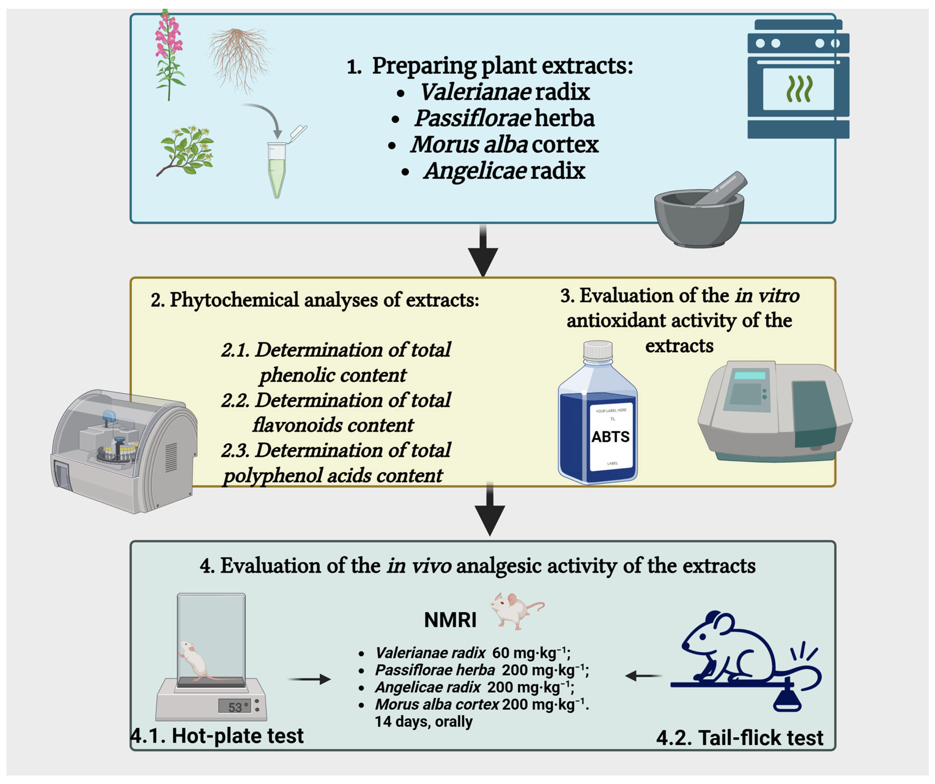
Figure 1. Overview of the experimental methodology.
Jmms 12 00039 g001
Figure 2. (A) Initial pain reaction latency in the hot-plate test. (B) Pain reaction latency in the hot-plate test after 7 days. (C) Pain reaction latency in the hot-plate test after 14 days. Values are expressed as mean ± S.E.M. M—control. * p < 0.05; ** p < 0.01; *** p < 0.001 vs. M. M—control group; PCT—paracetamol, 100 mg·kg−1; VO—Valeriana officinalis extract, 60 mg·kg−1; PI—Passiflora incarnata extract, 200 mg·kg−1; AA—Angelica archangelica extract, 200 mg·kg−1; MA—Morus alba extract, 200 mg·kg−1.
Jmms 12 00039 g002
Figure 3. (A) Initial pain reaction latency in the tail-flick test. (B) Pain reaction latency in the tail-flick test after 7 days. (C) Pain reaction latency in the tail-flick test after 14 days. Values are expressed as mean ± S.E.M. * p < 0.05; ** p < 0.01; vs. M. M—control group; PCT—paracetamol, 100 mg·kg−1; VO—Valeriana officinalis extract, 60 mg·kg−1; PI—Passiflora incarnata extract, 200 mg·kg−1; AA—Angelica archangelica extract, 200 mg·kg−1; MA—Morus alba extract, 200 mg·kg−1.
Jmms 12 00039 g003
Table 1. Quantitative analysis of the plant extracts.
Plant ExtractTP
(g/100 g Eq Expressed in Tannic Acid)
TF
(g/100 g Eq Expressed in Rutin)
TPA
(g/100 g Eq Expressed in Chlorogenic Acid)
AA4.27 ± 1.13ndnd
MA11.50 ± 0.052.79 ± 0.525.61 ± 0.43
PI6.36 ± 0.661.51 ± 0.060.85 ± 0.04
VO5.95 ± 0.090.64 ± 0.061.29 ± 0.11
AA—Angelica archangelica; MA—Morus alba; PI—Passiflora incarnata; TP—total polyphenolic content; TF—total flavonoid content; TPA—total polyphenolic acid content; VO—Valeriana officinalis. Results were expressed as mean ± SD (n = 5); nd—not detected.
Table 2. Determination of antioxidant activity using the ABTS method.
AAMAPIVOAscorbic Acid
IC50 (mg/mL)0.25990.06950.10440.12720.0165
AA—Angelica archangelica; IC50—inhibitory concentration 50; MA—Morus alba; PI—Passiflora incarnate; VO—Valeriana officinalis.
Disclaimer/Publisher’s Note: The statements, opinions and data contained in all publications are solely those of the individual author(s) and contributor(s) and not of MDPI and/or the editor(s). MDPI and/or the editor(s) disclaim responsibility for any injury to people or property resulting from any ideas, methods, instructions or products referred to in the content.