
Coronary Slow Flow Is Associated with Anxiety and Depression but Not Adverse Childhood Experiences and Alexithymia
 , , and
Abstract
1. Introduction
2. Methods
2.1. Study Design and Population
2.2. Coronary Angiography
2.3. Psychometric Measures
2.4. Statistical Analysis
3. Results
4. Discussion
5. Conclusions
Author Contributions
Funding
Institutional Review Board Statement
Informed Consent Statement
Data Availability Statement
Conflicts of Interest
References
- Zhu, Q.; Wang, S.; Huang, X.; Zhao, C.; Wang, Y.; Li, X.; Jia, D.; Ma, C. Understanding the pathogenesis of coronary slow flow: Recent advances. Trends Cardiovasc. Med. 2022, 34, 137–144. [Google Scholar] [CrossRef] [PubMed]
- Chalikias, G.; Tziakas, D. Slow Coronary Flow: Pathophysiology, Clinical Implications, and Therapeutic Management. Angiology 2021, 72, 808–818. [Google Scholar] [CrossRef] [PubMed]
- Tambe, A.A.; Demany, M.A.; Zimmerman, H.A.; Mascarenhas, E. Angina pectoris and slow flow velocity of dye in coronary arteries—A new angiographic finding. Am. Heart J. 1972, 84, 66–71. [Google Scholar] [CrossRef] [PubMed]
- Leone, M.C.; Gori, T.; Fineschi, M. The coronary slow flow phenomenon: A new cardiac “Y” syndrome? Clin. Hemorheol. Microcirc. 2008, 39, 185–190. [Google Scholar] [CrossRef]
- Aparicio, A.; Cuevas, J.; Moris, C.; Martin, M. Slow Coronary Blood Flow: Pathogenesis and Clinical Implications. Eur. Cardiol. 2022, 17, e08. [Google Scholar] [CrossRef]
- Levine, G.N.; Cohen, B.E.; Commodore-Mensah, Y.; Fleury, J.; Huffman, J.C.; Khalid, U.; Labarthe, D.R.; Lavretsky, H.; Michos, E.D.; Spatz, E.S.; et al. Psychological Health, Well-Being, and the Mind-Heart-Body Connection: A Scientific Statement From the American Heart Association. Circulation 2021, 143, e763–e783. [Google Scholar] [CrossRef]
- Murphy, B.; Le Grande, M.; Alvarenga, M.; Worcester, M.; Jackson, A. Anxiety and Depression After a Cardiac Event: Prevalence and Predictors. Front. Psychol. 2019, 10, 3010. [Google Scholar] [CrossRef]
- Greenage, M.; Kulaksizoglu, B.; Cilingiroglu, M.; Ali, R. The role of anxiety and emotional stress as a risk factor in treatment-resistant hypertension. Curr. Atheroscler. Rep. 2011, 13, 129–131. [Google Scholar] [CrossRef]
- Khan, F.M.; Kulaksizoglu, B.; Cilingiroglu, M. Depression and coronary heart disease. Curr. Atheroscler. Rep. 2010, 12, 105–109. [Google Scholar] [CrossRef]
- Kojima, M. Alexithymia as a prognostic risk factor for health problems: A brief review of epidemiological studies. Biopsychosoc. Med. 2012, 6, 21. [Google Scholar] [CrossRef]
- Suglia, S.F.; Koenen, K.C.; Boynton-Jarrett, R.; Chan, P.S.; Clark, C.J.; Danese, A.; Faith, M.S.; Goldstein, B.I.; Hayman, L.L.; Isasi, C.R.; et al. Childhood and Adolescent Adversity and Cardiometabolic Outcomes: A Scientific Statement From the American Heart Association. Circulation 2018, 137, e15–e28. [Google Scholar] [CrossRef] [PubMed]
- Keogh, T.M.; Howard, S.; Gallagher, S. Early Life Adversity and Blunted Cardiovascular Reactivity to Acute Psychological Stress: The Role of Current Depressive Symptoms. Psychosom. Med. 2022, 84, 170–178. [Google Scholar] [CrossRef] [PubMed]
- Peters, R.M.; Lumley, M.A. Relationship of alexithymia to cardiovascular disease risk factors among African Americans. Compr. Psychiatry 2007, 48, 34–41. [Google Scholar] [CrossRef] [PubMed]
- Yalvac, D.; Ozturk, S.; Sivri, N.; Kilic, Y.; Bulut, E.; Celik, A.; Barlas, Y.; Tengiz, I.; Yetkin, E. Effects of patients anxiety and depression scores on coronary flow in patients with normal coronary arteries. Int. J. Cardiol. 2015, 180, 55–57. [Google Scholar] [CrossRef]
- Durmaz, T.; Keles, T.; Erdogan, K.E.; Ayhan, H.; Bilen, E.; Bayram, N.A.; Akcay, M.; Oz, O.; Albayrak, Y.; Ozdemir, N.; et al. Coronary Slow Flow is Associated with Depression and Anxiety. Acta Cardiol. Sin. 2014, 30, 197–203. [Google Scholar]
- Elamragy, A.A.; Abdelhalim, A.A.; Arafa, M.E.; Baghdady, Y.M. Anxiety and depression relationship with coronary slow flow. PLoS ONE 2019, 14, e0221918. [Google Scholar] [CrossRef]
- Yavuz, F.; Alici, H.; Alici, D.; Inanc, I.H.; Ercan, S.; Davutoglu, V. The controversy about the association between depression and coronary slow flow phenomenon. Int. J. Cardiol. 2015, 186, 109–110. [Google Scholar] [CrossRef]
- Schussler, J.M. Effectiveness and safety of transradial artery access for cardiac catheterization. Bayl. Univ. Med. Cent. Proc. 2011, 24, 205–209. [Google Scholar] [CrossRef]
- Nuttall, F.Q. Body Mass Index: Obesity, BMI, and Health: A Critical Review. Nutr. Today 2015, 50, 117–128. [Google Scholar] [CrossRef]
- Mosteller, R.D. Simplified calculation of body-surface area. N. Engl. J. Med. 1987, 317, 1098. [Google Scholar]
- Gibson, C.M.; Cannon, C.P.; Daley, W.L.; Dodge, J.T., Jr.; Alexander, B., Jr.; Marble, S.J.; McCabe, C.H.; Raymond, L.; Fortin, T.; Poole, W.K.; et al. TIMI frame count: A quantitative method of assessing coronary artery flow. Circulation 1996, 93, 879–888. [Google Scholar] [CrossRef] [PubMed]
- Kunadian, V.; Harrigan, C.; Zorkun, C.; Palmer, A.M.; Ogando, K.J.; Biller, L.H.; Lord, E.E.; Williams, S.P.; Lew, M.E.; Ciaglo, L.N.; et al. Use of the TIMI frame count in the assessment of coronary artery blood flow and microvascular function over the past 15 years. J. Thromb. Thrombolysis 2009, 27, 316–328. [Google Scholar] [CrossRef] [PubMed]
- Çanga, A.; Kocaman, S.A.; Çetin, M.; Cicek, Y.; Emre, M. Relationship between leukocyte and subtype counts, low-grade inflammation and slow coronary flow phenomenon in patients with angiographically normal coronary arteries. Acta Cardiol. Sin. 2012, 28, 306–314. [Google Scholar]
- Zigmond, A.S.; Snaith, R.P. The hospital anxiety and depression scale. Acta Psychiatr. Scand. 1983, 67, 361–370. [Google Scholar] [CrossRef]
- Aydemir, Ö.; Guvenir, T.; Kuey, L.; Kultur, S. Validity and reliability of Turkish version of hospital anxiety and depression scale. Turk. Psikiyatri Derg. 1997, 8, 280–287. [Google Scholar]
- Bagby, R.M.; Parker, J.D.; Taylor, G.J. The twenty-item Toronto Alexithymia Scale—I. Item selection and cross-validation of the factor structure. J. Psychosom. Res. 1994, 38, 23–32. [Google Scholar] [CrossRef]
- Bagby, R.M.; Taylor, G.J.; Parker, J.D. The Twenty-item Toronto Alexithymia Scale—II. Convergent, discriminant, and concurrent validity. J. Psychosom. Res. 1994, 38, 33–40. [Google Scholar] [CrossRef]
- Güleç, H.; Köse, S.; Güleç, M.Y.; Çitak, S.; Evren, C.; Borckardt, J.; Sayar, K. Reliability and factorial validity of the Turkish version of the 20-item Toronto alexithymia scale (TAS-20). Psychiatry Clin. Psychopharmacol. 2009, 19, 214. [Google Scholar]
- Bernstein, D.P.; Fink, L.; Handelsman, L.; Foote, J.; Lovejoy, M.; Wenzel, K.; Sapareto, E.; Ruggiero, J. Initial reliability and validity of a new retrospective measure of child abuse and neglect. Am. J. Psychiatry 1994, 151, 1132–1136. [Google Scholar]
- Bernstein, D.P.; Stein, J.A.; Newcomb, M.D.; Walker, E.; Pogge, D.; Ahluvalia, T.; Stokes, J.; Handelsman, L.; Medrano, M.; Desmond, D.; et al. Development and validation of a brief screening version of the Childhood Trauma Questionnaire. Child. Abuse Negl. 2003, 27, 169–190. [Google Scholar] [CrossRef]
- Şar, V.; Öztürk, E.; İkikardeş, E. Validity and reliability of the Turkish version of Childhood Trauma Questionnaire. Turk. Klin. J. Med. Sci. 2012, 32, 1054–1063. [Google Scholar] [CrossRef]
- Honkalampi, K.; De Berardis, D.; Vellante, F.; Viinamäki, H. Relations between Alexithymia and Depressive and Anxiety Disorders and Personality. In Alexithymia: Advances in Research, Theory, and Clinical Practice; Taylor, G.J., Luminet, O., Bagby, R.M., Eds.; Cambridge University Press: Cambridge, UK, 2018; pp. 142–157. [Google Scholar]
- McLaughlin, K.A. Future Directions in Childhood Adversity and Youth Psychopathology. J. Clin. Child. Adolesc. Psychol. 2016, 45, 361–382. [Google Scholar] [CrossRef] [PubMed]
- Lemay, K.R.; Tulloch, H.E.; Pipe, A.L.; Reed, J.L. Establishing the Minimal Clinically Important Difference for the Hospital Anxiety and Depression Scale in Patients With Cardiovascular Disease. J. Cardiopulm. Rehabil. Prev. 2019, 39, E6–E11. [Google Scholar] [CrossRef]
- Christensen, A.V.; Dixon, J.K.; Juel, K.; Ekholm, O.; Rasmussen, T.B.; Borregaard, B.; Mols, R.E.; Thrysoe, L.; Thorup, C.B.; Berg, S.K. Psychometric properties of the Danish Hospital Anxiety and Depression Scale in patients with cardiac disease: Results from the DenHeart survey. Health Qual. Life Outcomes 2020, 18, 9. [Google Scholar] [CrossRef]
- Bambauer, K.Z.; Locke, S.E.; Aupont, O.; Mullan, M.G.; McLaughlin, T.J. Using the Hospital Anxiety and Depression Scale to screen for depression in cardiac patients. Gen. Hosp. Psychiatry 2005, 27, 275–284. [Google Scholar] [CrossRef]
- Wang, X.; Nie, S.P. The coronary slow flow phenomenon: Characteristics, mechanisms and implications. Cardiovasc. Diagn. Ther. 2011, 1, 37–43. [Google Scholar]
- Shui, Z.; Wang, Y.; Sun, M.; Gao, Y.; Liang, S.; Wang, Y.; Wang, X.; Yu, Q.; Zhang, S.; Liu, L. The effect of coronary slow flow on left atrial structure and function. Sci. Rep. 2021, 11, 7511. [Google Scholar] [CrossRef]
- Ozturk, H.M.; Ozturk, S.; Yetkin, E. Linkage between cardiovascular diseases and major depression: Contribution of platelet cells. Psychiatry Res. 2020, 287, 111026. [Google Scholar] [CrossRef]
- Sara, J.D.S.; Ahmad, A.; Toya, T.; Suarez Pardo, L.; Lerman, L.O.; Lerman, A. Anxiety Disorders Are Associated With Coronary Endothelial Dysfunction in Women With Chest Pain and Nonobstructive Coronary Artery Disease. J. Am. Heart Assoc. 2021, 10, e021722. [Google Scholar] [CrossRef]
- Oksuz, F.; Yarlioglues, M.; Ozturk, S.; Kaya, F.D.; Oksuz, E.; Murat, S.N.; Turak, O.; Celik, I.E.; Kilic, A.; Kurtul, A. Atrial electromechanical delay analysed by tissue Doppler echocardiography is prolonged in patients with generalised anxiety disorders. Kardiol. Pol. 2017, 75, 581–588. [Google Scholar] [CrossRef]
- Kim, Y.H.; Kim, S.H.; Lim, S.Y.; Cho, G.Y.; Baik, I.K.; Lim, H.E.; Na, J.O.; Han, S.W.; Ko, Y.H.; Shin, C. Relationship between depression and subclinical left ventricular changes in the general population. Heart 2012, 98, 1378–1383. [Google Scholar] [CrossRef] [PubMed]
- Celik, E.; Cay, S.; Sensoy, B.; Murat, S.; Oksuz, F.; Cankurt, T.; Ali Mendi, M. Heart Failure Functional Class Associated with Depression Severity But Not Anxiety Severity. Acta Cardiol. Sin. 2016, 32, 55–61. [Google Scholar] [PubMed]
- Berg, S.K.; Rasmussen, T.B.; Thrysoee, L.; Thorup, C.B.; Borregaard, B.; Christensen, A.V.; Mols, R.E.; Juel, K.; Ekholm, O. Mental health is a risk factor for poor outcomes in cardiac patients: Findings from the national DenHeart survey. J. Psychosom. Res. 2018, 112, 66–72. [Google Scholar] [CrossRef]
- Kyrou, I.; Kollia, N.; Panagiotakos, D.; Georgousopoulou, E.; Chrysohoou, C.; Tsigos, C.; Randeva, H.S.; Yannakoulia, M.; Stefanadis, C.; Papageorgiou, C.; et al. Association of depression and anxiety status with 10-year cardiovascular disease incidence among apparently healthy Greek adults: The ATTICA Study. Eur. J. Prev. Cardiol. 2017, 24, 145–152. [Google Scholar] [CrossRef]
- Cosci, F.; Fava, G.A. When Anxiety and Depression Coexist: The Role of Differential Diagnosis Using Clinimetric Criteria. Psychother. Psychosom. 2021, 90, 308–317. [Google Scholar] [CrossRef]
- Terock, J.; Klinger-Konig, J.; Janowitz, D.; Nauck, M.; Volzke, H.; Grabe, H.J. Alexithymia is associated with increased all-cause mortality risk in men, but not in women: A 10-year follow-up study. J. Psychosom. Res. 2021, 143, 110372. [Google Scholar] [CrossRef]
- Ossola, P.; Gerra, M.L.; Beltrani, M.; Marchesi, C. Alexithymia and Cardiac Outcome in Patients at First Acute Coronary Syndrome. Int. J. Behav. Med. 2019, 26, 673–679. [Google Scholar] [CrossRef]
- Marchesi, C.; Ossola, P.; Scagnelli, F.; Mellini, L.; Tonna, M.; Ardissino, D.; De Panfilis, C. The role of alexithymia in predicting incident depression in patients at first acute coronary syndrome. Compr. Psychiatry 2015, 62, 86–92. [Google Scholar] [CrossRef]
- Ho, F.K.; Celis-Morales, C.; Gray, S.R.; Petermann-Rocha, F.; Lyall, D.; Mackay, D.; Sattar, N.; Minnis, H.; Pell, J.P. Child maltreatment and cardiovascular disease: Quantifying mediation pathways using UK Biobank. BMC Med. 2020, 18, 143. [Google Scholar] [CrossRef]
- Kang, W.; Malvaso, A. Understanding cognitive deficits in people with coronary heart disease (CHD). J. Pers. Med. 2023, 13, 307. [Google Scholar] [CrossRef]
- Kang, W.; Malvaso, A. Mental health in coronary heart disease (CHD) patients: Findings from the UK Household Longitudinal Study (UKHLS). Healthcare 2023, 11, 1364. [Google Scholar] [CrossRef] [PubMed]
- Abaci, A.; Oguzhan, A.; Eryol, N.K.; Ergin, A. Effect of potential confounding factors on the thrombolysis in myocardial infarction (TIMI) trial frame count and its reproducibility. Circulation 1999, 100, 2219–2223. [Google Scholar] [CrossRef]

| Variables | Overall (n = 76) | NCF (n = 58) | CSF (n = 18) | p Value |
|---|---|---|---|---|
| Age, year | 55 ± 9 | 56 ± 8 | 53 ± 11 | 0.162 |
| Gender, male | 30 (39.5%) | 16 (27.6%) | 14 (77.8%) | <0.001 |
| Weight, kg | 85.8 ± 14.1 | 84.0 ± 13.9 | 89.9 + 13.3 | 0.122 |
| Height, cm | 165.3 ± 8.1 | 164.1 ± 7.8 | 169.1 ± 7.9 | 0.021 |
| Body mass index, kg/m2 | 31.3 ± 4.8 | 31.2 ± 4.9 | 31.3 ± 4.4 | 0.897 |
| Body surface area, m2 | 2.0 ± 0.1 | 2.0 ± 0.1 | 1.9 ± 0.1 | 0.480 |
| Current smoker | 29 (38.2%) | 20 (34.5%) | 9 (50.0%) | 0.236 |
| Hypertension | 42 (55.3%) | 33 (56.9%) | 9 (50.0%) | 0.607 |
| Diabetes mellitus | 21 (27.6%) | 18 (31.0%) | 3 (16.7%) | 0.234 |
| Hyperlipidemia | 42 (55.3%) | 36 (62.1%) | 6 (33.3%) | 0.032 |
| Medications | ||||
| Antiplatelets | 16 (21.1%) | 9 (15.8%) | 7 (41.2%) | 0.026 |
| Beta blocker | 14 (18.4%) | 9 (15.8%) | 5 (29.4%) | 0.208 |
| RAS blocker | 32 (42.1%) | 24 (42.1%) | 8 (47.1%) | 0.717 |
| Calcium channel blocker | 12 (15.8%) | 9 (15.8%) | 3 (17.6%) | 0.855 |
| Hypolipidemic | 8 (10.5%) | 6 (10.5%) | 2 (11.8%) | 0.885 |
| Antidiabetics | 19 (25.0%) | 17 (29.8%) | 2 (11.8%) | 0.135 |
| Diuretics | 23 (30.3%) | 19 (33.3%) | 4 (23.5%) | 0.443 |
| TIMI frame count measurements | ||||
| TFC-LAD corrected | 17.6 (12.4–22.9) | 14.7 (11.7–20.0) | 28.2 (17.6–35.2) | <0.001 |
| TFC-Cx | 20.0 (18.0–26.0) | 20.0 (15.5–22.0) | 28.0 (24.0–32.0) | <0.001 |
| TFC-RCA | 29.5 ± 13.7 | 23.6 ± 8.2 | 46.7 ± 12.3 | <0.001 |
| TFC-mean | 21.8 (17.5–26.9) | 19.6 (16.8–22.7) | 32.6 (27.7–38.7) | <0.001 |
| Variables | Overall (n = 76) | NCF (n = 58) | CSF (n = 18) | p Value |
|---|---|---|---|---|
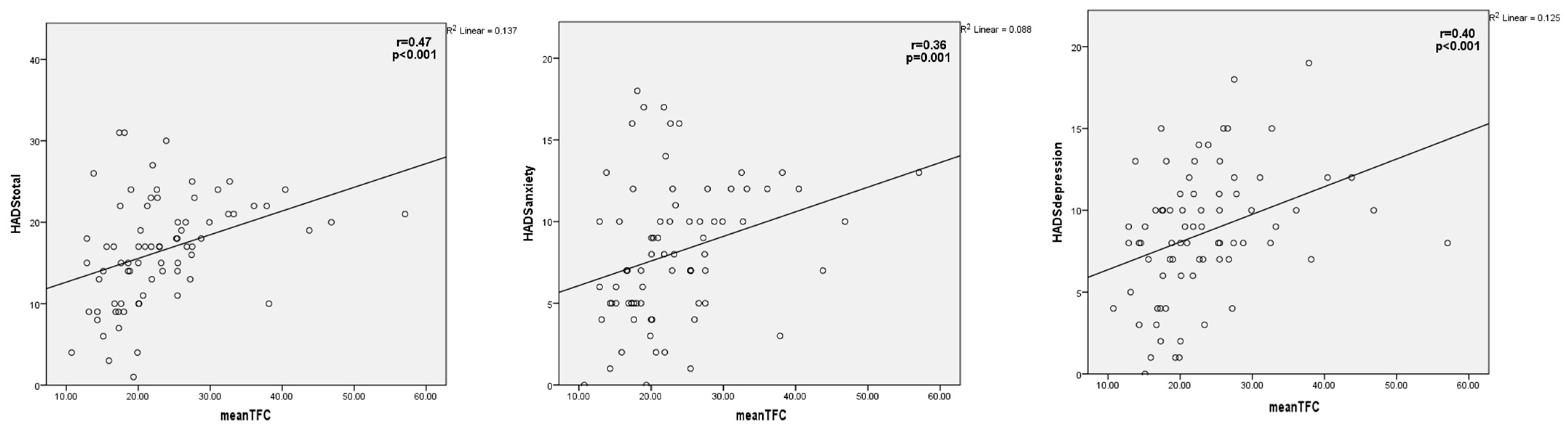
| HADS-Total score | 16.5 ± 6.6 | 15.3 ± 6.8 | 20.1 ± 4.1 | 0.001 |
| HADS-Anxiety | 8.1 ± 4.2 | 7.5 ± 4.4 | 9.8 ± 2.9 | 0.038 |
| HADS-Depression | 8.6 ± 4.1 | 7.9 ± 3.8 | 10.7 ± 3.7 | 0.010 |
| TAS-20 total score | 61.6 ± 11.5 | 61.6 ± 11.3 | 61.0 ± 11.9 | 0.846 |
| DIF | 20.0 ± 6.3 | 20.6 ± 6.0 | 18.0 ± 6.7 | 0.128 |
| DDF | 14.8 ± 3.8 | 14.9 ± 3.6 | 14.3 ± 4.2 | 0.592 |
| EOT | 26.4 ± 6.0 | 26.2 ± 5.9 | 26.4 ± 6.4 | 0.914 |
| CTQ score | 36.6 ± 10.8 | 37.3 ± 11.1 | 35.1 ± 12.0 | 0.464 |
| Physical abuse | 5 (5–5) | 5 (5–5) | 5 (5–5.25) | 0.516 |
| Emotional abuse | 5 (5–7) | 5 (5–7) | 5 (5–5.25) | 0.135 |
| Sexual abuse | 5.5 ± 1.6 | 5.4 ± 1.6 | 5.5 ± 1.6 | 0.825 |
| Physical negligence | 7.6 ± 3.0 | 7.9 ± 3.1 | 7.0 ± 3.0 | 0.281 |
| Emotional negligence | 10.5 ± 5.2 | 10.9 ± 5.2 | 9.0 ± 4.9 | 0.174 |
| Univariate | Multivariate * | |||
|---|---|---|---|---|
| Odds Ratio (95% CI) | p Value | Odds Ratio (95% CI) | p Value | |
| HADS-Total score | 1.13 (1.02–1.24) | 0.012 | 1.27 (1.08–1.50) | 0.003 |
| HADS-Anxiety score | 1.14 (1.00–1.30) | 0.043 | 1.25 (1.03–1.51) | 0.019 |
| HADS-Depression score | 1.21 (1.03–1.41) | 0.015 | 1.36 (1.06–1.74) | 0.014 |
| TAS-20 total score | 0.99 (0.95–1.04) | 0.844 | ||
| CTQ score | 0.98 (0.93–1.03) | 0.460 | ||
Disclaimer/Publisher’s Note: The statements, opinions and data contained in all publications are solely those of the individual author(s) and contributor(s) and not of MDPI and/or the editor(s). MDPI and/or the editor(s) disclaim responsibility for any injury to people or property resulting from any ideas, methods, instructions or products referred to in the content. |
© 2025 by the authors. Licensee MDPI, Basel, Switzerland. This article is an open access article distributed under the terms and conditions of the Creative Commons Attribution (CC BY) license (https://creativecommons.org/licenses/by/4.0/).
Share and Cite
Ozturk, H.M.; Inanc, I.H.; Cilingiroglu, M.; Turan, Y.; Kandemir, H.; Ozturk, S. Coronary Slow Flow Is Associated with Anxiety and Depression but Not Adverse Childhood Experiences and Alexithymia. J. Mind Med. Sci. 2025, 12, 19. https://doi.org/10.3390/jmms12010019
Ozturk HM, Inanc IH, Cilingiroglu M, Turan Y, Kandemir H, Ozturk S. Coronary Slow Flow Is Associated with Anxiety and Depression but Not Adverse Childhood Experiences and Alexithymia. Journal of Mind and Medical Sciences. 2025; 12(1):19. https://doi.org/10.3390/jmms12010019
Chicago/Turabian StyleOzturk, Hayriye Mihrimah, Ibrahim Halil Inanc, Mehmet Cilingiroglu, Yasar Turan, Huseyin Kandemir, and Selcuk Ozturk. 2025. "Coronary Slow Flow Is Associated with Anxiety and Depression but Not Adverse Childhood Experiences and Alexithymia" Journal of Mind and Medical Sciences 12, no. 1: 19. https://doi.org/10.3390/jmms12010019
APA StyleOzturk, H. M., Inanc, I. H., Cilingiroglu, M., Turan, Y., Kandemir, H., & Ozturk, S. (2025). Coronary Slow Flow Is Associated with Anxiety and Depression but Not Adverse Childhood Experiences and Alexithymia. Journal of Mind and Medical Sciences, 12(1), 19. https://doi.org/10.3390/jmms12010019