Anatomy-Weighted CTDIvol from Routine CT Metadata: A Patient-Specific, Multi-Vendor Study Using Deep-Learning Segmentation


Institute of One, LISIT Co., Ltd., Tokyo 150-0044, Japan
Tomography 2026, 12(9), 125; https://doi.org/10.3390/tomography12090125
Submission received: 6 August 2026
/
Revised: 28 August 2026
/
Accepted: 28 August 2026
/
Published: 30 August 2026
(This article belongs to the Section Artificial Intelligence in Medical Imaging)
Simple Summary
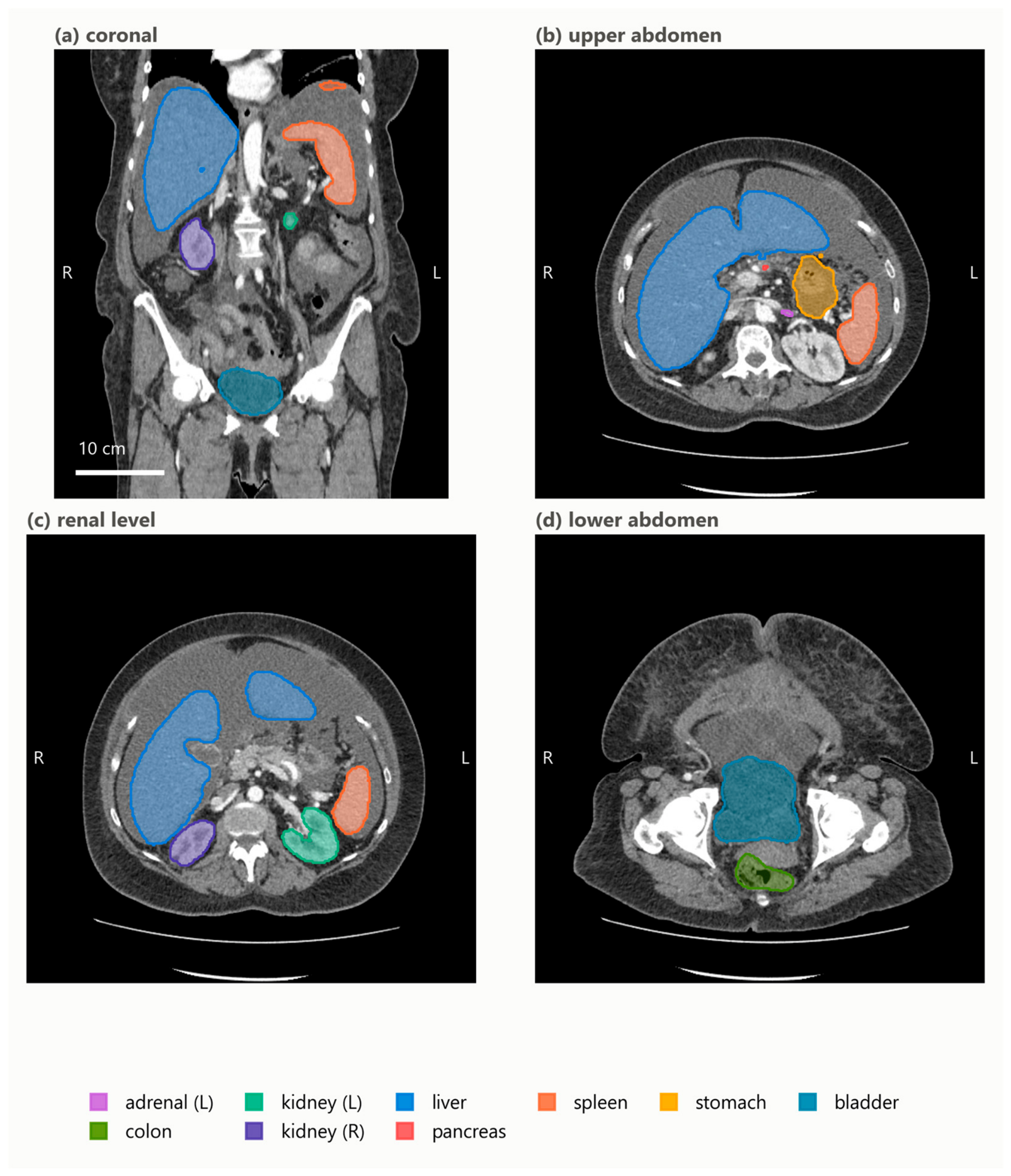
CT scanners report a single dose number for the whole scan, yet the dose is delivered unevenly across organs. We built an open, freely available software pipeline that uses deep-learning segmentation to outline each abdominal organ on routine CT images and then derives a patient-specific, organ-level dose index directly from data the scanner already records. Across scans from four manufacturers, we found that this organ-level information can be recovered for some scanners but is entirely absent for others because the values it needs were not kept in the archived scan records. All software, results, and data provenance are fully open, so the work can be reproduced and extended.
Abstract
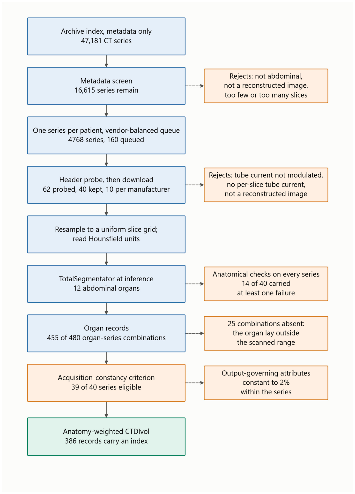
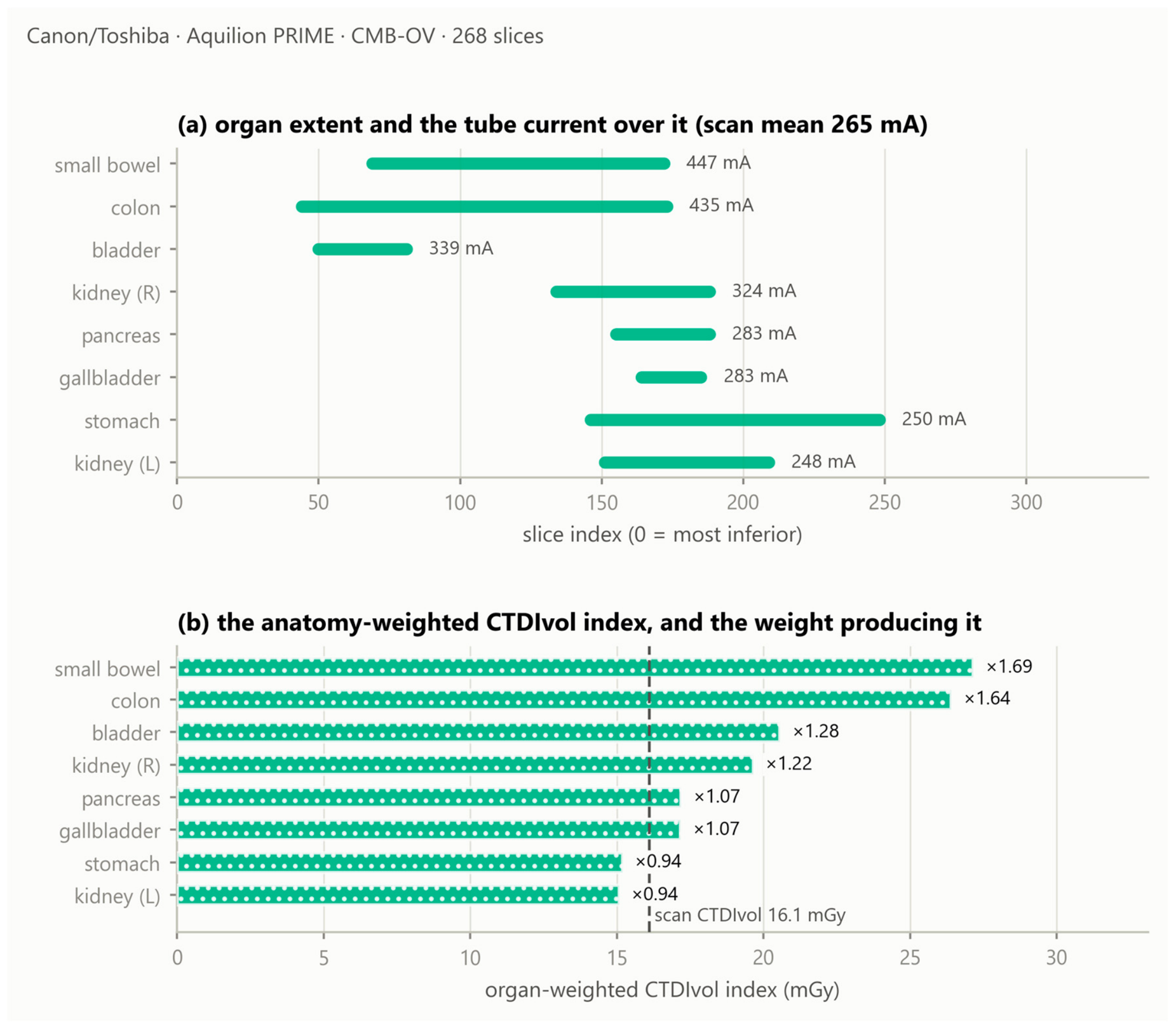
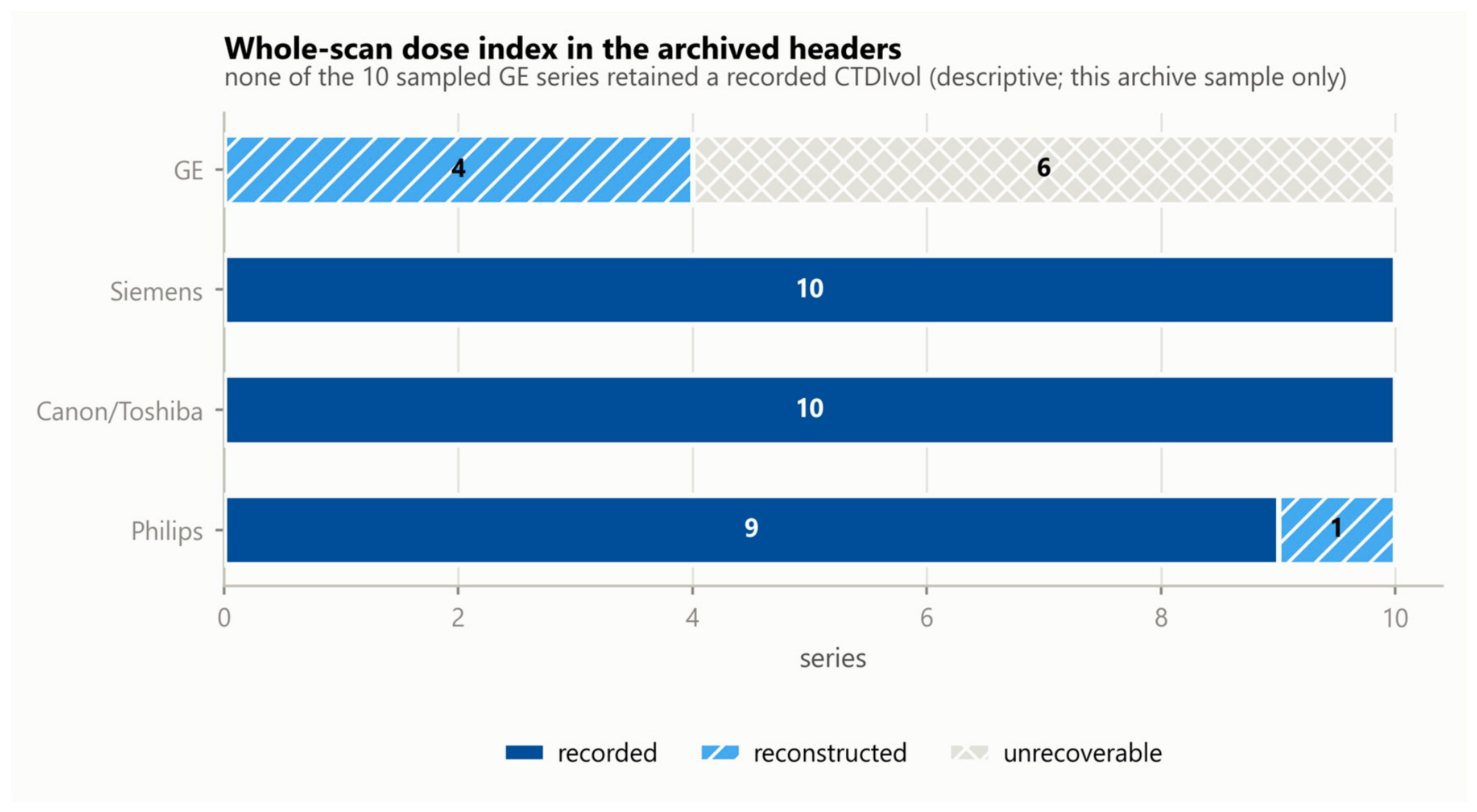
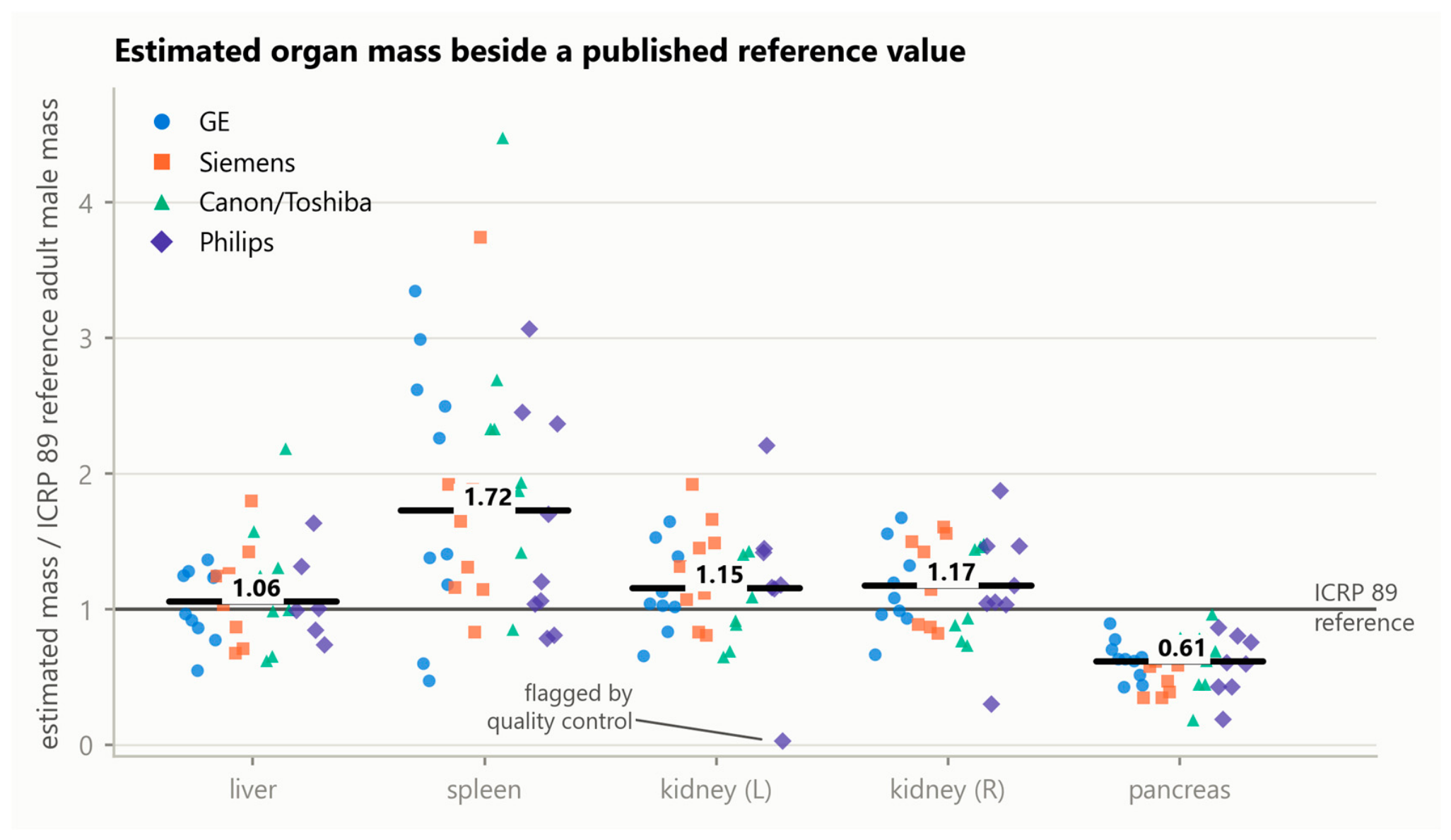
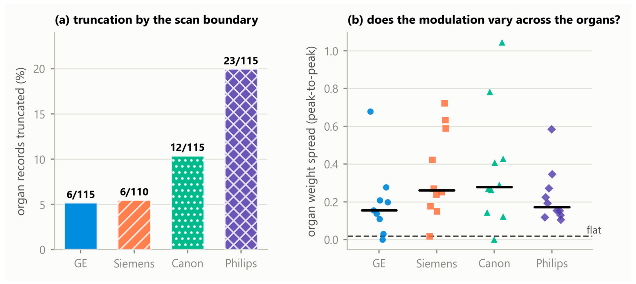
Background/Objectives: The volume computed tomography (CT) dose index (CTDIvol) is a scanner output, not an organ dose, and cannot express how tube-current modulation varies along a patient. An organ-specific weighted CTDIvol addressing this has been reported before, in single-institution cohorts and often from inputs routine archives do not retain. New here is not the quantity but what an open, multi-vendor operationalisation reveals: whether its inputs survive archive curation and what the fallback costs when they do not. Methods: Forty abdominal CT series, ten per manufacturer, were drawn from the Cancer Imaging Archive and twelve organs segmented with TotalSegmentator at inference. Of 480 requested organ–series combinations, 455 were produced. A rule-based acquisition-constancy criterion admitted 39 series. Results: Modulation weights spanned 0.59 to 1.69, so the index departs from the whole-scan CTDIvol by up to 70% within one acquisition. A recorded CTDIvol survived in 29 of 40 archived headers and was reconstructable in 5 and unavailable in 6, availability differing markedly between manufacturers. Forcing that reconstruction on series that did retain a value agreed to within 12% on three scanner models and diverged by 58% and 84% on two others. Estimated organ mass was broadly consistent with International Commission on Radiological Protection (ICRP) Publication 89 for liver and kidneys. Conclusions: This index is not an absorbed dose; the implementation is open.