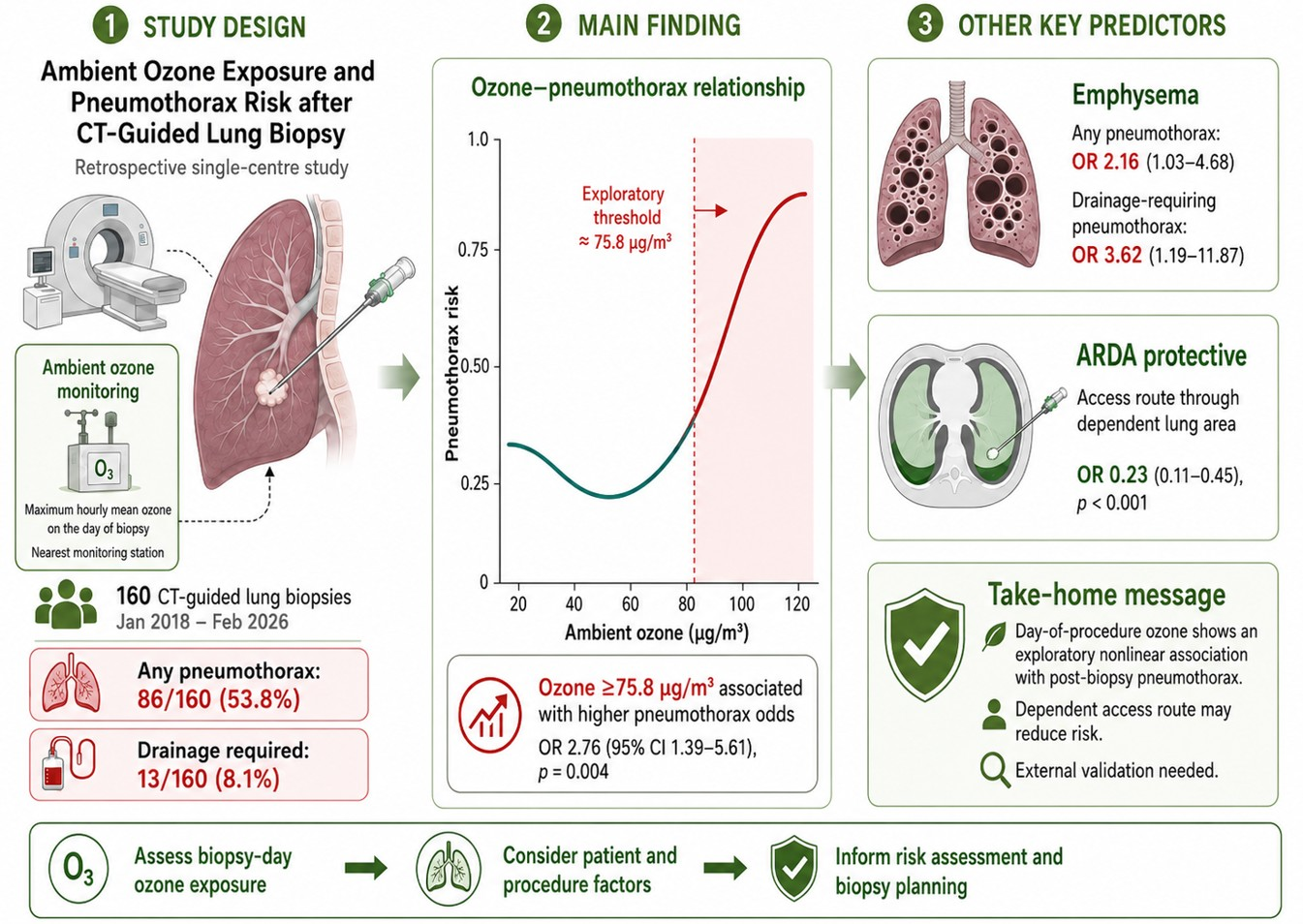
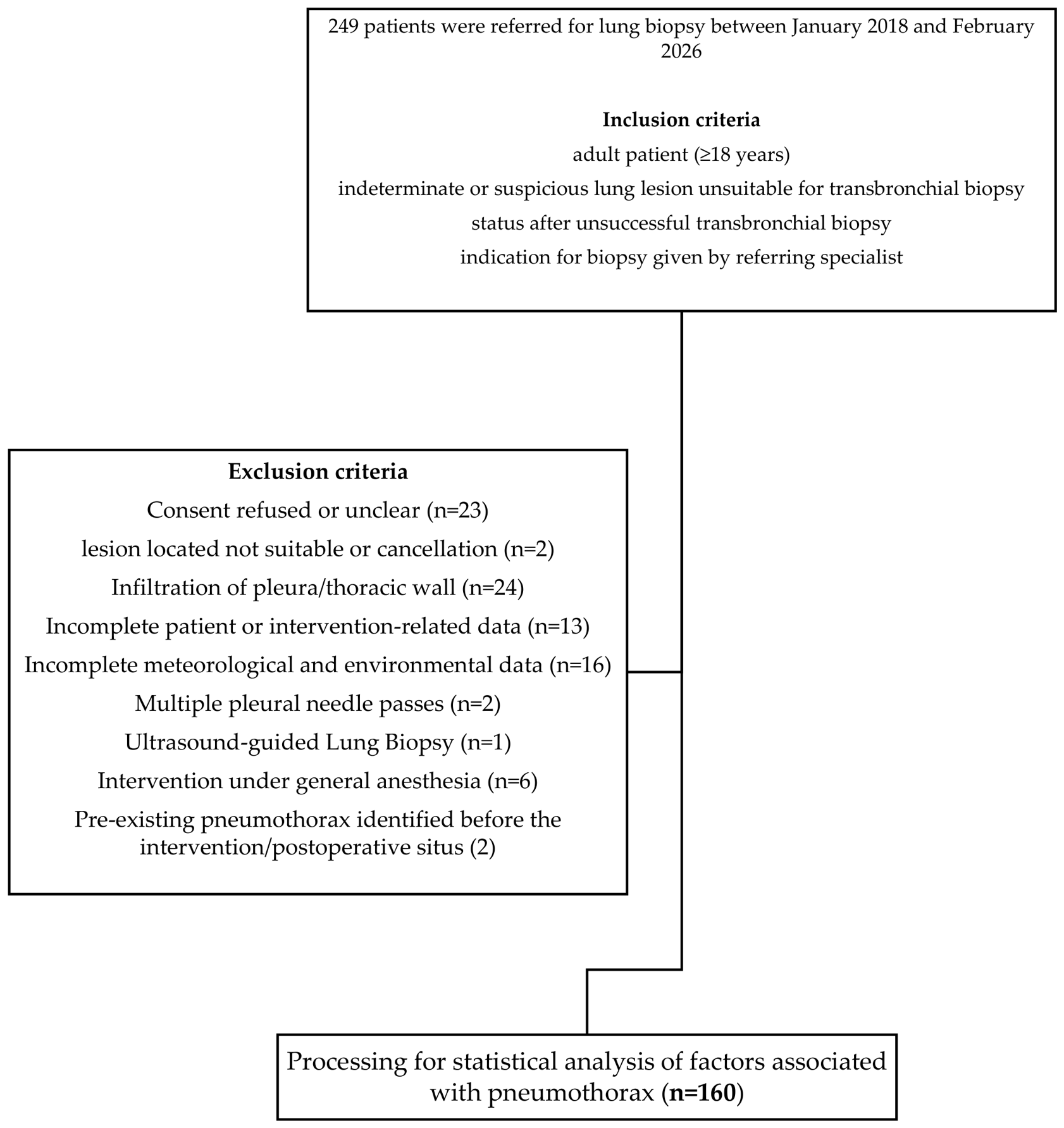
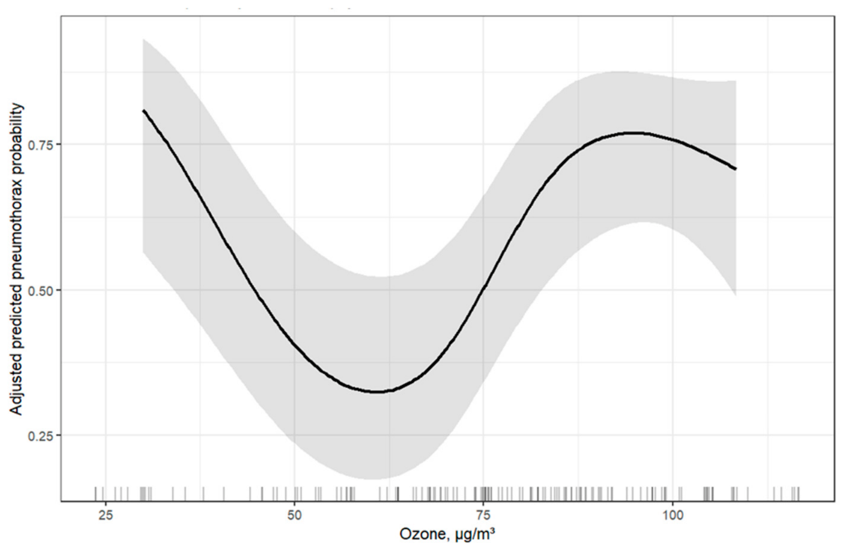
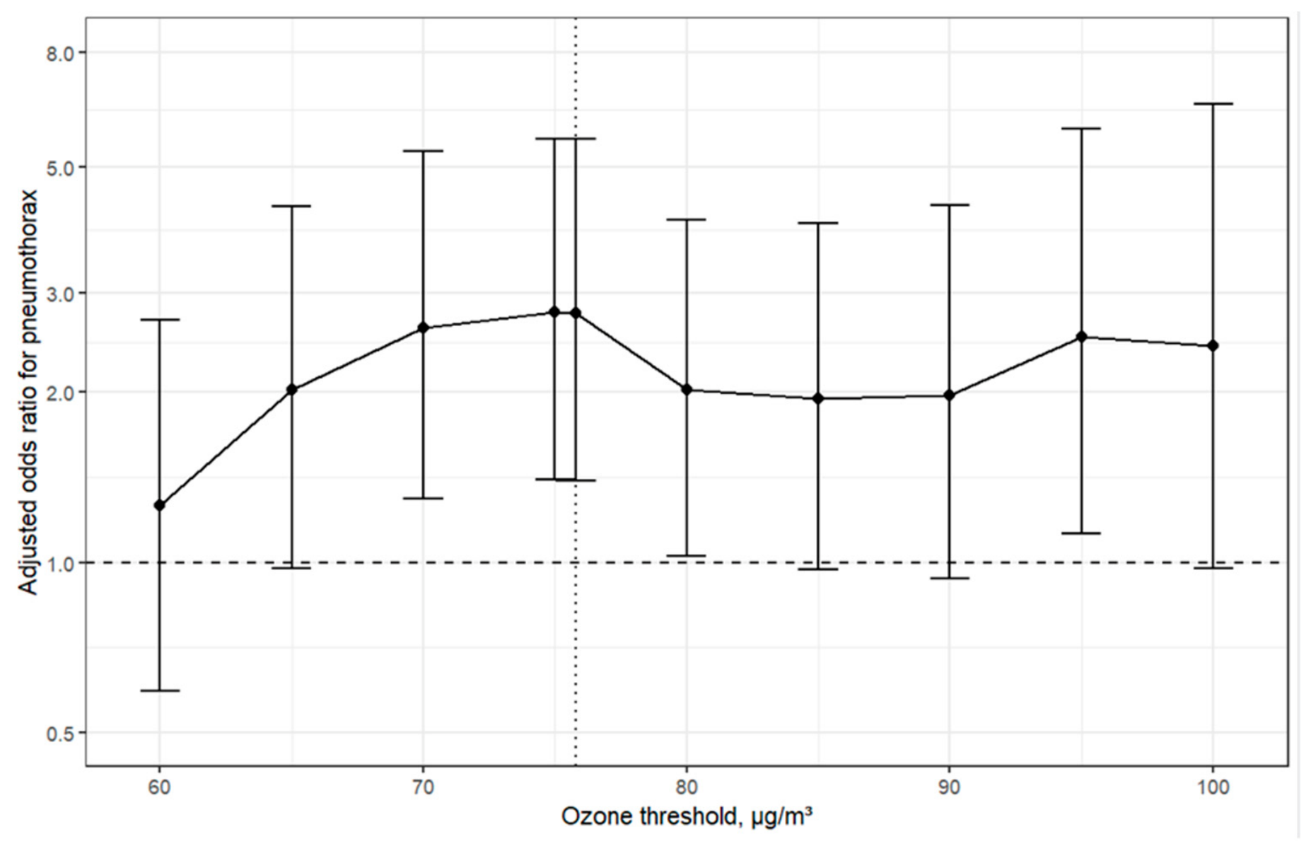
Ambient Ozone Exposure and Pneumothorax Risk After CT-Guided Lung Biopsy

 , , , ,
Simple Summary
Abstract
1. Introduction
2. Materials and Methods
2.1. Study Population
2.2. Baseline Evaluation and Biopsy Technique
2.3. Imaging Review and Procedural Variables
2.4. Meteorological and Air Pollution Data
2.5. Statistical Analysis
3. Results
3.1. Study Cohort and Baseline Characteristics
3.2. Primary Multivariable Models
3.3. Nonlinear Ozone Association and Threshold Sensitivity
3.4. Robustness Analyses
3.5. Drainage-Requiring Pneumothorax
4. Discussion
5. Conclusions
Supplementary Materials
Author Contributions
Funding
Institutional Review Board Statement
Informed Consent Statement
Data Availability Statement
Acknowledgments
Conflicts of Interest
Abbreviations
| ARDA | Access route through dependent lung area |
| CI | Confidence interval |
| CO | Carbon monoxide |
| COPD | Chronic obstructive pulmonary disease |
| CT | Computed tomography |
| CTLB | Computed tomography-guided lung biopsy |
| DPL | Distance from pleura to lesion |
| DSL | Distance from skin to lesion |
| EC | Elemental carbon |
| G | Gauge |
| IQR | Interquartile range |
| LD | Lateral decubitus |
| NO2 | Nitrogen dioxide |
| NOx | Nitrogen oxides |
| OR | Odds ratio |
| PM10 | Particulate matter with an aerodynamic diameter ≤ 10 μm |
| PM2.5 | Particulate matter with an aerodynamic diameter ≤ 2.5 μm |
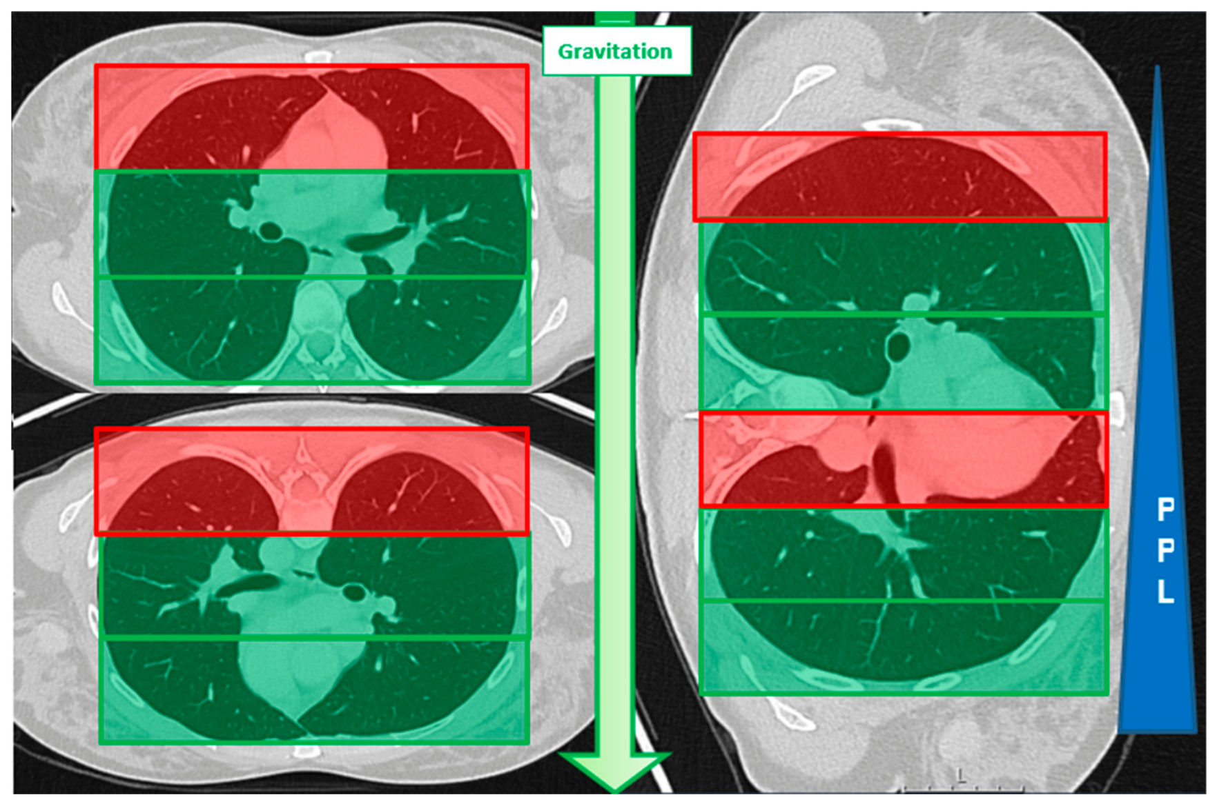
| PPL | Pleural pressure |
| ROC | Receiver operating characteristic |
| TLA | Three-letter acronym |
| UP | Upper lobe |
References
- Kim, D.Y.; Sun, J.S.; Kim, E.Y.; Park, K.J.; You, S. Diagnostic accuracy and safety of CT-guided percutaneous lung biopsy with a coaxial cutting needle for the diagnosis of lung cancer in patients with UIP pattern. Sci. Rep. 2022, 12, 15682. [Google Scholar] [CrossRef]
- Marx, T.; Bernard, N.; Parmentier, A.-L.; Puyraveau, M.; Martin, B.; Gantelet, M.; Pretalli, J.-B.; Dalphin, J.-C.; Mauny, F.; Desmettre, T. Short term association between air pollution (PM10, NO2 and O3) and secondary spontaneous pneumothorax. Sci. Rep. 2020, 10, 11823. [Google Scholar] [CrossRef] [PubMed]
- Yang, S.-R.; Schultheis, A.M.; Yu, H.; Mandelker, D.; Ladanyi, M.; Büttner, R. Precision medicine in non-small cell lung cancer: Current applications and future directions. Semin. Cancer Biol. 2022, 84, 184–198. [Google Scholar] [CrossRef] [PubMed]
- Khan, M.; Straub, R.; Moghaddam, S.; Maataoui, A.; Gurung, J.; Wagner, T.; Ackermann, H.; Thalhammer, A.; Vogl, T.; Jacobi, V. Variables affecting the risk of pneumothorax and intrapulmonal hemorrhage in CT-guided transthoracic biopsy. Eur. Radiol. 2008, 18, 1356–1363. [Google Scholar] [CrossRef] [PubMed]
- Swischuk, J.L.; Castaneda, F.; Patel, J.C.; Li, R.; Fraser, K.W.; Brady, T.M.; Bertino, R.E. Percutaneous transthoracic needle biopsy of the lung: Review of 612 lesions. J. Vasc. Interv. Radiol. 1998, 9, 347–352. [Google Scholar] [CrossRef] [PubMed]
- Tomiyama, N.; Yasuhara, Y.; Nakajima, Y.; Adachi, S.; Arai, Y.; Kusumoto, M.; Eguchi, K.; Kuriyama, K.; Sakai, F.; Noguchi, M. CT-guided needle biopsy of lung lesions: A survey of severe complication based on 9783 biopsies in Japan. Eur. J. Radiol. 2006, 59, 60–64. [Google Scholar] [CrossRef] [PubMed]
- Zielinska-Krawczyk, M.; Krenke, R.; Grabczak, E.M.; Light, R.W. Pleural manometry–historical background, rationale for use and methods of measurement. Respir. Med. 2018, 136, 21–28. [Google Scholar] [CrossRef] [PubMed]
- Covey, A.M.; Gandhi, R.; Brody, L.A.; Getrajdman, G.; Thaler, H.T.; Brown, K.T. Factors associated with pneumothorax and pneumothorax requiring treatment after percutaneous lung biopsy in 443 consecutive patients. J. Vasc. Interv. Radiol. 2004, 15, 479–483. [Google Scholar] [CrossRef] [PubMed]
- Poe, R.H.; Kallay, M.C.; Wicks, C.M.; Odoroff, C.L. Predicting risk of pneumothorax in needle biopsy of the lung. Chest 1984, 85, 232–235. [Google Scholar] [CrossRef] [PubMed]
- Ko, J.P.; Shepard, J.-A.O.; Drucker, E.A.; Aquino, S.L.; Sharma, A.; Sabloff, B.; Halpern, E.; McLoud, T.C. Factors influencing pneumothorax rate at lung biopsy: Are dwell time and angle of pleural puncture contributing factors? Radiology 2001, 218, 491–496. [Google Scholar] [CrossRef] [PubMed]
- Saji, H.; Nakamura, H.; Tsuchida, T.; Tsuboi, M.; Kawate, N.; Konaka, C.; Kato, H. The incidence and the risk of pneumothorax and chest tube placement after percutaneous CT-guided lung biopsy: The angle of the needle trajectory is a novel predictor. Chest 2002, 121, 1521–1526. [Google Scholar] [PubMed]
- Taslakian, B.; Koneru, V.; Babb, J.S.; Sridhar, D. Transthoracic needle biopsy of pulmonary nodules: Meteorological conditions and the risk of pneumothorax and chest tube placement. J. Clin. Med. 2019, 8, 727. [Google Scholar] [CrossRef] [PubMed]
- Drumm, O.; Joyce, E.A.; de Blacam, C.; Gleeson, T.; Kavanagh, J.; McCarthy, E.; McDermott, R.; Beddy, P. CT-guided lung biopsy: Effect of biopsy-side down position on pneumothorax and chest tube placement. Radiology 2019, 292, 190–196. [Google Scholar] [PubMed]
- Brönnimann, M.P.; Barroso, M.C.; Manser, L.; Gebauer, B.; Auer, T.A.; Collettini, F.; Dorn, P.; Huber, A.T.; Heverhagen, J.T.; Maurer, M.H. The role of gravitational effects and pre-puncture techniques in reducing pneumothorax during CT-guided lung biopsies. La Radiol. Medica 2025, 130, 1024–1038. [Google Scholar]
- Brönnimann, M.P.; McMurray, M.T.; Heverhagen, J.T.; Christe, A.; Wyss, C.; Peters, A.A.; Huber, A.T.; Dammann, F.; Obmann, V.C. Innovative strategies for minimizing hematoma risk in MRI-guided breast biopsies. Radiol. Oncol. 2025, 59, 91–99. [Google Scholar] [CrossRef] [PubMed]
- Asai, N.; Kawamura, Y.; Yamazaki, I.; Sogawa, K.; Ohkuni, Y.; O’uchi, T.; Kubo, A.; Yamaguchi, E.; Kaneko, N. Is emphysema a risk factor for pneumothorax in CT-guided lung biopsy? Springerplus 2013, 2, 1–6. [Google Scholar] [CrossRef]
- Theilig, D.; Petschelt, D.; Mayerhofer, A.; Hamm, B.; Gebauer, B.; Geisel, D. Impact of quantitative pulmonary emphysema score on the rate of pneumothorax and chest tube insertion in CT-guided lung biopsies. Sci. Rep. 2020, 10, 10978. [Google Scholar] [CrossRef] [PubMed]
- Li, R.; Jiang, N.; Liu, Q.; Huang, J.; Guo, X.; Liu, F.; Gao, Z. Impact of air pollutants on outpatient visits for acute respiratory outcomes. Int. J. Environ. Res. Public Health 2017, 14, 47. [Google Scholar] [CrossRef] [PubMed]
- Brauer, M.; Hoek, G.; Van Vliet, P.; Meliefste, K.; Fischer, P.H.; Wijga, A.; Koopman, L.P.; Neijens, H.J.; Gerritsen, J.; Kerkhof, M. Air pollution from traffic and the development of respiratory infections and asthmatic and allergic symptoms in children. Am. J. Respir. Crit. Care Med. 2002, 166, 1092–1098. [Google Scholar] [CrossRef] [PubMed]
- Lipscomb, T.; Harris, R.; Moeller, R.; Pletcher, J.; Haebler, R.; Ballachey, B.E. Histopathologic lesions in sea otters exposed to crude oil. Vet. Pathol. 1993, 30, 1–11. [Google Scholar] [CrossRef]
- Wang, J.-C.; Lin, C.-L.; Chen, C.-H.; Lin, C.-H. Air pollution exposure and risk of spontaneous pneumothorax in children: A longitudinal, nationwide study. Children 2022, 9, 61. [Google Scholar] [CrossRef] [PubMed]
- Abul, Y.; Karakurt, S.; Bostanci, K.; Yuksel, M.; Eryuksel, E.; Evman, S.; Celikel, T. Spontaneous pneumothorax and ozone levels: Is there a relation? Multidiscip. Respir. Med. 2011, 6, 16. [Google Scholar] [CrossRef] [PubMed]
- Bertolaccini, L.; Alemanno, L.; Rocco, G.; Cassardo, C. Air pollution, weather variations and primary spontaneous pneumothorax. J. Thorac. Dis. 2010, 2, 9. [Google Scholar] [PubMed]
- Bertolaccini, L.; Viti, A.; Boschetto, L.; Pasini, A.; Attanasio, A.; Terzi, A.; Cassardo, C. Analysis of spontaneous pneumothorax in the city of Cuneo: Environmental correlations with meteorological and air pollutant variables. Surg. Today 2015, 45, 625–629. [Google Scholar] [PubMed]
- Park, J.H.; Lee, S.H.; Yun, S.J.; Ryu, S.; Choi, S.W.; Kim, H.J.; Kang, T.K.; Oh, S.C.; Cho, S.J. Air pollutants and atmospheric pressure increased risk of ED visit for spontaneous pneumothorax. Am. J. Emerg. Med. 2018, 36, 2249–2253. [Google Scholar] [CrossRef] [PubMed]
- Marx, T.; Bernard, N.; Parmentier, A.-L.; Puyraveau, M.; Martin, B.; Gantelet, M.; Pretalli, J.-B.; Dalphin, J.-C.; Mauny, F.; Desmettre, T. Does air pollution really impact the onset of spontaneous pneumothorax? A French case-crossover study. Environ. Int. 2019, 127, 317–323. [Google Scholar] [CrossRef] [PubMed]
- Noppen, M.; Dekeukeleire, T.; Hanon, S.; Stratakos, G.; Amjadi, K.; Madsen, P.; Meysman, M.; D’Haese, J.; Vincken, W. Fluorescein-enhanced autofluorescence thoracoscopy in patients with primary spontaneous pneumothorax and normal subjects. Am. J. Respir. Crit. Care Med. 2006, 174, 26–30. [Google Scholar] [CrossRef] [PubMed]
- Tschopp, J.-M.; Marquette, C.-H. Spontaneous pneumothorax: Stop chest tube as first-line therapy. Eur. Respir. Rev. 2017, 26, 170124. [Google Scholar]
- Brönnimann, M.P.; Manser, L.; Christe, A.; Heverhagen, J.T.; Gebauer, B.; Auer, T.A.; Schnapauff, D.; Collettini, F.; Schroeder, C.; Dorn, P. Ground-Glass Opacities in the Access Route and Biopsy in Highly Perfused Dependent Areas of the Lungs as Risk Factors for Pulmonary Hemorrhage During CT-Guided Lung Biopsy: A Retrospective Study. Tomography 2025, 11, 35. [Google Scholar]
- Brönnimann, M.P.; Manser, L.; Maurer, M.H.; Gebauer, B.; Auer, T.A.; Schnapauff, D.; Collettini, F.; Nguyen, T.-L.; Komarek, A.; Krokidis, M.E. Enhanced Positioning Strategies to Reduce Pneumothorax in CT-Guided Lung Biopsies. Diagnostics 2024, 14, 2639. [Google Scholar] [CrossRef]
- Zidulka, A.; Braidy, T.F.; Rizzi, M.C.; Shiner, R.J. Position may stop pneumothorax progression in dogs. Am. Rev. Respir. Dis. 1982, 126, 51–53. [Google Scholar] [CrossRef] [PubMed]
- Swiss Federal Council. Ordinance on Air Pollution Control (OAPC) of 16 December 1985, Status as of 1 June 2023. Available online: https://www.fedlex.admin.ch/eli/cc/1986/208_208_208/en (accessed on 23 June 2026).
- Cercl’Air. Short-Term Air Pollution Index (KBI): Swiss Index System for Simplified Communication of Short-Term Air Pollution Considering Health Effects; Recommendation No. 27a; Cercl’Air: Basel, Switzerland, 2020; Available online: https://cerclair.ch/assets/pdf/27a_2020_12_23_D_Kurzzeit_Luftbelastungs-Index.pdf (accessed on 23 June 2026).
- Bulajich, B.; Subotich, D.; Mandarich, D.; Kljajich, R.V.; Gajich, M. Influence of atmospheric pressure, outdoor temperature, and weather phases on the onset of spontaneous pneumothorax. Ann. Epidemiol. 2005, 15, 185–190. [Google Scholar] [CrossRef] [PubMed]




| Characteristic | Overall | No Pneumothorax | Pneumothorax | p-Value 2 |
|---|---|---|---|---|
| N = 160 1 | N = 74 1 | N = 86 1 | ||
| Age (y) | 68.5 [60.0, 76.0] | 66.5 [55.0, 77.0] | 69.0 [63.0, 75.0] | 0.2 |
| Female | 65/160.0 (41%) | 34/74.0 (46%) | 31/86.0 (36%) | 0.3 |
| Patient Position | 0.089 | |||
| LD | 46/160.0 (29%) | 27/74.0 (36%) | 19/86.0 (22%) | |
| Prone | 47/160.0 (29%) | 17/74.0 (23%) | 30/86.0 (35%) | |
| Supine | 67/160.0 (42%) | 30/74.0 (41%) | 37/86.0 (43%) | |
| Lesion Size (mm) | 21.0 [14.0, 30.0] | 23.0 [14.0, 30.0] | 20.5 [14.0, 30.0] | 0.8 |
| Lesion Location in UP | 75/160.0 (47%) | 33/74.0 (45%) | 42/86.0 (49%) | 0.7 |
| DPL (mm) | 12.0 [0.0, 26.0] | 18.0 [0.0, 26.0] | 10.0 [0.0, 23.0] | 0.2 |
| DSL (mm) | 58.5 [45.0, 73.5] | 61.0 [46.0, 82.0] | 55.5 [44.0, 71.0] | 0.2 |
| Biopsy Angle (degrees) | 66.6 [48.0, 80.5] | 68.0 [50.0, 81.0] | 64.5 [47.0, 79.0] | 0.4 |
| Needle Size (G) | <0.001 | |||
| 16 | 10/160.0 (6%) | 2/74.0 (3%) | 8/86.0 (9%) | |
| 18 | 106/160.0 (66%) | 41/74.0 (55%) | 65/86.0 (76%) | |
| 20 | 44/160.0 (28%) | 31/74.0 (42%) | 13/86.0 (15%) | |
| Biopsy System | <0.001 | |||
| Side-cut | 71/160.0 (44%) | 20/74.0 (27%) | 51/86.0 (59%) | |
| Full-core | 89/160.0 (56%) | 54/74.0 (73%) | 35/86.0 (41%) | |
| Number of Samples (n) | 0.7 | |||
| 1 | 6/160.0 (4%) | 3/74.0 (4%) | 3/86.0 (3%) | |
| 2 | 33/160.0 (21%) | 14/74.0 (19%) | 19/86.0 (22%) | |
| 3 | 67/160.0 (42%) | 34/74.0 (46%) | 33/86.0 (38%) | |
| 4 | 36/160.0 (23%) | 15/74.0 (20%) | 21/86.0 (24%) | |
| 5 | 16/160.0 (10%) | 8/74.0 (11%) | 8/86.0 (9%) | |
| 6 | 2/160.0 (1%) | 0/74.0 (0%) | 2/86.0 (2%) | |
| Emphysema | 52/160.0 (33%) | 19/74.0 (26%) | 33/86.0 (38%) | 0.12 |
| ARDA | 70/160.0 (44%) | 46/74.0 (62%) | 24/86.0 (28%) | <0.001 |
| Respiratory Comorbidity | 0.8 | |||
| None/Unknown | 61/160.0 (38%) | 28/74.0 (38%) | 33/86.0 (38%) | |
| Asthma | 4/160.0 (3%) | 1/74.0 (1%) | 3/86.0 (3%) | |
| Smoking | 67/160.0 (42%) | 33/74.0 (45%) | 34/86.0 (40%) | |
| COPD | 28/160.0 (18%) | 12/74.0 (16%) | 16/86.0 (19%) | |
| Intervention After Noon | 58/160.0 (36%) | 25/74.0 (34%) | 33/86.0 (38%) | 0.7 |
| Season | 0.4 | |||
| Spring | 49/160.0 (31%) | 23/74.0 (31%) | 26/86.0 (30%) | |
| Summer | 42/160.0 (26%) | 15/74.0 (20%) | 27/86.0 (31%) | |
| Fall | 32/160.0 (20%) | 18/74.0 (24%) | 14/86.0 (16%) | |
| Winter | 37/160.0 (23%) | 18/74.0 (24%) | 19/86.0 (22%) | |
| Urban Residence | 54/160.0 (34%) | 25/74.0 (34%) | 29/86.0 (34%) | >0.9 |
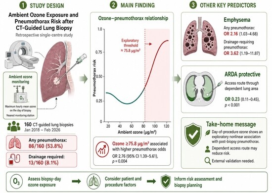
| Ozone, μg/m3 | 76.0 [57.4, 95.3] | 69.8 [56.8, 87.7] | 82.2 [63.4, 100.9] | 0.025 |
| Ozone ≥ 75.8 μg/m3 | 81/160.0 (51%) | 28/74.0 (38%) | 53/86.0 (62%) | 0.004 |
| Model | Variable | OR 95% CI | p-Value |
|---|---|---|---|
| Linear ozone model | Ozone per 10 μg/m3 | 1.09 (0.97–1.23) | 0.167 |
| Linear ozone model | Emphysema | 1.96 (0.95–4.15) | 0.072 |
| Linear ozone model | ARDA | 0.23 (0.12–0.45) | 0.000 |
| Threshold ozone model | Ozone ≥ 75.8 μg/m3 | 2.76 (1.39–5.61) | 0.004 |
| Threshold ozone model | Emphysema | 2.16 (1.03–4.68) | 0.047 |
| Threshold ozone model | ARDA | 0.23 (0.11–0.45) | 0.000 |
| Firth threshold model | Ozone ≥ 75.8 μg/m3 | 2.68 (1.37–5.39) | 0.004 |
| Firth threshold model | Emphysema | 2.1 (1.01–4.50) | 0.047 |
| Firth threshold model | ARDA | 0.24 (0.12–0.46) | 0.000 |
| Model | Ozone ≥ 75.8 μg/m3 | Emphysema | ARDA |
|---|---|---|---|
| Base model | 2.76 (1.39–5.61); p = 0.004 | 2.16 (1.03–4.68); p = 0.046 | 0.23 (0.11–0.45); p ≤ 0.001 |
| Needle size adjusted | 2.87 (1.40–6.07); p = 0.005 | 2.18 (0.99–4.94); p = 0.056 | 0.23 (0.11–0.48); p ≤ 0.001 |
| Biopsy system adjusted | 2.93 (1.43–6.18); p = 0.004 | 2.19 (1.01–4.93); p = 0.051 | 0.27 (0.13–0.56); p ≤ 0.001 |
| Operator adjusted | 2.53 (1.11–5.94); p = 0.029 | 1.66 (0.71–3.93); p = 0.243 | 0.22 (0.10–0.49); p ≤ 0.001 |
| Season adjusted | 2.96 (1.32–6.87); p = 0.009 | 2.11 (0.99–4.63); p = 0.056 | 0.23 (0.11–0.46); p ≤ 0.001 |
| Variable | OR (95% CI) | p-Value |
|---|---|---|
| Ozone ≥ 75.8 μg/m3 | 1.63 (0.53–5.41) | 0.399 |
| Emphysema | 3.62 (1.19–11.87) | 0.023 |
| ARDA | 0.85 (0.26–2.63) | 0.778 |
Disclaimer/Publisher’s Note: The statements, opinions and data contained in all publications are solely those of the individual author(s) and contributor(s) and not of MDPI and/or the editor(s). MDPI and/or the editor(s) disclaim responsibility for any injury to people or property resulting from any ideas, methods, instructions or products referred to in the content. |
© 2026 by the authors. Licensee MDPI, Basel, Switzerland. This article is an open access article distributed under the terms and conditions of the Creative Commons Attribution (CC BY) license.
Share and Cite
Afilal, N.; Komarek, A.; Dieckmeyer, M.; Can, E.; Jonczyk, M.; Heverhagen, J.T.; Brönnimann, M.P. Ambient Ozone Exposure and Pneumothorax Risk After CT-Guided Lung Biopsy. Tomography 2026, 12, 98. https://doi.org/10.3390/tomography12070098
Afilal N, Komarek A, Dieckmeyer M, Can E, Jonczyk M, Heverhagen JT, Brönnimann MP. Ambient Ozone Exposure and Pneumothorax Risk After CT-Guided Lung Biopsy. Tomography. 2026; 12(7):98. https://doi.org/10.3390/tomography12070098
Chicago/Turabian StyleAfilal, Nour, Alois Komarek, Michael Dieckmeyer, Elif Can, Martin Jonczyk, Johannes T. Heverhagen, and Michael P. Brönnimann. 2026. "Ambient Ozone Exposure and Pneumothorax Risk After CT-Guided Lung Biopsy" Tomography 12, no. 7: 98. https://doi.org/10.3390/tomography12070098
APA StyleAfilal, N., Komarek, A., Dieckmeyer, M., Can, E., Jonczyk, M., Heverhagen, J. T., & Brönnimann, M. P. (2026). Ambient Ozone Exposure and Pneumothorax Risk After CT-Guided Lung Biopsy. Tomography, 12(7), 98. https://doi.org/10.3390/tomography12070098