A Scoping Review of Machine-Learning Derived Radiomic Analysis of CT and PET Imaging to Investigate Atherosclerotic Cardiovascular Disease
Abstract
1. Introduction
2. Methodology
2.1. Eligibility Criteria
2.2. Sources and Search Strategy
2.3. Data Extraction and Reporting
2.4. Quality Assessment
3. Results
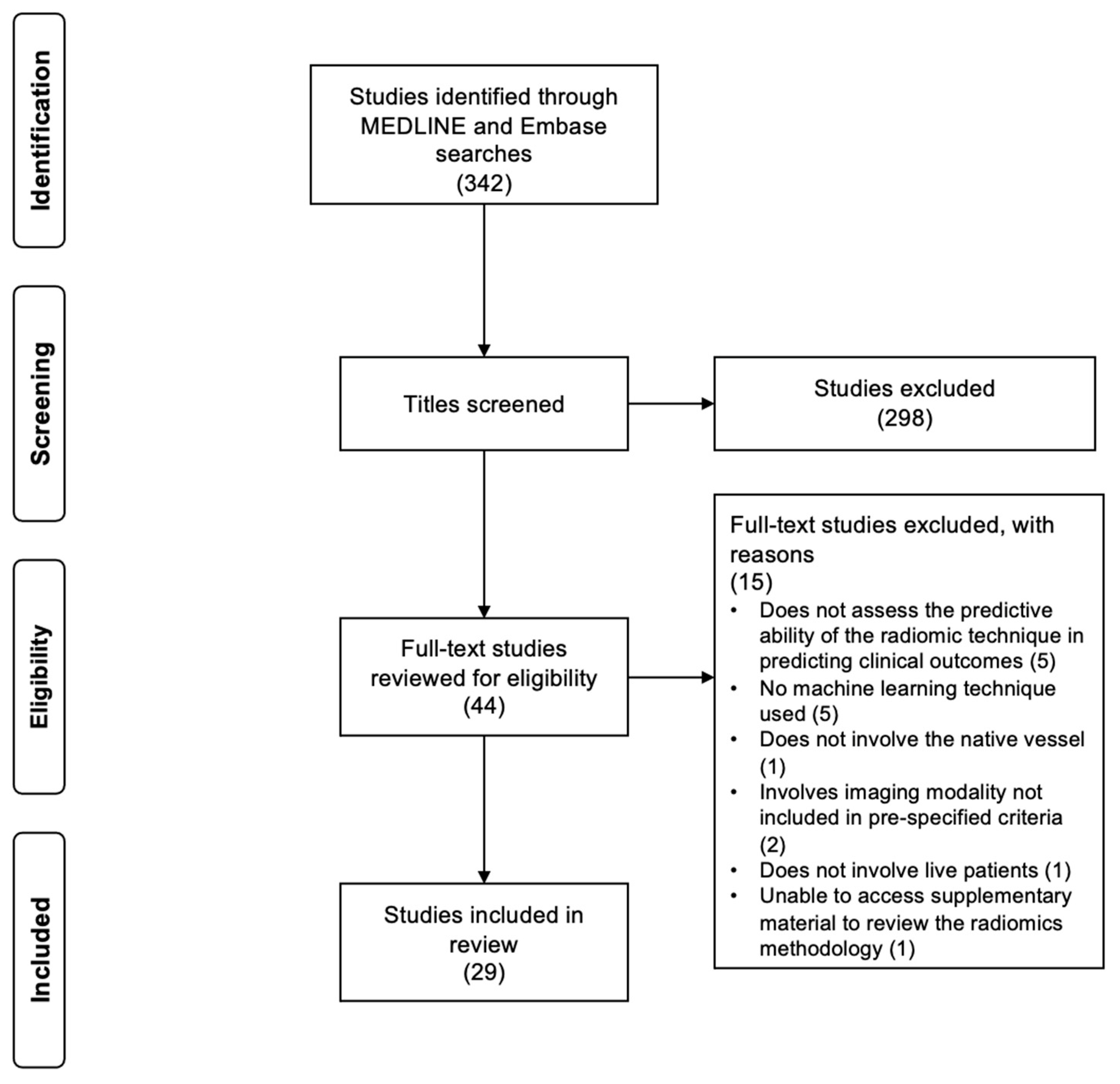
3.1. Literature Search
3.2. Study Characteristics
3.3. Image Acquisition
| Study | Modality | Radiomics Architecture | Segmentation and Processing | Performance Evaluation |
|---|---|---|---|---|
| Carotid studies | ||||
| Chen et al. [14] | CT angiography | Adherence to radiomics guidelines: nil Feature extraction software: 3D Slicer (https://www.slicer.org/, accessed on 27 August 2024) | Segmentation: manual segmentation of the coronary plaque and semiautomated segmentation of the PVAT using 3D Slicer (https://www.slicer.org/, accessed on 27 August 2024) Features extracted: shape, first order, GLCM, GLDM, GLSZM, GLRLM and NGTDM Machine learning techniques: SVM | Performance assessment: AUC from the ROC, accuracy, sensitivity, specificity, PPV, and NPV Internal validation: dataset split into training set (n = 100) and validation set (n = 44). Tenfold cross validation No external validation |
| Cilla et al. [15] | CT angiography | Adherence to radiomics guidelines: radiomic feature extraction performed in accordance with IBSI Feature extraction software: Moddicom (radiomics software package for R, https://github.com/kbolab/moddicom, accessed on 27 August 2024) | Segmentation: manual segmentation Features extracted: first order, shape, GLCM, GLRLM, GLSZM, NGTDM and GLDM Machine learning techniques: logistic regression, SVM, CART | Performance assessment: AUC from the ROC, AUC, class-specific accuracy (proportion of both true positive and true negatives amongst all cases), PPV, sensitivity and F-measure Internal validation: fivefold cross validation applied to each machine learning model No external validation |
| Ebrahimian et al. [26] | Dual-energy CT angiography | Adherence to radiomics guidelines: nil Feature extraction software: PyRadiomics integrated into Dual-Energy Tumour Analysis prototype software (eXamine, Siemens Healthineers, Forcheim, Germany) | Segmentation: automated segmentation using Dual-Energy Tumour Analysis prototype software (eXamine, Siemens Healthineers, Forcheim, Germany) Features extracted: shape, first-order, GLCM, NGTDM, GLSZM, GLRLM, GLDM, and higher-order features Machine learning techniques: multinomial logistic regression | Performance assessment: AUC from the ROC Internal validation: DNM No external validation |
| Kafouris et al. [36] | PET/CT using 0.14 mCi/kg 18F-FDG | Adherence to radiomics guidelines: features extracted according to IBSI guidelines Feature extraction software: in-house software based on Matlab platform (Version 9.3, Matlab R2017b, Natick, MA, USA) | Segmentation: manual segmentation around the carotid artery wall Features extracted: first order, GLCM, GLRLM, GLSZM and NGTDM Machine learning techniques: univariate logistic regression | Performance assessment: AUC from the ROC Internal validation: bootstrapping generating 200 bootstrap samples No external validation |
| Liu et al. [37] | CT angiography | Adherence to radiomics guidelines: nil Feature extraction software: Radcloud platform (Huiying Medical Technology, Beijing, China) | Segmentation: manual segmentation of the coronary plaque using ITK-SNAP software (version 3.7, http://www.itksnap.org/, accessed on 27 August 2024) Features extracted: shape, first order, GLDM, GLRLM, GLCM, GLSZM and NGTDM Machine learning techniques: LASSO used to construct a ‘radiomics score’ | Performance assessment: AUC from the ROC Internal validation: dataset split into training set (n = 135) and validation set (n = 58) External validation using 87 patients |
| Nie et al. [38] | CT angiography | Adherence to radiomics guidelines: nil Feature extraction software: Shukun AI Scientific Research Platform (Shukun Technology, Beijing, China) | Segmentation: automated segmentation of the PVAT using Perivascular Fat Analysis Software (Shukun Technology, Beijing, China) Features extracted: first order, shape, GLCM, GLDM, GLRLM, GLSZM and NGTDM Machine learning techniques: Bagging DecisionTree, XGBoost, random forest, SVM and quadratic discriminant analysis | Performance assessment: AUC from the ROC Internal validation: dataset split into training set (n = 163) and test set (n = 40) No external validation |
| Le et al. [39] | CT angiography | Adherence to radiomics guidelines: nil Feature extraction software: PyRadiomics (version 3.0, https://pyradiomics.readthedocs.io/, accessed on 27 August 2024) | Segmentation: manual segmentation using TexRad (Feedback Medical Ltd., London, UK) Features extracted: first order, GLCM, GLRLM, GLSZM, GLDM, and NGTDM Machine learning techniques: decision tree, random forest, LASSO, Elastic Net regression (weight for L1 and L2 penalties = 0.5), neural network, and XGBoost | Performance assessment: AUC from the ROC Internal validation: fivefold cross validation No external validation |
| Shan et al. [40] | CT angiography | Adherence to radiomics guidelines: nil Feature extraction software: PyRadiomics integrated into Python | Segmentation: semi-automated segmentation using 3D Slicer Features extracted: shape, first order, GLDM, GLRLM, GLCM, GLSZM and NGTDM Machine learning techniques: logistic regression, SVM, random forest, light gradient boosting machine, AdaBoost, XGBoost, and multi-layer perception | Performance assessment: AUC from the ROC Internal validation: dataset split into training set and validation set in a ratio of 7:3 No external validation |
| Shi et al. [41] | CT angiography | Adherence to radiomics guidelines: nil Feature extraction software: The Deepwise Multimodal Research Platform (version 2.0, Beijing Deepwise & League of PHD Technology Co. Ltd, Beijing, China) | Segmentation: manual segmentation of the coronary plaque using The Deepwise Multimodal Research Platform (version 2.0, Beijing Deepwise & League of PHD Technology Co. Ltd, Beijing, China) Features extracted: shape, first order, GLDM, GLRLM, GLCM, GLSZM and NGTDM Machine learning techniques: analysis of variance F-value, mutual information and linear models penalised with the L1 norm | Performance assessment: AUC from the ROC, calibration, and decision curve analyses Internal validation: fivefold cross validation applied to each machine learning model No external validation |
| Xia et al. [42] | CT angiography | Adherence to radiomics guidelines: nil Feature extraction software: PyRadiomics (version 2.4) integrated into Python | Segmentation: manual segmentation of the coronary plaque using 3D Slicer (version 4.11) Features extracted: shape, first order, GLCM, GLSZM, GLRLM, NGTDM and GLDM Machine learning techniques: random forest, XGBoost, logistic regression, SVM and k-nearest neighbour | Performance assessment: predictive value of the model assessed using AUC from the ROC Internal validation: dataset split into training set (n = 165) and validation set (n = 66). Fivefold cross validation used on the training set No external validation |
| Coronary studies | ||||
| Chen et al. [16] | CT coronary angiography | Adherence to radiomics guidelines: nil Feature extraction software: Perivascular Fat Analysis Tool | Segmentation: semi-automated segmentation of the PCAT using Perivascular Fat Analysis Tool Features extracted: shape, first order, GLDM, GLCM, GLRLM, GLSZM and NGTDM Machine learning techniques: multivariate logistic regression used to construct a ‘radiomics score’ | Performance assessment: AUC from the ROC Internal validation: dataset split into training set (n = 108) and validation set (n = 47). Fivefold cross validation performed No external validation |
| Chen et al. [17] | CT coronary angiography | Adherence to radiomics guidelines: features extracted according to IBSI guidelines Feature extraction software: Radiomics, Syngo.Via FRONTIER (version 1.2.1, Siemens Healthineers, Forcheim, Germany) | Segmentation: manual segmentation using Radiomics, Syngo.Via FRONTIER (version 1.2.1, Siemens Healthineers, Forcheim, Germany) Features extracted: shape, first order, GLCM, GLSZM, GLRLM, GLDM and NGTDM Machine learning techniques: multivariable logistic regression and XGBoost used to construct the algorithm | Performance assessment: predictive value of the model assessed using AUC from the ROC Internal validation: dataset split into training set and validation set in a ratio of 7:3. Fivefold cross validation used on the training set (n = 137) External validation using 159 patients |
| Feng et al. [18] | CT coronary angiography | Adherence to radiomics guidelines: nil Feature extraction software: Radiomics, Syngo.Via FRONTIER (version 1.3.0) | Segmentation: semi-automated segmentation of the plaque using Coronary Plaque Analysis Syngo.Via Frontier (version 5.0.2, Siemens Healthineers, Forcheim, Germany) Features extracted: shape, first order and texture Machine learning techniques: random forest model and logistic regression used to construct the radiomics model | Performance assessment: AUC from the ROC, sensitivity, specificity, and accuracy Internal validation: dataset split into training set (n = 280) and validation set (n =120) No external validation |
| Homayounieh et al. [19] | CT coronary angiography | Adherence to radiomics guidelines: nil Feature extraction software: Radiomics, Syngo.Via FRONTIER | Segmentation: automated segmentation using Radiomics, Syngo.Via FRONTIER Features extracted: shape, first order, GLCM, GLRLM, GLSZM, NGTDM and GLDM Machine learning techniques: multiple logistic regression and kernel Fisher discriminant analysis | Performance assessment: AUC from the ROC Internal validation: nil No external validation |
| Hou et al. [20] | CT coronary angiography | Adherence to radiomics guidelines: nil Feature extraction software: DNM | Segmentation: semi-automated segmentation of the PCAT Features extracted: first order, GLCM, GLRLM, GLSZM, GLDM and NGTDM Machine learning techniques: SVM, k-nearest neighbour, Light GBM, and random forest | Performance assessment: AUC from the ROC Internal validation: dataset split into training set (n = 123) and validation set (n = 54). Tenfold cross validation used on the training set No external validation |
| Hu et al. [21] | CT coronary angiography | Adherence to radiomics guidelines: nil Feature extraction software: PyRadiomics library integrated into an unknown software | Segmentation: manual segmentation using ITK-SNAP software (version 3.6.0) Features extracted: first order, shape, texture, higher order Machine learning techniques: logistic regression | Performance assessment: AUC from the ROC, sensitivity, specificity, PPV, NPV, positive likelihood ratio, negative likelihood ratio Internal validation: dataset split into training set (n = 88) and validation set (n = 31) No external validation |
| Jing et al. [22] | CT coronary angiography | Adherence to radiomics guidelines: nil Feature extraction software: PyRadiomics library integrated into Pericoronary Adipose Tissue Analysis Software (Shukun Technology, Beijing, China) | Segmentation: automated segmentation using CoronaryDoc software (Shukun Technology, Beijing, China) Features extracted: first order and texture features Machine learning techniques: SVM, ridge regression classifier and logistic regression | Performance assessment: AUC from the ROC, accuracy, specificity, sensitivity, PPV, and NPVs Internal validation: dataset split into training set and validation set at a ratio of 2:1. Fivefold cross validation performed No external validation |
| Kim et al. [23] | CT coronary angiography | Adherence to radiomics guidelines: features extracted according to IBSI guidelines Feature extraction software: PyRadiomics integrated into Python | Segmentation: semi-automated segmentation of the PCAT using in-house Python software Features extracted: shape, first order, GLCM, GLDM, GLRLM, GLSZM and NGTDM Machine learning techniques: multivariate logistic regression | Performance assessment: predictive value of the model assessed using AUC from the ROC Internal validation: stratified threefold cross validation performed No external validation |
| Kwiecinski et al. [24] | PET/CT performed using 250 MBq 18F-NaF | Adherence to radiomics guidelines: nil Feature extraction software: Radiomics Image Analysis (version 1.4.2, https://github.com/neuroconductor/RIA, accessed on 27 August 2024) on R | Segmentation: automated segmentation of the PET/CT using coronary microcalcification activity. Semi-automated segmentation of the plaques from the CTCA using Autoplaque (version 2.5, Cedars-Sinai Medical Center, Los Angeles, CA, USA) Features extracted: DNM type of features extracted Machine learning techniques: univariable and multivariable logistic regression, linear regression and random forest | Performance assessment: nil Internal validation: DNM No external validation |
| Lee et al. [25] | CT coronary angiography | Adherence to radiomics guidelines: nil Feature extraction software: PyRadiomics integrated into Python | Segmentation: semi-automated segmentation of the coronary plaque using QAngioCT Research Edition (version 2.1.9.1, Medis Medical Imaging, Leiden, Netherlands) Features extracted: first order, GLCM, GLRLM, GLSZM, GLDM and NGTDM Machine learning techniques: multivariable Cox regression model | Performance assessment: AUC from the ROC Internal validation: dataset split into training set and validation set in a ratio of 8:2 No external validation |
| Li et al. [27] | CT coronary angiography | Adherence to radiomics guidelines: nil Feature extraction software: PyRadiomics integrated into Python | Segmentation: manual segmentation of the coronary plaque Features extracted: shape, first order, GLCM, GLDM, GLRLM, GLSZM and NGTDM Machine learning techniques: Naïve Bayes, decision tree, random forest, gradient boosting decision tree, SVM, multilayer perceptron, logistic regression, and k-nearest neighbours | Performance assessment: AUC from the ROC Internal validation: dataset split into training set (n = 36) and validation set (n = 8). Fivefold cross validation performed on the training set No external validation |
| Li et al. [28] | CT coronary angiography | Adherence to radiomics guidelines: nil Feature extraction software: PyRadiomics integrated into Research Portal (version 1.1, United Imaging Intelligence Co. Ltd., Shanghai, China) | Segmentation: automated segmentation of the coronary plaque using Research Portal (version 1.1) Features extracted: shape, first order, GLCM, GLRLM, GLSZM, NGTDM and GLDM Machine learning techniques: DNM | Performance assessment: AUC from the ROC Internal validation: dataset split into training set and validation set in a ratio of 8:2. Fivefold cross validation performed External validation using 50 patients |
| Lin et al. [29] | CT coronary angiography | Adherence to radiomics guidelines: nil Feature extraction software: Radiomics Image Analysis software package (version 1.4.1) on R | Segmentation: automated segmentation of the PCAT using Autoplaque software (version 2.5) Features extracted: shape, first order features, GLCM and GLRLM Machine learning techniques: XGBoost | Performance assessment: AUC from the ROC Internal validation: tenfold cross validation No external validation |
| Lin et al. [30] | CT coronary angiography | Adherence to radiomics guidelines: nil Feature extraction software: Radiomics Image Analysis software package (version 1.4.2) on R | Segmentation: semi-automated segmentation of the coronary plaque using Autoplaque (version 2.5) Features extracted: shape, first order, GLCM and GLRLM Machine learning techniques: XGBoost | Performance assessment: AUC from the ROC Internal validation: tenfold cross validation External validation on 19 patients |
| Oikonomou et al. [31] (study 2 and 3) | CT coronary angiography | Adherence to radiomics guidelines: nil Feature extraction software: PyRadiomics integrated into 3D Slicer | Segmentation: manual segmentation of the PVAT Features extracted: shape, first order, GLCM, GLDM, GLRLM, GLSZM, NGTDM and higher order Machine learning techniques: random forest | Performance assessment: predictive value of the model assessed using AUC from ROC Internal validation: dataset split into training set and validation set in a ratio of 4:1. Fivefold cross validation performed External validation performed on the validation dataset |
| Si et al. [32] | CT coronary angiography | Adherence to radiomics guidelines: nil Feature extraction software: Research Portal (version 1.1) | Segmentation: automated segmentation using the VB-net model Features extracted: shape, first order, GLCM, GLRLM, GLSZM, GLDM and NGTDM Machine learning techniques: logistic regression | Performance assessment: AUC from the ROC Internal validation: dataset split into training set and validation set in a ratio of 7:3. Fivefold cross validation performed No external validation |
| Wen et al. [33] | CT coronary angiography | Adherence to radiomics guidelines: nil Feature extraction software: PyRadiomics integrated into 3D Slicer (version 4.10.2) | Segmentation: manual segmentation of the PCAT using 3D slicer Features extracted: first order, GLCM, GLRLM, GLSZM, GLDM and higher order Machine learning techniques: logistic regression, decision tree and SVM | Performance assessment: AUC from the ROC Internal validation: dataset split into training set and validation set in a ratio of 4:1 No external validation |
| You et al. [34] | CT coronary angiography | Adherence to radiomics guidelines: nil Feature extraction software: Artificial Intelligence Kit (GE Healthcare, Chicago, IL, USA) | Segmentation: semi-automated segmentation of the epicardial adipose tissue using EATseg software (https://github.com/MountainAndMorning/EATSeg, accessed on 27 August 2024) and 3D slicer (version 4.11) Processing: nil Features extracted: first order, GLCM, GLSZM, GLRLM, NGTDM and GLDM Machine learning techniques: logistic regression | Performance assessment: AUC from the ROC Internal validation: dataset split into training set and validation set in a ratio of 7:3 No external validation |
| Yu et al. [35] | CT coronary angiography | Adherence to radiomics guidelines: nil Feature extraction software: PyRadiomics integrated into an in-house software | Segmentation: automated segmentation using CoronaryDoc, FAI Analysis Tool (version 5.1.2, Shukun Technology, Beijing, China) Features extracted: first order, GLCM, GLSZM, GLRLM, NGTDM and GLDM Machine learning techniques: SVM | Performance assessment: AUC from the ROC Internal validation: dataset split into training set and validation set in a ratio of 2:1. Fivefold cross validation performed applied to training set No external validation |
3.4. Segmentation
3.5. Processing
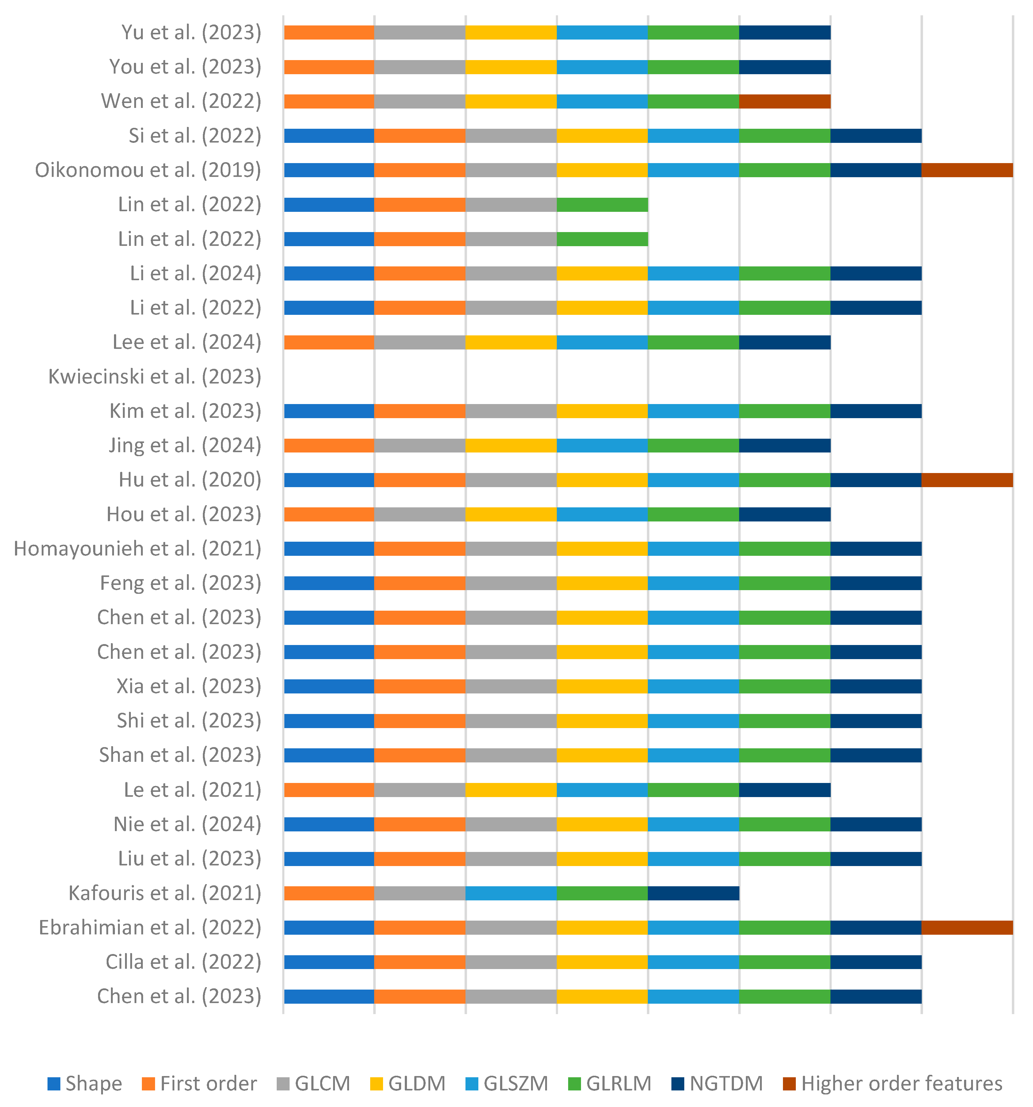
3.6. Radiomic Feature Extraction
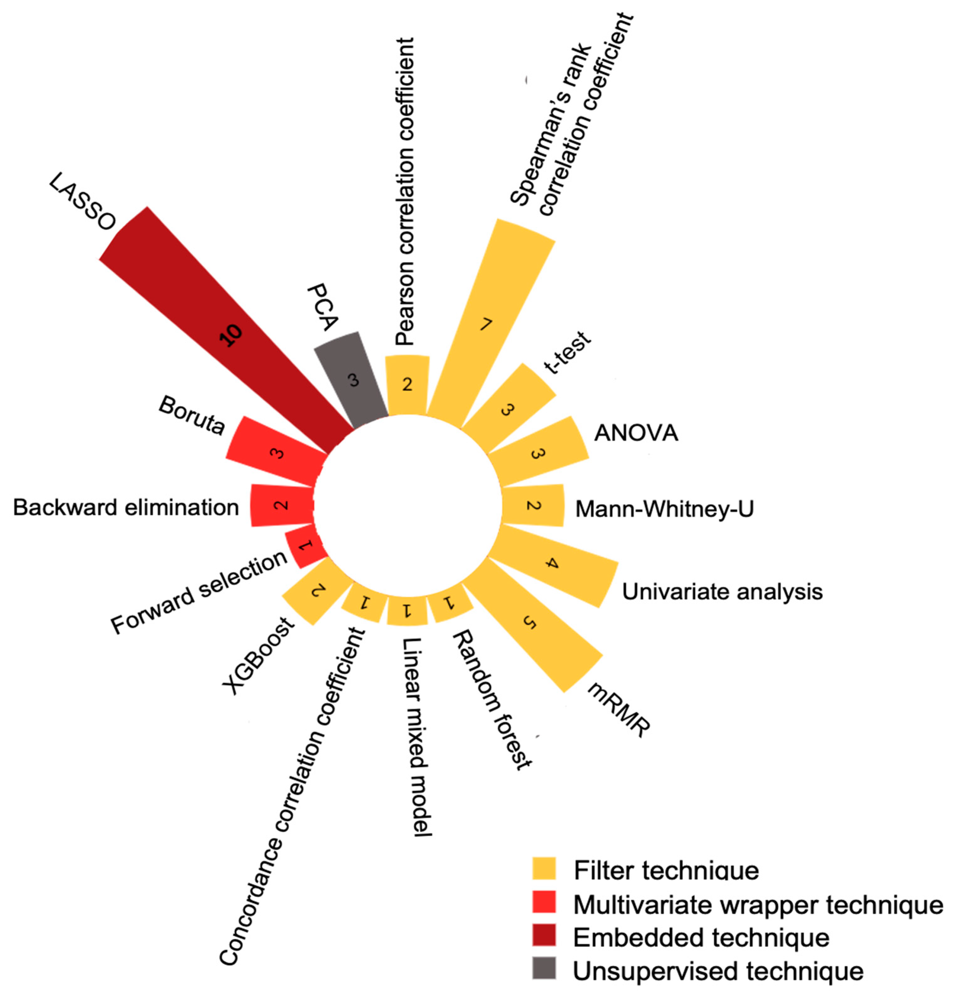
3.7. Dimensionality Reduction and Feature Selection
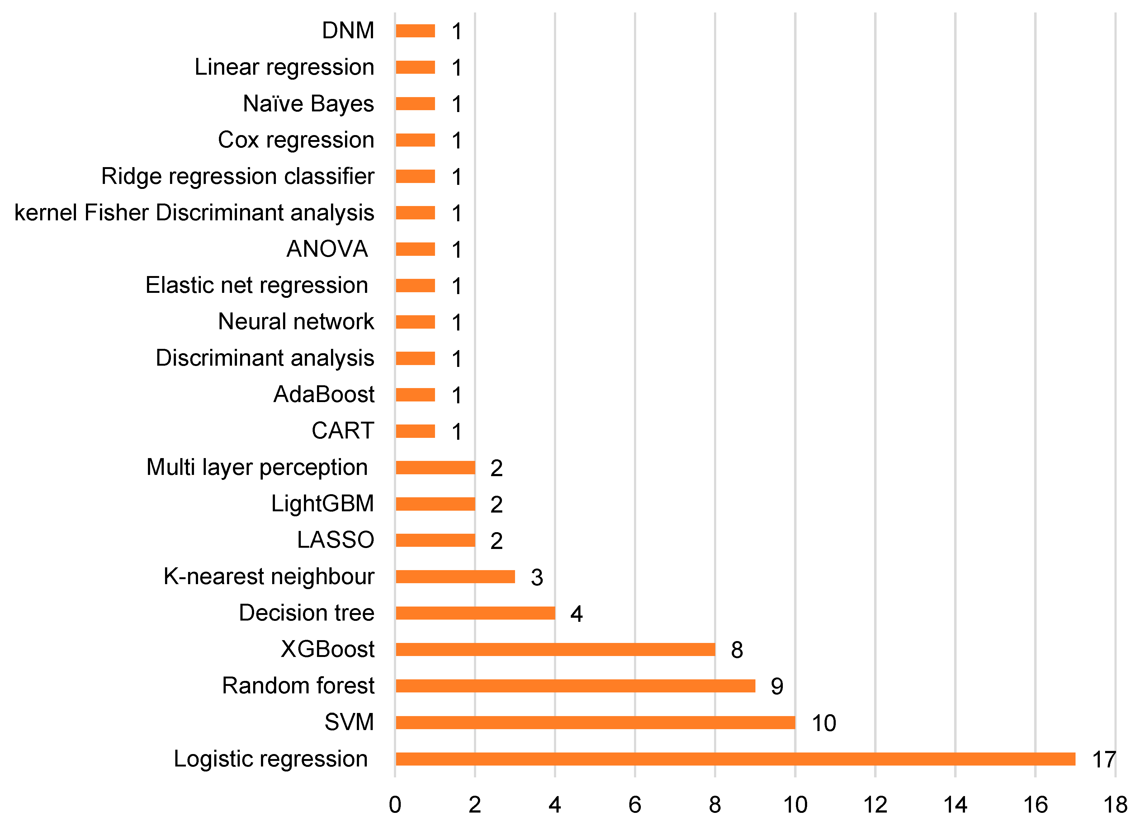
3.8. Machine Learning Methods
3.9. Performance Evaluation and Validation
4. Discussion
Limitations and Areas for Further Research
5. Conclusions
Supplementary Materials
Author Contributions
Funding
Conflicts of Interest
References
- Roth, G.A.; Mensah, G.A.; Johnson, C.O.; Addolorato, G.; Ammirati, E.; Baddour, L.M.; Barengo, N.C.; Beaton, A.Z.; Benjamin, E.J.; Benziger, C.P.; et al. Global Burden of Cardiovascular Diseases and Risk Factors, 1990–2019: Update From the GBD 2019 Study. J. Am. Coll. Cardiol. 2020, 76, 2982–3021. [Google Scholar] [CrossRef]
- Lyle, A.N.; Taylor, W.R. The pathophysiological basis of vascular disease. Lab. Investig. 2019, 99, 284–289. [Google Scholar] [CrossRef] [PubMed]
- Shaw, S.Y. Molecular imaging in cardiovascular disease: Targets and opportunities. Nat. Rev. Cardiol. 2009, 6, 569–579. [Google Scholar] [CrossRef] [PubMed]
- Knuuti, J.; Wijns, W.; Saraste, A.; Capodanno, D.; Barbato, E.; Funck-Brentano, C.; Prescott, E.; Storey, R.F.; Deaton, C.; Cuisset, T.; et al. 2019 ESC Guidelines for the diagnosis and management of chronic coronary syndromes: The Task Force for the diagnosis and management of chronic coronary syndromes of the European Society of Cardiology (ESC). Eur. Heart J. 2020, 41, 407–477. [Google Scholar] [CrossRef] [PubMed]
- van Timmeren, J.E.; Cester, D.; Tanadini-Lang, S.; Alkadhi, H.; Baessler, B. Radiomics in medical imaging—“how-to” guide and critical reflection. Insights Imaging 2020, 11, 91. [Google Scholar] [CrossRef]
- Koçak, B.; Durmaz, E.Ş.; Ateş, E.; Kılıçkesmez, Ö. Radiomics with artificial intelligence: A practical guide for beginners. Diagnostic Interv. Radiol. 2019, 25, 485. [Google Scholar] [CrossRef]
- Gillies, R.J.; Kinahan, P.E.; Hricak, H. Radiomics: Images Are More than Pictures, They Are Data. Radiology 2015, 278, 563–577. [Google Scholar] [CrossRef]
- Tricco, A.C.; Lillie, E.; Zarin, W.; O’Brien, K.K.; Colquhoun, H.; Levac, D.; Moher, D.; Peters, M.D.J.; Horsley, T.; Weeks, L.; et al. PRISMA Extension for Scoping Reviews (PRISMA-ScR): Checklist and Explanation. Ann. Intern. Med. 2018, 169, 467–473. [Google Scholar] [CrossRef]
- Sadeghi, M.M.; Glover, D.K.; Lanza, G.M.; Fayad, Z.A.; Johnson, L.L. Imaging Atherosclerosis and Vulnerable Plaque. J. Nucl. Med. 2010, 51 (Suppl. S1), 51S LP-65S. [Google Scholar] [CrossRef]
- Wolters Kluwer. Ovid. 2023. Available online: https://ovidsp.ovid.com/ (accessed on 15 March 2023).
- Dionisio, F.C.F.; Oliveira, L.S.; Hernandes, M.d.A.; Engel, E.E.; de Azevedo-Marques, P.M.; Nogueira-Barbosa, M.H. Manual versus semiautomatic segmentation of soft-tissue sarcomas on magnetic resonance imaging: Evaluation of similarity and comparison of segmentation times. Radiol. Bras. 2021, 54, 155–164. [Google Scholar] [CrossRef]
- Kocak, B.; Akinci D’Antonoli, T.; Mercaldo, N.; Alberich-Bayarri, A.; Baessler, B.; Ambrosini, I.; Andreychenko, A.E.; Bakas, S.; Beets-Tan, R.G.H.; Bressem, K.; et al. METhodological RadiomICs Score (METRICS): A quality scoring tool for radiomics research endorsed by, E.u.S.o.M.I.I. Insights Imaging 2024, 15, 8. [Google Scholar] [CrossRef]
- Wells, G.; Shea, B.; O’Connell, D.; Peterson, J.; Welch, V.; Losos, M.; Tugwell, P. The Newcastle-Ottawa Scale (NOS) for Assessing the Quality of Nonrandomised Studies in meta-Analyses. Available online: https://www.ohri.ca/programs/clinical_epidemiology/oxford.asp (accessed on 19 August 2024).
- Chen, C.; Tang, W.; Chen, Y.; Xu, W.; Yu, N.; Liu, C.; Li, Z.; Tang, Z.; Zhang, X. Computed tomography angiography-based radiomics model to identify high-risk carotid plaques. Quant. Imaging Med. Surg. 2023, 13, 6089–6104. [Google Scholar] [CrossRef]
- Cilla, S.; Macchia, G.; Lenkowicz, J.; Tran, E.H.; Pierro, A.; Petrella, L.; Fanelli, M.; Sardu, C.; Re, A.; Boldrini, L.; et al. CT angiography-based radiomics as a tool for carotid plaque characterization: A pilot study. Radiol. Med. 2022, 127, 743–753. [Google Scholar] [CrossRef] [PubMed]
- Chen, M.; Hu, J.; Chen, C.; Hao, G.; Hu, S.; Xu, J.; Hu, C. Radiomics analysis of pericoronary adipose tissue based on plain CT for preliminary screening of coronary artery disease in patients with type 2 diabetes mellitus. Acta Radiol. 2023, 64, 2704–2713. [Google Scholar] [CrossRef]
- Chen, Q.; Xie, G.; Tang, C.X.; Yang, L.; Xu, P.; Gao, X.; Lu, M.; Fu, Y.; Huo, Y.; Zheng, S.; et al. Development and Validation of CCTA-based Radiomics Signature for Predicting Coronary Plaques With Rapid Progression. Circ. Cardiovasc. Imaging 2023, 16, e015340. [Google Scholar] [CrossRef] [PubMed]
- Feng, C.; Chen, R.; Dong, S.; Deng, W.; Lin, S.; Zhu, X.; Liu, W.; Xu, Y.; Li, X.; Zhu, Y.; et al. Predicting coronary plaque progression with conventional plaque parameters and radiomics features derived from coronary CT angiography. Eur. Radiol. 2023, 33, 8513–8520. [Google Scholar] [CrossRef]
- Homayounieh, F.; Yan, P.; Digumarthy, S.R.; Kruger, U.; Wang, G.; Kalra, M.K. Prediction of Coronary Calcification and Stenosis: Role of Radiomics From Low-Dose CT. Acad. Radiol. 2021, 28, 972–979. [Google Scholar] [CrossRef] [PubMed]
- Hou, J.; Zheng, G.; Han, L.; Shu, Z.; Wang, H.; Yuan, Z.; Peng, J.; Gong, X. Coronary computed tomography angiography imaging features combined with computed tomography-fractional flow reserve, pericoronary fat attenuation index, and radiomics for the prediction of myocardial ischemia. J. Nucl. Cardiol. 2023, 30, 1838–1850. [Google Scholar] [CrossRef]
- Hu, W.; Wu, X.; Dong, D.; Cui, L.-B.; Jiang, M.; Zhang, J.; Wang, Y.; Wang, X.; Gao, L.; Tian, J.; et al. Novel radiomics features from CCTA images for the functional evaluation of significant ischaemic lesions based on the coronary fractional flow reserve score. Int. J. Cardiovasc. Imaging 2020, 36, 2039–2050. [Google Scholar] [CrossRef]
- Jing, M.; Xi, H.; Sun, J.; Zhu, H.; Deng, L.; Han, T.; Zhang, B.; Zhang, Y.; Zhou, J. Differentiation of acute coronary syndrome with radiomics of pericoronary adipose tissue. Br. J. Radiol. 2024, 97, 850–858. [Google Scholar] [CrossRef]
- Kim, J.N.; Gomez-Perez, L.; Zimin, V.N.; Makhlouf, M.H.E.; Al-Kindi, S.; Wilson, D.L.; Lee, J. Pericoronary Adipose Tissue Radiomics from Coronary Computed Tomography Angiography Identifies Vulnerable Plaques. Bioengineering 2023, 10, 360. [Google Scholar] [CrossRef] [PubMed]
- Kwiecinski, J.; Kolossváry, M.; Tzolos, E.; Meah, M.N.; Adamson, P.D.; Joshi, N.V.; Williams, M.C.; van Beek, E.J.R.; Berman, D.S.; Maurovich-Horvat, P.; et al. Latent Coronary Plaque Morphology From Computed Tomography Angiography, Molecular Disease Activity on Positron Emission Tomography, and Clinical Outcomes. Arterioscler. Thromb. Vasc. Biol. 2023, 43, e279–e290. [Google Scholar] [CrossRef] [PubMed]
- Lee, S.-E.; Hong, Y.; Hong, J.; Jung, J.; Sung, J.M.; Andreini, D.; Al-Mallah, M.H.; Budoff, M.J.; Cademartiri, F.; Chinnaiyan, K.; et al. Prediction of the development of new coronary atherosclerotic plaques with radiomics. J. Cardiovasc. Comput. Tomogr. 2024, 18, 274–280. [Google Scholar] [CrossRef] [PubMed]
- Ebrahimian, S.; Homayounieh, F.; Singh, R.; Primak, A.; Kalra, M.K.; Romero, J. Spectral segmentation and radiomic features predict carotid stenosis and ipsilateral ischemic burden from DECT angiography. Diagnostic. Interv. Radiol. 2022, 28, 264–274. [Google Scholar] [CrossRef]
- Li, X.; Yin, W.; Sun, Y.; Kang, H.; Luo, J.; Chen, K.; Hou, Z.; Gao, Y.; Ren, X.; Yu, Y. Identification of pathology-confirmed vulnerable atherosclerotic lesions by coronary computed tomography angiography using radiomics analysis. Eur. Radiol. 2022, 32, 4003–4013. [Google Scholar] [CrossRef]
- Li, J.; Ren, L.; Guo, H.; Yang, H.; Cui, J.; Zhang, Y. Radiomics-based discrimination of coronary chronic total occlusion and subtotal occlusion on coronary computed tomography angiography. BMC Med. Imaging 2024, 24, 84. [Google Scholar] [CrossRef]
- Lin, A.; Kolossváry, M.; Yuvaraj, J.; Cadet, S.; McElhinney, P.A.; Jiang, C.; Nerlekar, N.; Nicholls, S.J.; Slomka, P.J.; Maurovich-Horvat, P.; et al. Myocardial Infarction Associates With a Distinct Pericoronary Adipose Tissue Radiomic Phenotype: A Prospective Case-Control Study. JACC Cardiovasc. Imaging 2020, 13, 2371–2383. [Google Scholar] [CrossRef]
- Lin, A.; Kolossváry, M.; Cadet, S.; McElhinney, P.; Goeller, M.; Han, D.; Yuvaraj, J.; Nerlekar, N.; Slomka, P.J.; Marwan, M.; et al. Radiomics-Based Precision Phenotyping Identifies Unstable Coronary Plaques From Computed Tomography Angiography. JACC Cardiovasc. Imaging 2022, 15, 859–871. [Google Scholar] [CrossRef]
- Oikonomou, E.K.; Williams, M.C.; Kotanidis, C.P.; Desai, M.Y.; Marwan, M.; Antonopoulos, A.S.; Thomas, K.E.; Thomas, S.; Akoumianakis, I.; Fan, L.M.; et al. A novel machine learning-derived radiotranscriptomic signature of perivascular fat improves cardiac risk prediction using coronary CT angiography. Eur. Heart J. 2019, 40, 3529–3543. [Google Scholar] [CrossRef]
- Si, N.; Shi, K.; Li, N.; Dong, X.; Zhu, C.; Guo, Y.; Hu, J.; Cui, J.; Yang, F.; Zhang, T. Identification of patients with acute myocardial infarction based on coronary CT angiography: The value of pericoronary adipose tissue radiomics. Eur. Radiol. 2022, 32, 6868–6877. [Google Scholar] [CrossRef]
- Wen, D.; Xu, Z.; An, R.; Ren, J.; Jia, Y.; Li, J.; Zheng, M. Predicting haemodynamic significance of coronary stenosis with radiomics-based pericoronary adipose tissue characteristics. Clin. Radiol. 2022, 77, e154–e161. [Google Scholar] [CrossRef]
- You, H.; Zhang, R.; Hu, J.; Sun, Y.; Li, X.; Hou, J.; Pei, Y.; Zhao, L.; Zhang, L.; Yang, B.; et al. Performance of Radiomics Models Based on Coronary Computed Tomography Angiography in Predicting The Risk of Major Adverse Cardiovascular Events Within 3 Years: A Comparison Between the Pericoronary Adipose Tissue Model and the Epicardial Adipose Tissue Mo. Acad. Radiol. 2023, 30, 390–401. [Google Scholar] [CrossRef] [PubMed]
- Yu, L.; Chen, X.; Ling, R.; Yu, Y.; Yang, W.; Sun, J.; Zhang, J. Radiomics features of pericoronary adipose tissue improve CT-FFR performance in predicting hemodynamically significant coronary artery stenosis. Eur. Radiol. 2023, 33, 2004–2014. [Google Scholar] [CrossRef]
- Kafouris, P.P.; Koutagiar, I.P.; Georgakopoulos, A.T.; Spyrou, G.M.; Visvikis, D.; Anagnostopoulos, C.D. Fluorine-18 fluorodeoxyglucose positron emission tomography-based textural features for prediction of event prone carotid atherosclerotic plaques. J. Nucl. Cardiol. 2021, 28, 1861–1871. [Google Scholar] [CrossRef]
- Liu, M.; Chang, N.; Zhang, S.; Du, Y.; Zhang, X.; Ren, W.; Sun, J.; Bai, J.; Wang, L.; Zhang, G. Identification of vulnerable carotid plaque with CT-based radiomics nomogram. Clin. Radiol. 2023, 78, e856–e863. [Google Scholar] [CrossRef]
- Nie, J.-Y.; Chen, W.-X.; Zhu, Z.; Zhang, M.-Y.; Zheng, Y.-J.; Wu, Q.-D. Initial experience with radiomics of carotid perivascular adipose tissue in identifying symptomatic plaque. Front. Neurol. 2024, 15, 1340202. [Google Scholar] [CrossRef] [PubMed]
- Le, E.P.V.; Rundo, L.; Tarkin, J.M.; Evans, N.R.; Chowdhury, M.M.; Coughlin, P.A.; Pavey, H.; Wall, C.; Zaccagna, F.; Gallagher, F.A.; et al. Assessing robustness of carotid artery CT angiography radiomics in the identification of culprit lesions in cerebrovascular events. Sci. Rep. 2021, 11, 3499. [Google Scholar] [CrossRef] [PubMed]
- Shan, D.; Wang, S.; Wang, J.; Lu, J.; Ren, J.; Chen, J.; Wang, D.; Qi, P. Computed tomography angiography-based radiomics model for predicting carotid atherosclerotic plaque vulnerability. Front. Neurol. 2023, 14, 1151326. [Google Scholar] [CrossRef]
- Shi, J.; Sun, Y.; Hou, J.; Li, X.; Fan, J.; Zhang, L.; Zhang, R.; You, H.; Wang, Z.; Zhang, A.; et al. Radiomics Signatures of Carotid Plaque on Computed Tomography Angiography. Clin. Neuroradiol. 2023, 33, 931–941. [Google Scholar] [CrossRef]
- Xia, H.; Yuan, L.; Zhao, W.; Zhang, C.; Zhao, L.; Hou, J.; Luan, Y.; Bi, Y.; Feng, Y. Predicting transient ischemic attack risk in patients with mild carotid stenosis using machine learning and CT radiomics. Front. Neurol. 2023, 14, 1105616. [Google Scholar] [CrossRef]
- Moher, D.; Liberati, A.; Tetzlaff, J.; Altman, D.G. Preferred reporting items for systematic reviews and meta-analyses: The PRISMA statement. BMJ 2009, 339, b2535. [Google Scholar] [CrossRef] [PubMed]
- Evans, N.R.; Tarkin, J.M.; Chowdhury, M.M.; Le, E.P.V.; Coughlin, P.A.; Rudd, J.H.F.; Warburton, E.A. Dual-Tracer Positron-Emission Tomography for Identification of Culprit Carotid Plaques and Pathophysiology In Vivo. Circ. Cardiovasc. Imaging 2020, 13, e009539. [Google Scholar] [CrossRef] [PubMed]
- Tarkin, J.M.; Joshi, F.R.; Evans, N.R.; Chowdhury, M.M.; Figg, N.L.; Shah, A.V.; Starks, L.T.; Martin-Garrido, A.; Manavaki, R.; Yu, E.; et al. Detection of Atherosclerotic Inflammation by 68Ga-DOTATATE PET Compared to [18F]FDG PET Imaging. J. Am. Coll. Cardiol. 2017, 69, 1774–1791. [Google Scholar] [CrossRef] [PubMed]
- Joshi, F.R.; Manavaki, R.; Fryer, T.D.; Figg, N.L.; Sluimer, J.C.; Aigbirhio, F.I.; Davenport, A.P.; Kirkpatrick, P.J.; Warburton, E.A.; Rudd, J.H.F. Vascular Imaging With 18F-Fluorodeoxyglucose Positron Emission Tomography Is Influenced by Hypoxia. J. Am. Coll. Cardiol. 2017, 69, 1873–1874. [Google Scholar] [CrossRef]
- Naylor, R.; Rantner, B.; Ancetti, S.; de Borst, G.J.; De Carlo, M.; Halliday, A.; Kakkos, S.K.; Markus, H.S.; McCabe, D.J.H.; Sillesen, H.; et al. Editor’s Choice—European Society for Vascular Surgery (ESVS) 2023 Clinical Practice Guidelines on the Management of Atherosclerotic Carotid and Vertebral Artery Disease. Eur. J. Vasc. Endovasc. Surg. 2023, 65, 7–111. [Google Scholar] [CrossRef]
- Zwanenburg, A.; Vallières, M.; Abdalah, M.A.; Aerts, H.J.W.L.; Andrearczyk, V.; Apte, A.; Ashrafinia, S.; Bakas, S.; Beukinga, R.J.; Boellaard, R.; et al. The Image Biomarker Standardization Initiative: Standardized Quantitative Radiomics for High-Throughput Image-based Phenotyping. Radiology 2020, 295, 328–338. [Google Scholar] [CrossRef]
- Pinto dos Santos, D.; Dietzel, M.; Baessler, B. A decade of radiomics research: Are images really data or just patterns in the noise? Eur. Radiol. 2021, 31, 1–4. [Google Scholar] [CrossRef]
- Shafiq-ul-Hassan, M.; Zhang, G.G.; Latifi, K.; Ullah, G.; Hunt, D.C.; Balagurunathan, Y.; Abdalah, M.A.; Schabath, M.B.; Goldgof, D.G.; Mackin, D.; et al. Intrinsic dependencies of CT radiomic features on voxel size and number of gray levels. Med. Phys. 2017, 44, 1050–1062. [Google Scholar] [CrossRef]
- Larue, R.T.H.M.; van Timmeren, J.E.; de Jong, E.E.C.; Feliciani, G.; Leijenaar, R.T.H.; Schreurs, W.M.J.; Sosef, M.N.; Raat, F.H.P.J.; van der Zande, F.H.R.; Das, M.; et al. Influence of gray level discretization on radiomic feature stability for different CT scanners, tube currents and slice thicknesses: A comprehensive phantom study. Acta Oncol. 2017, 56, 1544–1553. [Google Scholar] [CrossRef]
- Mackin, D.; Ger, R.; Dodge, C.; Fave, X.; Chi, P.-C.; Zhang, L.; Yang, J.; Bache, S.; Dodge, C.; Jones, A.K.; et al. Effect of tube current on computed tomography radiomic features. Sci. Rep. 2018, 8, 2354. [Google Scholar] [CrossRef]
- Escudero Sanchez, L.; Rundo, L.; Gill, A.B.; Hoare, M.; Mendes Serrao, E.; Sala, E. Robustness of radiomic features in CT images with different slice thickness, comparing liver tumour and muscle. Sci. Rep. 2021, 11, 8262. [Google Scholar] [CrossRef]
- He, L.; Huang, Y.; Ma, Z.; Liang, C.; Liang, C.; Liu, Z. Effects of contrast-enhancement, reconstruction slice thickness and convolution kernel on the diagnostic performance of radiomics signature in solitary pulmonary nodule. Sci. Rep. 2016, 6, 34921. [Google Scholar] [CrossRef]
- The Royal College of Physicians; The British Society of Cardiovascular Imaging; The Royal College of Radiologists. Standards of Practice of Computed Tomography Coronary Angiography (CTCA) in Adult Patients. The Royal College of Radiologists. London.. 2014. Available online: https://www.rcr.ac.uk/our-services/all-our-publications/clinical-radiology-publications (accessed on 2 August 2024).
- Mackin, D.; Fave, X.; Zhang, L.; Fried, D.; Yang, J.; Taylor, B.; Rodriguez-Rivera, E.; Dodge, C.; Jones, A.K.; Court, L. Measuring Computed Tomography Scanner Variability of Radiomics Features. Invest. Radiol. 2015, 50, 757–765. [Google Scholar] [CrossRef] [PubMed]
- Giesen, A.; Mouselimis, D.; Weichsel, L.; Giannopoulos, A.A.; Schmermund, A.; Nunninger, M.; Schuetz, M.; André, F.; Frey, N.; Korosoglou, G. Pericoronary adipose tissue attenuation is associated with non-calcified plaque burden in patients with chronic coronary syndromes. J. Cardiovasc. Comput. Tomogr. 2023, 17, 384–392. [Google Scholar] [CrossRef]
- Yuvaraj, J.; Lin, A.; Nerlekar, N.; Munnur, R.K.; Cameron, J.D.; Dey, D.; Nicholls, S.J.; Wong, D.T.L. Pericoronary Adipose Tissue Attenuation Is Associated with High-Risk Plaque and Subsequent Acute Coronary Syndrome in Patients with Stable Coronary Artery Disease. Cells 2021, 10, 1143. [Google Scholar] [CrossRef] [PubMed]
- Yu, M.; Dai, X.; Deng, J.; Lu, Z.; Shen, C.; Zhang, J. Diagnostic performance of perivascular fat attenuation index to predict hemodynamic significance of coronary stenosis: A preliminary coronary computed tomography angiography study. Eur. Radiol. 2020, 30, 673–681. [Google Scholar] [CrossRef] [PubMed]
- Gresser, E.; Woźnicki, P.; Messmer, K.; Schreier, A.; Kunz, W.G.; Ingrisch, M.; Stief, C.; Ricke, J.; Nörenberg, D.; Buchner, A.; et al. Radiomics Signature Using Manual Versus Automated Segmentation for Lymph Node Staging of Bladder Cancer. Eur. Urol. Focus 2023, 9, 145–153. [Google Scholar] [CrossRef]
- Lin, Y.-C.; Lin, G.; Pandey, S.; Yeh, C.-H.; Wang, J.-J.; Lin, C.-Y.; Ho, T.-Y.; Ko, S.-F.; Ng, S.-H. Fully automated segmentation and radiomics feature extraction of hypopharyngeal cancer on MRI using deep learning. Eur. Radiol. 2023, 33, 6548–6556. [Google Scholar] [CrossRef]
- Traverso, A.; Wee, L.; Dekker, A.; Gillies, R. Repeatability and Reproducibility of Radiomic Features: A Systematic Review. Int. J. Radiat. Oncol. Biol. Phys. 2018, 102, 1143–1158. [Google Scholar] [CrossRef]
- Kocak, B.; Baessler, B.; Bakas, S.; Cuocolo, R.; Fedorov, A.; Maier-Hein, L.; Mercaldo, N.; Müller, H.; Orlhac, F.; Pinto dos Santos, D.; et al. CheckList for EvaluAtion of Radiomics research (CLEAR): A step-by-step reporting guideline for authors reviewers endorsed by ESR and EuSoMII. Insights Imaging 2023, 14, 75. [Google Scholar] [CrossRef]
- Lambin, P.; Leijenaar, R.T.H.; Deist, T.M.; Peerlings, J.; de Jong, E.E.C.; van Timmeren, J.; Sanduleanu, S.; Larue, R.T.H.M.; Even, A.J.G.; Jochems, A.; et al. Radiomics: The bridge between medical imaging and personalized medicine. Nat. Rev. Clin. Oncol. 2017, 14, 749–762. [Google Scholar] [CrossRef] [PubMed]
- Munn, Z.; Peters, M.D.J.; Stern, C.; Tufanaru, C.; McArthur, A.; Aromataris, E. Systematic review or scoping review? Guidance for authors when choosing between a systematic or scoping review approach. BMC Med. Res. Methodol. 2018, 18, 143. [Google Scholar] [CrossRef] [PubMed]
- Di Pilla, A.; Nero, C.; Specchia, M.L.; Ciccarone, F.; Boldrini, L.; Lenkowicz, J.; Alberghetti, B.; Fagotti, A.; Testa, A.C.; Valentini, V.; et al. A cost-effectiveness analysis of an integrated clinical-radiogenomic screening program for the identification of BRCA 1/2 carriers (e-PROBE study). Sci. Rep. 2024, 14, 928. [Google Scholar] [CrossRef] [PubMed]

| Study | Patient Demographics | Age (Years) | Eligibility Criteria | Comorbidities (Number of Patients) |
|---|---|---|---|---|
| Carotid studies | ||||
| Chen et al. [14] Single-centre study | Overall: 144 Male: 110 | 70.9 ± 9.1 | Inclusion criteria: diagnosis of extracranial carotid stenosis between 30–99% on CTA images, sufficient information to ascertain cerebral ischemia symptoms in the medical records, and adequate information regarding vascular risk factors in the medical records Exclusion criteria: cardiogenic stroke, simultaneous bilateral anterior circulation events, complications of radiation therapy and vasculitis, stroke involving the posterior circulation only, inadequate image quality | Hypertension: 111 Hyperlipidaemia: 69 Smoker: 65 Diabetes mellitus: 52 CAD: 40 |
| Cilla et al. [15] Single-centre study | Overall: 30 Male: 19 | 72.96 (50–86) | Inclusion criteria: patients aged 18–75 years requiring carotid endarterectomy for >70% stenosis Exclusion criteria: patients requiring combined aorto-coronary bypass surgery and carotid endarterectomy | Hypertension: 28 Hyperlipidaemia: 17 CAD: 12 Diabetes mellitus: 9 Chronic kidney disease: 3 Peripheral arterial disease: 2 Abdominal aorta aneurysm: 1 |
| Ebrahimian et al. [26] Single-centre study | Overall: 85 Male: 56 | 73 ± 10 | Inclusion criteria: patients undergoing dual-energy CTA of the neck to investigate common or internal carotid artery stenosis Exclusion criteria: patients scanned using other scanners, previous revascularisation surgery, metallic implants or stents, dental implants, motion artefact on imaging | DNM |
| Kafouris et al. [36] Single-centre study | Overall: 21 Male: 18 | 70.4 ± 7.0 | Inclusion criteria: patients undergoing carotid endarterectomy for stenosis > 70% Exclusion criteria: cardiological ischaemic events < 6 months ago; active infection, inflammatory or neoplastic disease, uncontrolled diabetes mellitus, multiple significant stenoses across the carotid arteries | Hypertension: 18 Hyperlipidaemia: 15 Smoker: 11 Diabetes mellitus: 9 CAD: 4 |
| Liu et al. [37] Multi-centre study | Overall: 280 Male: 201 | Symptomatic patients Training group: 63.8 ± 7.2 Validation group: 63.0 ± 7.1 External test group: 62.8 ± 7.5 Asymptomatic patients Training group: 65.3 ± 8.8 Validation group: 61.0 ± 8.0 External test group: 63.4 ± 8.6 | Inclusion criteria: extracranial carotid artery stenosis secondary to atherosclerosis disease Exclusion criteria: history of carotid stenting and endarterectomy, cardiac thrombus, carotid occlusion, poor image quality, symptomatic bilateral carotid stenosis | Hypertension: 209 Smoker: 202 CAD: 159 Hyperlipidaemia: 132 Diabetes mellitus: 99 |
| Nie et al. [38] Single-centre study | Overall: 203 Male: 115 | 71.9 ± 9.6 | Inclusion criteria: extracranial carotid atherosclerosis Exclusion criteria: ischemic stroke or TIA caused by intracranial carotid stenosis >50%, ischemic stroke or TIA occurred >2 weeks before CTA, posterior circulation symptoms, history of intervention to the cervicocerebral artery, cerebral haemorrhage, meningioma, craniotomy, arteriovenous fistula, temporal lobectomy, moyamoya disease, reversible cerebral vasoconstriction syndrome, arteritis, carotid artery dissection, carotid artery aneurysm, carotid artery web, poor image quality, incomplete clinical information | Hypertension: 155 Diabetes mellitus: 72 Smoker: 55 Hyperlipidaemia: 50 |
| Le et al. [39] Single-centre study | Overall: 41 Male: 32 | 74.1 ± 8.4 | Inclusion criteria: bilateral carotid atherosclerosis (Evans et al. [44]), nil inclusion criteria (Tarkin et al. [45]), DNM (Joshi et al. [46]) Exclusion criteria: atrial fibrillation (Evans et al. [44]), nil exclusion criteria (Tarkin et al. [45]), DNM (Joshi et al. [46]) | Stroke: 30 Smoker: 29 (includes current and ex-smokers) Hypertension 27 TIA: 11 Diabetes mellitus: 8 |
| Shan et al. [40] Single-centre study | Overall: 74 Male: 63 | 66.9 ± 8.8 | Inclusion criteria: patients aged >18 years with carotid atherosclerotic plaque diagnosed on CTA and contrast-enhanced ultrasound Exclusion criteria: incomplete clinical information, poor image quality | Hypertension: 52 Smoker: 41 Diabetes mellitus: 29 |
| Shi et al. [41] Single-centre study | Overall: 167 Male: 131 | 66.2 ± 7.7 | Inclusion criteria: patients with suspected stroke who underwent head and neck CTA and brain MRI Exclusion criteria: incomplete clinical information, negative carotid CTA, cerebral haemorrhage, intra-cranial tumour, intra-cranial trauma, previous brain surgery, posterior circulation stroke, suspected cardioembolic | Hypertension: 115 Smoker: 91 Hyperlipidaemia: 73 Diabetes mellitus: 48 CAD: 23 |
| Xia et al. [42] Single-centre study | Overall: 179 Male: 125 | 65.4 ± DNM | Inclusion criteria: patients undergoing carotid CTA with carotid artery stenosis of 30–50% Exclusion criteria: carotid artery dissection or aneurysm, intracranial vascular disease (e.g., intracranial atherosclerosis with stenosis < 50%, vasculitis, aneurysm), posterior circulation stroke, intracerebral haemorrhage; other causes of haemorrhagic stroke (e.g., cardioembolic source and chest embolism); patients with other neurological diseases such as brain tumours or demyelinating disease | DNM |
| Coronary studies | ||||
| Chen et al. [16] Single-centre study | Overall: 155 Male: 81 | 62 ± 10 | Inclusion criteria: patients with suspected CAD who underwent plain CT and CTCA Exclusion criteria: patients without diabetes, previous history of coronary artery disease, history of cardiac or coronary surgery, anomalous origin of coronary artery, coronary malformation, coronary artery aneurysm, coronary artery calcium score >600, poor image quality | Hypertension: 113 Hyperlipidaemia: 54 Smoker: 31 |
| Chen et al. [17] Multi-centre study | Overall: 214 Male: 163 | Development group: 63 ± 11 Validation group: 65 ± 10 | Inclusion criteria: minimum of 2 CTCA studies 6 months apart, baseline coronary artery stenosis was 25% to 70% Exclusion criteria: patients undergoing coronary artery bypass grafting or percutaneous coronary intervention before or during the study, missing or insufficient imaging data, poor image quality, different tube voltage settings used between the CTCA examinations | Hypertension: 147 Diabetes mellitus: 68 Hyperlipidaemia: 33 Smoker: 30 |
| Feng et al. [18] Single-centre study | Overall: 280 Male: 184 | Progression group: 70.1 ± 10.5 Non-progression group: 70.2 ± 10.0 | Inclusion criteria: ≥2 CTCA examination ≥2 years apart with >2 mm atherosclerotic lesion on the baseline imaging, consistent imaging technique during both scans Exclusion criteria: incomplete clinical information, poor imaging quality, coronary revascularisation before or during the study | Hypertension: 223 Diabetes mellitus: 87 Smoker: 76 |
| Homayounieh et al. [19] Single-centre study | Overall: 106 Male: 68 | 64 ± 7 | Inclusion criteria: patients undergoing low-dose CT for lung cancer screening received CTCA within 12 months Exclusion criteria: coronary stents, prior cardiac surgery, metal artefacts in the cardiac region | Hyperlipidaemia: 91 Hypertension: 84 Smoker: 45 Diabetes mellitus: 28 |
| Hou et al. [20] Single-centre study | Overall: 96 Male: 68 | 62.6 ± 13.4 | Inclusion criteria: patients with suspected or known CAD who underwent CTCA and SPECT-myocardial perfusion imaging Exclusion criteria: poor image quality, no lesion on CTCA, previous ACS or revascularisation, MPI was conducted over 30 days after CTCA, failed automatic image segmentation | Hypertension: 61 Diabetes mellitus: 32 Smoker: 30 Hyperlipidaemia: 24 |
| Hu et al. [21] Single-centre study | Overall: 109 Male: 81 | Training group FFR ≤ 0.8 patients: 62.5 ± 8.3 FFR > 0.8 patients: 61.2 ± 8.2 Validation group FFR ≤ 0.8 patients: 71.3 ± 7.8 FFR > 0.8 patients: 66.6 ± 6.4 | Inclusion criteria: patients who experienced non-emergency invasive coronary angiography and FFR within 30 days after CTCA examination, and target lesions were located in the epicardial coronary artery with a diameter > 2 mm Exclusion criteria: prior stent implantation, inadequate image quality, unsuccessful image segmentation, stenosis <30% or >90% in the target lesion, tandem lesions that precluded identification of the culprit lesion, previous cardiac resynchronisation or catheter ablation therapy, complex congenital heart disease, severe cardiac insufficiency or liver and kidney dysfunction, contraindication to iodine contrast and coronary microangiopathy | Hypertension: 81 Diabetes mellitus: 40 Hyperlipidaemia: 78 Smoker: 33 |
| Jing et al. [22] Single-centre study | Overall: 620 Male: 336 | Training group CAD patients: 53 (47–58) CCS patients: 63 (55–69) ACS patients: 59.7 ± 11.9 Testing group No CAD patients: 54 (49–58.3) CCS patients: 58 (53–69.8) ACS patients: 60.7 ± 10.9 | Inclusion criteria: no history of ACS or coronary bypass surgery or stenting, absence of atrial fibrillation, no severe renal impairment (eGFR > 30ml/m/1.73 m2, no contraindication to iodine contrast; CTCA within 3 days followed by invasive coronary angiography Exclusion criteria: incomplete imaging and clinical data, coronary artery malformations, artificial valve, cardiac pacemaker, myocarditis, vasculitis, inadequate image quality | Hyperlipidaemia: 379 Hypertension: 362 Smoker: 286 Diabetes mellitus: 182 |
| Kim et al. [23] Single-centre study | Overall: 25 Male: 19 | 63 ± 11 | Inclusion criteria: patients that underwent both CTCA and IVOCT for the investigation of coronary plaques Exclusion criteria: history of myocardial infarction, previous coronary stent implantation, inadequate CTCA or IVOCT images | Hyperlipidaemia: 24 Diabetes mellitus: 20 Hypertension: 11 Chronic kidney disease: 11 |
| Kwiecinski et al. [24] Multi-centre study | Overall: 260 Male: 216 | 65 ± 9 | Inclusion criteria: patients with established CAD Exclusion criteria: coronary artery stenting | Hyperlipidaemia: 235 Smoker: 172 Hypertension: 153 Diabetes mellitus: 54 Peripheral arterial disease: 14 |
| Lee et al. [25] Multi-centre study | Overall: 1162 Male: 647 | 60.3 ± 9.2 | Inclusion criteria: patients that underwent clinically indicated CTCA Exclusion criteria: inadequate imaging quality, coronary revascularisation before or during the study, failure to extract radiomic features, coronary plaque at baseline | Hypertension: 600 Smoker: 431 Hyperlipidaemia: 420 Diabetes mellitus: 231 |
| Li et al. [27] Single-centre study | Overall: 44 Male: 40 | Training group: 53.0 ± 9.0 Validation group: 48.5 ± 11.6 | Inclusion criteria: patients with CAD and end-stage heart failure who underwent CTCA prior to surgery Exclusion criteria: contraindications to CTCA, inadequate image quality | Hyperlipidaemia: 29 Smoker: 21 Hypertension: 17 Diabetes mellitus: 12 |
| Li et al. [28] Multi-centre study | Overall: 132 Male: 91 | Subtotal occlusion patients: 65 (55–71) Chronic total occlusion patients: 63 (58–73) | Inclusion criteria: patients with subtotal or chronic total coronary artery occlusion who underwent both CTCA and invasive coronary angiography Exclusion criteria: patients who underwent bypass surgery or percutaneous coronary intervention for occluded arteries, >2 week interval between CTCA and invasive coronary angiography, multiple occlusive lesions, excessive calcification precluding lumen analysis, inadequate image quality | Hypertension: 80 Diabetes mellitus: 48 Smoker: 48 |
| Lin et al. [29] Single-centre study | Overall: 180 Male: 156 | Acute MI patients: 58.4 (51.6–73.7) Stable CAD patients: 60.0 (52.0–68.5) No CAD patients: 59.5 (52.0–69.0) | Inclusion criteria: patients with post-thrombolysis STEMI or non-STEMI and had a culprit lesion identified on invasive coronary angiography Exclusion criteria: previous MI or revascularisation, clinical instability, severe renal impairment (eGFR < 30 ml/m/1.73 m2), allergy to iodinated contrast | Hypertension: 127 Diabetes mellitus: 40 Hyperlipidaemia: 98 Smoker: 63 |
| Lin et al. [30] Single-centre study | Overall: 120 Male: 104 | Acute MI patients: 59.9 ± 11.6 Stable CAD patients: 60.2 ± 11.3 | Inclusion criteria: patients with acute MI undergoing CTCA and invasive coronary angiography Exclusion criteria: previous MI or revascularisation, clinical instability, severe renal impairment (eGFR < 30 ml/m/1.73 m2), allergy to iodinated contrast | Hypertension: 85 Hyperlipidaemia: 67 Smoker: 44 Diabetes mellitus: 28 |
| Oikonomou et al. [31] Multi-centre study | Study 2 Overall: 202 Male: 134 | MACE group: 64 (55–72) Non-MACE group: 62 (53–70) | Inclusion criteria: study 2—patients undergoing clinically indicated CTCA, study 3—patients undergoing CTCA after acute MI or stable CAD Exclusion criteria: DNM | Hypertension: 129 Hyperlipidaemia: 80 Smoker: 56 Diabetes mellitus: 34 |
| Study 3 Overall: 88 Male: 65 | Stable CAD group: 62 (51–70) Acute MI group: 62 (53–72) | Smoker: 55 Hypertension: 42 Hyperlipidaemia: 41 Diabetes mellitus: 13 | ||
| Si et al. [32] Single-centre study | Overall: 210 Male: 148 | 62.5 ± 10.4 | Inclusion criteria: patients with acute MI Exclusion criteria: DNM | Hyperlipidaemia: 145 Hypertension: 111 Diabetes mellitus: 69 Smoker: 74 |
| Wen et al. [33] Single-centre study | Overall: 92 Male: 66 | 58.3 ± 10.3 | Inclusion criteria: patients suspected with CAD undergoing CTCA and invasive coronary angiography and FFR examination, <30-day interval between CTCA and FFR measurement Exclusion criteria: previous revascularisation, inadequate CTCA image quality, incomplete CTCA acquisition | Hypertension: 43 Hyperlipidaemia: 39 Smoker: 37 Diabetes mellitus: 8 |
| You et al. [34] Multi-centre study | Overall: 288 Male: 175 | Training group MACE patients: 59.1 ± 10.4 Non-MACE patients: 59.6 ± 9.6 Validation group MACE patients: 60.4 ± 10.0 Non-MACE patients: 61.4 ± 8.4 | Inclusion criteria: patients who underwent CTCA—half of the cohort had a major adverse cardiovascular event within 3 years Exclusion criteria: previous PCI or CABG, revascularisation surgery within 6 weeks after CTCA, incomplete clinical information, inadequate imaging quality, previous MI, cardiomyopathy, valvular heart disease, congenital heart disease, chest malignancy | Hypertension: 193 Diabetes mellitus: 107 Smoker: 94 Hyperlipidaemia: 26 |
| Yu et al. [35] Single-centre study | Overall: 146 Male: 102 | 65.5 ± 8.3 | Inclusion criteria: patients with known CAD who had CTCA, invasive coronary angiography, and FFR within 1 month Exclusion criteria: previous revascularisation, tandem coronary lesions, previous MI, inadequate CTCA quality | Hypertension: 105 Hyperlipidaemia: 59 Diabetes mellitus: 56 Smoker: 50 |
Disclaimer/Publisher’s Note: The statements, opinions and data contained in all publications are solely those of the individual author(s) and contributor(s) and not of MDPI and/or the editor(s). MDPI and/or the editor(s) disclaim responsibility for any injury to people or property resulting from any ideas, methods, instructions or products referred to in the content. |
© 2024 by the authors. Licensee MDPI, Basel, Switzerland. This article is an open access article distributed under the terms and conditions of the Creative Commons Attribution (CC BY) license (https://creativecommons.org/licenses/by/4.0/).
Share and Cite
Badesha, A.S.; Frood, R.; Bailey, M.A.; Coughlin, P.M.; Scarsbrook, A.F. A Scoping Review of Machine-Learning Derived Radiomic Analysis of CT and PET Imaging to Investigate Atherosclerotic Cardiovascular Disease. Tomography 2024, 10, 1455-1487. https://doi.org/10.3390/tomography10090108
Badesha AS, Frood R, Bailey MA, Coughlin PM, Scarsbrook AF. A Scoping Review of Machine-Learning Derived Radiomic Analysis of CT and PET Imaging to Investigate Atherosclerotic Cardiovascular Disease. Tomography. 2024; 10(9):1455-1487. https://doi.org/10.3390/tomography10090108
Chicago/Turabian StyleBadesha, Arshpreet Singh, Russell Frood, Marc A. Bailey, Patrick M. Coughlin, and Andrew F. Scarsbrook. 2024. "A Scoping Review of Machine-Learning Derived Radiomic Analysis of CT and PET Imaging to Investigate Atherosclerotic Cardiovascular Disease" Tomography 10, no. 9: 1455-1487. https://doi.org/10.3390/tomography10090108
APA StyleBadesha, A. S., Frood, R., Bailey, M. A., Coughlin, P. M., & Scarsbrook, A. F. (2024). A Scoping Review of Machine-Learning Derived Radiomic Analysis of CT and PET Imaging to Investigate Atherosclerotic Cardiovascular Disease. Tomography, 10(9), 1455-1487. https://doi.org/10.3390/tomography10090108


