Communication

Stapled Peptides: An Innovative and Ultimate Future Drug Offering a Highly Powerful and Potent Therapeutic Alternative

1
Research Institute of Pharmaceutical Sciences, College of Pharmacy, Sookmyung Women’s University, Seoul 04310, Republic of Korea
2
College of Pharmacy, Duksung Women’s University, Seoul 01369, Republic of Korea
*
Author to whom correspondence should be addressed.
Biomimetics 2024, 9(9), 537; https://doi.org/10.3390/biomimetics9090537
Submission received: 23 July 2024 / Revised: 28 August 2024 / Accepted: 30 August 2024 / Published: 5 September 2024

Abstract

:
Peptide-based therapeutics have traditionally faced challenges, including instability in the bloodstream and limited cell membrane permeability. However, recent advancements in α-helix stapled peptide modification techniques have rekindled interest in their efficacy. Notably, these developments ensure a highly effective method for improving peptide stability and enhancing cell membrane penetration. Particularly in the realm of antimicrobial peptides (AMPs), the application of stapled peptide techniques has significantly increased peptide stability and has been successfully applied to many peptides. Furthermore, constraining the secondary structure of peptides has also been proven to enhance their biological activity. In this review, the entire process through which hydrocarbon-stapled antimicrobial peptides attain improved drug-like properties is examined. First, the essential secondary structural elements required for their activity as drugs are validated, specific residues are identified using alanine scanning, and stapling techniques are strategically incorporated at precise locations. Additionally, the mechanisms by which these structure-based stapled peptides function as AMPs are explored, providing a comprehensive and engaging discussion.

1. Introduction

The growing resistance of microorganisms to antibiotics is a significant global health concern [1]. Despite the discovery of many new antibiotics, including vancomycin, in the 2000s, humanity continues to suffer from MRSA (Methicillin-Resistant Staphylococcus Aureus) and VRSA (Vancomycin-Resistant Staphylococcus Aureus) infections [2]. The proliferation of drug-resistant strains is largely attributed to the worldwide overuse of antibiotics in humans [3]. Consequently, the current pharmaceutical industry shows great interest in developing new antibiotics targeting these resistant strains [4].
Antibiotics have been heralded as saviors, especially noted for their crucial role during World War II and in combating various diseases [5]. Since the discovery of penicillin in 1928, countless lives have been saved by antibiotics, and they continue to provide new hope to many [6]. However, the emergence of MDR (Multi-Drug-Resistant) strains, including MRSA and VRSA, poses a significant threat to global health care [7]. Additionally, the development of new antibiotics has slowed dramatically since the 20th century, necessitating alternative solutions to combat drug-resistant bacteria [8].
Antimicrobial peptides (AMPs) are relatively short chains, usually comprising 12 to 40 amino acid residues [9]. These peptides typically display a positive charge due to an abundance of basic amino acids such as arginine, lysine, and histidine [10]. Their hydrophobic residues are crucial for penetrating the bacterial membrane [11]. While most α-helical AMPs are unstructured in solution, they adopt a more organized structure upon interacting with bacterial membranes [12]. The evaluation of their preferred secondary structures, antimicrobial activities, and membrane-disrupting capabilities is essential [13]. AMPs can exhibit a micelle-like structure with hydrophobic residues on one side and hydrophilic residues on the other [14]. For example, human antimicrobial peptides such as LL-37 have the ability to form various structures through oligomerization. LL-37 oligomerizes into helical bundles stabilized by hydrophobic interactions and hydrogen bonding between helical segments. Additionally, under certain conditions, it can assemble into fibril-like structures, demonstrating the polymorphism of its oligomerization [15]. Furthermore, the stapling process, as described below, can significantly influence oligomerization and self-assembly. The stapling process increases structural rigidity, facilitating the peptide’s ability to self-assemble into more stable and organized nanostructures. This is closely associated with enhanced antimicrobial activity against pathogens and may play a positive role in amplifying biological activity [16].
The term AMP refers to small polypeptides produced by all living organisms to protect the host from pathogenic microbes, similar to antibiotics [17]. Like antibiotics, AMPs are naturally produced by microorganisms [18]. Due to the vast diversity of microorganisms on Earth, AMPs display remarkable structural and functional diversity and have mechanisms of action different from existing antibiotics [19]. This diversity can make AMPs a valuable alternative to conventional antibiotics for treating MDR bacterial infections [20].
However, AMPs also have undesirable characteristics, such as susceptibility to proteolytic digestion, toxicity to eukaryotic cells, and inefficient delivery to target sites [21]. Solving these issues is crucial for the development of AMPs as new antibiotics [22]. Efforts in the scientific community focus on optimizing AMPs through specific amino acid substitutions, de novo design, and prodrugs to overcome challenges like size reduction and hydrophobicity control [23].
Peptide-based drugs, in general, offer significant advantages, including high bioavailability and flexible conformational structures [24]. Despite their short half-lives due to enzymatic degradation, peptides have potential clinical applications if their proteolytic stability can be improved [25]. Peptides typically have a higher molecular weight (500–5000 Da) than small molecules, offering a larger surface area for interaction with protein targets, leading to fewer side effects and lower toxicity [26]. Additionally, their relatively small size compared to proteins can reduce manufacturing costs [27].
Various approaches have been proposed to enhance the stability and efficacy of peptide drugs, with peptide stapling being particularly promising [28] (Figure 1). This method involves forming a covalent bridge between amino acid chains, which stabilizes the peptide’s active conformation and protects it from enzymatic degradation [29]. Stapled peptides potentially offer greater drug-like properties than small molecules [30]. Since peptides are usually administered via injection, they can achieve fast systemic absorption, bypass first-pass metabolism, and allow for precise targeting and pharmacokinetic monitoring [31].
Stapled peptides have revolutionized the concept of undruggable targets, overcoming concerns about the large binding interfaces of helical protein fragments compared to small molecules [32]. These techniques enhance the properties of antimicrobial peptides, making them highly efficacious and pathway specific [33].
Specifically, hydrocarbon peptide stapling has proven effective in reinforcing α-helicity, improving stability and selectivity [34]. This technique brings previously uncontacted amino acids into appropriate crosslinks, enhancing cell penetration, proteolytic stability, and biological activity [35]. Stapled peptides, particularly in the context of AMPs, stabilize the helical structure and enhance antimicrobial activity [36].
Taken together, AMPs and stapled peptides share a connection through their structural and functional properties. AMPs are short, naturally occurring peptides that play a crucial role in the immune response by disrupting the membranes of pathogenic microorganisms. Similarly, stapled peptides, which are synthetically modified peptides with stabilized α-helices through hydrocarbon staples, enhance proteolytic stability, cell permeability, and target specificity. The structural rigidity and improved bioavailability of stapled peptides make them promising candidates for mimicking the action of AMPs, particularly in targeting membrane proteins and disrupting cellular processes in a similar manner to how AMPs target microbial membranes. Therefore, the design of stapled peptides can be inspired by the functional principles of AMPs, aiming to create potent and selective therapeutics that leverage the inherent antimicrobial mechanisms.
While many reviews discuss stapled peptides, most focus on the mechanical aspects of the stapling strategy. This review aims to minimize redundant explanations and instead emphasize the significance and effectiveness of stapled peptides from a protein structure-based perspective, offering fresh insights into their potential as druggable biomimetics.

2. Structure-Based Approach

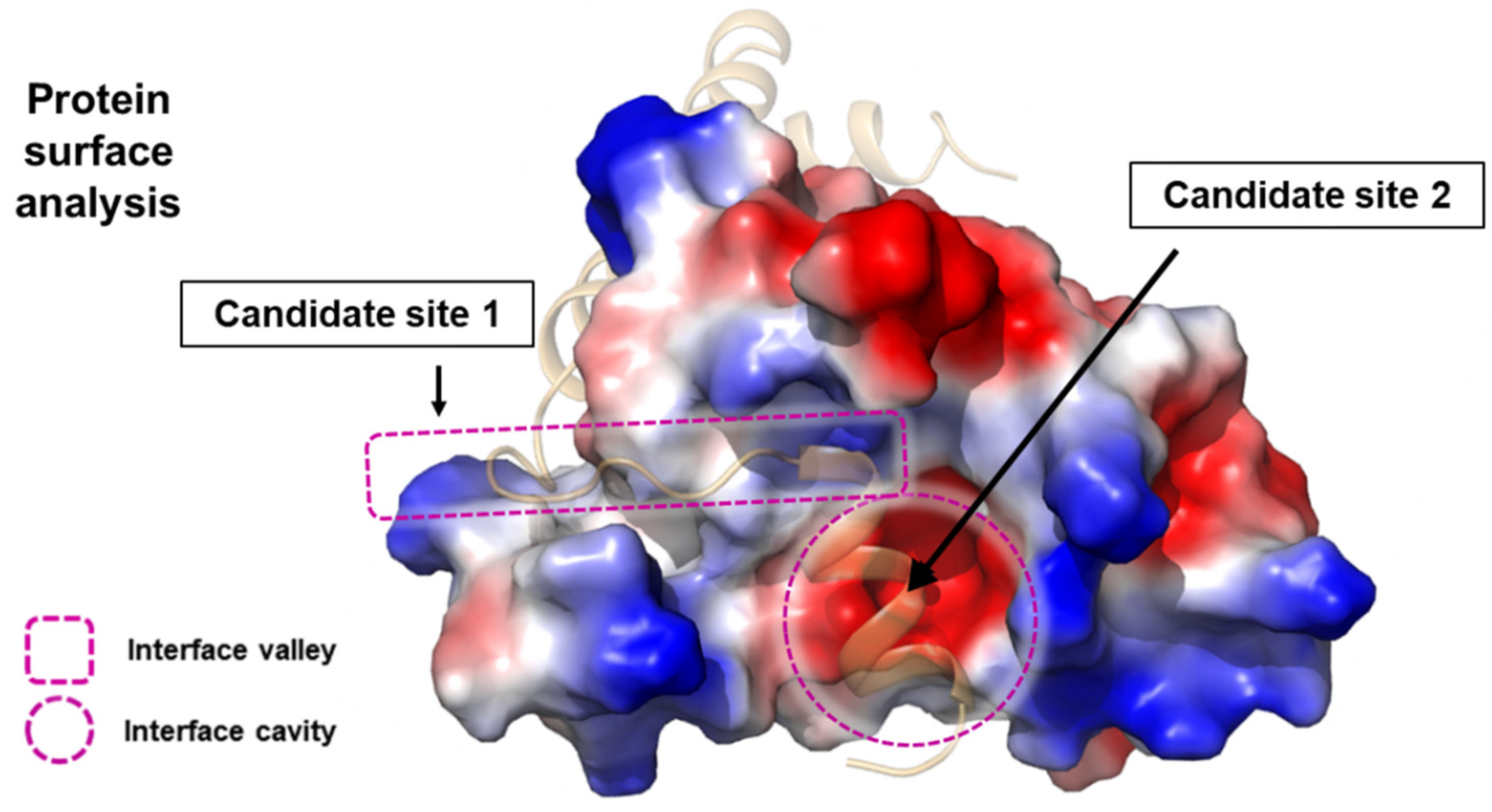
A high-resolution structure can reveal critical positions where introducing a rigid α-helical structure might be essential for tight binding between two proteins (Figure 2) [37]. Such structures provide a detailed view of the molecular interactions and the spatial arrangement of amino acids, allowing for precise identification of regions where structural reinforcement could enhance binding affinity and specificity. If there is an α-helix passing through an obvious pocket or valley within the target protein, this region can be ideal for targeting, as it often represents a key interaction interface [38].
Once the peptide chain that forms the core of the interaction is identified, one can design a stapled peptide consisting of that chain to stabilize its structure and enhance its binding properties [39]. This involves introducing modifications to the peptide, such as incorporating non-natural amino acids or employing specific chemical linkers to create a covalent bond between side chains, thus forming a stable cross-link. These modifications enforce the peptide into an α-helical conformation, which is often more resistant to proteolytic degradation and possesses improved cell permeability compared to its linear counterpart [40].
The process of designing such a stapled peptide typically starts with the selection of suitable sites for modification. Computational modeling and molecular dynamics simulations can be employed to predict the impact of different modifications on the peptide’s structure and function. Once potential sites are identified, synthetic chemistry techniques are used to incorporate the modified amino acids into the peptide chain. The choice of linker type and length is crucial, as it must be compatible with the desired α-helical structure and the specific geometry of the target site [41].

3. Selection of Stapling Residues

To stabilize the peptide’s secondary structure, staples must connect two side chains situated on the same face of the helix [37]. It is essential to recognize that the α-helix comprises 3.6 residues per turn. Therefore, the residues selected for stapling should adhere to specific positions: they must be located at i and i+4 (one-loop staple), i+7 (two-loop staple), or even i+11 (three-loop staple) (Figure 1) [28]. When systematically designed in this manner, stapled peptides can enhance protease resistance, improve pharmacokinetic properties, and increase biological activity [42].
As previously mentioned, to induce a peptide to adopt an α-helical structure, it is essential to link the side chains of two amino acids to form a stapled peptide [28]. The number of stapling bridges within the same peptide does not necessarily have to be limited to one. For longer peptides, it is feasible to use double, triple, or even quadruple stapling, utilizing four amino acids to create two distinct side braces. Typically, double stapling alone is sufficient to confer the desired helicity to the peptide, generally achieving over 80% helicity [43].
For the design of hydrocarbon peptide stapling, it is crucial to select residues that are vital for maintaining the helical structure and are located in regions likely to form contact faces (usually lysine) [30]. Since stapling can alter the physicochemical properties of the original amino acids, residues essential for preserving the helical structure are typically excluded from stapling [32]. Generally, the spacing between residues selected for stapling corresponds to one helix turn or two helix turns, which means choosing the i-th residue, the i+4-th residue, and the i+7-th residue. For maximizing helicity, the i+11-th residue may also be chosen. A reasonable approach for determining the residues for stapling involves performing an alanine scanning and using the results to guide the selection process [43].

4. Alanine Scanning

An α-helix typically contains anywhere from a few to several dozen amino acids. In a conventional α-helix, each amino acid residue is arranged at an approximate 100° relative to the axis, creating a spiral shape. A single turn of the helix generally includes about 3.6 amino acid residues on average [44].
The region of the α-helix where stapling will be applied is determined through structural analysis, as described in the previous section. To ensure that the implemented peptide functions effectively as a helix when synthesized, it is crucial to select appropriate residues for stapling [45].
For the peptide to interact effectively with its partner, as it does in its native form, it is essential to preserve amino acid residues that are critical for this interaction. In other words, residues significant for recognition should not be modified by the stapling technique [46]. Although each amino acid contributes differently to the formation of the α-helix, methionine, alanine, leucine, glutamate, and lysine are generally vital for helix formation. Thus, the residues chosen for the stapling technique should be those that are essential for maintaining the α-helix structure but are not critical for the interaction [47].
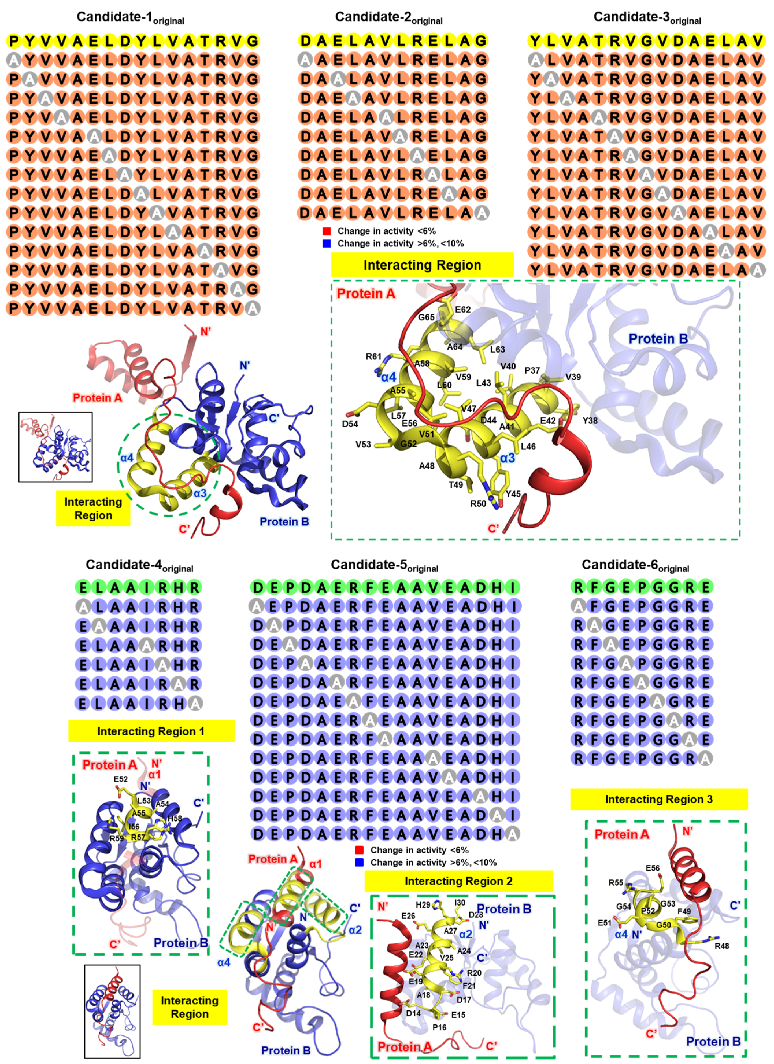
Due to the potential discrepancy between theoretical predictions and actual results, it is often beneficial to conduct alanine scanning for thorough validation (Figure 3). Specifically, this involves sequentially replacing each amino acid in the peptide sequence with alanine and assessing the activity of each resulting peptide, using appropriate in vitro or in vivo methods [43]. This approach helps identify the positions where amino acid substitutions have minimal impact on activity, allowing those positions to be designated for the stapling technique [40].
Our argument for the potential of stapled peptides is critically supported by examples based on the VapBC26 and VapBC30 proteins derived from Mycobacterium tuberculosis. In this study, researchers designed peptidomimetics to inhibit the VapBC26 and VapBC30 complexes by mimicking their binding interfaces. These peptidomimetics, particularly those mimicking the α3 and α4 helices of VapC26 and the α1 helix of VapB30, successfully disrupted the protein complexes, thereby increasing RNase activity and inhibiting bacterial growth, ultimately enhancing druggability. The peptidomimetics, named ‘V26-SP-8’ and ‘V30-SP-8’, created through α-helix stapling, demonstrated improved cell penetration, stability, and efficacy, even surpassing traditional antibiotics like vancomycin. These findings suggest that stapled peptides could be a promising strategy for developing novel drugs with high druggability [49]. Like this, stapling has proven to be a valuable tool in peptidomimetics by stabilizing the α-helical structure, enhancing proteolytic stability, and improving cell permeability. These modifications increase the therapeutic potential of peptidomimetics, making them more effective AMPs and offering new possibilities in drug design.

5. CD Spectroscopy

After selecting residues for stapling through alanine scanning to preserve biological or chemical activity while maintaining α-helicity, it is essential to confirm the formation of the α-helical secondary structure. If amino acids like proline, which disrupt α-helicity, do not significantly impact activity and are located at the ends of the peptide sequence, they may be removed for better results in α-helicity measurements using circular dichroism (CD) spectroscopy [50].
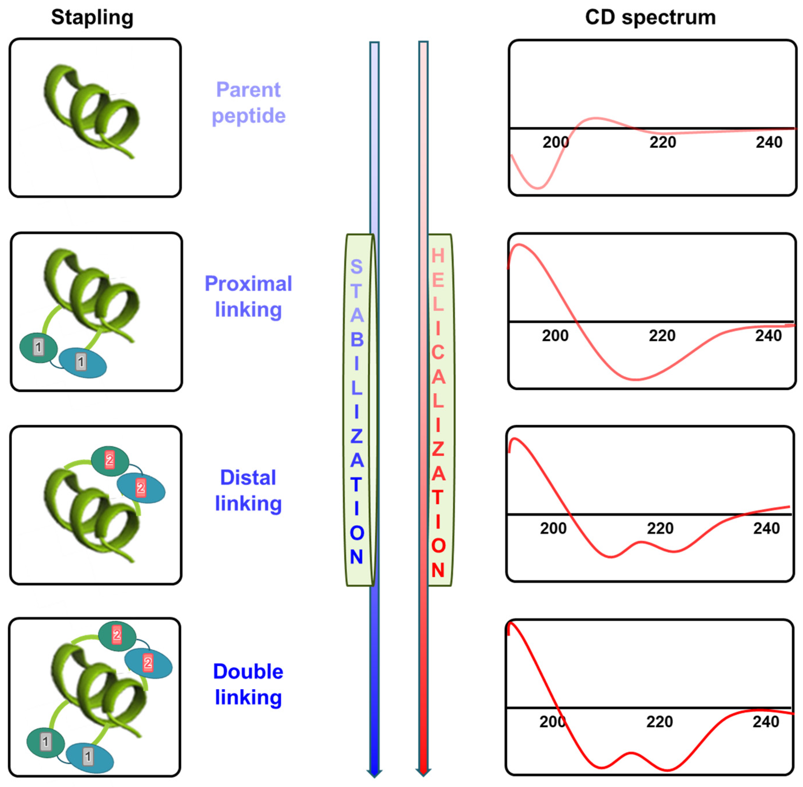
CD spectroscopy is primarily used to investigate structural changes in stapled peptides and estimate their helicity (Figure 4). Peptides and proteins exhibit distinct CD spectra based on their predominant secondary structure [51]. Peptides in an unstructured conformation show a strong minimum at 195 nm in the CD spectrum, while those in an α-helical conformation typically exhibit a strong positive peak at 190–195 nm and dual minima at 208 and 222 nm. Specifically, the peak at 222 nm is a hallmark feature of the α-helix structure. Therefore, when interpreting CD data, the greater the intensity of the positive peak at 190–195 nm and the negative peaks at 208 nm and 222 nm, the higher the quantitative degree of α-helix formation. For instance, after measuring the CD spectrum, the α-helical content can be quantified by comparing it with standard α-helix reference data, allowing for the estimation of the proportion of α-helical structure present. Successful stapling, especially when more distal regions are linked or multiple sites are stapled, often leads to significant increases in helicity due to α-helix stabilization [52]. This α-helicity can be quantified using simple software like CDNN [53].

6. Stability Confirmation Post-Synthesis

To assess the proteolytic stability of stapled peptides, trypsin degradation tests are commonly employed [45]. Trypsin is frequently used in these protease tests because it primarily cleaves at the carboxyl side of charged amino acids such as Lys and Arg [54]. If the results indicate that the stapled peptide exhibits greater stability to protease degradation than its native linear counterparts, the stapling is considered successful [55].

7. Conclusions

In drug discovery, proteins that engage in intracellular interactions with other proteins are widely regarded as highly biologically appealing targets. As the building blocks of life, proteins play a pivotal role in all aspects of cellular systems, regulating numerous physiological enzymatic activities through structural stabilization [56]. This is true not only for large proteins but also for small peptides with fewer than 50 amino acids, which perform critical enzymatic activities as antibacterial agents, hormones, and neurotransmitters essential to every living organism [57].
Stapling peptides combine the broad target recognition capabilities of protein therapeutics with robust cell-penetrating ability. The successful design and evaluation of potent stapled peptide interactions demonstrate that stapling can significantly enhance the pharmacologic performance of peptides [30]. This includes increasing target affinity, proteolytic resistance, and serum half-life, while also conferring high levels of cell penetration [45].
Since intracellular protein–protein interaction-derived stapled peptides represent a recent advancement, the successful implementation of stapled peptide technology requires meticulous observation of protein–protein interactions and the optimization of a multistep process. This involves varying the positions and number of staples to determine the optimal output [58].
Stapled peptides offer a novel therapeutic alternative capable of inhibiting the function of proteins, such as enzymes, that were previously difficult to target using classical small molecules. Theoretically, the emergence of an α-helical structure through stapling increases protease resistance by blocking the protease enzyme’s access to target sites on peptide chains, thereby enhancing peptide stability [59]. This ultimately leads to improved delivery success rates for peptide drugs.
Although practical examples are still limited, the growing number of reports on stapled peptides as potent and specific inhibitors of protein–protein interactions suggests that they could provide crucial information for drug development in the future.

Author Contributions

Conceptualization, D.-H.K. and S.-M.K.; software, D.-H.K. and S.-M.K.; validation, D.-H.K. and S.-M.K.; formal analysis, D.-H.K.; investigation, S.-M.K.; writing—original draft preparation, D.-H.K. and S.-M.K.; writing—review and editing, D.-H.K. and S.-M.K.; visualization, D.-H.K. and S.-M.K.; supervision, S.-M.K.; project administration, S.-M.K. All authors have read and agreed to the published version of the manuscript.

Funding

This research received no external funding.

Institutional Review Board Statement

Not applicable.

Informed Consent Statement

Not applicable.

Data Availability Statement

Data are contained within the article.

Conflicts of Interest

The authors declare no conflicts of interest.

References

  1. Sharma, S.; Chauhan, A.; Ranjan, A.; Mathkor, D.M.; Haque, S.; Ramniwas, S.; Tuli, H.S.; Jindal, T.; Yadav, V. Emerging challenges in antimicrobial resistance: Implications for pathogenic microorganisms, novel antibiotics, and their impact on sustainability. Front. Microbiol. 2024, 15, 1403168. [Google Scholar] [CrossRef] [PubMed]
  2. Belete, M.A.; Gedefie, A.; Alemayehu, E.; Debash, H.; Mohammed, O.; Gebretsadik, D.; Ebrahim, H.; Tilahun, M. The prevalence of vancomycin-resistant Staphylococcus aureus in Ethiopia: A systematic review and meta-analysis. Antimicrob. Resist. Infect. Control 2023, 12, 86. [Google Scholar] [CrossRef] [PubMed]
  3. Hull, R.C.; Wright, R.C.T.; Sayers, J.R.; Sutton, J.A.F.; Rzaska, J.; Foster, S.J.; Brockhurst, M.A.; Condliffe, A.M. Antibiotics Limit Adaptation of Drug-Resistant Staphylococcus aureus to Hypoxia. Antimicrob. Agents Chemother. 2022, 66, e0092622. [Google Scholar] [CrossRef] [PubMed]
  4. Li, M.; Li, J.; Li, J.; Zhang, J.; Zhao, Y.; Li, W.; Zhang, Y.; Hu, J.; Xie, X.; Zhang, D.; et al. Design, synthesis, and evaluation of novel pleuromutilin aryl acrylate derivatives as promising broad-spectrum antibiotics especially for combatting multi-drug resistant gram-negative bacteria. Eur. J. Med. Chem. 2023, 259, 115653. [Google Scholar] [CrossRef]
  5. Christensen, S.B. Drugs That Changed Society: History and Current Status of the Early Antibiotics: Salvarsan, Sulfonamides, and beta-Lactams. Molecules 2021, 26, 6057. [Google Scholar] [CrossRef]
  6. Thakuria, B.; Lahon, K. The Beta Lactam Antibiotics as an Empirical Therapy in a Developing Country: An Update on Their Current Status and Recommendations to Counter the Resistance against Them. J. Clin. Diagn. Res. 2013, 7, 1207–1214. [Google Scholar] [CrossRef]
  7. Kanannejad, Z.; Pourvali, A.; Esmaeilzadeh, H.; Shokouhi Shoormasti, R.; Reza Fazlollahi, M.; Fallahpour, M.; Zaremehrjardi, F. Diagnosis and selection of alternative antibiotics in beta-lactams hypersensitivity reactions: Current recommendations and challenges. Int. Immunopharmacol. 2023, 122, 110573. [Google Scholar] [CrossRef]
  8. de Araujo, A.C.J.; Freitas, P.R.; Araujo, I.M.; Siqueira, G.M.; de Oliveira Borges, J.A.; Alves, D.S.; Miranda, G.M.; Dos Santos Nascimento, I.J.; de Araujo-Junior, J.X.; da Silva-Junior, E.F.; et al. Potentiating-antibiotic activity and absorption, distribution, metabolism, excretion and toxicity properties (ADMET) analysis of synthetic thiadiazines against multi-drug resistant (MDR) strains. Fundam. Clin. Pharmacol. 2024, 38, 84–98. [Google Scholar] [CrossRef]
  9. Carballo, G.M.; Vazquez, K.G.; Garcia-Gonzalez, L.A.; Rio, G.D.; Brizuela, C.A. Embedded-AMP: A Multi-Thread Computational Method for the Systematic Identification of Antimicrobial Peptides Embedded in Proteome Sequences. Antibiotics 2023, 12, 139. [Google Scholar] [CrossRef]
  10. Morales-Martinez, A.; Bertrand, B.; Hernandez-Meza, J.M.; Garduno-Juarez, R.; Silva-Sanchez, J.; Munoz-Garay, C. Membrane fluidity, composition, and charge affect the activity and selectivity of the AMP ascaphin-8. Biophys. J. 2022, 121, 3034–3048. [Google Scholar] [CrossRef]
  11. Park, Y.; Hahm, K.S. Novel short AMP: Design and activity study. Protein Pept. Lett. 2012, 19, 652–656. [Google Scholar] [CrossRef] [PubMed]
  12. Caselli, L.; Kohler, S.; Schirone, D.; Humphreys, B.; Malmsten, M. Conformational control of antimicrobial peptide amphiphilicity: Consequences for boosting membrane interactions and antimicrobial effects of photocatalytic TiO(2) nanoparticles. Phys. Chem. Chem. Phys. 2024, 26, 16529–16539. [Google Scholar] [CrossRef] [PubMed]
  13. Lee, H.; Lee, S.; Lee, I.; Nam, H. AMP-BERT: Prediction of antimicrobial peptide function based on a BERT model. Protein Sci. 2023, 32, e4529. [Google Scholar] [CrossRef]
  14. Abdullah, S.J.; Mu, Y.; Bhattacharjya, S. Structures, Interactions and Activity of the N-Terminal Truncated Variants of Antimicrobial Peptide Thanatin. Antibiotics 2024, 13, 74. [Google Scholar] [CrossRef] [PubMed]
  15. Sancho-Vaello, E.; Francois, P.; Bonetti, E.J.; Lilie, H.; Finger, S.; Gil-Ortiz, F.; Gil-Carton, D.; Zeth, K. Structural remodeling and oligomerization of human cathelicidin on membranes suggest fibril-like structures as active species. Sci. Rep. 2017, 7, 15371. [Google Scholar] [CrossRef]
  16. Nielsen, J.E.; Alford, M.A.; Yung, D.B.Y.; Molchanova, N.; Fortkort, J.A.; Lin, J.S.; Diamond, G.; Hancock, R.E.W.; Jenssen, H.; Pletzer, D.; et al. Self-Assembly of Antimicrobial Peptoids Impacts Their Biological Effects on ESKAPE Bacterial Pathogens. ACS Infect. Dis. 2022, 8, 533–545. [Google Scholar] [CrossRef]
  17. Pirtskhalava, M.; Vishnepolsky, B.; Grigolava, M.; Managadze, G. Physicochemical Features and Peculiarities of Interaction of AMP with the Membrane. Pharmaceuticals 2021, 14, 471. [Google Scholar] [CrossRef]
  18. Wang, R.; Wang, T.; Zhuo, L.; Wei, J.; Fu, X.; Zou, Q.; Yao, X. Diff-AMP: Tailored designed antimicrobial peptide framework with all-in-one generation, identification, prediction and optimization. Brief. Bioinform. 2024, 25, bbae078. [Google Scholar] [CrossRef]
  19. Shabir, U.; Ali, S.; Magray, A.R.; Ganai, B.A.; Firdous, P.; Hassan, T.; Nazir, R. Fish antimicrobial peptides (AMP’s) as essential and promising molecular therapeutic agents: A review. Microb. Pathog. 2018, 114, 50–56. [Google Scholar] [CrossRef]
  20. Monteiro, C.; Fernandes, H.; Oliveira, D.; Vale, N.; Barbosa, M.; Gomes, P.; MC, L.M. AMP-Chitosan Coating with Bactericidal Activity in the Presence of Human Plasma Proteins. Molecules 2020, 25, 3046. [Google Scholar] [CrossRef]
  21. Kravchenko, S.V.; Domnin, P.A.; Grishin, S.Y.; Vershinin, N.A.; Gurina, E.V.; Zakharova, A.A.; Azev, V.N.; Mustaeva, L.G.; Gorbunova, E.Y.; Kobyakova, M.I.; et al. Enhancing the Antimicrobial Properties of Peptides through Cell-Penetrating Peptide Conjugation: A Comprehensive Assessment. Int. J. Mol. Sci. 2023, 24, 16723. [Google Scholar] [CrossRef] [PubMed]
  22. Zhang, O.L.; Niu, J.Y.; Yu, O.Y.; Mei, M.L.; Jakubovics, N.S.; Chu, C.H. Development of a Novel Peptide with Antimicrobial and Mineralising Properties for Caries Management. Pharmaceutics 2023, 15, 2560. [Google Scholar] [CrossRef]
  23. Soylemez, U.G.; Yousef, M.; Bakir-Gungor, B. Novel Antimicrobial Peptide Design Using Motif Match Score Representation. IEEE/ACM Trans. Comput. Biol. Bioinform. 2024, 1–12. [Google Scholar] [CrossRef] [PubMed]
  24. Chandarana, C.; Juwarwala, I.; Shetty, S.; Bose, A. Peptide Drugs: Current Status and Treatment for the Treatment of Various Diseases. Curr. Drug Res. Rev. 2024, 16, 381–394. [Google Scholar] [CrossRef] [PubMed]
  25. Nicze, M.; Borowka, M.; Dec, A.; Niemiec, A.; Buldak, L.; Okopien, B. The Current and Promising Oral Delivery Methods for Protein- and Peptide-Based Drugs. Int. J. Mol. Sci. 2024, 25, 815. [Google Scholar] [CrossRef] [PubMed]
  26. Guo, S.; Wang, J.; Wang, Q.; Wang, J.; Qin, S.; Li, W. Advances in peptide-based drug delivery systems. Heliyon 2024, 10, e26009. [Google Scholar] [CrossRef]
  27. Rizvi, S.F.A.; Zhang, H.; Fang, Q. Engineering peptide drug therapeutics through chemical conjugation and implication in clinics. Med. Res. Rev. 2024. [Google Scholar] [CrossRef]
  28. Moiola, M.; Memeo, M.G.; Quadrelli, P. Stapled Peptides-A Useful Improvement for Peptide-Based Drugs. Molecules 2019, 24, 3654. [Google Scholar] [CrossRef]
  29. Wang, C.; Zhang, W.; Xu, L.; Tu, J.; Su, S.; Li, Q.; Zhang, T.; Zheng, L.; Wang, H.; Zhuang, X.; et al. Discovery of a Double-Stapled Short Peptide as a Long-Acting HIV-1 Inactivator with Potential for Oral Bioavailability. J. Med. Chem. 2024, 67, 9991–10004. [Google Scholar] [CrossRef]
  30. Dongrui, Z.; Miyamoto, M.; Yokoo, H.; Demizu, Y. Innovative peptide architectures: Advancements in foldamers and stapled peptides for drug discovery. Expert. Opin. Drug Discov. 2024, 19, 699–723. [Google Scholar] [CrossRef]
  31. Holdbrook, D.A.; Marzinek, J.K.; Boncel, S.; Boags, A.; Tan, Y.S.; Huber, R.G.; Verma, C.S.; Bond, P.J. The nanotube express: Delivering a stapled peptide to the cell surface. J. Colloid. Interface Sci. 2021, 604, 670–679. [Google Scholar] [CrossRef] [PubMed]
  32. Anananuchatkul, T.; Chang, I.V.; Miki, T.; Tsutsumi, H.; Mihara, H. Construction of a Stapled alpha-Helix Peptide Library Displayed on Phage for the Screening of Galectin-3-Binding Peptide Ligands. ACS Omega 2020, 5, 5666–5674. [Google Scholar] [CrossRef]
  33. Cornillie, S.P.; Bruno, B.J.; Lim, C.S.; Cheatham, T.E., 3rd. Computational Modeling of Stapled Peptides toward a Treatment Strategy for CML and Broader Implications in the Design of Lengthy Peptide Therapeutics. J. Phys. Chem. B 2018, 122, 3864–3875. [Google Scholar] [CrossRef] [PubMed]
  34. Gallagher, E.E.; Menon, A.; Chmiel, A.F.; Deprey, K.; Kritzer, J.A.; Garner, A.L. A cell-penetrant lactam-stapled peptide for targeting eIF4E protein-protein interactions. Eur. J. Med. Chem. 2020, 205, 112655. [Google Scholar] [CrossRef]
  35. Ma, B.; Liu, D.; Zheng, M.; Wang, Z.; Zhang, D.; Jian, Y.; Ma, J.; Fan, Y.; Chen, Y.; Gao, Y.; et al. Development of a Double-Stapled Peptide Stabilizing Both alpha-Helix and beta-Sheet Structures for Degrading Transcription Factor AR-V7. JACS Au 2024, 4, 816–827. [Google Scholar] [CrossRef] [PubMed]
  36. Yang, F.; Yin, F.; Li, Z. Helical Stabilization of Peptide Macrocycles by Stapled Architectures. Methods Mol. Biol. 2022, 2371, 391–409. [Google Scholar] [CrossRef]
  37. Zhang, J.; Dong, S. In-Bridge Stereochemistry: A Determinant of Stapled Peptide Conformation and Activity. Chembiochem 2024, 25, e202300747. [Google Scholar] [CrossRef]
  38. Fathi, F.; Alizadeh, B.; Tabarzad, M.V.; Tabarzad, M. Important structural features of antimicrobial peptides towards specific activity: Trends in the development of efficient therapeutics. Bioorg. Chem. 2024, 149, 107524. [Google Scholar] [CrossRef]
  39. Dougherty, P.G.; Wen, J.; Pan, X.; Koley, A.; Ren, J.G.; Sahni, A.; Basu, R.; Salim, H.; Appiah Kubi, G.; Qian, Z.; et al. Enhancing the Cell Permeability of Stapled Peptides with a Cyclic Cell-Penetrating Peptide. J. Med. Chem. 2019, 62, 10098–10107. [Google Scholar] [CrossRef]
  40. Ito, T.; Hashimoto, W.; Ohoka, N.; Misawa, T.; Inoue, T.; Kawano, R.; Demizu, Y. Structure-Activity Relationship Study of Helix-Stabilized Antimicrobial Peptides Containing Nonproteinogenic Amino Acids. ACS Biomater. Sci. Eng. 2023, 9, 4654–4661. [Google Scholar] [CrossRef]
  41. Feurstein, C.; Meyer, V.; Jung, S. Structure-Activity Predictions From Computational Mining of Protein Databases to Assist Modular Design of Antimicrobial Peptides. Front. Microbiol. 2022, 13, 812903. [Google Scholar] [CrossRef]
  42. Hillman, R.A.; Nadraws, J.W.; Bertucci, M.A. The Hydrocarbon Staple & Beyond: Recent Advances Towards Stapled Peptide Therapeutics that Target Protein-Protein Interactions. Curr. Top. Med. Chem. 2018, 18, 611–624. [Google Scholar] [CrossRef]
  43. Tan, Y.S.; Lane, D.P.; Verma, C.S. Stapled peptide design: Principles and roles of computation. Drug Discov. Today 2016, 21, 1642–1653. [Google Scholar] [CrossRef] [PubMed]
  44. Zhen, B.; Geng, C.; Yang, Y.; Liang, H.; Jiang, Y.; Li, X.; Ye, G. Systematic alanine and stapling mutational analysis of antimicrobial peptide Chem-KVL. Bioorg. Med. Chem. Lett. 2024, 107, 129794. [Google Scholar] [CrossRef] [PubMed]
  45. You, Y.; Liu, H.; Zhu, Y.; Zheng, H. Rational design of stapled antimicrobial peptides. Amino Acids 2023, 55, 421–442. [Google Scholar] [CrossRef] [PubMed]
  46. Hirano, M.; Saito, C.; Goto, C.; Yokoo, H.; Kawano, R.; Misawa, T.; Demizu, Y. Rational Design of Helix-Stabilized Antimicrobial Peptide Foldamers Containing alpha,alpha-Disubstituted Amino Acids or Side-Chain Stapling. Chempluschem 2020, 85, 2731–2736. [Google Scholar] [CrossRef]
  47. Valiente, P.A.; Becerra, D.; Kim, P.M. A Method to Calculate the Relative Binding Free Energy Differences of alpha-Helical Stapled Peptides. J. Org. Chem. 2020, 85, 1644–1651. [Google Scholar] [CrossRef]
  48. Bittrich, S.; Segura, J.; Duarte, J.M.; Burley, S.K.; Rose, Y. RCSB protein Data Bank: Exploring protein 3D similarities via comprehensive structural alignments. Bioinformatics 2024, 40, btae370. [Google Scholar] [CrossRef]
  49. Kang, S.M. Focused Overview of Mycobacterium tuberculosis VapBC Toxin-Antitoxin Systems Regarding Their Structural and Functional Aspects: Including Insights on Biomimetic Peptides. Biomimetics 2023, 8, 412. [Google Scholar] [CrossRef]
  50. Tu, L.; Wang, D.; Li, Z. Design and Synthetic Strategies for Helical Peptides. Methods Mol. Biol. 2019, 2001, 107–131. [Google Scholar] [CrossRef]
  51. Makura, Y.; Ueda, A.; Kato, T.; Iyoshi, A.; Higuchi, M.; Doi, M.; Tanaka, M. X-ray Crystallographic Structure of alpha-Helical Peptide Stabilized by Hydrocarbon Stapling at i,i + 1 Positions. Int. J. Mol. Sci. 2021, 22, 5364. [Google Scholar] [CrossRef] [PubMed]
  52. McWhinnie, F.S.; Sepp, K.; Wilson, C.; Kunath, T.; Hupp, T.R.; Baker, T.S.; Houston, D.R.; Hulme, A.N. Mono-Substituted Hydrocarbon Diastereomer Combinations Reveal Stapled Peptides with High Structural Fidelity. Chemistry 2018, 24, 2094–2097. [Google Scholar] [CrossRef] [PubMed]
  53. Lighezan, L.; Georgieva, R.; Neagu, A. The secondary structure and the thermal unfolding parameters of the S-layer protein from Lactobacillus salivarius. Eur. Biophys. J. 2016, 45, 491–509. [Google Scholar] [CrossRef] [PubMed]
  54. Glibowicka, M.; He, S.; Deber, C.M. Enhanced proteolytic resistance of cationic antimicrobial peptides through lysine side chain analogs and cyclization. Biochem. Biophys. Res. Commun. 2022, 612, 105–109. [Google Scholar] [CrossRef]
  55. Grison, C.M.; Burslem, G.M.; Miles, J.A.; Pilsl, L.K.A.; Yeo, D.J.; Imani, Z.; Warriner, S.L.; Webb, M.E.; Wilson, A.J. Double quick, double click reversible peptide “stapling”. Chem. Sci. 2017, 8, 5166–5171. [Google Scholar] [CrossRef]
  56. Nero, T.L.; Parker, M.W.; Morton, C.J. Protein structure and computational drug discovery. Biochem. Soc. Trans. 2018, 46, 1367–1379. [Google Scholar] [CrossRef]
  57. Winter, A.; Higueruelo, A.P.; Marsh, M.; Sigurdardottir, A.; Pitt, W.R.; Blundell, T.L. Biophysical and computational fragment-based approaches to targeting protein-protein interactions: Applications in structure-guided drug discovery. Q. Rev. Biophys. 2012, 45, 383–426. [Google Scholar] [CrossRef]
  58. Zhang, Y.; Guo, J.; Cheng, J.; Zhang, Z.; Kang, F.; Wu, X.; Chu, Q. High-Throughput Screening of Stapled Helical Peptides in Drug Discovery. J. Med. Chem. 2023, 66, 95–106. [Google Scholar] [CrossRef]
  59. Al Musaimi, O.; Lombardi, L.; Williams, D.R.; Albericio, F. Strategies for Improving Peptide Stability and Delivery. Pharmaceuticals 2022, 15, 1283. [Google Scholar] [CrossRef]
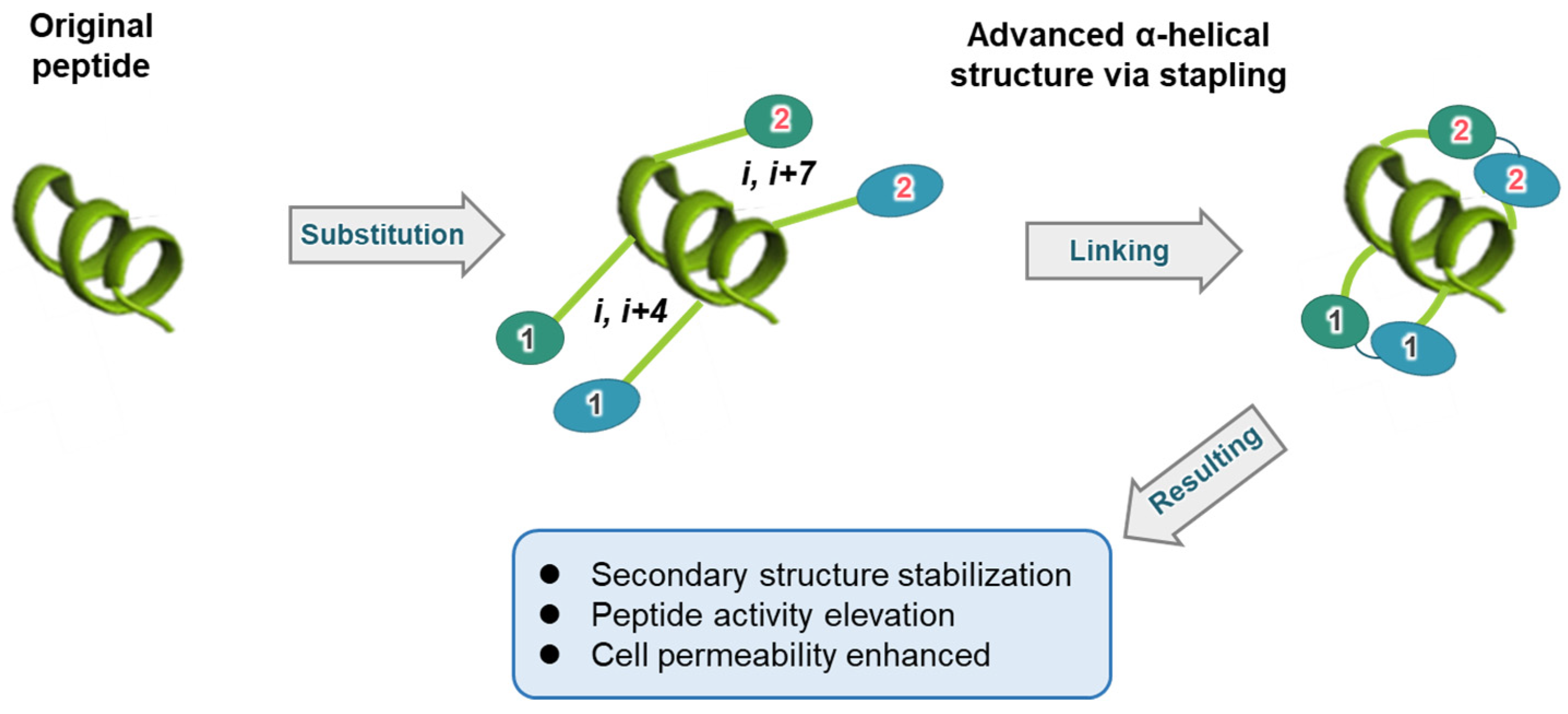
Figure 1. Schematic diagram of peptide helicalization using stapling. The process of the original peptide undergoing helicalization through stapling is depicted. Amino acid residues are denoted by the letter ‘i’, and the types of linkers are indicated using Arabic numerals, with the linked residues connected accordingly.
Biomimetics 09 00537 g001
Figure 2. An example of selecting parent peptide positions using protein structure information. Look at the two proteins in the figure, which bind to each other. One protein is depicted in a surface form, while the other is shown as a faint ribbon, revealing its secondary structure. Suppose we need to design a peptide that must bind to the protein displayed in surface form. In that case, Site 1 and Site 2, based on this protein–protein interaction, can serve as effective alternatives by utilizing the interface valley and cavity.
Biomimetics 09 00537 g002
Figure 3. Example of structure-based alanine scanning. For convenience, the proteins used in this description are labeled as A and B. As described in the main text, this process involves identifying stapling spots that maintain the α-helix structure while minimally affecting key interactions. Candidate peptides, in which amino acid residues are sequentially substituted with alanine, are ultimately selected based on the preservation of their activity compared to the parent peptide. This figure was created using the template structures 5X3T and 4XGQ from the Protein Data Bank [48].
Biomimetics 09 00537 g003
Figure 4. Diagram of stabilization and improved helicity through stapling. The diagram illustrates the stabilization and enhancement of helicity due to stapling. As stabilization progresses, the CD spectrum exhibits two distinct minima. This trend highlights the characteristic improvement of secondary structure clarity with repeated linking.
Biomimetics 09 00537 g004
Disclaimer/Publisher’s Note: The statements, opinions and data contained in all publications are solely those of the individual author(s) and contributor(s) and not of MDPI and/or the editor(s). MDPI and/or the editor(s) disclaim responsibility for any injury to people or property resulting from any ideas, methods, instructions or products referred to in the content.