Review

Current Applications and Future Perspectives of Artificial Intelligence in Face-Driven Orthodontics: A Scoping Review


by
Barbora Heribanová
1,*,
Katarína Janáková
1,
Juraj Tomášik
1,
Daniela Tichá
1,
Štefan Harsányi
2 and
Andrej Thurzo
1,*
1
Department of Orthodontics, Regenerative and Forensic Dentistry, Faculty of Medicine and KORFS, Comenius University in Bratislava, Dvorakovo Nabrezie 4, 81102 Bratislava, Slovakia
2
Institute of Medical Biology, Genetics and Clinical Genetics, Faculty of Medicine, Comenius University in Bratislava, Sasinkova 4, 81108 Bratislava, Slovakia
*
Authors to whom correspondence should be addressed.
Biomimetics 2026, 11(2), 146; https://doi.org/10.3390/biomimetics11020146
Submission received: 30 December 2025 / Revised: 4 February 2026 / Accepted: 13 February 2026 / Published: 16 February 2026
(This article belongs to the Special Issue Dentistry and Craniofacial District: The Role of Biomimetics 2026)

Abstract

Artificial Intelligence (AI) has introduced transformative possibilities in orthodontics by enhancing diagnostic precision, treatment planning, and aesthetic outcomes. In face-driven orthodontics, treatment objectives extend beyond achieving proper occlusion to optimizing facial balance and harmony. With the growing patient demand for aesthetic improvements, AI technologies enable clinicians to integrate facial analysis and dynamic soft-tissue evaluation into personalized treatment approaches. Research in this scoping review analyzed current applications of AI in face-driven orthodontics, focusing on diagnosis, soft-tissue assessment, and individualized treatment planning. A comprehensive search was conducted in PubMed and Scopus for studies published between 2021 and 2025. The review followed the PRISMA-ScR guidelines. Of 54 initially identified studies, 24 met the inclusion criteria after title, abstract, and full-text screening. Extracted data were organized according to the main application areas of AI in face-driven orthodontics. Most studies focused on AI-assisted facial analysis, 3D reconstruction, and treatment simulation. Deep learning models demonstrated high performance in soft-tissue prediction, aesthetic evaluation, and diagnostic accuracy. However, heterogeneity in datasets, a lack of standardized validation protocols, limited external validation across included studies and limited clinical applicability were identified as key limitations. AI-based facial analysis supports a shift toward individualized, aesthetics-oriented orthodontic planning. Although current evidence highlights its potential for improving diagnostic precision and treatment outcomes, further validation through large-scale clinical studies is essential for broader implementation in everyday practice.

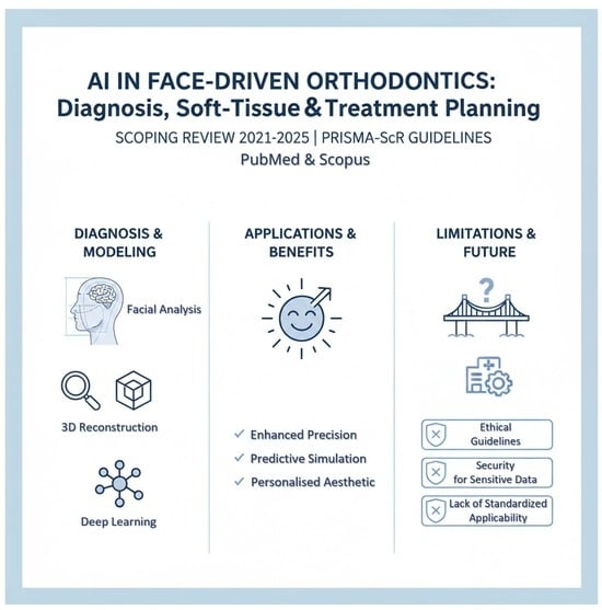
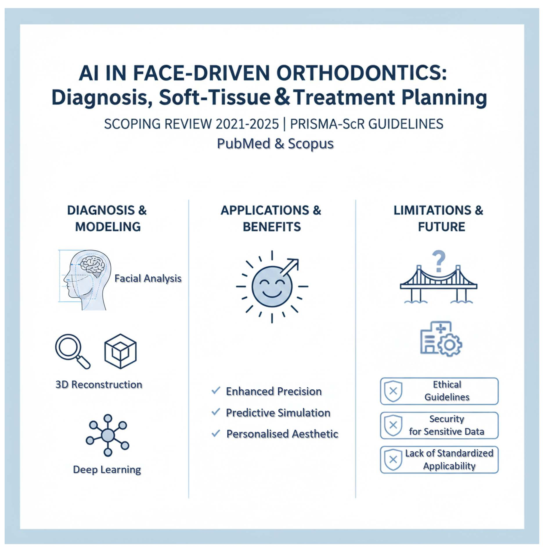
Graphical Abstract

1. Introduction

Artificial Intelligence (AI) has rapidly evolved into a transformative force across medicine and dentistry, driving innovation in diagnosis, treatment planning, and personalized care. The aim of this scoping review is to explore current applications of Artificial Intelligence in face-driven orthodontics, focusing on diagnosis, treatment planning, and facial aesthetic outcomes in clinical practice. The aim is also to answer the question: What applications of AI are used in face-driven orthodontics and what is the use in orthodontic diagnosis and treatment planning? In addition, this review outlines the existing limitations of AI use in clinical orthodontics.
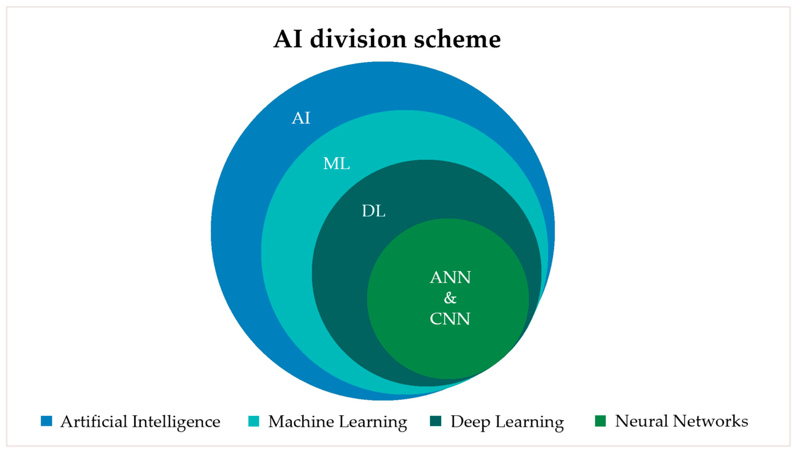
There have been significant advances in the application of Artificial Intelligence (AI) technologies. Machine learning (ML) is a branch of Artificial Intelligence that enables computer systems to learn from data and improve their performance on specific tasks without being explicitly programmed. Deep learning (DL) is a subset of machine learning that employs multi-layered artificial neural networks to automatically identify complex patterns within large datasets. The AI division scheme is shown in Figure 1. While traditional machine learning often relies on manually engineered features, deep learning models are capable of autonomously learning hierarchical representations directly from raw data. Deep learning includes Artificial Neural Networks (ANN) and Convolutional Neural Networks (CNN). Artificial Neural Networks (ANNs) are typically composed of fully connected (dense) layers. Convolutional Neural Networks (CNNs) incorporate convolutional layers for feature extraction, pooling layers for hierarchical spatial abstraction, and often conclude with dense layers to perform tasks such as classification or regression [1,2]. These advances have made AI-based systems powerful analytical tools in various areas of medicine, paving the way for their clinical translation in orthodontics and craniofacial research. Within orthodontics, a growing focus has been placed on aligning functional correction with overall facial harmony, giving rise to the concept of face-driven orthodontics [2].
Face-driven orthodontics focuses on matching proper occlusion with overall facial harmony [2]. It emphasizes the integration of attractiveness considerations into treatment planning, ensuring that orthodontic outcomes are consistent with the patient’s expectations for improved facial attractiveness [3]. Linking Artificial Intelligence to this approach increases diagnostic accuracy, personalizes treatment planning, and optimizes clinical procedures, thereby improving overall patient satisfaction and treatment outcomes [4,5,6]. Consequently, the combination of AI and face-driven orthodontics represents a paradigm shift from conventional occlusal-based treatment planning toward a holistic, patient-centered, and data-driven aesthetic analysis. Unlike traditional orthodontics, which primarily emphasizes dental alignment and skeletal relationships, face-driven approaches prioritize facial soft-tissue morphology and prediction, dynamic facial expressions, and patient-specific aesthetic goals as primary diagnostic and therapeutic determinants. Therefore, methodological differences include the integration of facial analysis, soft-tissue prediction, and outcome simulation into the diagnostic workflow, shifting clinical priorities toward individualized aesthetic optimization alongside functional stability.
As technological capabilities continue to expand, AI is expected to play an increasingly critical role in optimizing orthodontic workflows and improving aesthetic outcomes. As the field of orthodontics continues to advance, ongoing research and development of AI technologies will play a key role in addressing these challenges while fostering innovations that combine both function and beauty [7,8,9,10,11,12]. This review therefore aims to provide a comprehensive overview of current AI applications in face-driven orthodontics, identify existing limitations, and highlight potential directions for future research and clinical implementation.
From a biomimetics perspective, artificial intelligence represents a computational paradigm inspired by human cognitive and perceptual processes, particularly those of the brain and visual system. Machine learning and deep learning models emulate biological mechanisms such as neural signal processing, pattern recognition, learning from experience, and adaptive decision-making, which are fundamental to human facial perception and aesthetic judgment. In face-driven orthodontics, AI systems are trained to replicate expert-level clinical reasoning by integrating complex facial cues, soft-tissue morphology, and dynamic expressions into diagnostic and treatment-planning workflows. This biologically inspired modeling enables the simulation of human aesthetic assessment and predictive adaptation to individual facial characteristics, thereby aligning orthodontic decision-making with principles of biomimetic design. Consequently, AI-driven facial analysis and outcome prediction can be regarded as a form of digital biomimetics, in which computational models mimic human biological intelligence to support personalized, functionally stable, and aesthetically harmonious craniofacial treatment planning.

2. Materials and Methods

The studies included in this scoping review focused on the application of Artificial Intelligence technologies in face-driven orthodontic treatment. The review was conducted in accordance with the PRISMA-ScR guidelines, as confirmed by the completed checklist and flow diagram.
Search strategy
Relevant studies were retrieved from the PubMed and Scopus databases. The search was carried out on 1 February 2025 at 05:20 pm and included articles published between 2021 and 2025. The search terms included the keywords “orthodontics”, “face-driven”, “soft tissue”, “Artificial Intelligence”, and “AI”. Database-specific search strategies were applied: in PubMed, the search was conducted using Title/Abstract fields and relevant MeSH terms, with filters applied for publication years (2021–2025) and language (English); in Scopus, the search was performed using TITLE-ABS-KEY fields, with year and language limitations applied as stated in the inclusion criteria. The search query was as follows:
For PubMed:
(“Orthodontics”[MeSH] OR orthodontic*[tiab]) AND (face-driven[tiab] OR “soft tissue”[tiab]) AND (“Artificial Intelligence”[MeSH] OR artificial intelligence[tiab])
For Scopus:
TITLE-ABS-KEY (orthodontics OR orthodontic) AND TITLE-ABS-KEY
(“face-driven” OR “soft tissue” OR facial OR face) AND TITLE-ABS-KEY (“artificial intelligence” OR “AI”)
Study design
Inclusion criteria were: original research papers, reviews and comparative studies written in English language and published between 2021–2025. Only studies involving orthodontic patients or human facial data relevant to orthodontic diagnosis or treatment, studies describing, developing or validating AI-based models applied to facial analysis, soft-tissue evaluation, 3D facial reconstruction, aesthetic outcome evaluation or treatment simulation within face-driven orthodontics were included in the search.
Exclusion criteria included: studies published outside of the time frame 2021–2025 and written in a language other than English. Studies other than reviews and research papers, and not related to orthodontics or not involving facial or soft-tissue analysis were excluded from the search. Studies describing AI applications unrelated to diagnosis, treatment planning, or aesthetic evaluation (e.g., administrative or non-clinical applications), and studies lacking a clear description of an AI-based model or algorithm were also excluded from the search.
Initial screening was performed by reviewing titles and abstracts to identify potentially eligible studies. The research was performed at the same time jointly by two reviewers. There were no significant disagreements, everything was resolved through discussion and consensus, as the inclusion criteria clearly indicated the suitability of the selected articles, which was also agreed upon by both independent reviewers conducting the research. Based on the content of the included studies, focal thematic areas were identified and used to structure data extraction and synthesis. List of the variables extracted includes:
-
type of AI model
-
clinical usability
-
data modality
Research question
The research question was formulated using the PCC model:
Population (P) = orthodontic patients
Concept (C) = AI models based on Artificial Intelligence in face-driven orthodontics
Context (C) = clinical application and research
Research question: What applications of Artificial Intelligence are used in face-driven orthodontics and what is the use in orthodontic diagnosis and treatment planning?

3. Results

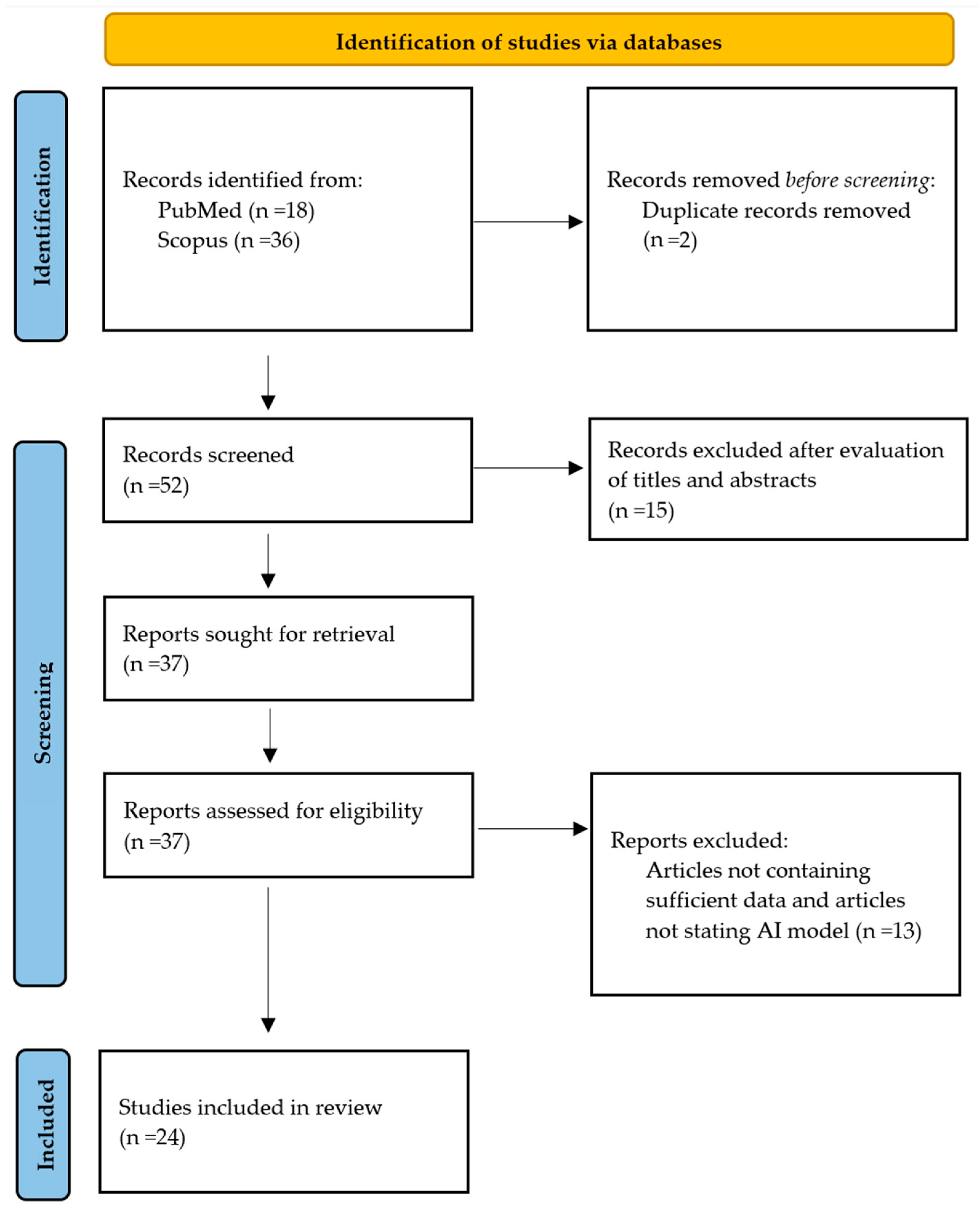
After an initial search, 54 potentially relevant studies were identified. After removing duplicate records, 52 studies remained. Based on the title and abstract screening, 15 studies were excluded due to non-compliance with inclusion criteria. At the full-text review stage, 13 studies were excluded due to lack of orthodontic relevance, absence of facial or soft-tissue analysis, focus on non-clinical AI applications, or insufficient description of an AI-based diagnostic or treatment-planning model, were not written in English language or published between 2021 and 2025. The remaining 24 studies underwent assessment and met all predefined eligibility criteria. This scoping review was conducted in accordance with the PRISMA-ScR (Preferred Reporting Items for Systematic Reviews and Meta-Analyses extension for Scoping Reviews) checklist, as shown in Figure 2.
In total, 24 studies were subjected to full-text assessment when available and were included in the final review in Table 1, organized according to their primary application areas within face-driven orthodontics.
A simplified representation of the studies according to their use in clinical practice and type of AI model is shown in Table 2.
Within the diagnostic domain, included studies addressed two-dimensional (2D) facial analysis, three-dimensional (3D) facial symmetry and asymmetry assessment, and the diagnosis of facial dysmorphology. These investigations primarily relied on 2D facial photographs, 3D facial scans, and cone-beam computed tomography (CBCT) imaging data.
Within the landmark identification domain, studies focused on automated facial landmark detection, quantitative soft-tissue analysis, identification of skeletal abnormalities, automatic cephalometric analysis, and three-dimensional template-based determination of cephalometric landmarks. The datasets used in these studies consisted mainly of 2D facial photographs, lateral cephalograms, and CBCT images.
Within the treatment planning domain, included studies examined the prediction of lateral facial profile changes following orthodontic treatment, feasibility analyses of intelligent predictive models, prediction of extraction versus non-extraction treatment decisions, digital 3D smile design, evaluation of pre- and post-orthognathic surgical changes, visualization tools for predicting soft-tissue outcomes after orthognathic surgery, assessment of facial soft-tissue changes following bimaxillary orthognathic surgery in patients with cleft lip and palate, prediction of soft-tissue and alveolar bone changes after orthodontic treatment, and comparisons of facial growth prediction models. These studies utilized a wide range of imaging modalities, including lateral cephalograms, 2D facial photographs, 3D facial scans, four-dimensional (4D) video recordings, and CBCT images.
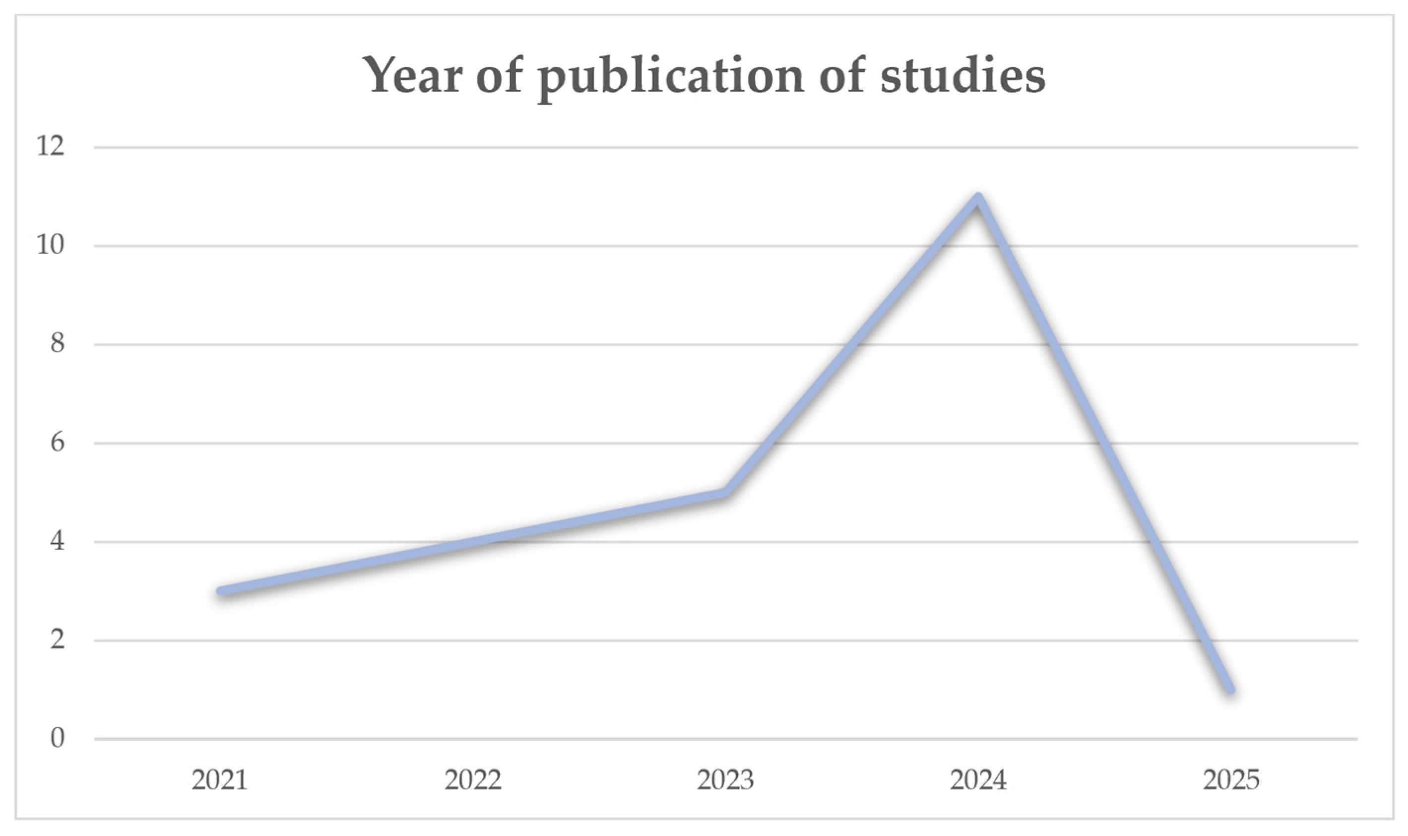
Each year the number of publications on the topic increases, which is represented by Figure 3, but as the review is conducted at the beginning of 2025, there has been only one study this year that has met the inclusion criteria so far. That is the reason for the decrease in the graph.
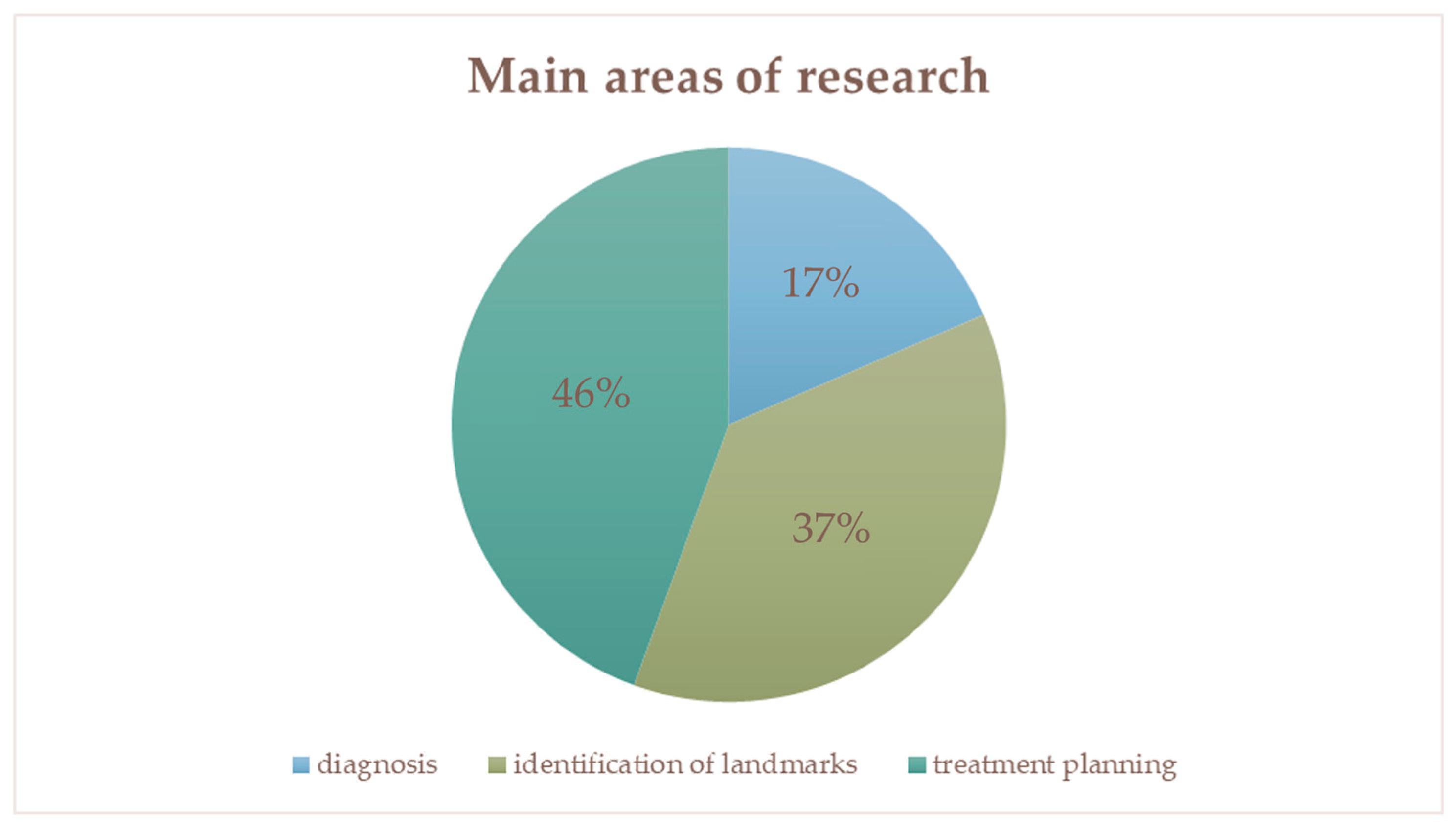
Figure 4 represents the distribution of studies by main area of research–diagnosis, identification of landmarks and treatment planning.
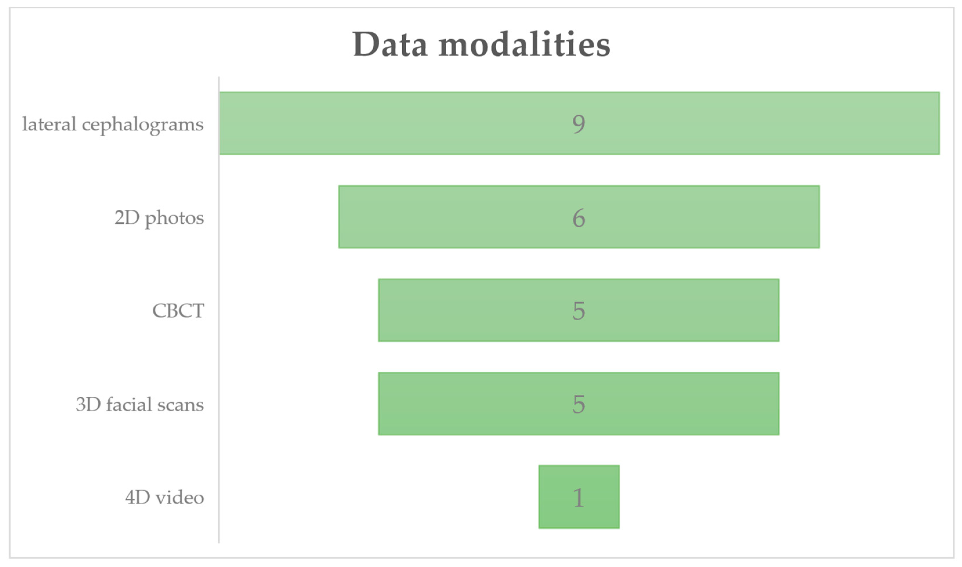
Figure 5 summarizes the distribution of the included studies based on imaging data utilized, encompassing 2D facial photographs, 3D facial scans, 4D dynamic video recordings, lateral cephalograms and CBCT imaging.

4. Discussion

Artificial Intelligence (AI) in face-driven orthodontics represents a significant advancement, emphasizing the interplay of harmonious outcomes and treatment efficacy. Aesthetics in dentistry has recently become a motivation for patients and often serves as the main reason for seeking out a specialist. Its importance is increasingly recognized in orthodontics, with studies showing that patients prioritize improved appearance when making treatment decisions. The aim of this scoping review was to explore current applications of Artificial Intelligence in face-driven orthodontics, focusing on diagnosis, treatment planning, and facial aesthetic outcomes in clinical practice.
Recent advances in Artificial Intelligence have enabled the processing of large volumes of imaging and clinical data with increasing precision, facilitating the integration of facial aesthetics into orthodontic diagnosis and treatment planning. The findings of this scoping review indicate that AI applications in face-driven orthodontics cluster primarily into three domains: treatment planning and outcome prediction, facial landmark detection and soft-tissue analysis, and automated diagnosis. Among these, treatment planning and outcome prediction emerged as the most extensively explored application, reflecting growing clinical interest in forecasting facial aesthetic changes and supporting goal-oriented orthodontic decision-making [5,37]. Across studies, AI-based landmark detection applied to lateral cephalograms, CBCT scans, 2D photographs and 3D facial scans demonstrated accuracy and precision comparable to expert clinicians, offering substantial gains in efficiency and reproducibility. However, common limitations that could affect the resulting performance could be different measurement methods, landmark definition variability, head posture standardization, and inter-device variability [38,39,40,41]. Machine learning and deep learning approaches, including artificial neural networks and convolutional neural networks, dominated this field, with some studies employing gradient boosting algorithms for structured datasets. While these models generally reported high performance, most relied on internal validation strategies, and external validation remained limited [42].
The studies suggest that intelligent diagnostics perform extremely well, with accuracy and precision similar to that of trained experts. Automation reduces the possibility of human error and enables the analysis of large data sets. These systems can simplify tasks and provide results quickly, which can save dentists time and help them perform their duties more efficiently [42,43]. The model of intelligent diagnosis methods may become increasingly accurate as data accumulates in the near future [6].
Artificial Intelligence can record facial features, enabling a comprehensive perception of the patient that provides additional information for treatment strategies [44]. The more the analysis of the face as a whole is taken into account, the more orthodontists will realize the importance of planning the ideal smile design from the beginning of treatment [12]. Artificial Intelligence technologies are being used to perform facial analyses that go beyond the simple assessment of proportionality and symmetry. These tools can identify facial characteristics that are essential for the development of customized treatment plans.
Harmonic results are most effectively assessed using complex three-dimensional (3D) analyses of facial structures that take into account image depth, different layers such as bone, muscle, fat tissue, and skin. This systematic approach allows professionals to select the optimal methods to improve facial appearance in orthodontic therapy. The creation of precise 3D models, for example, through facial scans, supports clinical decision-making so that the chosen treatment takes into account the individual patient’s facial proportions and meets their requirements about visual appearance [37,45].
Although this approach has several advantages over traditional manual procedures, its implementation in clinical practice is still limited, probably due to two main reasons: lack of technical expertise and the high costs associated with the equipment [46]. However, it is now possible to record a facial scan using a mobile phone or tablet [39,47,48].
In the recent past, the best practice was to use AI algorithms in cephalometric analysis, but the need to double-check the outcome data manually is still present [5]. A deep learning-based automatic soft tissue analysis model performs landmark detection and measurement calculations on, for example, orthodontic facial photographs, to achieve a more comprehensive quantitative soft tissue assessment. The system can automatically detect 43 landmarks on frontal images and 17 landmarks on lateral images of the face. The models can assist maxillofacial orthopedists in efficient and accurate quantitative soft tissue assessment in clinical practice. Between model prediction and manual measurements, there was no statistically significant difference [26]. The soft tissue profile of the face provides only limited information and is not sufficient by itself to thoroughly plan the final appearance of a smile customized to a given face [39].
Facial symmetry is increasingly important in today’s orthodontic treatment. However, the boundary of asymmetry is not clearly defined. Stereophotogrammetry has a distinct advantage in measuring facial asymmetry. Facial asymmetry can be quantified using three-dimensional technology, and it is also possible to investigate whether conventional assessment of facial asymmetry agrees with analysis using 3D technology, which has been reported to be reliable. By measuring the original face and its mirror image, it is possible to compare the correspondence of the surface of the individual regions, as well as the whole face, to calculate the degree of symmetry. Significant diagnostic values are in the area of the lips, chin, lateral parts of the mandible and cheeks. The most significant influence on facial symmetry has the mandible [42,49].
Video recordings of a patient talking and smiling can provide valuable information about the visual identity of the conversation and the dynamics of the smile that traditional still images do not provide [50]. Four-dimensional recordings are the basis for motion-simulating designs that overcome many limitations. However, it should be noted that this AI virtual smile design can sometimes be unrealistic and unattainable due to overlooking skeletal relationships, occlusion, and the shape of dental arches. Nevertheless, the use of new modern digital tools contributes to their updating and progressive increase in reliability [51,52,53]. By examining facial proportions and linear distances between areas, AI tools can provide insights that were previously difficult to quantify due to subjective assessment. These include assessments of facial proportions based on standard aesthetic models such as the golden ratio or Marquardt’s mask, although their generalizability remains limited [39]. This analytical ability also allows orthodontists to predict the need for tooth extraction or surgery. It is important to take into account the unique anatomy of an individual’s dentition to reflect the patient’s individual beauty, thereby providing a more holistic approach to orthodontic care [5,6,37].
A study revealed that the AI-enhanced images were perceived as more attractive, compared to the original real photographs, underscoring the effectiveness of AI in meeting aesthetic requirements. In addition, the use of AI tools allows that specific adjustments can be made based on individual patient preferences such as lip fullness and eye size, contributing to higher satisfaction rates [12,37]. AI also facilitates remote monitoring of patient‘s progress, improving adherence to the treatment plan and allowing for early interventions if necessary [52,54].
Artificial Intelligence has the potential to substantially advance face-driven orthodontics by enhancing diagnostic precision, treatment planning, and aesthetic outcome prediction. However, the ethical implications of AI implementation are particularly pronounced in this field due to the use of facial images and three- and four-dimensional facial data, which constitute highly sensitive biometric information [3,55,56].
Responsible integration of AI into orthodontic workflows requires clear governance frameworks addressing informed consent, secondary data use, secure data storage, and accountability for AI-assisted decisions [55]. Ethical oversight should be operationalized through human-in-the-loop models, ensuring that qualified orthodontists remain responsible for validating AI-generated outputs before clinical application [6,57,58,59].

5. Conclusions

Artificial Intelligence systems are becoming increasingly adept at simulating tooth movement and changes in the surrounding soft tissues. Such a simulation, or illustrative virtual reality, helps to improve understanding, motivation and, thus, patient involvement in the treatment process. Future developments may allow for real-time adjustments based on patient progress, optimizing treatment sequencing and potentially reducing the overall treatment duration [6,60]. Properly executed treatment achieves harmony between soft and hard tissues [50,60,61]. Several studies still undergo internal validation.
AI in face-driven orthodontics shows significant potential in enhancing diagnostic precision and aesthetic treatment outcomes, yet it must remain a tool under human clinical supervision [60,62].
Despite these advances, the integration of AI into face-driven orthodontics raises ethical considerations and challenges, particularly regarding the oversight of AI applications in patient care [55,56,63]. Also important is the need for appropriate communication among orthodontists to ensure a cohesive approach to treatment planning in which technology and patient-centered care are seamlessly integrated [54,64,65].
Despite the advances made, current AI technologies still have limitations that cannot be overlooked. Although AI can help significantly, it lacks the nuanced critical and emotional intelligence inherent in human clinicians. This limitation underscores the fact that AI cannot completely replace the expertise and judgment of experienced doctors, but clinicians who effectively integrate AI tools are likely to outperform those who do not adapt to digital advances [6,62,66,67].

Author Contributions

Conceptualization, B.H. and A.T.; methodology, B.H. and J.T.; literature search and data curation, B.H. and D.T.; formal analysis, B.H.; writing—original draft preparation, B.H.; writing—review and editing, J.T., D.T., K.J. and Š.H.; supervision, A.T. All authors have read and agreed to the published version of the manuscript.

Funding

The article was supported and co-financed by the European Union within the Programme Slovakia, project: Simulation and Training Center for General Medicine and Dentistry CU, 401101B343, and also by the Office of the Deputy Prime Minister of Slovakia, grant number Industrial Research and Experimental Development Projects in the Field of Biotechnologies PK 1/2025–ScaniFy.

Institutional Review Board Statement

Not applicable.

Informed Consent Statement

Not applicable.

Data Availability Statement

No new data was created or analysed in this study. Data sharing is not applicable to this article.

Acknowledgments

During the preparation of this manuscript, the authors used generative AI tools for language refinement and stylistic editing. The authors reviewed and edited the output and take full responsibility for the content of this publication.

Conflicts of Interest

The authors declare no conflicts of interest. The funders had no role in the design of the study; in the collection, analyses, or interpretation of data; in the writing of the manuscript; or in the decision to publish the results.

Abbreviations

The following abbreviations are used in this manuscript:
AIArtificial Intelligence
MLMachine learning
DLDeep learning
ANNArtificial neural networks
CNNConvolutional neural networks
GBGradient boosting
3DThree-dimensional
PCCPopulation, Concept, Context

References

  1. Kulikowski, C.A. An Opening Chapter of the First Generation of Artificial Intelligence in Medicine: The First Rutgers AIM Workshop, June 1975. Yearb. Med. Inform. 2015, 10, 227–233. [Google Scholar] [CrossRef]
  2. Bichu, Y.M.; Hansa, I.; Bichu, A.Y.; Premjani, P.; Flores-Mir, C.; Vaid, N.R. Applications of Artificial Intelligence and Machine Learning in Orthodontics: A Scoping Review. Prog. Orthod. 2021, 22, 18. [Google Scholar] [CrossRef]
  3. Corfmat, M.; Martineau, J.T.; Régis, C. High-Reward, High-Risk Technologies? An Ethical and Legal Account of AI Development in Healthcare. BMC Med. Ethics 2025, 26, 4. [Google Scholar] [CrossRef]
  4. Thomas, M. Orthodontics in the “Art” of Aesthetics. Int. J. Orthod. Milwaukee 2015, 26, 23–28. [Google Scholar]
  5. Tomášik, J.; Zsoldos, M.; Oravcová, Ľ.; Lifková, M.; Pavleová, G.; Strunga, M.; Thurzo, A. AI and Face-Driven Orthodontics: A Scoping Review of Digital Advances in Diagnosis and Treatment Planning. AI 2024, 5, 158–176. [Google Scholar] [CrossRef]
  6. Thakur, S.M.; Shenoy, U.; Hazare, A.; Karia, H.; Khorgade, P.; Nandeshwar, N.; Bhattacharya, S.; Thakur, S.M.; Shenoy, U.; Hazare, A.; et al. Transforming Orthodontics with Artificial Intelligence: A Comprehensive Review. J. Adv. Dent. Pract. Res. 2024, 3, 56–64. [Google Scholar] [CrossRef]
  7. AI In Orthodontics: Leveraging Technology for Improved Orthodontic Outcomes-Lean Orthodontics®. Available online: https://leanorthodontics.com/en/blog/ai-in-orthodontics-leveraging-technology-for-improved-orthodontic-outcomes/ (accessed on 6 February 2025).
  8. Du, W.; Bi, W.; Liu, Y.; Zhu, Z.; Tai, Y.; Luo, E. Machine Learning-Based Decision Support System for Orthognathic Diagnosis and Treatment Planning. BMC Oral Health 2024, 24, 286. [Google Scholar] [CrossRef]
  9. Jheon, A.H.; Oberoi, S.; Solem, R.C.; Kapila, S. Moving towards Precision Orthodontics: An Evolving Paradigm Shift in the Planning and Delivery of Customized Orthodontic Therapy. Orthod. Craniofac. Res. 2017, 20, 106–113. [Google Scholar] [CrossRef] [PubMed]
  10. Kapila, S.; Vora, S.R.; Rengasamy Venugopalan, S.; Elnagar, M.H.; Akyalcin, S. Connecting the Dots towards Precision Orthodontics. Orthod. Craniofac. Res. 2023, 26, 8–19. [Google Scholar] [CrossRef] [PubMed]
  11. Kato, R.M.; Parizotto, J.d.O.L.; Oliveira, P.H.J.d.; Gonçalves, J.R. Artificial Intelligence in Orthognathic Surgery—A Narrative Review of Surgical Digital Tools and 3D Orthognathic Surgical Planning. J. Calif. Dent. Assoc. 2023, 51, 1. [Google Scholar] [CrossRef]
  12. Facially Driven Interdisciplinary Integrated Digital Orthodontics|Compendium. Available online: https://compendiumlive.com/2021/11/facially-driven-interdisciplinary-integrated-digital-orthodontics (accessed on 8 February 2025).
  13. Rousseau, M.; Retrouvey, J.M. Machine Learning in Orthodontics: Automated Facial Analysis of Vertical Dimension for Increased Precision and Efficiency. Am. J. Orthod. Dentofac. Orthop. 2022, 161, 445–450. [Google Scholar] [CrossRef]
  14. Cai, J.; Deng, Y.; Min, Z.; Zhang, Y.; Zhao, Z.; Jing, D. Revealing the Representative Facial Traits of Different Sagittal Skeletal Types: Decipher What Artificial Intelligence Can See by Grad-CAM. J. Dent. 2023, 138, 104701. [Google Scholar] [CrossRef]
  15. Gong, B.; Chang, Q.; Shi, T.; Wang, S.; Wang, Y.; Zuo, F.; Xie, X.; Bai, Y. Research of Orthodontic Soft Tissue Profile Prediction Based on Conditional Generative Adversarial Networks. J. Dent. 2025, 154, 105570. [Google Scholar] [CrossRef]
  16. Ma, Y.; Li, Y.; Liu, X.; Gao, J.; Wang, A.; Chen, H.; Liu, Z.; Jin, Z. Future Perspectives of Digital Twin Technology in Orthodontics. Displays 2024, 85, 102818. [Google Scholar] [CrossRef]
  17. Zhu, Y.; Zhang, L.; Liu, S.; Wen, A.; Gao, Z.; Qin, Q.; Gao, L.; Zhao, Y.; Wang, Y. Automatic Three-Dimensional Facial Symmetry Reference Plane Construction Based on Facial Planar Reflective Symmetry Net. J. Dent. 2024, 147, 105043. [Google Scholar] [CrossRef]
  18. Mason, T.; Kelly, K.M.; Eckert, G.; Dean, J.A.; Dundar, M.M.; Turkkahraman, H. A Machine Learning Model for Orthodontic Extraction/Non-Extraction Decision in a Racially and Ethnically Diverse Patient Population. Int. Orthod. 2023, 21, 100759. [Google Scholar] [CrossRef]
  19. Lyu, L.; Zhang, M.J.; Wen, A.N.; Wang, S.; Zhao, Y.J.; Yong, W.; Yu, T.T.; Liu, D. 3D Facial Mask for Facial Asymmetry Diagnosis. Heliyon 2024, 10, e26734. [Google Scholar] [CrossRef]
  20. Zilio, C.; Tel, A.; Perrotti, G.; Testori, T.; Sembronio, S.; Robiony, M. Validation of ‘Total Face Approach’ (TFA) Three-Dimensional Cephalometry for the Diagnosis of Dentofacial Dysmorphisms and Correlation with Clinical Diagnosis. Int. J. Oral Maxillofac. Surg. 2024, 54, 420–429. [Google Scholar] [CrossRef] [PubMed]
  21. Alikhasi, M.; Yousefi, P.; Afrashtehfar, K.I. Smile Design: Mechanical Considerations. Dent. Clin. N. Am. 2022, 66, 477–487. [Google Scholar] [CrossRef]
  22. Ahn, J.; Nguyen, T.P.; Kim, Y.J.; Kim, T.; Yoon, J. Automated Analysis of Three-Dimensional CBCT Images Taken in Natural Head Position That Combines Facial Profile Processing and Multiple Deep-Learning Models. Comput. Methods Programs Biomed. 2022, 226, 107123. [Google Scholar] [CrossRef] [PubMed]
  23. Duangsuwan, J.; Raocharernporn, S.; Thiradilok, S.; Manopatanakul, S. Computerized Three-Dimensional Cephalometric Template for Thai Adults. Heliyon 2023, 9, e15077. [Google Scholar] [CrossRef] [PubMed]
  24. Koseoglu, M.; Ramachandran, R.A.; Ozdemir, H.; Ariani, M.D.; Bayindir, F.; Sukotjo, C. Automated Facial Landmark Measurement Using Machine Learning: A Feasibility Study. J. Prosthet. Dent. 2024, 134, 1148–1155. [Google Scholar] [CrossRef] [PubMed]
  25. Akgün, F.A.; Fındık, Y.; Solak, S.; Uçar, M.H.B.; Büyükçavuş, M.H.; Baykul, T. Face Comparison Analysis of Patients with Orthognathic Surgery Treatment Using Cloud Computing–Based Face Recognition Application Programming Interfaces. Am. J. Orthod. Dentofac. Orthop. 2023, 163, 710–719. [Google Scholar] [CrossRef] [PubMed]
  26. Chang, Q.; Bai, Y.; Wang, S.; Wang, F.; Wang, Y.; Zuo, F.; Xie, X. Automatic Soft-Tissue Analysis on Orthodontic Frontal and Lateral Facial Photographs Based on Deep Learning. Orthod. Craniofac. Res. 2024, 27, 893–902. [Google Scholar] [CrossRef] [PubMed]
  27. Salazar, D.; Rossouw, P.E.; Javed, F.; Michelogiannakis, D. Artificial Intelligence for Treatment Planning and Soft Tissue Outcome Prediction of Orthognathic Treatment: A Systematic Review. J. Orthod. 2024, 51, 107–119. [Google Scholar] [CrossRef]
  28. Seo, J.; Yang, I.H.; Choi, J.Y.; Lee, J.H.; Baek, S.H. Three-Dimensional Facial Soft Tissue Changes After Orthognathic Surgery in Cleft Patients Using Artificial Intelligence-Assisted Landmark Autodigitization. J. Craniofac. Surg. 2021, 32, 2695–2700. [Google Scholar] [CrossRef]
  29. Cho, S.J.; Moon, J.H.; Ko, D.Y.; Lee, J.M.; Park, J.A.; Donatelli, R.E.; Lee, S.J. Orthodontic Treatment Outcome Predictive Performance Differences between Artificial Intelligence and Conventional Methods. Angle Orthod. 2024, 94, 557. [Google Scholar] [CrossRef]
  30. Mercier, J.P.; Rossi, C.; Sanchez, I.N.; Renovales, I.D.; Sahagún, P.M.P.; Templier, L. Reliability and Accuracy of Artificial Intelligence-Based Software for Cephalometric Diagnosis. A Diagnostic Study. BMC Oral Health 2024, 24, 1309. [Google Scholar] [CrossRef]
  31. Ye, H.; Cheng, Z.; Ungvijanpunya, N.; Chen, W.; Cao, L.; Gou, Y. Is Automatic Cephalometric Software Using Artificial Intelligence Better than Orthodontist Experts in Landmark Identification? BMC Oral Health 2023, 23, 467. [Google Scholar] [CrossRef]
  32. Park, J.A.; Moon, J.H.; Lee, J.M.; Cho, S.J.; Seo, B.M.; Donatelli, R.E.; Lee, S.J. Does Artificial Intelligence Predict Orthognathic Surgical Outcomes Better than Conventional Linear Regression Methods? Angle Orthod. 2024, 94, 549–556. [Google Scholar] [CrossRef]
  33. Moon, J.H.; Shin, H.K.; Lee, J.M.; Cho, S.J.; Park, J.A.; Donatelli, R.E.; Lee, S.J. Comparison of Individualized Facial Growth Prediction Models Based on the Partial Least Squares and Artificial Intelligence. Angle Orthod. 2023, 94, 207. [Google Scholar] [CrossRef]
  34. Jeon, S.; Lee, K.C. Comparison of Cephalometric Measurements between Conventional and Automatic Cephalometric Analysis Using Convolutional Neural Network. Prog. Orthod. 2021, 22, 14. [Google Scholar] [CrossRef] [PubMed]
  35. ter Horst, R.; van Weert, H.; Loonen, T.; Bergé, S.; Vinayahalingam, S.; Baan, F.; Maal, T.; de Jong, G.; Xi, T. Three-Dimensional Virtual Planning in Mandibular Advancement Surgery: Soft Tissue Prediction Based on Deep Learning. J. Craniomaxillofac. Surg. 2021, 49, 775–782. [Google Scholar] [CrossRef]
  36. Sadek, M.; Alaskari, O.; Hamdan, A. Accuracy of Web-Based Automated versus Digital Manual Cephalometric Landmark Identification. Clin. Oral Investig. 2024, 28, 621. [Google Scholar] [CrossRef]
  37. Tomášik, J.; Zsoldos, M.; Majdáková, K.; Fleischmann, A.; Oravcová, Ľ.; Sónak Ballová, D.; Thurzo, A. The Potential of AI-Powered Face Enhancement Technologies in Face-Driven Orthodontic Treatment Planning. Appl. Sci. 2024, 14, 7837. [Google Scholar] [CrossRef]
  38. Gracea, R.S.; Winderickx, N.; Vanheers, M.; Hendrickx, J.; Preda, F.; Shujaat, S.; Perula, M.C.d.L.; Jacobs, R. Artificial Intelligence for Orthodontic Diagnosis and Treatment Planning: A Scoping Review. J. Dent. 2025, 152, 105442. [Google Scholar] [CrossRef]
  39. Thurzo, A.; Vitovič, P. Moderná Diagnostika, Simulácie a 3D Tlač v Zubnom Lekárstve; Comenius University in Bratislava: Bratislava, Slovakia, 2021; ISBN 978-80-223-5211-6. [Google Scholar] [CrossRef]
  40. Hung, K.F.; Ai, Q.Y.H.; Leung, Y.Y.; Yeung, A.W.K. Potential and Impact of Artificial Intelligence Algorithms in Dento-Maxillofacial Radiology. Clin. Oral Investig. 2022, 26, 5535–5555. [Google Scholar] [CrossRef]
  41. Nordblom, N.F.; Büttner, M.; Schwendicke, F. Artificial Intelligence in Orthodontics: Critical Review. J. Dent. Res. 2024, 103, 577–584. [Google Scholar] [CrossRef] [PubMed]
  42. Fracchia, D.E.; Bignotti, D.; Lai, S.; Cubeddu, S.; Curreli, F.; Lombardo, M.; Verdecchia, A.; Spinas, E. Reproducibility of AI in cephalometric landmark detection: A preliminary study. Diagnostics 2025, 15, 2521. [Google Scholar] [CrossRef]
  43. Khanagar, S.B.; Al-Ehaideb, A.; Vishwanathaiah, S.; Maganur, P.C.; Patil, S.; Naik, S.; Baeshen, H.A.; Sarode, S.S. Scope and Performance of Artificial Intelligence Technology in Orthodontic Diagnosis, Treatment Planning, and Clinical Decision-Making—A Systematic Review. J. Dent. Sci. 2021, 16, 482–492. [Google Scholar] [CrossRef] [PubMed]
  44. Tartaglia, G.M.; Grandi, G.; Mian, F.; Sforza, C.; Ferrario, V.F. Non-Invasive 3D Facial Analysis and Surface Electromyography during Functional Pre-Orthodontic Therapy: A Preliminary Report. J. Appl. Oral Sci. 2009, 17, 487–494. [Google Scholar] [CrossRef][Green Version]
  45. Nanda, V.; Gutman, B.; Bar, E.; Alghamdi, S.; Tetradis, S.; Lusis, A.J.; Eskin, E.; Moon, W. Quantitative Analysis of 3-Dimensional Facial Soft Tissue Photographic Images: Technical Methods and Clinical Application. Prog. Orthod. 2015, 16, 21. [Google Scholar] [CrossRef][Green Version]
  46. Shujaat, S.; Bornstein, M.M.; Price, J.B.; Jacobs, R. Integration of Imaging Modalities in Digital Dental Workflows—Possibilities, Limitations, and Potential Future Developments. Dentomaxillofac. Radiol. 2021, 50, 20210268. [Google Scholar] [CrossRef] [PubMed]
  47. Tichá, D.; Tomášik, J.; Oravcová, Ľ.; Thurzo, A. Three-Dimensionally-Printed Polymer and Composite Materials for Dental Applications with Focus on Orthodontics. Polymers 2024, 16, 3151. [Google Scholar] [CrossRef] [PubMed]
  48. Lepišová, M.; Tomášik, J.; Oravcová, Ľ.; Thurzo, A. Three-Dimensional-Printed Elements Based on Polymer and Composite Materials in Dentistry: A Narrative Review. Bratisl. Med. J. 2025, 126, 14–27. [Google Scholar] [CrossRef]
  49. Wu, Z.; Gao, X.; Long, H.; Lai, W. Quantitative Analysis of Facial Symmetry by Using Three-Dimensional Technology. BMC Oral Health 2022, 22, 277. [Google Scholar] [CrossRef]
  50. Kelley, R.H.G.; Flores-Mir, C.; Ayala Puente, J.; Ferrando Cascales, Á.; Friedländer, I.M.; Cascales, R.F. Protocol for Facially Guided Digital Orthodontic Diagnosis and Treatment Planning. AJO-DO Clin. Companion 2024, 4, 370–375. [Google Scholar] [CrossRef]
  51. Marchand, L.; Touati, R.; Fehmer, V.; Ducret, M.; Sailer, I. Latest Advances in Augmented Reality Technology and Its Integration into the Digital Workflow. Int. J. Comput. Dent. 2020, 23, 397–408. [Google Scholar]
  52. Kurbad, A. The Use of ‘Extended Reality’(Augmented Reality) in Esthetic Treatment Planning Der Einsatz Der “erweiterten Realität “(Augmented Reality) in Der Ästhetischen Planung. Int. J. Comput. Dent. 2020, 23, 149–160. [Google Scholar]
  53. Paľovčík, M.; Tomášik, J.; Zsoldos, M.; Thurzo, A. 3D-Printed Accessories and Auxiliaries in Orthodontic Treatment. Appl. Sci. 2025, 15, 78. [Google Scholar] [CrossRef]
  54. Liu, J.; Zhang, C.; Shan, Z. Application of Artificial Intelligence in Orthodontics: Current State and Future Perspectives. Healthcare 2023, 11, 2760. [Google Scholar] [CrossRef] [PubMed]
  55. Schwendicke, F.; Samek, W.; Krois, J. Artificial Intelligence in Dentistry: Chances and Challenges. J. Dent. Res. 2020, 99, 769–774. [Google Scholar] [CrossRef]
  56. Martinez-Martin, N. What Are Important Ethical Implications of Using Facial Recognition Technology in Health Care? AMA J. Ethics 2019, 21, 180–187. [Google Scholar] [CrossRef]
  57. Thurzo, A.; Thurzo, V. Embedding Fear in Medical AI: A Risk-Averse Framework for Safety and Ethics. AI 2025, 6, 101. [Google Scholar] [CrossRef]
  58. Thurzo, A. Provable AI Ethics and Explainability in Medical and Educational AI Agents: Trustworthy Ethical Firewall. Electronics 2025, 14, 1294. [Google Scholar] [CrossRef]
  59. Yamashiro, T.; Ko, C.-C. Artificial Intelligence and Machine Learning in Orthodontics. Orthod. Craniofac. Res. 2021, 24, 3–5. [Google Scholar] [CrossRef]
  60. Bardhan, S.; Nagar, G.; Adapala, K.; Wani, M.A.; Tomer, A.; Kundal, S.; Sharma, M. Validation of an AI model for automated detection of alveolar bone changes post-orthodontics using cone-beam computed tomography. J. Orthod. Craniofacial Res. 2025, in press. [Google Scholar] [CrossRef]
  61. Jazmati, H.M.; Hajeer, M.Y.; Ajaj, M.A. Assessment of Facial Soft Tissue Dimensions in Adult Patients with Different Sagittal Skeletal Classes using Cone Beam Computed Tomography. J. Contemp. Dent. Pract. 2016, 17, 542–548. [Google Scholar] [CrossRef]
  62. Kusaibati, A.M.; Sultan, K.; Hajeer, M.Y.; Burhan, A.S.; Alam, M.K. Adult patient expectations and satisfaction: Can they be influenced by viewing the three-dimensional predicted outcome before fixed orthodontic treatment of dental crowding? J. World Fed. Orthod. 2023, 12, 269–279. [Google Scholar] [CrossRef] [PubMed]
  63. Jagtap, R. Dentistry-Overview of Artificial and Augmented Intelligence Uses in Dentistry; SCDI White Paper No. 1106; American Dental Association: Chicago, IL, USA, 2022. [Google Scholar]
  64. Polónyiová, K.; Kolesárová, M.; Vitovič, P.; Hnilicová, S. Relationships Between Student Satisfaction, Self-Efficacy, Knowledge and Skills Learnt During Simulation Courses for Medical Students. Bratisl. Med. J. 2025, 126, 2251–2258. [Google Scholar] [CrossRef]
  65. Fawaz, P.; El Sayegh, P.; Vande Vannet, B. What Is the Current State of Artificial Intelligence Applications in Dentistry and Orthodontics? J. Stomatol. Oral Maxillofac. Surg. 2023, 12, 124. [Google Scholar] [CrossRef] [PubMed]
  66. Grauer, D. Quality in Orthodontics: The Role of Customized Appliances. J. Esthet. Restor. Dent. 2021, 33, 253–258. [Google Scholar] [CrossRef] [PubMed]
  67. Bor, S.; Ciğerim, S.Ç.; Kotan, S. Comparison of AI-assisted cephalometric analysis and orthodontist-performed digital tracing analysis. Prog. Orthod. 2024, 25, 41. [Google Scholar] [CrossRef]
Figure 1. Graphic visualization of AI division scheme.
Biomimetics 11 00146 g001
Figure 2. Selection of studies via PRISMA-ScR flow diagram.
Biomimetics 11 00146 g002
Figure 3. Graphic visualization of the rising trend of articles about the use of AI in face-driven orthodontics.
Biomimetics 11 00146 g003
Figure 4. The divided main areas of research: Diagnosis 17% (n = 4), landmark identification 37% (n = 9), and treatment planning 46% (n = 11). The values given in the graph are percentages.
Biomimetics 11 00146 g004
Figure 5. Division of studies according to the data modalities used in their research. Several studies used more than one data modality.
Biomimetics 11 00146 g005
Table 1. Overview of included studies.
ReferenceTitle of the StudyYear of
Publication
Type of AI ModelUse in
Clinical
Practice
Data
Modality
[13]Machine learning in orthodontics: Automated facial analysis of vertical dimension for increased precision and efficiency2022ML2D facial
analysis
2D photos
[14]Revealing the representative facial traits of different sagittal skeletal types: decipher what artificial intelligence can see by Grad-CAM2023DLidentification of skeletal abnormality on the basis of soft tissues only2D photos
[15]Research of orthodontic soft tissue profile prediction based on conditional generative adversarial networks2025DLpredicting changes in lateral appearance after orthodontic treatmentlateral
cephalograms
[16]Future perspectives of digital twin technology in orthodontics2024MLfeasibility analysis of an intelligent predictive model3D
facial scans
[17]Automatic three-dimensional facial symmetry reference plane construction based on facial planar reflective symmetry net2024DL3D facial symmetry analysis3D
facial scans
[18]A machine learning model for orthodontic extraction/non-extraction decision in a racially and ethnically diverse patient population2023MLthe ability to predict extraction/non-extraction decisionslateral
cephalograms
[19]3D face mask for facial asymmetry diagnosis2024ML3D asymmetry assessment3D
facial scans
[20]Validation of ‘total face approach’ (TFA) three-dimensional cephalometry for the diagnosis of dentofacial dysmorphisms and correlation with clinical diagnosis2024MLdiagnosis of dysmorphiaCBCT
[21]Smile Design: Mechanical Considerations2022MLdigital 3D smile design2D photos
&
3D facial scans
&
4D video
[22]Automated analysis of three-dimensional CBCT images taken in natural head position that combines facial profile processing and multiple deep-learning models2022DLautomatic cephalometric analysisCBCT
[23]Computerized three-dimensional cephalometric template for Thai adults2023MLdetermination of cephalometric landmarks by creating 3D templatesCBCT
[24]Automated facial landmark measurement using machine learning: A feasibility study2024MLdetection of facial landmarks2D photos
[25]Face comparison analysis of patients with orthognathic surgery treatment using cloud computing-based face recognition application programming interfaces2023DLdifferences between before and after orthognathic surgery2D photos
[26]Automatic soft-tissue analysis on orthodontic frontal and lateral facial photographs based on deep learning2024DLautomatic soft tissue analysis2D photos
[27]Artificial intelligence for treatment planning and soft tissue outcome prediction of orthognathic treatment: A systematic review2024MLvisualization tool for predicting soft tissue outcomes after orthognathic treatmentCBCT
[28]Three-Dimensional Facial Soft Tissue Changes After Orthognathic Surgery in Cleft Patients Using Artificial Intelligence-Assisted Landmark Autodigitization2021MLfacial soft tissue changes after bimaxillary orthognathic surgery in patients with cleft lip and palateCBCT
[29]Orthodontic treatment outcome predictive performance differences between artificial intelligence and conventional methods2024MLprediction of soft tissue and alveolar bone changes after orthodontic treatmentlateral
cephalograms
[30]Reliability and accuracy of Artificial intelligence-based software for cephalometric diagnosis. A diagnostic study2024MLautomatic cephalometric analysislateral
cephalograms
[31]Is automatic cephalometric software using artificial intelligence better than orthodontist experts in landmark identification?2023MLautomatic cephalometric analysislateral
cephalograms
[32]Does artificial intelligence predict orthognathic surgical outcomes better than conventional linear regression methods?2024DLpredicting orthognathic surgery outcomeslateral
cephalograms
[33]Comparison of individualized facial growth prediction models based on the partial least squares and artificial intelligence2023DLcomparison of facial growth prediction modelslateral
cephalograms
[34]Comparison of cephalometric measurements between conventional and automatic cephalometric analysis using convolutional neural network2021MLautomatic identification of anatomical landmarkslateral
cephalograms
[35]Three-dimensional virtual planning in mandibular advancement surgery: soft tissue prediction based on deep learning2021DLpredicting the virtual soft tissue profile after mandibular surgery3D facial scans
[36]Accuracy of web-based automated versus digital manual cephalometric landmark identification2024DLidentification of cephalometric landmarkslateral
cephalograms
Table 2. Overview of included studies that have been divided into three groups based on the purpose of AI.
Use in Clinical PracticeType of AI ModelReferences
DiagnosisML = 3, DL = 1[13,17,19,20]
Identification of landmarksML = 5, DL = 4[14,22,23,24,26,30,31,34,36]
Treatment planningML = 6, DL = 5[15,16,18,21,25,27,28,29,32,33,35]
Disclaimer/Publisher’s Note: The statements, opinions and data contained in all publications are solely those of the individual author(s) and contributor(s) and not of MDPI and/or the editor(s). MDPI and/or the editor(s) disclaim responsibility for any injury to people or property resulting from any ideas, methods, instructions or products referred to in the content.