1. Introduction
Allergic diseases are increasingly prevalent worldwide, with a higher incidence in developed countries. Food allergy is one of the most common non-communicable diseases in childhood [
1], with an incidence of 4–8.5% to 10% according to various sources [
1,
2]. It occurs mainly in the age group 0–4 years, with the most common food allergens according to the literature being cow’s milk, peanuts/nuts, eggs and seafood [
1]. However, their frequency differs from region to region. In the European Union, there are fourteen foods recognized as major food allergens, of which eight are responsible for approximately 95% of food allergy cases: cow’s milk, eggs, fish, peanuts, shellfish, soybeans, nuts and wheat. The following incidence of food allergies has been reported among the adult population, according to the literature data: in the United States, approximately 9% of adults are diagnosed with at least one food allergy, while in Europe, it affects 6% among adults [
2].
Studies show a contradictory relationship between maternal exposure to allergens and early sensitization of the fetus to allergens. The latter is judged based on umbilical cord blood tests or by tracking whether atopic disease will develop in the offspring over time [
3]. Pregnancy is a unique condition in which the maternal immune system provides tolerance to paternal antigens to ensure fetal wear while maintaining the functionality of the mother’s immune response to protect against infectious agents so as to provide protection for the mother and the fetus [
4]. Physiologically, pregnancy is explained by the theory of a predominance of anti-inflammatory “Th-2” tolerogenic response, which protects against pregnancy complications if it continues beyond the first trimester [
4]. On the other hand, the placenta is a unique organ for pregnancy, formed jointly by structures of the mother and the fetus and aiding in the exchange of substances between them [
5]. An implantable blastocyst—which has an envelope of two cell layers, the inner cytotrophoblast and the outer syncytiotrophoblast—erodes the maternal endometrial blood vessels, causing the maternal blood to contact the syncytiotrophoblast. The chorionic villi, which develop from the trophoblastic cell layers as outgrowths of the cytotrophoblast, then infiltrate the uterine stroma during implantation and, together with the endometrium covering them (decidua basalis), form the placenta [
3]. There are two possible pathways for the passage of an intrauterine allergen—through the maternal decidual vessels via the amniochorionic membranes in the amniotic fluid and through the placenta from maternal to fetal circulation [
3]. Transplacental transport provides exposure to a wide variety of antigens, ensuring the main function of the placenta—the exchange of nutrients and waste metabolites between mother and fetus [
3]. Pastor-Vargas et al. investigated the presence of allergens in utero by analyzing samples of amniotic fluid at two stages of pregnancy: the 15–20th week and at birth. The authors found the presence of milk, fruits, eggs, fish, nuts and wheat in the amniotic fluid [
6], and allergens were found in all samples examined. In addition, samples taken through amniocentesis had a higher concentration of the described allergens than samples taken at birth [
6]. Amniotic fluid contains levels of IgE similar to those in the mother’s bloodstream. This is probably due to the passage of maternal IgE from the placenta decidua to the amniotic fluid. The fetus comes into contact with these components during aspiration of amniotic fluid, as well as through direct skin contact [
7]. However, there are also reports that most of the transportable allergens accumulate in the placenta instead of entering the fetal circulation [
7]. However, the hemochorionic type of the human placenta allows for easier transfer of nutrients and antibodies from the mother’s blood to the fetal circulation through the direct contact of maternal blood with the fetal chorion, and during the third trimester of pregnancy, IgG is actively transferred through the placenta into the fetal serum using the neonatal FcRN [
8].
It is also important to note that from the second trimester of pregnancy, the cells of the developing immune system in the fetal intestine are able to present antigens to T-lymphocytes, as well as to express IgE and IgG receptors with high and low affinity [
7].
By the end of the second trimester, however, fetal T cells predominantly differentiate in the T regulatory cell (Treg) direction and push the immune response in the direction of tolerance. Thus, they do not generate an anti-maternal immune response [
9].
In summary, the functionality of the normal immune response is associated with the generation of immunoglobulins of different classes with different biological functionality. Intrauterine the process is strictly regulated both by the duration of pregnancy (development and maturation of fetal immune system) and by maternal factors (transport across the placenta and active exposure to antigens in amniotic fluid).
In an attempt to trace this connection, the objective of our study is to track intrauterine sensitization to essential food proteins at birth in umbilical cord blood in mothers with established peripheral blood eosinophilia and in their infants using allergen-specific IgE and IgG.
3. Results
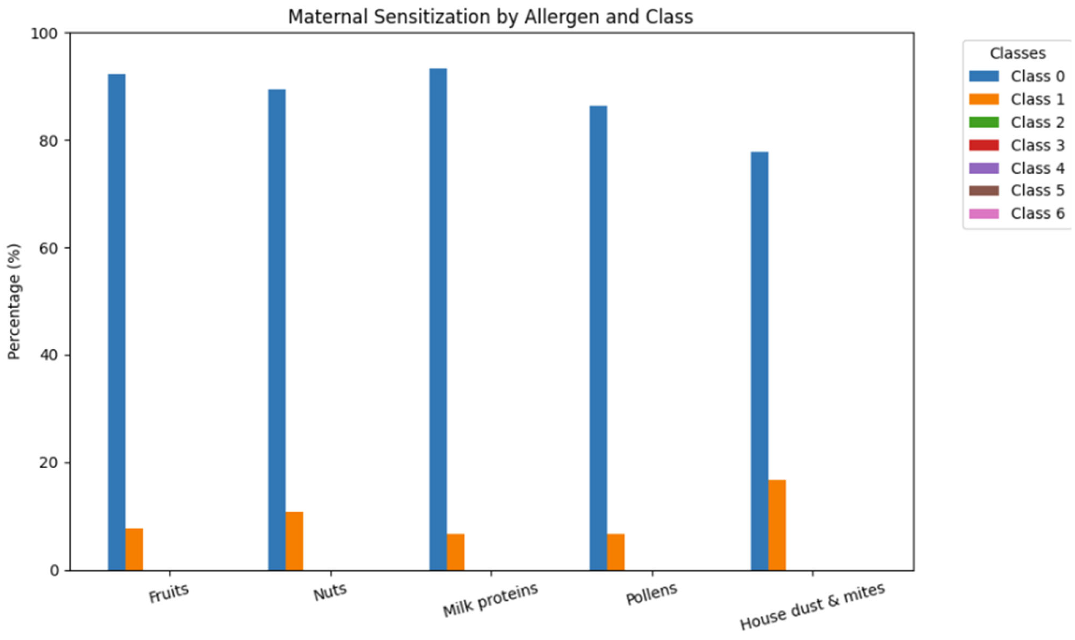
The results for sensitization in mothers are shown in
Figure 2.
Maternal sensitization to allergens includes the following: to fruits—f49 Apple, f84 Kiwi, and f237 Apricot; to nuts—f13 Peanut, f17 Hazelnut, and f20 Almond; to milk proteins—f2 Cow‘s milk, f76 Alpha-Lactalbumin, f77 Beta-Lactalbumin, and f78 Casein; to pollen—g1 Sweet vernal grass, g3 Orchard grass, g6 Timothy grass, g12 Cultivated rye, t2 Alder, t3 Birch, t4 Hazel, t7 Oak, t23 Cy-press, w1 Common ragweed, w6 Mugwort, and w9 English plantain; to house dust and mites—d1 Dermatophagoides pter., d2 Dermatophagoides farina, and h1 House dust (Greer). The results are presented based on IgE class according to the scale described in
Table 1.
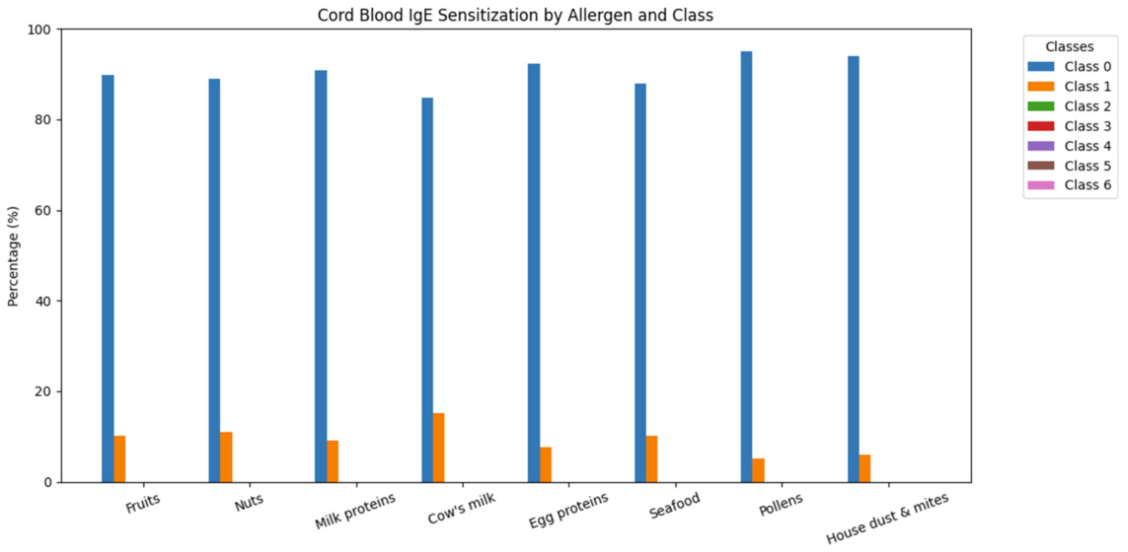
Cord blood sensitization includes the following: to fruits—f49 Apple, f84 Kiwi, and f237 Apricot; to nuts—f13 Peanut, f17 Hazelnut, and f20 Almond; to milk proteins—f76 Alpha-Lactalbumin, f77 Beta-Lactalbumin, and f78 Casein; to egg proteins—f1 Egg white and f75 Egg yolk; to seafood—f3 Codfish, f23 Crab, and f24 Shrimp/Prawn; to pollens—g1 Sweet vernal grass, g3 Orchard grass, g6 Timothy grass, g12 Cultivated rye, t2 Alder, t3 Birch, t4 Hazel, t7 Oak, t23 Cy-press, w1 Common ragweed, w6 Mugwort, and w9 English plantain; to house dust and mites—d1 Dermatophagoides pter., d2 Dermatophagoides farina, and h1 House dust (Greer). Both maternal and cord blood sensitization was predominantly classified as Class 0, with a small proportion of Class 1 responses. Higher sensitization classes (Class ≥ 2) were negligible and did not contribute meaningfully to the overall distribution. The results are presented based on IgE class according to the scale described in
Table 1, as seen in
Figure 2 and
Figure 3:
In
n = 5 umbilical cord blood samples, a specific IgE was found to pollen and/or house dust mites (
Figure 3). All of the umbilical cord blood samples were collected from mothers with allergic rhinitis.
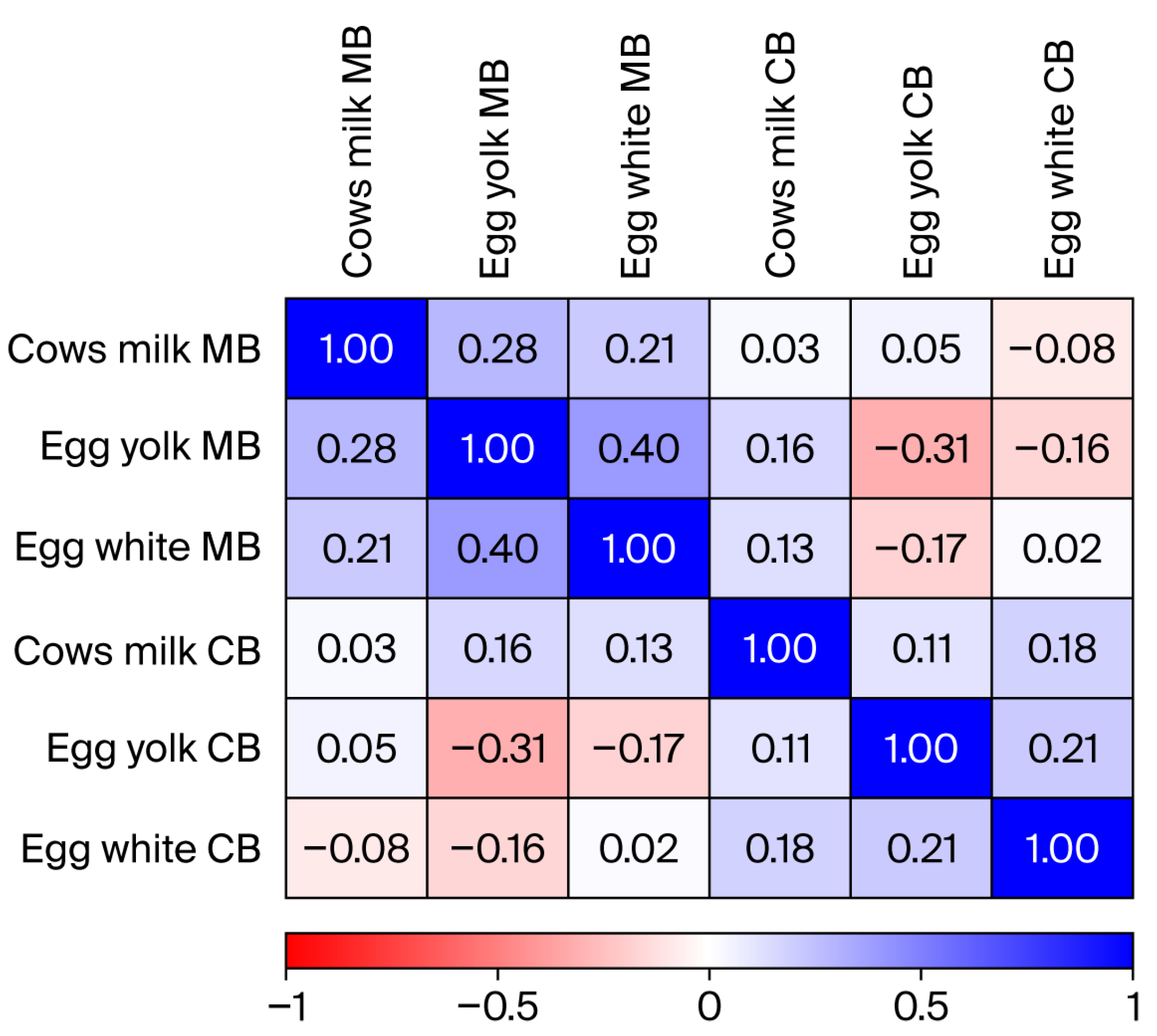
The correlation analysis of specific IgE in umbilical cord blood and maternal blood is presented in
Figure 4.
When examining the correlation between the presence of specific IgE to egg proteins and cow’s milk (
Figure 4), we did not find a significant correlation between specific IgE to cow’s milk (
p = 0.857), egg white (
p = 0.926) and egg yolk (
p = 0.096) in umbilical cord blood and maternal blood samples taken immediately before birth.
The IgG-specific response to various food antigens as part of the normal immune response in the mother is shown in
Figure 5. In addition, the figure shows the specific IgG to the same food antigens (which have crossed the placental barrier via the neonatal Fcn receptor (FcnR) in umbilical cord blood. The latter could be considered as food exposure related to the mothers’ diet, as the assessment was conducted in relation to exposure to foods that could be potential allergens in isotype switching to IgE class.
Nutritional exposure and reactivity in mothers and their babies is shown in
Figure 5.
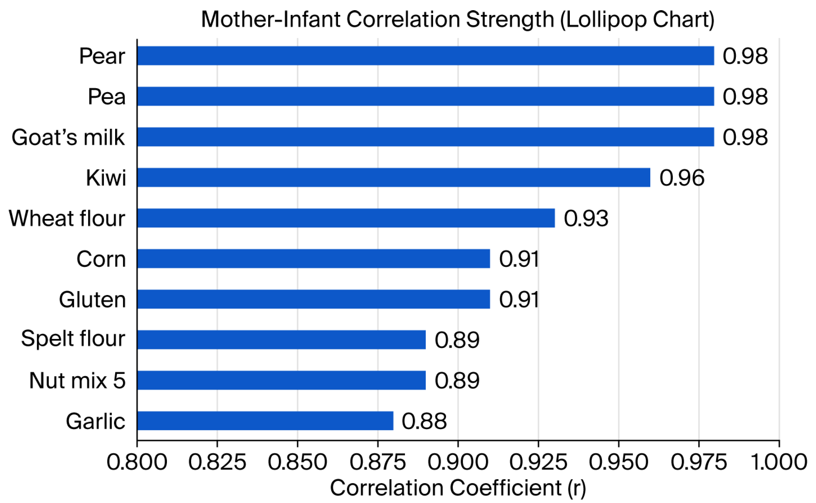
The antigens with the highest correlation between mothers and their babies are shown in
Figure 5.
Figure 5 presents the allergens/antigens with the highest correlation (Pearson’s correlation analysis) between the established IgG in mothers and their babies. The latter is indicative of both the mothers’ eating habits during pregnancy and the active transport of food-specific antibodies through the placenta.
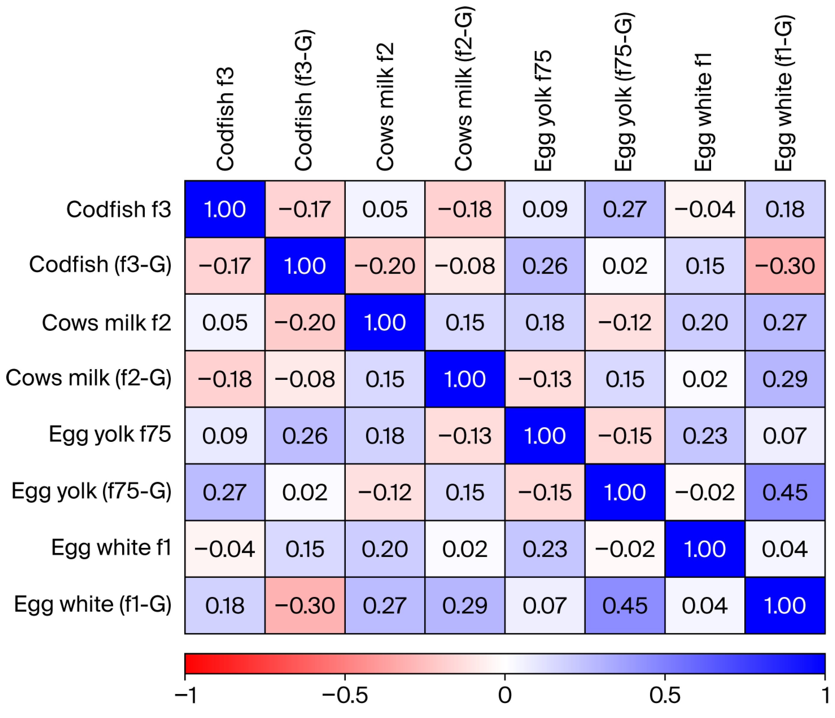
The co-correlation analysis of the specific IgE and IgG in umbilical cord blood showed no dependence between the two variables (
Figure 6).
Figure 6 presents Spearman’s correlation analysis between specific IgE and IgG in umbilical cord blood samples. We did not find a significant correlation between the two variables. This suggests that IgG and IgE responses probably occur independently of each other. This is probably related to the normal mechanisms of immune response generation but may also be due to the small number of patients included in our study. The term “atopic mother” included 12 women with previously established allergic rhinitis, in addition to the eosinophilia characteristic of all mothers during pregnancy.
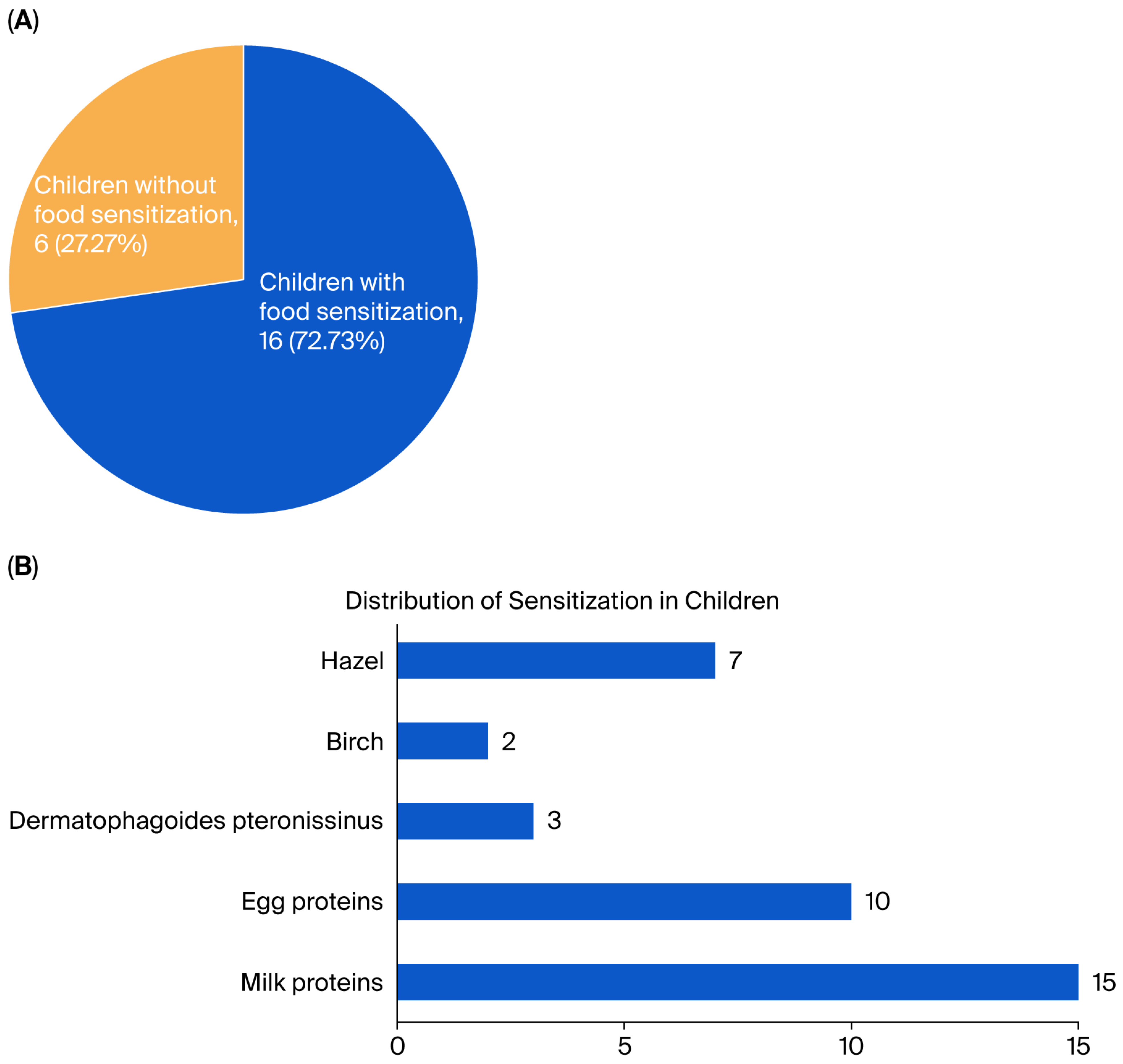
Figure 7A presents data on reported sensitization (presence of specific IgE to one or more food antigens at 6–8 months after birth (second timepoint) in children born to mothers with eosinophilia during pregnancy.
Figure 7B shows the distribution of sensitization among children is presented.
When assessing sensitization in children aged 6–8 months, 16 of them, 72.73%, were sensitized to dietary proteins (most commonly egg and milk proteins), whereas for 6 of them, 27.27%, sensitization was not detected during this study in the specified time range. Statistical analysis showed that there was a statistically significant difference between the observed frequencies compared to the expected frequencies in unequal proportions (χ2, p = 0.0347) and the studied group. Considering that all children were born to mothers with eosinophilia, we could assume that maternal eosinophilia itself is a risk factor for sensitization in the offspring. However, such a statement is not reliable given the small number of patients in our study (n = 22). Additional observations are needed to confirm this relationship.
4. Discussion
From the second trimester of pregnancy, cells from the developing immune system of the fetus are capable of presenting antigens to T-lymphocytes [
7]. Human IgG has been shown to increase the translocation of allergens such as Bet v 1 and β-lactoglobulin across the placental barrier in an in vitro model [
7], and these allergens can provoke fetal IgE production independently of maternal IgE [
7]. There is evidence that the passage of allergens is increased in functional disorders of the placenta, an increase in the dose of the allergen and a decrease in the molecular weight (MW) of allergens [
3]. Altered placental function has been documented in women with asthma [
3]. Factors such as the solubility of allergenic molecules into lipids, the polarity of molecules and the existence of specific transport mechanisms are also important. Most substances with a low MW (<500 Da) simply diffuse through the placental tissue [
3]. Active transport of maternal IgG is an ideal means of transferring allergens to the fetus. For example, the inhaled feline allergen, Fel d 1, has been found in an IgG complex in up to 40% of infants’ umbilical cord sera [
3]. In the group we studied, with
n = 5 umbilical cord blood samples, we found a specific IgE to Timothy grass pollen and/or house dust mites. These samples are from babies whose mothers, in addition to peripheral eosinophilia, have already established allergic rhinitis. Transplacental transfer of a free allergen has been described as early as the 26th week of gestation. One of the putative pathways is based on placental fibrinoids. They are formed at sites of syncytiotrophoblastic degeneration, which are typical of all placentas at all stages of pregnancy and are permeable to macromolecules [
3]. However, such a perfusion model cannot explain the differences in the transfer rate of allergens with different molecular weights. A paracellular porous pathway and an endocytic mechanism of free antigen passage have also been described. However, there are still no definitive data confirming that exposure to maternal allergens is associated with the development of allergy in the first year of life [
3].
The most potent immunological transport through the placenta during pregnancy takes place through the FcRN, an MHC class I-like Fc receptor that is expressed by the syncytiotrophoblast and can bind all high-affinity IgG subtypes under appropriate conditions. The receptor provides the transcytosis of IgG from the maternal circulation to the fetal capillaries of the placental villi [
3,
10]. For its part, the cytotrophoblast does not express IgG receptors and, when intact during the first trimester of pregnancy, restricts the passage of IgG through the placenta [
3]. In humans, the FcRN receptor is expressed in the early stages of pregnancy and increases progressively from the beginning of the second trimester to the end of pregnancy, with the majority of IgG antibodies transferred by the 34th week of gestation [
10]. Accordingly, we found a strong positive correlation between specific IgG in maternal blood and umbilical cord blood. It is also important to note that there is also a quantitative correspondence with regard to the IgG class detected in mothers and their babies. In all of the studied subjects, the specific IgG classes moved in class 1–3, independent of the nutritional exposure of the mothers. It could be speculated that such a phenomenon is due to tolerogenicity during pregnancy. Fetal endothelial cells represent the next barrier to the passage of IgG to the fetal circulation. A candidate for a transport molecule expressed by these cells is Fc gamma receptor IIb (FcgRIIb). The latter binds to immune complexes but cannot bind to monomeric IgG. Expression of FcgRIIb increases significantly after the 20th week of gestation, when IgG transfer begins [
3]. Maternal IgG to respiratory allergens (house dust mites, birch pollen, cat) and food allergens (eggs, cow’s milk) have been found to be transferred in utero [
9]. The latter is of utmost importance because a distinctive feature of the immature adaptive immune response is its limited capacity to switch between classes before reaching 6 months of age and reaching levels typical of adults up to 3 years of age [
3].
FcRN selectively binds to IgG, but it has been shown that IgE can naturally form IgG complexes in mice and humans [
11]. In mouse models, IgE from sensitized mothers is transferred to fetuses in utero through an FcRN IgG-dependent process [
11]. In addition, maternal cells may pass through the placenta before birth and can settle long-term in the tissues of the offspring. On this basis, it is recommended to avoid the allergen during pregnancy [
10,
11]. In addition to sensitizing fet12al mast cells against allergens, the passed maternal IgE induces the maturation of fetal macrophages and potentiates their survival in vivo [
11] as binding of IgE to mast cells in an antigen-independent manner is sufficient for these changes. However, given the short half-life of IgE, the process must be supported by new synthesis [
9]. The opposite has been proven: the elimination of the original IgE in mice leads to a decrease in the number of mast cells and the possibility of developing anaphylaxis. Since IgE-linked mast cells are present in fetal tissues in the second trimester in women with allergies, it is possible for mast cells to become mature and gain functionality even before birth [
11,
12,
13]. Our study did not find a statistically significant correlation. When examining the relationship between the presence of specific IgE to egg proteins and cow’s milk, we did not find a significant correlation between specific IgE to cow’s milk (
p = 0.857), egg white (
p = 0.926) and egg yolk (
p = 0.096) in umbilical cord blood and maternal blood samples taken immediately before birth. It is also important to emphasize that none of the pregnant women with eosinophilia had been on an elimination diet for the foods described during or before pregnancy. Such a result fully corresponds to the different biological activity of IgE and IgG with the same specificity. Maternal IgG (specific) passes to the baby and high maternal IG to a specific allergen may reduce babies IgE production. On the other hand, monomeric IgE cannot pass through the placenta. This strongly supports the hypothesis of intrauterine sensitization.
Examining IgE in umbilical cord blood, Klaus Bønnelykke used the term “fingerprint” due to a close match between the specific IgE in the mother’s blood and in the umbilical cord blood. The author found specific IgE only in newborns for whom the mother had the same specific IgE as the level of specific IgE in umbilical cord blood strongly correlated with the maternal level, and did not show such a close correlation with the paternal level of IgE [
14]. We found a correlation between specific IgE in mothers and their babies in cases of maternal sensitization to grass pollen and/or house dust and accompanying clinically manifested allergic rhinitis before pregnancy, in which IgE with the same specificity was found in the umbilical cord blood of the heavy babies. However, the small number of cases is the main limiting factor in the analysis of the results.
In fact, the level of specific IgE can be reduced to about 50% 4–6 years after the last exposure to the allergen [
15]. This is due to the ongoing synthesis of IgE by long-lived IgE-producing plasma cells [
15]. Thus, maternal IgE may be both sufficient and necessary for the development of allergic inflammation of both the respiratory tract and the skin of the offspring, the latter occurring at the first exposure to allergens of the child [
12], since the sensitization phase has taken place intrauterinely. In addition, allergen-specific IgE has also been detected in breast milk, possibly through the previously described FcRN-dependent IgE-IgG transfer, which may continue during early childhood [
8,
11]. We investigated the correlation between IgE and IgG synthesized against codfish, cow’s milk, egg yolk and egg white. The results showed no correlation between the two variables. However, the small number of patients (
n = 22) makes the claim unreliable. The generation of antibodies with the same specificity (common Fab fragment) but with different Fc regions is part of the diversity and breadth of the immune response, which can generate tolerance or develop an allergy depending on the stimuli of the microenvironment.
In the absence of inflammatory and other signals that induce the maturation of dendritic cells, it is likely that the encounter with an allergen in the fetal period will lead to tolerance rather than sensitization to the allergen [
3]. Tracking the IgE/IgG ratio would be a better indicator in this context, but such an approach would require measurement at at least two time points during continuous allergen exposure. However, such measurement was not included in our study.
Therefore, the offspring initially rely on maternal IgG. There is evidence that children with maternal transfer of IgG above a certain threshold avoid IgE sensitization to the homologous aeroallergen, and it should be emphasized that IgG and IgE bind to different allergenic epitopes and do not always sufficiently impede their respective binding to Fc [
12]. In mouse models of allergic diseases, maternal IgG-allergenic immune complexes transmitted through breast milk are sufficient to eliminate the inflammatory response in the offspring [
12]. Direct evidence also comes from oral immunotherapy (OIT) studies with peanut milk proteins [
2], pointing to a remarkable increase in plasma levels of specific IgG in reducing symptoms and inducing nutritional tolerance in allergic patients [
15]. A Dutch study showed that maintaining tolerance in atopic children and adults to a cow’s milk allergy was associated with increased levels of milk-specific IgG4 in combination with low specific IgE [
16]. Thus, low (absent) maternal specific IgG can potentiate IgE production in the baby under appropriate microenvironmental conditions. Maternal allergen-specific IgG can be detected in the serum of children up to 6 months of age [
8]. However, there are data indicating the opposite. IgG antibodies can form immune complexes with allergens in foods and thus cause inflammatory reactions when increased in proportion to the duration of exposure. Their functionality can by realized by two main mechanisms: (1) steric blockade of antigenic epitopes or (2) signaling by the inhibitory FC receptor Fcγr2β [
13,
17,
18]. Currently, it is not recommended to delay the introduction of solid foods in infants, as their earlier introduction induces the development of tolerance [
12]. The latter also supports the lack of benefits of carrying out elimination diets with allergenic foods during pregnancy [
12].
In the presence of allergic disease, the balance of maternal Th1/Th2 cytokines modulates the immune response in the offspring. For example, the severity of asthma in mothers during pregnancy correlates positively with the risk of developing asthma in the child and blockage of maternal IL-4 in allergic mothers was associated with a reduction in the development of allergic asthma in mouse models [
11]. There are conflicting data regarding whether cytokines can cross the placenta. There is partial evidence of IL-6 passage [
11]; transport of IL-4 and IL-13 has not been confirmed [
11].
In contrast, however, maternal IL-5 is transferred to the fetus. Studies in mouse models have shown that IL-5 leads to sensory hyperinnervation of the respiratory tract in the offspring, which this characteristic persists into adulthood. The passed IL-5 potentiates the production of fetal eosinophils, which, together with nerve growth factors, affect the processes of innervation formation pathogenetically related to the realization of potential hyperreactivity in the offspring [
11]. We followed the development of sensitization in the period 6–8 months (after weaning) in 22 mothers with established peripheral eosinophilia. When assessing sensitization in children aged 6–8 months, 16 of them, 72.73%, were sensitized to dietary proteins (most commonly egg and milk proteins), whereas for 6 of them, 27.27%, sensitization was not detected during this study in the specified time range. Statistical analysis showed that maternal eosinophilia in peripheral blood is a risk factor for the development of allergy in the offspring (χ
2,
p = 0.0347). The distribution of sensitization in our children’s group is as follows: in 15 children, sensitization to milk proteins is observed; in 10 children, sensitization to eggs; and in 7, sensitization to hazelnut. It is important to emphasize that in five children, sensitization to inhalant allergens (birch and
Dermatophagoides pteronysinus) is observed at 6–8 months. This result may be due to the influence of cytokines (the third signal in the immune response), but the small number of patients (
n = 22) does not confirm the definitiveness of the result. Our study has several other weaknesses, such as the lack of a control group of mothers without eosinophilia and the small number of patients. We could not present a more in-depth analysis of mothers with eosinophilia and clinically manifested rhinitis. However, our results outlined the role of maternal eosinophilia as a predictive factor for the development of sensitization in the offspring. In addition, we did not assess the number of eosinophils in the umbilical cord samples. This would have provided additional indirect information about the influence of maternal eosinophilia and the accompanying sensitization on the development of sensitization in the fetus. However, this omission highlights the need for further work to clarify this indicator.
In conclusion, although we were able to follow a small number of patients, our data support the hypothesis of the development of intrauterine sensitization. Furthermore, our study is the first to directly track specific IgE and specific IgG in mothers and their children.




 ,
,