Review

Pruritus in Autoimmune Demyelinating Diseases of the Central Nervous System: A Review

by
Christian Messina
1,* and
Mariateresa Zuccarello
2
1
Azienda Sanitaria Provinciale Catania, ASPCT, 95100 Catania, Italy
2
Department of Medical and Surgical Sciences, and Advanced Technologies, “G.F. Ingrassia”, Azienda Ospedaliera Universitaria “G. Rodolico-San Marco”, University of Catania, 95124 Catania, Italy
*
Author to whom correspondence should be addressed.
Allergies 2025, 5(4), 32; https://doi.org/10.3390/allergies5040032
Submission received: 15 April 2025 / Revised: 29 August 2025 / Accepted: 22 September 2025 / Published: 23 September 2025
(This article belongs to the Section Dermatology)

Abstract

Pruritus (itching) is an underrecognized but often debilitating symptom in patients with central nervous system (CNS) demyelinating diseases, including multiple sclerosis (MS) and neuromyelitis optica spectrum disorder (NMOSD). It is often considered a paroxysmal symptom. Although less studied than pain or spasticity, pruritus can significantly impair the quality of life. This review aims to provide a comprehensive overview of the pathophysiological mechanisms underlying pruritus in demyelinating CNS disorders, its clinical presentations, and the available treatment options. We explore the central origins of neuropathic itch, focusing on spinal cord, brainstem, and cerebral lesions, with particular emphasis on white matter involvement and spinothalamic tract dysfunction. In addition, we review pruritus triggered or exacerbated by disease-modifying therapies (DMTs) used in MS and NMOSD.

1. Introduction

Pruritus (itching) is an unpleasant sensation on the skin, eliciting the will to scratch [1]. Chronic pruritus (CP) refers to daily itching, lasting more than 6 weeks [1]. Among its various etiologies, pruritus represents a diagnostic challenge. A thorough dermatological and neurological examination, supported by ancillary investigations such as complete blood tests, computerized tomography scan, Magnetic Resonance Imaging (MRI), or skin biopsy, may be required to establish the diagnosis. To aid in clinical assessment, the Neuropathic Pruritus 5 (NP5) screening questionnaire was developed [2]. This tool includes five yes/no items, each scoring one point: whether pruritus is associated with twinges, burning sensations, exacerbation by activity or stress, or relief with exposure to a cold environment [2]. A score of two or more positive responses suggests neuropathic pruritus, with a reported sensitivity of 76% and specificity of 77% [2]. Depending on the presence or absence of skin disruption, itching can be classified into pruritus on diseased or non-diseased skin [1]. The first group comprises patients with dermatological disorders, such as atopic dermatitis, psoriasis, dry skin, neoplasms, and urticaria, where pruritus arises in the context of visible or primary skin disease such as psoriasis, atopic dermatitis, dry skin, neoplasms and urticaria [1]. On the other hand, the second group includes patients affected by non-primarily dermatological conditions, such as systemic disorders affecting the liver, kidney, or blood, central or peripheral neurological disorders, psychogenic disorders, mixed etiology, and pruritus of undetermined origin [1]. Multiple sclerosis (MS) is an autoimmune-mediated neurodegenerative disease of the central nervous system (CNS) characterized by inflammatory demyelination with axonal transection [3]. MS onset is usually between 20 and 40 years old, and it is the most common non-traumatic disabling disease affecting young adults [3,4]. On the other hand, neuromyelitis optica spectrum disorder (NMOSD) is a rare demyelinating disorder of CNS, which mainly affects the optic nerve and spinal cord [5]. Paroxysmal symptoms (PSs) are defined as transient sensory, motor, or autonomic symptoms that are caused by ectopic impulses generated at the site of demyelination, lasting less than 24 h [6]. Itching is just one of many PSs and may be one of the first signs of MS, appearing with or without other abnormal sensations, such as pins and needles and burning or stabbing pains [6]. In this review, all causes of pruritus in patients with autoimmune demyelinating disorders of the CNS are explored, with a focus on identifying the pathophysiological mechanisms underlying each etiology. Furthermore, all potential treatment options for pruritus in demyelinating disorders of the CNS are discussed.

2. Multiple Sclerosis (MS), Paroxysmal Symptoms (PSs) and Pruritus

MS is a chronic autoimmune disease affecting the CNS with demyelination, inflammation, neuronal loss, and gliosis (scarring), causing varying degrees of damage to both myelin and axons [7]. MS affects 2.3 million people worldwide and is commonly diagnosed in individuals within the working-age population, with females experiencing it more often than males [3,4,7]. Its etiology remains unknown. Autoreactive T cells spread into lymphatic tissues, undergo expansion, attach themselves to adhesion molecules that are upregulated, and begin to generate matrix metalloproteinases (MMPs), leading to the breakdown of the blood–brain barrier (BBB). In CNS, they encounter antigen-presenting cells (APCs) and begin dividing into two types: proinflammatory T helper 1 (Th1) cells and anti-inflammatory Th2 cells [7]. The latter are responsible for releasing cytokines that attack macrophages and microglial cells. Moreover, activated B cells produce autoreactive antibodies that cross the damaged section of the BBB, essentially triggering the formation of myelin autoantibodies, and initiate the complement cascade, which attacks myelin again, leading to demyelination [7]. These inflammatory processes most likely cause relapses [7]. As a result of these chronic mechanisms, nervous tissue in the CNS is disrupted by transection and axonal injury, which leads to MS progression [7]. Upon entry into CNS, autoreactive T cells release proinflammatory cytokines such as interleukins (IL-1 family, IL-2, IL-12, IL-17, IL-23), TNF-α, and interferon-gamma [7]. These mediators contribute to further disruption of the BBB, facilitating the infiltration of macrophages, complement components, B cells, and antibodies [7]. Within CNS, T cell activation leads to an amplified release of proinflammatory cytokines as well as neurotoxic mediators such as reactive oxygen species and nitric oxide [7]. These immune responses involve interactions with resident microglia, astrocytes, and macrophages [7]. Moreover, cytokine regulation, particularly of IL-4, IL-5, IL-10, and TGF-β, has been associated with the activity of CD4+, CD8+, and Th1 cells [7]. IL-4 and IL-5, typically secreted by Th2 cells, are involved in promoting humoral immunity and B cell activation. However, in MS, a Th1/Th17-dominant response prevails, and the relative deficiency of IL-4 and IL-5 may fail to counterbalance pro-inflammatory pathways [7,8]. IL-10 and TGF-β, primarily produced by regulatory T cells (Tregs), are crucial for suppressing autoreactive T cell proliferation and maintaining peripheral tolerance [7]. Their activity is mediated via IL-10R and TGF-βR expressed on effector T cells, antigen-presenting cells, and microglia [7]. In MS patients, a functional impairment or reduced production of IL-10 and TGF-β has been associated with a failure to control CNS-directed autoimmunity, allowing for persistent inflammation, demyelination, and axonal damage [7]. CD4+ Th1 cells—producers of IFN-γ—and CD8+ cytotoxic T lymphocytes further contribute to CNS injury by directly targeting oligodendrocytes and promoting macrophage/microglial activation [7,8]. Over time, these chronic processes give rise to the characteristic clinical features of multiple sclerosis and may contribute to disease progression in certain patients. However, about 1.6–17% of patients with MS could experience paroxysmal symptoms (PSs) during the course of disease, 24% of which occur as the initial manifestations of the disease [6,9]. PSs are defined as transient sensory, motor, or autonomic symptoms, such as trigeminal neuralgia, dysarthria, ataxia, paresthesia, pain, or akinesia, that are caused by ectopic impulses generated at the site of demyelination, lasting less than 24 h, in contrast to a relapse, which lasts more than 24 h [6]. The pathogenesis of PSs remains only partially understood, and several hypotheses have been proposed to explain this distinctive clinical phenomenon [9]. The most widely accepted theory, initially proposed by Osterman et al., suggests that partially demyelinated axons may generate ectopic spontaneous nerve impulses, which can propagate transversely to neighboring axons via ephaptic transmission, ultimately resulting in paroxysmal attacks [9]. An alternative explanation involves the concept of “inflammatory irritation”, based on the positive therapeutic response to corticosteroids reported in many cases [9]. Another potential mechanism implicates ion channel dysfunction in partially demyelinated axons, which could increase axonal sensitivity to minor physiological fluctuations, such as reductions in ionized calcium due to hyperventilation, a recognized trigger of PSs [9]. In 1976, pruritus was identified as one of the PSs in MS by Osterman, and further cases have been documented in the literature over time [6,10]. In patients with MS, pruritus has been reported as either an initial presenting symptom or as a manifestation during disease relapse [11]. In some cases, it may also arise during remission phases [11]. Pruritus associated with MS typically presents with a sudden onset, is localized and intense, lasts from a few seconds to several minutes, and is often accompanied by superficial sensory disturbances and/or pain [12]. Paroxysmal pruritic sensations may emerge concurrently with neurological recovery during treatment with magnetic fields, suggesting that itching could potentially serve as a clinical indicator of symptomatic improvement in MS [11].

3. Neuromyelitis Optica Spectrum Disorders (NMOSD), Paroxysmal Symptoms (PSs) and Pruritus

NMOSD is a rare demyelinating inflammatory disease of the CNS, previously considered a subtype of MS [5]. Aquaporin-4 (AQP4) is a bidirectional water channel, with the highest expression in astrocytes, which controls the flow of water in the brain and spinal cord and is involved in glutamate reuptake and neuroexcitation [5]. An unidentified trigger initiates the production of AQP4-IgG by B cells [5]. Several peripheral immune cell populations are believed to contribute to the systemic inflammatory milieu observed in NMOSD [5]. In this context, disruption of the blood–brain barrier (BBB) permits the infiltration of inflammatory cells, cytokines, and pathogenic antibodies into the CNS [5]. AQP4-IgG binds to the extracellular domain of AQP4, predominantly expressed on astrocytic endfeet, leading to antibody-dependent cellular cytotoxicity [5]. This antibody–antigen interaction also activates the classical complement cascade, ultimately forming the membrane attack complex (MAC), which induces astrocyte injury and death [5]. Subsequent secondary demyelination is mediated by complement-dependent cytotoxicity [5]. The pathogenesis of NMOSD is thus characterized by both peripheral and central inflammation, orchestrated by a complex interplay involving B cells, complement proteins, T lymphocytes, neutrophils, eosinophils, microglia, and interleukin-6 [5]. A central role is played by B cells, which produce pathogenic autoantibodies against AQP4-IgG [13]. Upon binding to AQP4 on astrocytes, AQP4-IgG triggers classical complement activation via C1q, leading to the formation of the MAC, astrocyte damage, and subsequent secondary demyelination and neuronal loss [13]. Complement activation also recruits neutrophils and eosinophils, which infiltrate the CNS and contribute to tissue injury through the release of proteases and reactive oxygen species [13]. IL-6, produced by various immune cells, including monocytes, astrocytes, and B cells, promotes survival and differentiation of plasmablasts via IL-6R and plays a pivotal role in amplifying AQP4-IgG production [13]. IL-6 also facilitates the breakdown of the BBB and enhances Th17 differentiation [13]. Infiltrating Th17 and Th1 CD4+ T cells further sustain the inflammatory milieu, while resident microglia contribute to lesion propagation by releasing pro-inflammatory cytokines and presenting antigens [13]. Together, these mechanisms underline the astrocytopathy and antibody-mediated pathology that distinguish NMOSD from MS [13]. In NMOSD, the primary target of autoimmune attack is the astrocyte, with demyelination as a secondary phenomenon that results from the highly destructive pathogenic process [5], and the optic nerves and spinal cord are the most frequently affected neurological sites [5]. While the underlying basis for this selective vulnerability remains unclear, it may be partly explained by the higher expression levels of AQP4 in these regions [5]. Additionally, increased permeability of the BBB at these sites may facilitate greater access of pathogenic AQP4-IgG to the CNS [5]. In recent years, PSs have been increasingly recognized as more prevalent in NMOSD compared to MS, with the estimated prevalence ranging from 14% to 43% [14]. Notably, a more recent survey reported PSs in up to 71.9% of patients with NMOSD [14]. Recent evidence indicates that PSs often arise during or shortly after acute relapses of NMOSD, particularly following episodes of myelitis [14]. This finding aligns with earlier reports describing the occurrence of painful tonic spasms and other transient symptoms in close temporal relation to spinal cord lesions [14]. One hypothesis proposed to explain this association involves ephaptic transmission resulting from damage to centripetal fibers, leading to the aberrant activation of adjacent corticospinal tracts [14]. Neuropathic pruritus has been reported in only 4.5% of MS patients; however, in two case series of individuals with NMOSD, its prevalence was notably higher, ranging from 12% to 27% [12]. This increased frequency in NMOSD may be attributed to characteristic central cord lesions involving the dorsal horn—an area densely populated with pruritus-related mediators—or to lesions affecting the periaqueductal gray matter [12]. Another possible explanation is that neurons implicated in the modulation of neuropathic pruritus are particularly enriched in aquaporin-4 (AQP4) channels, rendering them more susceptible to the pathogenic mechanisms of NMOSD [12]. Itching in NMOSD is similar in character to that described in MS patients [12].

4. Pruritus and Pathogenetic Mechanisms

Pruritus or itching is an unpleasant sensation that causes a desire to scratch, which negatively affects psychological and physical aspects of life [15,16]. It is the most common symptom of skin diseases, and the most common reason for patients to consult dermatologists [15,16]. Itching can be continuous or intermittent, and local or generalized [15,16]. Pruritus may originate from dermatological causes, such as inflammatory dermatoses (atopic dermatitis, psoriasis, dry skin, etc.), infectious dermatosis (pediculosis, scabies, insect bites, etc.), autoimmune dermatoses (bullous pemphigoid, dermatomyositis, etc.), genodermatoses (Darier’s disease, Hailey–Hailey disease, ichthyoses, Sjögren–Larsson syndrome, epidermolysis bullosa, neurofibromatosis type 1), dermatoses of pregnancy (polymorphic eruption of pregnancy, pemphigoid gestationis, prurigo gestationis) and cancer (cutaneous T cell-lymphoma, cutaneous B cell lymphoma, leukemic infiltrates of the skin) [1]. Additional causes of pruritus may be associated with underlying systemic conditions, such as endocrine and metabolic diseases, infectious diseases, hematological and lymphoproliferative diseases, visceral neoplasms, and drugs [1]. Pruritus from neurogenic origins (without neuronal damage) and neuropathic disorders (with neuronal damage) have also been described [1], as well as pruritus of somatoform nature, characterized by chronic itch in the absence of identifiable dermatologic, neurologic, or systemic causes, often in association with psychological or functional disorders [1]. Although the precise mechanisms underlying pruritus have yet to be fully elucidated, current research highlights the involvement of several key mediators in both the initiation and exacerbation of itch [16]. These mediators appear to play distinct roles depending on the specific pruritic condition [16]. In addition, various signaling pathways and neurotransmitters have been implicated in the transmission of itch sensations [16]. Mediator-related pruritus refers to itching caused by the release and activity of various chemical substances, which act as pruritogenic mediators. Mediator-related pruritus implies that itching is associated with the mediation of several substances. Among amines, histamine is the most widely recognized mediator. It is primarily released by mast cells in response to immunological or inflammatory triggers [16,17]. Histamine exerts its pruritogenic effects through binding to H1 and H4 receptors, which are expressed on primary sensory neurons and immune cells [16,17]. Activation of these receptors contributes to the excitation of peripheral itch-specific nerve fibers, initiating the transmission of pruritic signals to the central nervous system. Among amines, histamine is the most well-known chemical mediator involved in pruritus, due to its ability to bind H1 and H4 receptors, which play important roles in the appearance of itching [16,17]. Moreover, another amine, serotonin (5-HT), can contribute to pruritus through both peripheral and central mechanisms [16,17]. Peripherally, serotonin can stimulate mast cells to release histamine, thereby indirectly promoting itch. Centrally, serotonin modulates pruritus via its interaction with opioid pathways and specific serotonin receptors (e.g., 5-HT2 and 5-HT3), which are expressed on neurons within the spinal cord and brain [16,17]. These receptors participate in the integration and amplification of pruritic signals along the ascending pathways, particularly through their influence on interneurons and spinothalamic tract neurons [16]. Serotonin is primarily released by enterochromaffin cells, platelets, and certain neurons, and its dual action underscores its complex role in both histaminergic and non-histaminergic itch pathways [16]. Moreover, serotonin may induce pruritus through peripheral mediation, by encouraging mast cells to release histamine, and central nervous system mediation, through opioid participation [16]. Furthermore, proteases play a significant role in the induction of histamine-independent pruritus by activating protease-activated receptors (PARs), particularly PAR2 and PAR4 [16,17]. These receptors are G-protein-coupled and are expressed on primary sensory neurons, keratinocytes, and various immune cells [16]. Proteases such as tryptase (released by mast cells) and endogenous enzymes like kallikreins or cathepsins can cleave and activate PARs, leading to the excitation of peripheral C-fibers involved in itch transmission [16]. The activation of PAR2 has been linked to chronic pruritus and is implicated in inflammatory skin diseases and neuropathic itch conditions [16]. This mechanism does not rely on histamine release, making it a key component of non-histaminergic pruritic pathways [16]. Furthermore, proteases appear to play a role in the induction of histamine-independent pruritus through the activation of protease-activated receptors (PARs), especially PAR2 and PAR4 [16]. Interleukins (ILs) are a family of cytokines—secreted proteins and signaling molecules—that can act as potent mediators of pruritus by triggering and amplifying itch responses [16,17]. Among them, IL-2, IL-3, IL-4, IL-6, and IL-10 have been implicated in pruritic mechanisms, particularly in inflammatory and immune-mediated conditions [16,17]. These cytokines are primarily produced by T lymphocytes, mast cells, and other immune cells upon immune system activation [16]. They act on receptors expressed on various target cells, including sensory neurons, keratinocytes, and additional immune cell subsets [16]. For example, IL-4 and IL-13 have been shown to sensitize peripheral neurons to pruritogens and to enhance itch via the JAK-STAT signaling pathway [16,17]. While the exact role of each interleukin in neuropathic itch is still under investigation, their involvement suggests a complex neuroimmune interaction at the interface of skin, immune system, and nervous system. Additionally, several peptides and phospholipid-derived metabolites are recognized as important mediators of pruritus, either by directly activating sensory neurons or by modulating the release of other pruritogenic substances. Among the peptides, bradykinin, substance P, calcitonin gene-related peptide (CGRP), neurotrophins (such as nerve growth factor, NGF), and opioid peptides have all been implicated in itch signaling [16,17]. These peptides are typically produced by immune cells (e.g., mast cells, eosinophils), keratinocytes, or peripheral neurons, and act on receptors expressed on sensory nerve endings [16]. For example, substance P acts on the neurokinin-1 receptor (NK1R), while CGRP can bind to CGRP receptors and modulate neurogenic inflammation [16]. Opioid peptides act via µ- and κ-opioid receptors, with µ-opioid receptor activation often associated with itch induction, and κ-opioid receptor activation potentially counteracting itch [16]. In addition, phospholipid metabolites such as endocannabinoids (e.g., anandamide), eicosanoids (e.g., prostaglandins, leukotrienes), and platelet-activating factors (PAF) have been shown to modulate itch through both pro- and anti-pruritic mechanisms [16]. These mediators are released by various immune cells and interact with specific receptors on cutaneous sensory neurons (e.g., cannabinoid receptors CB1 and CB2, prostaglandin receptors, and PAF receptors), ultimately influencing neuronal excitability and pruriceptive signal transduction. Additionally, peptides, such as bradykinin, substance P, CGRP, neurotrophins and opioid peptides, and phospholipid metabolites, such as endocannabinoids, eicosanoids and platelet-activating factors (PAF), are considered pruritus mediators [16,17]. Significant advances in understanding itch signaling have shed light on the pathogenesis of pruritus. Currently, two main signaling pathways involved in pruritus have been identified: the histamine-dependent (histaminergic) pathway and the histamine-independent (non-histaminergic) pathway [16]. Furthermore, pruritus can be generated within the CNS without the need for peripheral stimulation [16]. In the histaminergic pathway, histamine activates phospholipase C-β3 (PLC-β3) and phospholipase C (PLC) through binding to its specific receptors, particularly the H1 receptor and the H4 receptor [16]. This activation triggers the downstream signaling of the transient receptor potential vanilloid 1 (TRPV1) ion channel [16]. Subsequently, itch signals are transmitted to the CNS via cutaneous nerve fibers, ultimately resulting in the sensation of itching [16]. In contrast, the non-histaminergic pathway involves a variety of pruritogens, such as cowhage, chloroquine, bovine adrenal medulla 8-22 peptide (BAM8-22), SLIGRL, and β-alanine [16]. Cowhage initially stimulates PAR2, which in turn sensitizes PLC [16]. This cascade activates downstream targets, including TRPV1 and transient receptor potential cation channel A1 (TRPA1) [16]. Ultimately, itch signals are relayed to the CNS via cutaneous mechanosensory afferent fibers, generating the itching sensation [16]. Despite the fact that cowhage activates polymodal nociceptors via PAR2 and downstream TRPV1/TRPA1 signaling, scratching these same nociceptors can paradoxically inhibit the sensation of itch [18]. This phenomenon is thought to involve segmental inhibition within the spinal cord: nociceptive input from scratching activates inhibitory interneurons that suppress the transmission of pruritic signals in the dorsal horn, a process analogous to the gate control theory of pain [18]. Thus, mechanical stimulation from scratching may engage a negative feedback loop that transiently interrupts itch perception, even when the original stimulus is mediated by nociceptive pathways [18]. Additionally, the Mas-related G-protein-coupled receptors (Mrgprs) are activated by chloroquine, SLIGRL, BAM8-22, and β-alanine, coupling to Gβγ or PLC, among others [16,19]. This results in the activation of TRPA1 and TRPV1 channels [16,19]. Mrgpr-positive neurons detect the itch signals, which are then transmitted via afferent fibers to the spinal cord [16,19]. These signals are modulated by the gastrin-releasing peptide (GRP)–GRP receptor (GRPR) and the B-type natriuretic peptide (BNP)–natriuretic peptide receptor A (NPRA) systems, ultimately leading to the perception of itch [16]. Itch stimuli initially trigger the release of various pruritogenic mediators, including inflammatory mediators, neuromediators, and neuropeptides, from skin cells such as immune cells and keratinocytes [16]. These mediators then bind to their specific receptors, leading to the activation of itch-specific sensory neurons. The itch signals are subsequently transmitted through either mechanically insensitive C-fibers, which are part of the histamine-dependent (histaminergic) pathway, or mechanically sensitive C-type fibers, which are involved in the histamine-independent (non-histaminergic) pathway [16]. The neural signals responsible for itch perception are initiated at the level of peripheral nerve endings and are transmitted centrally through a well-characterized pathway [16]. These signals are first conveyed by specialized primary afferent neurons, particularly unmyelinated C-fibers and thinly myelinated Aδ-fibers, which are selectively tuned to pruritic stimuli [16]. The impulses travel to the cell bodies of these neurons, located in the dorsal root ganglia (DRG), where signal modulation and integration can occur [16]. From there, the signals enter the spinal cord and ascend along the contralateral spinothalamic tract, a key component of the anterolateral system responsible for transmitting nociceptive and pruriceptive inputs [16]. As the signals ascend, they pass through the brainstem and project to the ventral posterior nucleus of the thalamus, which serves as a critical relay center [16]. The thalamus then distributes the information to multiple cortical areas involved in the multidimensional experience of itch. These include the primary somatosensory cortex (for localization and intensity), the secondary somatosensory cortex and insular cortex (for affective and interoceptive components), and regions such as the anterior cingulate cortex and prefrontal cortex, which are associated with the emotional and motivational responses to itch, including the urge to scratch [16]. This distributed processing underlies the complex and subjective nature of itch perception. The signals travel from the peripheral fibers through the dorsal root ganglion (DRG) of the spinal cord, along the spinothalamic tract to the thalamus, and finally reach the cerebral cortex, where the sensation of itch is perceived [16]. Figure 1 summarizes molecular mechanisms underlying pruritus.

5. Spinal Cord and Pruritus

He and colleagues reported that 21.0% of NMOSD patients and 2.1% of MS patients reported pruritus during the course of the disease, especially in those with myelitis [20]. Moreover, they found a high distribution of pruritus correlated to the dermatomal distribution of involved spinal cord segments or medulla [20]. These findings suggest that dorsal horns or trigeminal spinal nucleus might be involved in the pathogenesis of itching [20]. Furthermore, in most patients experiencing pruritus, lesions were found to be centrally located within the spinal cord, suggesting that involvement of the spinal cord grey matter may represent the primary underlying cause of itch [20,21]. In a case series, Yamamoto et al. reported the case of a 38-year-old woman complaining of paroxysmal itching occurring in the neck and left upper extremity corresponding to the cervical spinal segments bilateral C3 and left C4 to C6, with a C3 to C4 lesion found in the left dorsal area of the spinal cord in an axial image in Magnetic Resonance Imaging (MRI) in T2-weighted spin-echo images [22]. Those hyperintense areas within the cervical spinal cord diminished following adrenocorticosteroid therapy, accompanied by the resolution of paroxysmal pruritus [22]. Akyiama et al. showed that, in the upper cervical spinal cord (C1–C2), the majority of neurons localized in the dorsomedial region of the superficial dorsal horns co-expressing neurokinin-1 receptors (NK1R) may play a key role in mediating chronic itch and contribute to ascending somatosensory pathways [23]. However, in previous research, the spinothalamic tract (STT) was found to be involved in pruritus, particularly pruriceptive STT neurons located in the superficial dorsal horn, rather than STT neurons located in the deep dorsal horn [24]. Previous studies in rodent models have shown that itch-related neural pathways ascend to the brain through the superficial layers of the spinal dorsal horn, specifically involving laminae I and II [25]. Another pathway includes the spinoparabrachial pathway [26]. Grey matter involvement of the spinal cord appears to be a key factor underlying pruritus, which is likely secondary to spinal neuronopathy rather than demyelination [27]. The higher prevalence of centrally located spinal cord lesions affecting dorsal horn neurons in NMOSD compared to MS may explain the significantly increased incidence of pruritus in NMOSD [27]. Furthermore, in some NMOSD patients, the distribution of pruritus did not follow the dermatomal pattern of the affected spinal cord segments, suggesting the possible involvement of itch-processing pathways, such as pruriceptive neurons within the spinothalamic tract [27]. Notably, the majority of NMOSD patients with pruritus also reported neuropathic pain in the same region, supporting the notion that lesion localization may concurrently influence both neuropathic itch and pain symptoms [27]. Thus, it can be speculated that lesions involving the spinal cord grey matter—both superficial and deep dorsal horns—may contribute to the development of pruritus.

6. Brainstem and Pruritus

In a multicenter study of 258 patients with brainstem lesions and NMOSD, 12.4% reported having pruritus [28]. The involvement of itch-specific neurons and spinal cord pathways has been proposed as a potential underlying mechanism [20,28]. It is also plausible that similar processes occur within the brainstem, affecting itch-specific neurons of the trigeminal sensory pathway or ascending spinal pathways [20,28]. The presence of pruritus in conjunction with other brainstem-related symptoms—such as vomiting or hiccups—further supports brainstem localization [28]. Alternatively, the extension of spinal cord lesions into the brainstem may also play a contributory role [20,28]. In a previously reported case, a 34-year-old woman presented with an itching sensation in the right postauricular region without an apparent rash and a diagnosis of NMOSD [29]. MRI revealed a T2-hyperintense lesion with partial enhancement extending from the lower medulla oblongata to the upper cervical spinal cord [29]. Her symptoms markedly improved following intravenous and oral corticosteroid therapy [29]. Findings from animal models indicate that descending fibers originating from the periaqueductal gray matter play a crucial role in modulating and inhibiting afferent itch signals [25]. In patients with NMOSD who experience pruritus, spinal cord lesions are commonly present, along with potential pathological alterations in the periaqueductal gray matter, which are changes that may evade detection through conventional MRI [25]. NMO-IgG antibodies target astrocytes and ependymal cells surrounding the cerebral aqueduct, thereby disrupting the local physicochemical environment of inhibitory neurons within the periaqueductal gray [25]. This disruption may lead to neuronal degeneration, apoptosis, or necrosis, ultimately impairing the neurons’ ability to generate sufficient inhibitory signals [25]. As a result, the loss of inhibitory control may contribute to the manifestation of clinically intractable pruritus [25]. Moreover, Ingrasci et al. showed a strong association between facial or scalp itch in MS patients and the likelihood of having T2 hyperintense lesions within the anterior pons/ventromedial medulla, supporting a correlation between facial or scalp pruritus and pathological alterations in the sensory nerve fibers of the medial lemniscus—responsible for innervating the scalp—as well as in those of the principal sensory trigeminal nucleus, which innervates the face [6]. Thus, it can be speculated that lesions involving both the medulla oblongata and the midbrain—particularly within the periaqueductal gray matter—may contribute to the development of pruritus.

7. Cerebellum and Pruritus

The cerebellum, traditionally associated with motor coordination, is increasingly recognized for its role in sensory integration as well as cognitive and affective processes. Neuroanatomical evidence indicates that it projects to sensorimotor and reward-related cortical areas via the thalamus, while receiving afferents from these same regions through the pontine nuclei and inferior olives [30]. In the context of pruritus, the cerebellum may contribute to modulating the urge to scratch—a behavior that not only induces counter-irritation to suppress itch but also activates reward circuits, providing a reinforcing and potentially addictive sensation of relief [30]. Although cases of pruritus associated with cerebellar lesions have not yet been reported in the literature, some studies suggest that the cerebellum may be involved in modulating the scratching response and the associated sensation of pleasure [26,31,32]. In a previous study, seed-based resting-state functional connectivity (rs-FC) analysis was employed to investigate alterations in brain neural circuits in patients with chronic spontaneous urticaria (CSU) [33]. The results revealed enhanced rs-FC between the thalamus, cerebellum, and brain regions involved in the scratching response [33]. Another study reported significant activation of the cerebellar hemispheres during pruritic episodes, suggesting a role in mediating the urge to scratch [33]. These findings support the hypothesis that cerebellar efferent pathways projecting to reward and sensorimotor regions may contribute to the neuropathophysiology of CSU-related pruritus [33]. Therefore, lesions affecting the cerebellum could potentially contribute to the development of pruritus.

8. Brain and Pruritus

Following processing in the spinal cord, pruritic signals are transmitted via the spinothalamic tract to the thalamus and through the spinoparabrachial pathway to the parabrachial nucleus [34,35]. From these relay centers, the signals are projected to multiple brain regions involved in itch perception and modulation [34,35]. The primary and secondary somatosensory cortices are primarily responsible for the localization, intensity encoding, and recognition of itch [34,35]. Additional areas consistently activated include the midcingulate cortex, which is linked to the affective motivational aspects of itch, and both the anterior cingulate cortex (ACC) and insular cortex, which are associated with unpleasant sensory experiences [34,35]. Motor-related regions such as the premotor cortex, supplementary motor area, striatum, and cerebellum are thought to contribute to the generation or inhibition of scratching behavior [34,35]. Furthermore, the prefrontal cortex is implicated in the cognitive and decision-making processes associated with itch [34,35]. Notably, ’itch-selective’ regions have been identified in the precuneus and posterior cingulate cortex (PCC), which are associated with memory and attention processes and may be specifically activated by pruritic stimuli but not by painful stimuli [34,35]. Sabah and colleagues described a case of a female patient with MS who presented with severe pruritus localized to the lower extremities [36]. A brain MRI revealed multiple small, round periventricular plaques in both cerebral hemispheres, as well as along the longitudinal axis of the corpus callosum, whereas the spinal cord MRI was unremarkable [36]. Recently, pruritus has been reported in 33.33% of MS patients with thalamic lesions and in 14.8% of those with cortical lesions, although this difference did not reach statistical significance [6]. Nonetheless, dysfunction of the right anterior insula and its associated regions—such as the right precentral gyrus, cingulate cortex, and bilateral amygdala—appears to disrupt the neural networks involved in itch scratch regulation, potentially leading to compulsive itching or skin-picking behaviors in patients with frontotemporal dementia (FTD) [37]. Itch perception originates from the activation of small unmyelinated nerve endings in the skin by pruritogens, which transmit signals to the ipsilateral dorsal root ganglia of the spinal cord [38]. These signals are subsequently relayed across synapses to the contralateral spinothalamic tract, ascend to the thalamus, and are ultimately projected to neurons in the primary sensory cortex for cortical processing [38]. Therefore, any disruption along this pathway—including lesions affecting the thalamus or the primary sensory cortex—may potentially lead to abnormal itch perception. In patients with CSU, increased amplitude of low-frequency fluctuations (ALFF) has been observed in the right ventral striatum and putamen, with these values correlating positively with pruritus intensity [39,40]. Structural MRI also revealed greater gray matter volume (GMV) in these same regions compared to healthy controls [39,40]. Seed-based rs-FC analysis further showed reduced connectivity between the right ventral striatum and right occipital cortex, as well as between the right putamen and left precentral gyrus [39,40]. The ventral striatum is known to mediate reward and reinforcement processes and has been implicated in the relief of itch via scratching, particularly in emotionally salient contexts [39,40]. Similarly, the putamen is involved in the modulation of the itch-scratch cycle [39,40]. Notably, increased GMV in the striatum has also been documented in chronic pain, suggesting a potential shared mechanism, such as central sensitization, underlying both chronic itch and pain [39,40]. These findings indicate that the ventral striatum and putamen may contribute not only to the perception and relief of itch, but also to anticipatory mechanisms related to pruritic stimuli [39,40].

9. The Role of Astrocytes in Pruritus in Demyelinating Diseases of CNS

Astrocytes have long been recognized as contributors to chronic itch [41,42]. The occurrence of intractable pruritus in NMOSD suggests that reactive astrocytes may also play a role in the pathogenesis of acute itch [41,42]. In NMOSD, astrocyte dysfunction is likely to lead to neuronal injury, which in turn triggers pruritic symptoms [41,42]. The primary target of NMO-associated autoantibodies is AQP4, a water channel protein predominantly expressed on the end-feet of astrocytes throughout the CNS—particularly in the spinal cord, optic nerves, and perivascular regions—and to a lesser extent in ependymal cells [41,42]. Therefore, it is likely that AQP4-mediated astrocytic damage may contribute to the development of itching.
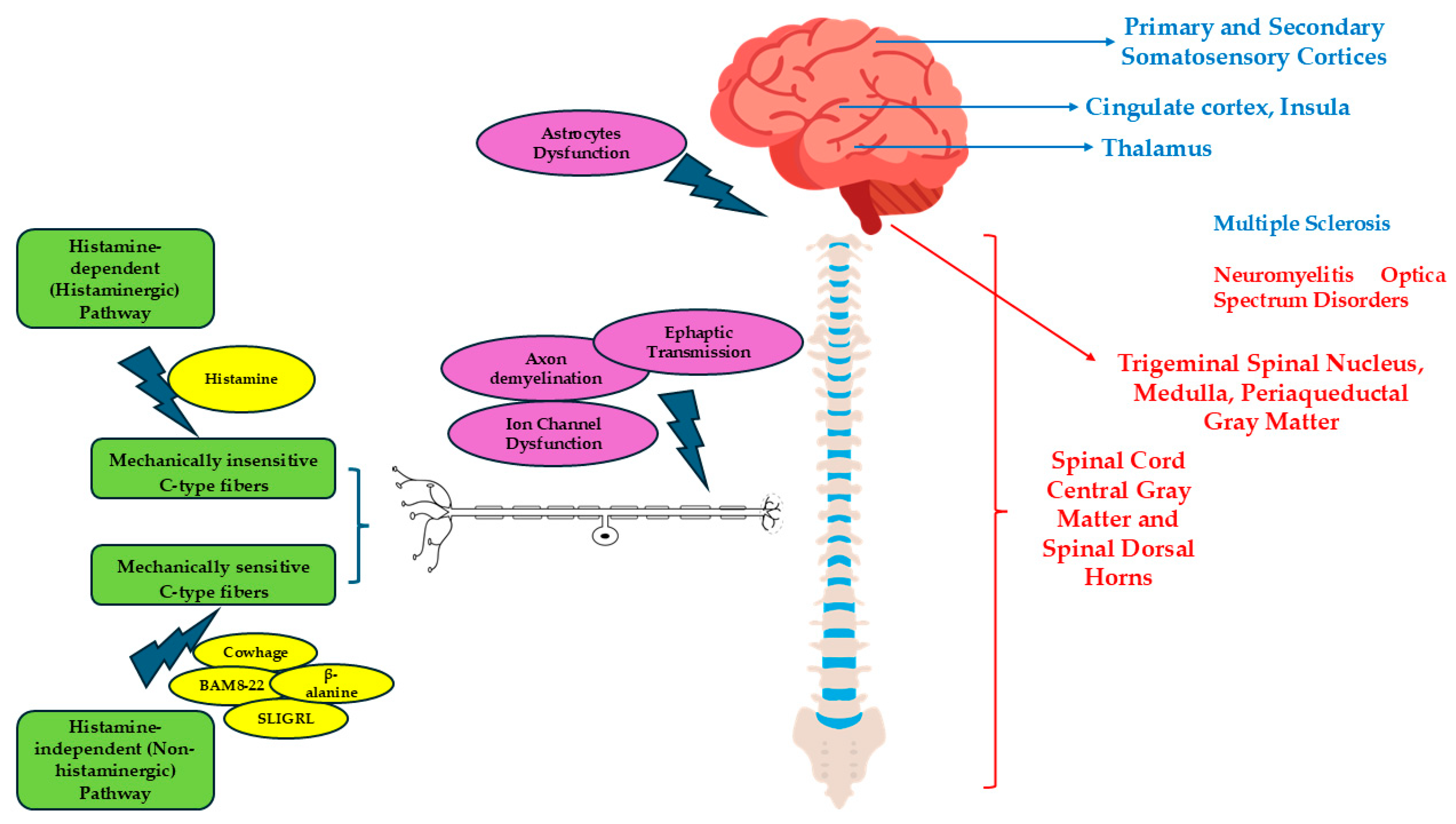
Figure 2 shows the pruritogenic pathways and central processing of itch in MS and NMOSD.
A comparative overview of pruritus characteristics in MS and NMOSD is presented in Table 1.

10. Drugs and Pruritus

Injection-site reactions (ISRs), such as erythema, edema, pain and pruritus, are the most common adverse reactions (ARs) following Interferon-beta administration [43]. They are often moderate and generalized cutaneous ARs, leading to itchy eczematous dermatitis [43]. Generalized cutaneous ARs, such as itchy eczematous dermatitis, are extremely rare across all disease-modifying therapies (DMTs) for multiple sclerosis. Among the available treatments, dermatologic side effects have been reported more frequently with teriflunomide, as also mentioned in its Summary of Product Characteristics (SPC). It has been previously reported that natalizumab, a monoclonal anti-a4-integrin antibody, may cause an acquired perforating dermatosis with pruritus and skin rash [44,45]. Furthermore, pruritus has been reported as an AR following administration of glatiramer acetate, fingolimod, teriflunomide, alemtuzumab, cladribine, dimethyl fumarate, ocrelizumab, and rituximab [46,47,48]. Moreover, acute attack therapy, such as methylprednisolone infusion and therapeutic plasma exchange, may lead to pruritus [49,50].

11. Pruritus in Other Autoimmune Demyelinating Disorders of the CNS

In a recent study, pruritus was observed in a patient with recurrent myelitis during a relapse of anti-myelin oligodendrocyte glycoprotein (MOG)-associated disease (MOGAD) [51]. Bauer and Sacheli reported the case of a patient experiencing left leg weakness with full left arm paresthesia, itching over the entire left face, and blurred vision in the left eye, after an influenza vaccination (H1N1, H3N2, B/Yamagata and B/Victoria) [52]. Scattered foci of bright T2 signal changes were seen in the peri- and non-periventricular regions and the deep hemispheric centrum, peritrigonal, and cortical/subcortical regions, suggesting a demyelinating disorder of the CNS but without satisfying the diagnostic criteria for clinically isolated syndrome (CIS) and acute disseminated encephalomyelitis (ADEM) [52].

12. Management and Treatment of Pruritus in Demyelinating Diseases of the CNS

Itching is often intense, debilitating, and challenging to manage. Conventional antipruritic therapies are generally ineffective, and no standardized treatment protocol has been established to date. In case of acute attack or contrast-enhancement lesions, administration of high-dose methylprednisolone may relieve the itching sensation [36]. H1-antihistamines are among the most extensively studied pharmacological agents for the management of pruritus [1,53]. While sedating first-generation antihistamines are considered highly effective in cases of non-neuropathic pruritus, they generally show limited efficacy in neuropathic itch [1,53]. Nonetheless, their sedative properties may offer indirect benefits, such as improving sleep quality, reducing nocturnal scratching, and alleviating scratch-induced cutaneous inflammation [1,53]. Importantly, their use is contraindicated in patients over 65 years of age due to the increased risk of adverse cardiovascular events [1,53]. Systemic sodium channel blockers such as carbamazepine and oxcarbazepine have been suggested as a potential first-line treatment for pruritus in multiple sclerosis, with reports indicating a favorable clinical response [53,54]. In addition, phenytoin, phenobarbital, and synacthen have been explored as alternative therapeutic options in the management of neuropathic pruritus, with a fair degree of symptom relief [9]. Gabapentin and pregabalin, which modulate the trafficking of alpha-2-delta calcium channels and exert supraspinal effects, have previously been reported to provide benefits [53]; they are commonly prescribed, particularly for patients with brachioradial pruritus [53]. Portugal et al. described the case of a 58-year-old male patient with primary progressive MS and a longstanding history of refractory bilateral focal pruritus, whose marked symptomatic improvement was achieved following local intradermal administration of botulinum toxin type A [55]. Also, topical treatments such as glucocorticosteroids or tacrolimus, although not effective against the primary causes of neuropathic itch, can alleviate the associated inflammation that exacerbates it [53]. In some cases, they may also help differentiate neuropathic itch from inflammatory itch based on the presence or absence of symptom improvement [53]. The significant association between pruritus and psychological comorbidities in demyelinating diseases of the CNS, such as anxiety and depression, suggests that psychological interventions could potentially benefit affected patients [6]. This highlights a promising avenue for future research aimed at evaluating their efficacy in alleviating itch. Addressing scratch-induced lesions is crucial to prevent further itching, infection, scarring, and disfigurement [53] and one of the simplest methods is trimming the fingernails. The use of thermoplastic mesh dressings may also promote wound healing by limiting additional scratching [53]. Since scratching often provides temporary relief of neuropathic itch by increasing sensory input, this behavior can become self-reinforcing, making it essential to break the scratching cycle as part of the treatment [53]. Cognitive behavioral therapy, physiotherapy, and meditation can assist patients in resisting the urge to scratch, alleviate depression or aggression, and reduce self-inflicted injury [53]. Many individuals benefit from occlusive therapy, which involves covering the itchy areas to reduce visual triggers and limit scratching [53], which not only protects the skin from further damage and sun exposure but also enhances the effectiveness of topical treatments [53]. Additionally, the mechanosensory input provided by tight bandaging may help relieve itch by increasing inhibitory signaling in the dorsal horn of the spinal cord [53]. In cases of refractory neuropathic itch, decompressive neurosurgery may be considered [53]. In a case report, transcranial direct stimulation was shown to alleviate one patient’s persistent itch for three months, although it did not have an effect on pain [56]. An overview of current pharmacological and non-pharmacological treatment options for pruritus in MS and NMOSD is provided in Table 2 and Table 3.

13. Conclusions

Pruritus is a symptom that significantly impacts a patient’s quality of life and can lead to dermatological lesions. Therefore, in cases of intractable pruritus, it is crucial to investigate not only the dermatological or systemic conditions but also demyelinating diseases of the CNS. Although specific treatment guidelines for pruritus in CNS demyelinating diseases are still lacking, various studies have reported benefits following the use of medications such as carbamazepine. However, further research is needed to establish a consensus on the management of pruritus in these conditions.

Author Contributions

Conceptualization, C.M.; resources, C.M.; writing—original draft preparation, C.M. and M.Z.; writing—review and editing, M.Z.; visualization, C.M.; supervision, C.M. All authors have read and agreed to the published version of the manuscript.

Funding

This research received no external funding.

Institutional Review Board Statement

Not applicable.

Informed Consent Statement

Not applicable.

Data Availability Statement

No new data were created or analyzed in this study. Data sharing is not applicable to this article.

Conflicts of Interest

The authors declare no conflicts of interest.

References

  1. Rajagopalan, M.; Saraswat, A.; Godse, K.; Shankar, D.S.K.; Kandhari, S.; Shenoi, S.D.; Tahiliani, S.; Zawar, V.V. Diagnosis and management of chronic pruritus: An expert consensus review. Indian J. Dermatol. 2017, 62, 7–17. [Google Scholar] [CrossRef]
  2. Huguen, J.; Brenaut, E.; Clerc, C.J.; Poizeau, F.; Marcorelles, P.; Quereux, G.; Dupuy, A.; Misery, L. Comparison of Characteristics of Neuropathic and Non-neuropathic Pruritus to Develop a Tool for the Diagnosis of Neuropathic Pruritus: The NP5. Front. Med. 2019, 6, 79. [Google Scholar] [CrossRef]
  3. Jakimovski, D.; Bittner, S.; Zivadinov, R.; Morrow, S.A.; Benedict, R.H.; Zipp, F.; Weinstock-Guttman, B. Multiple sclerosis. Lancet 2024, 403, 183–202. [Google Scholar] [CrossRef] [PubMed]
  4. Dobson, R.; Giovannoni, G. Multiple sclerosis—A review. Eur. J. Neurol. 2019, 26, 27–40. [Google Scholar] [CrossRef]
  5. Siriratnam, P.; Huda, S.; Butzkueven, H.; van der Walt, A.; Jokubaitis, V.; Monif, M. A comprehensive review of the advances in neuromyelitis optica spectrum disorder. Autoimmun. Rev. 2023, 22, 103465. [Google Scholar] [CrossRef] [PubMed]
  6. Ingrasci, G.; Tornes, L.; Brown, A.; Delgado, S.; Hernandez, J.; Yap, Q.V.; Yosipovitch, G. Chronic pruritus in multiple sclerosis and clinical correlates. J. Eur. Acad. Dermatol. Venereol. 2023, 37, 154–159. [Google Scholar] [CrossRef] [PubMed]
  7. Haki, M.; AL-Biati, H.A.; Al-Tameemi, Z.S.; Ali, I.S.; Al-hussaniy, H.A. Review of multiple sclerosis: Epidemiology, etiology, pathophysiology, and treatment. Medicine 2024, 103, e37297. [Google Scholar] [CrossRef]
  8. Correale, J.; Farez, M.F. The Role of Astrocytes in Multiple Sclerosis Progression. Front. Neurol. 2015, 6, 180. [Google Scholar] [CrossRef]
  9. Freiha, J.; Riachi, N.; Chalah, M.A.; Zoghaib, R.; Ayache, S.S.; Ahdab, R. Paroxysmal Symptoms in Multiple Sclerosis—A Review of the Literature. J. Clin. Med. 2020, 9, 3100. [Google Scholar] [CrossRef]
  10. Osterman, P.O. Paroxysmal Itching in Multiple Sclerosis. Int. J. Dermatol. 1979, 18, 626–627. [Google Scholar] [CrossRef]
  11. Sandyk, R. Paroxysmal Itching in Multiple Sclerosis During Treatment with External Magnetic Fields. Int. J. Neurosci. 1994, 75, 65–71. [Google Scholar] [CrossRef] [PubMed]
  12. Zhao, S.; Mutch, K.; Elsone, L.; Miller, J.; Jacob, A. An unusual case of ‘itchy paralysis’: Neuromyelitis optica presenting with severe neuropathic itch. Pract. Neurol. 2015, 15, 149–151. [Google Scholar] [CrossRef]
  13. Mora Cuervo, D.L.; Hansel, G.; Sato, D.K. Immunobiology of neuromyelitis optica spectrum disorders. Curr. Opin. Neurobiol. 2022, 76, 102618. [Google Scholar] [CrossRef]
  14. Lotan, I.; Bacon, T.; Kister, I.; Levy, M. Paroxysmal symptoms in neuromyelitis optica spectrum disorder: Results from an online patient survey. Mult. Scler. Relat. Disord. 2020, 46, 102578. [Google Scholar] [CrossRef]
  15. Ikoma, A.; Rukwied, R.; Ständer, S.; Steinhoff, M.; Miyachi, Y.; Schmelz, M. Neurophysiology of Pruritus. Arch. Dermatol. 2003, 139, 1475–1478. [Google Scholar] [CrossRef] [PubMed]
  16. Song, J.; Xian, D.; Yang, L.; Xiong, X.; Lai, R.; Zhong, J. Pruritus: Progress toward Pathogenesis and Treatment. Biomed. Res. Int. 2018, 2018, 9625936. [Google Scholar] [CrossRef]
  17. Luo, J.; Feng, J.; Liu, S.; Walters, E.T.; Hu, H. Molecular and cellular mechanisms that initiate pain and itch. Cell. Mol. Life Sci. 2015, 72, 3201–3223. [Google Scholar] [CrossRef]
  18. Akiyama, T.; Carstens, E. Neural processing of itch. Neuroscience 2013, 250, 697–714. [Google Scholar] [CrossRef]
  19. Wilson, S.R.; Gerhold, K.A.; Bifolck-Fisher, A.; Liu, Q.; Patel, K.N.; Dong, X.; Bautista, D.M. TRPA1 is required for histamine-independent, Mas-related G protein–coupled receptor–mediated itch. Nat. Neurosci. 2011, 14, 595–602. [Google Scholar] [CrossRef] [PubMed]
  20. He, M.; Wu, L.; Huang, D.; Yau, V.; Yu, S. Pruritus in neuromyelitis optica spectrum disorders and multiple sclerosis. J. Clin. Neurosci. 2020, 79, 108–112. [Google Scholar] [CrossRef]
  21. Elsone, L.; Townsend, T.; Mutch, K.; Das, K.; Boggild, M.; Nurmikko, T.; Jacob, A. Neuropathic pruritus (itch) in neuromyelitis optica. Mult. Scler. J. 2013, 19, 475–479. [Google Scholar] [CrossRef]
  22. Yamamoto, K.; Kawazawa, S.; Takase, Y.; Fukusako, T.; Morimatsu, M. Paroxysmal itching and magnetic resonance imaging of the spinal cord in multiple sclerosis. Rinsho Shinkeigaku 1989, 29, 1345–1351. [Google Scholar]
  23. Akiyama, T.; Nguyen, T.; Curtis, E.; Nishida, K.; Devireddy, J.; Delahanty, J.; Carstens, M.L.; Carstens, E. A central role for spinal dorsal horn neurons that express neurokinin-1 receptors in chronic itch. Pain 2015, 156, 1240–1246. [Google Scholar] [CrossRef]
  24. Davidson, S.; Zhang, X.; Khasabov, S.G.; Moser, H.R.; Honda, C.N.; Simone, D.A.; Giesler, G.J., Jr. Pruriceptive spinothalamic tract neurons: Physiological properties and projection targets in the primate. J. Neurophysiol. 2012, 108, 1711–1723. [Google Scholar] [CrossRef] [PubMed]
  25. Xiao, L.; Qiu, W.; Lu, Z.; Li, R.; Hu, X. Intractable pruritus in neuromyelitis optica. Neurol. Sci. 2016, 37, 949–954. [Google Scholar] [CrossRef] [PubMed]
  26. Saini, L.; Gunasekaran, P.K.; Tiwari, S.; Krishna, D.; Laxmi, V.; Jindal, P.; Kumar, P. Paroxysmal Neuropathic Pruritus in Patients With Chiari Malformation Type I: A Rare Phenotype. Pediatr. Neurol. 2023, 140, 65–67. [Google Scholar] [CrossRef]
  27. Misery, L.; Genestet, S.; Zagnoli, F. Neuromyelitis Optica and Skin. Dermatology 2022, 238, 823–828. [Google Scholar] [CrossRef]
  28. Kremer, L.; Mealy, M.; Jacob, A.; Nakashima, I.; Cabre, P.; Bigi, S.; Paul, F.; Jarius, S.; Aktas, O.; Elsone, L.; et al. Brainstem manifestations in neuromyelitis optica: A multicenter study of 258 patients. Mult. Scler. J. 2014, 20, 843–847. [Google Scholar] [CrossRef]
  29. Matsuura, J.; Kimura, A.; Kasai, T.; Yoshida, T.; Nakagawa, M.; Mizuno, T. A Case of Neuromyelitis Optica with Relapse Symptoms from Paroxysmal Pruritus. Brain Nerve 2015, 67, 1057–1060. [Google Scholar] [CrossRef]
  30. Yosipovitch, G.; Ishiuji, Y.; Patel, T.S.; Hicks, M.I.; Oshiro, Y.; Kraft, R.A.; Winnicki, E.; Coghill, R.C. The Brain Processing of Scratching. J. Investig. Dermatol. 2008, 128, 1806–1811. [Google Scholar] [CrossRef] [PubMed]
  31. Mochizuki, H.; Kakigi, R. Central mechanisms of itch. Clin. Neurophysiol. 2015, 126, 1650–1660. [Google Scholar] [CrossRef]
  32. Mochizuki, H.; Tanaka, S.; Morita, T.; Wasaka, T.; Sadato, N.; Kakigi, R. The cerebral representation of scratching-induced pleasantness. J. Neurophysiol. 2014, 111, 488–498. [Google Scholar] [CrossRef] [PubMed]
  33. Zhang, L.; Zou, Z.; Yu, S.; Xiao, X.; Shi, Y.; Cao, W.; Liu, Y.; Zheng, H.; Zheng, Q.; Zhou, S.; et al. Functional connectivity impairment of thalamus-cerebellum-scratching neural circuits in pruritus of chronic spontaneous urticaria. Front. Neurosci. 2022, 16, 1026200. [Google Scholar] [CrossRef] [PubMed]
  34. Mahmoud, O.; Oladipo, O.; Mahmoud, R.H.; Yosipovitch, G. Itch: From the skin to the brain—Peripheral and central neural sensitization in chronic itch. Front. Mol. Neurosci. 2023, 16, 1272230. [Google Scholar] [CrossRef] [PubMed]
  35. Yosipovitch, G.; Rosen, J.D.; Hashimoto, T. Itch: From mechanism to (novel) therapeutic approaches. J. Allergy Clin. Immunol. 2018, 142, 1375–1390. [Google Scholar] [CrossRef]
  36. Kataria, S.; Neupane, K.; Ahmed, Z.; Rehman, U.; Asif, S. An Unusual Presentation of Multiple Sclerosis in a Middle-Aged Woman: A Case Report and Literature Review. Cureus 2020, 12, e11017. [Google Scholar] [CrossRef]
  37. Hadad, R.; Mandelli, M.L.; Rankin, K.P.; Toohey, C.; Sturm, V.E.; Javandel, S.; Milicic, A.; Knudtson, M.; Allen, I.E.; Hoffmann, N.; et al. Itching Frequency and Neuroanatomic Correlates in Frontotemporal Lobar Degeneration. JAMA Neurol. 2024, 81, 977–984. [Google Scholar] [CrossRef]
  38. Wong, L.-S.; Wu, T.; Lee, C.-H. Inflammatory and Noninflammatory Itch: Implications in Pathophysiology-Directed Treatments. Int. J. Mol. Sci. 2017, 18, 1485. [Google Scholar] [CrossRef]
  39. Wang, Y.; Fang, J.L.; Cui, B.; Liu, J.; Song, P.; Lang, C.; Bao, Y.; Sum, R.; Xu, C.; Ding, X.; et al. The functional and structural alterations of the striatum in chronic spontaneous urticaria. Sci. Rep. 2018, 8, 1725. [Google Scholar] [CrossRef]
  40. Harte, S.E.; Harris, R.E.; Clauw, D.J. The neurobiology of central sensitization. J. Appl. Biobehav. Res. 2018, 23, e12137. [Google Scholar] [CrossRef]
  41. Tsuda, M. Astrocytes in the spinal dorsal horn and chronic itch. Neurosci. Res. 2018, 126, 9–14. [Google Scholar] [CrossRef]
  42. Shiratori-Hayashi, M.; Tsuda, M. Role of reactive astrocytes in the spinal dorsal horn under chronic itch conditions. J. Pharmacol. Sci. 2020, 144, 147–150. [Google Scholar] [CrossRef] [PubMed]
  43. Maurelli, M.; Bergamaschi, R.; Antonini, A.; Fargnoli, M.C.; Puma, E.; Mallucci, G.; Totaro, R.; Girolomoni, G. Interferon-beta injection site reactions in patients with multiple sclerosis. J. Dermatol. Treat. 2018, 29, 831–834. [Google Scholar] [CrossRef]
  44. Piqué-Duran, E.; Eguía, P.; García-Vázquez, O. Acquired perforating dermatosis associated with natalizumab. J. Am. Acad. Dermatol. 2013, 68, e185–e187. [Google Scholar] [CrossRef]
  45. Fisher, R.M.; Hadley, G.; Ieremia, E.; Moswela, O.; Zaki, F.; DeLuca, G.C.; McPherson, T. Natalizumab-induced acquired perforating dermatosis. Clin. Exp. Dermatol. 2021, 46, 1373–1375. [Google Scholar] [CrossRef] [PubMed]
  46. Barbieri, M.A.; Sorbara, E.E.; Battaglia, A.; Cicala, G.; Rizzo, V.; Spina, E.; Cutroneo, P.M. Adverse Drug Reactions with Drugs Used in Multiple Sclerosis: An Analysis from the Italian Pharmacovigilance Database. Front. Pharmacol. 2022, 13, 808370. [Google Scholar] [CrossRef]
  47. He, D.; Guo, R.; Zhang, F.; Zhang, C.; Dong, S.; Zhou, H. Rituximab for relapsing-remitting multiple sclerosis. Cochrane Database Syst. Rev. 2013, 2013, CD009130. [Google Scholar] [CrossRef]
  48. Liang, G.; Chai, J.; Ng, H.S.; Tremlett, H. Safety of dimethyl fumarate for multiple sclerosis: A systematic review and meta-analysis. Mult. Scler. Relat. Disord. 2020, 46, 102566. [Google Scholar] [CrossRef]
  49. Fateen, T.; Sultana, N.; Sarwar, M.; Saqlain, N. Complications of Therapeutic Plasma Exchange in pediatric patients: An experience at a tertiary care hospital. Pak. J. Med. Sci. 2023, 39, 994. [Google Scholar] [CrossRef]
  50. Moreno Escobosa, M.C.; Cruz Granados, S.; Moya Quesada, M.C.; Amat López, J. Anaphylaxis due to methylprednisolone. J. Investig. Allergol. Clin. Immunol. 2008, 18, 407–408. [Google Scholar] [PubMed]
  51. Pujari, S.S.; Kulkarni, R.V.; Nadgir, D.B.; Ojha, P.K.; Nagendra, S.; Aglave, V.; Nadgir, R.D.; Sant, H.; Palasdeokar, N.; Nirhale, S.; et al. Myelin Oligodendrocyte Glycoprotein (MOG)-IgG Associated Demyelinating Disease. Ann. Indian Acad. Neurol. 2021, 24, 69–77. [Google Scholar] [CrossRef] [PubMed]
  52. Bauer, R. Influenza Vaccine-Induced CNS Demyelination in a 50-Year-Old Male. Am. J. Case Rep. 2014, 15, 368–373. [Google Scholar] [CrossRef]
  53. Steinhoff, M.; Schmelz, M.; Szabó, I.L.; Oaklander, A.L. Clinical presentation, management, and pathophysiology of neuropathic itch. Lancet Neurol. 2018, 17, 709–720. [Google Scholar] [CrossRef]
  54. Yamamoto, M.; Yabuki, S.; Hayabara, T.; Otsuki, S. Paroxysmal itching in multiple sclerosis: A report of three cases. J. Neurol. Neurosurg. Psychiatry 1981, 44, 19–22. [Google Scholar] [CrossRef]
  55. Portugal, D.M.; Ferreira, E.F.; Camões-Barbosa, A. Botulinum toxin type A therapy for bilateral focal neuropathic pruritus in multiple sclerosis: A case report. Int. J. Rehabil. Res. 2021, 44, 382–383. [Google Scholar] [CrossRef] [PubMed]
  56. Dhand, A.; Aminoff, M.J. The neurology of itch. Brain 2014, 137, 313–322. [Google Scholar] [CrossRef] [PubMed]
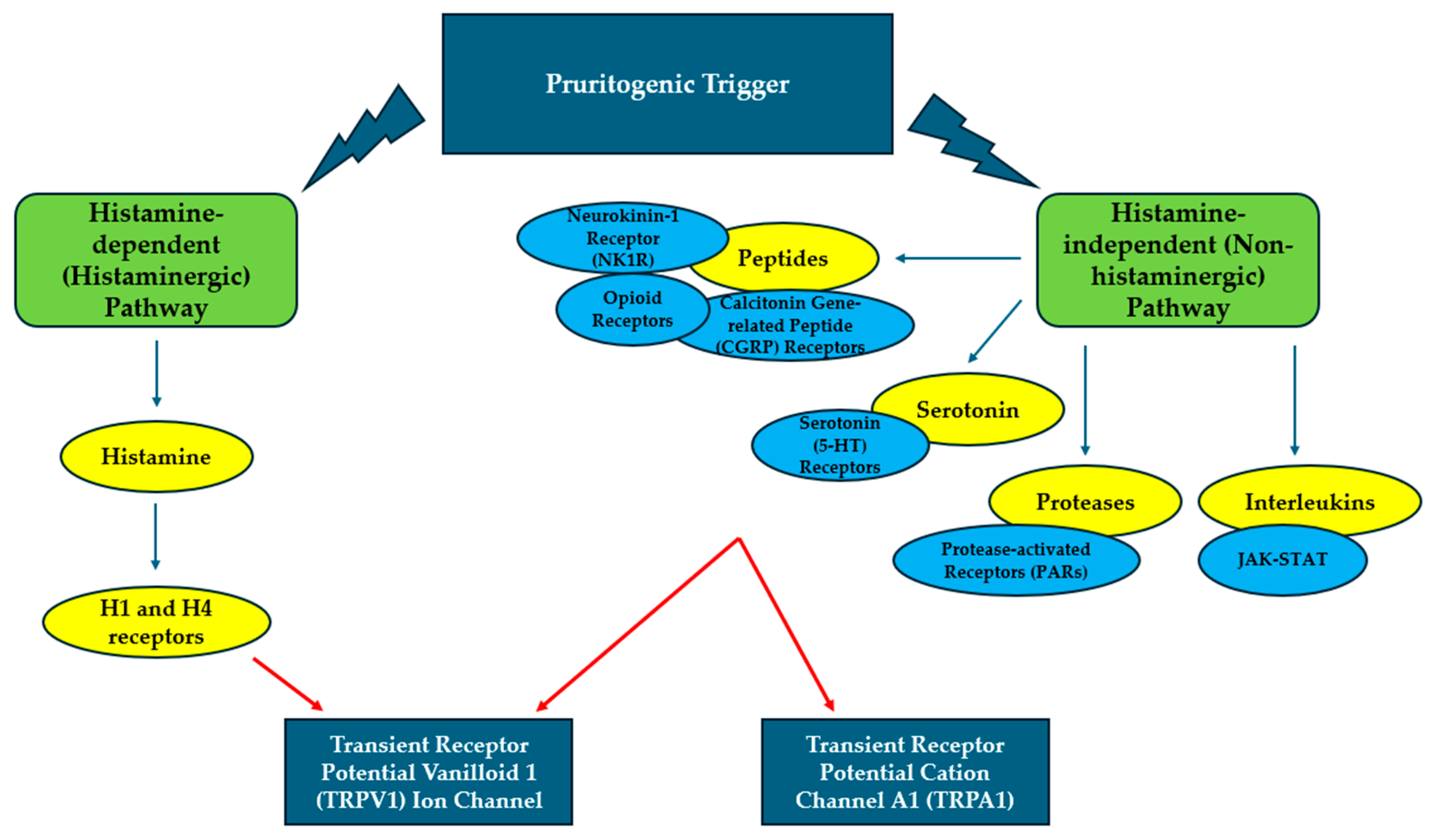
Figure 1. Schematic summary of the molecular mechanisms underlying pruritus. The figure illustrates key mediators, receptors, and pathways involved in the initiation and transmission of pruritic signals. Two distinct mechanisms mediate itch perception: the histaminergic pathway, triggered by histamine binding to histamine receptor 1 (H1R) and histamine receptor 4 (H4R), which activates phospholipase C beta-3 (PLC-β3) and phospholipase C (PLC) and subsequently the transient receptor potential vanilloid 1 (TRPV1) ion channel; and the non-histaminergic pathway, induced by pruritogens such as cowhage, chloroquine, bovine adrenal medulla peptide 8-22 (BAM8-22), Ser-Leu-Ile-Gly-Arg-Leu (SLIGRL), and β-alanine via protease-activated receptor 2 (PAR2) and Mas-related G-protein-coupled receptors (Mrgprs), leading to activation of transient receptor potential ankyrin 1 (TRPA1) and TRPV1 channels. Green rectangles represent the two main pathways (histaminergic and non-histaminergic), yellow ovals indicate the mediating signaling routes, blue ovals represent the mediating molecules involved in the itch cascade, and blue rectangles correspond to the activation receptors located at the terminal stage of the mechanism.
Allergies 05 00032 g001
Figure 2. Pruritogenic pathways and central processing of itch in MS and NMOSD. External pruritogenic stimuli may activate either the histamine-dependent (histaminergic) or the histamine-independent (non-histaminergic) pathway. In the former, histamine directly conveys the signal to the CNS via mechanically insensitive C-fibers. In the latter, various non-histaminergic pruritogens—including cowhage, chloroquine, BAM8-22, SLIGRL, and β-alanine—activate mechanically sensitive C-type fibers. Both pathways transmit signals through the dorsal root ganglion (DRG), ascending via the spinothalamic tract, through the brainstem to the thalamus, and ultimately to the cerebral cortex, where the itch is perceived. Internal mechanisms, such as axonal demyelination, ephaptic transmission, ion channel dysfunction, and astrocyte pathology, can also lead to neuropathic pruritus. Regions typically affected in MS are highlighted in blue, while those commonly involved in NMOSD are marked in red.
Allergies 05 00032 g002
Table 1. Comparison of Pruritus Characteristics in MS and NMOSD.
MSNMOSD
Frequency of neuropathic pruritus4.5% of patients12–27% of patients
Clinical features of pruritusSudden onset, localized, intense, lasting from a few seconds to several minutes, often accompanied by superficial sensory disturbances and/or painSudden onset, localized, intense, lasting from a few seconds to several minutes, often accompanied by superficial sensory disturbances and/or pain
Clinical significance of pruritusInitial presenting symptom, or a manifestation during disease relapses. Rarely, it occurs during remissionsInitial presenting symptom, or a manifestation during disease relapses
Dermatomal/Non-dermatomal DistributionPredominantly dermatomal distributionBoth dermatomal and non-dermatomal distribution
Location of lesionsPotentially throughout the CNS
  • Central cord lesions involving the dorsal horns
  • Periaqueductal grey matter
  • Medulla oblongata
Neuronal pathways involved
  • Ephaptic transmission from demyelinated axons
  • Inflammatory irritation
  • Ion channel dysfunction in demyelinated axons
  • Aberrant activation of adjacent corticospinal tracts and ephaptic transmission from damaged centripetal fibers
  • Spinoparabrachial pathway dysfunction
  • Astrocytic dysfunction
  • NK1R hyperactivity
Response to treatmentGood response to therapyPartial or insufficient response to therapy
Table 2. Available Pharmacological Treatments for Neuropathic Pruritus.
EffectivenessNotes
Systemic drugs
High Dose MethylprednisoloneGoodTo be administered in case of acute attack or contrast-enhancing lesions
H1-antihistaminesHigh in non-neuropathic pruritus, limited in neuropathic pruritus
  • Indirect benefits, such as improving sleep quality, reducing nocturnal scratching, and alleviating scratch-induced cutaneous inflammation
  • Contraindicated in patients >65 years
Systemic sodium channel blockers (Carbamazepine, Oxcarbazepine)High (considered the first-line therapy)Recommended in cases of neuropathic pruritus in MS
Calcium channel (α2δ subunit) modulators (Pregabalin, Gabapentin) High
  • Exertion of supraspinal effects
  • Recommended in cases of brachioradial pruritus
Antidepressants (Amitriptyline, Paroxetine, Sertraline) GoodRecommended in case of anxiety/depression coexistence
PhenytoinVariable
PhenobarbitalVariable
SynachtenVariable
Local drugs
Intradermal administration of botulinum toxin type AEffective in one caseEmployed in refractory bilateral focal pruritus
Topical SteroidsLow
  • It can alleviate the associated inflammation
  • It may help differentiate neuropathic itch from inflammatory itch
Topical TacrolimusLow
  • It can alleviate the associated inflammation
  • It may help differentiate neuropathic itch from inflammatory itch
TRPV1 receptor desensitization (Capsaicin cream)GoodBurning on initial application
Table 3. Available Non-Pharmacological Treatments for Neuropathic Pruritus.
EffectivenessNotes
Trimming the fingernailsHigh
Thermoplastic mesh dressingsHighIt may also promote wound healing
Cognitive behavioral therapyHighIt can assist patients in resisting the urge to scratch, alleviate depression or aggression, and reduce self-inflicted injury
PhysiotherapyHighIt can assist patients in resisting the urge to scratch, alleviate depression or aggression, and reduce self-inflicted injury
MeditationHighIt can assist patients in resisting the urge to scratch, alleviate depression or aggression, and reduce self-inflicted injury
Occlusive therapyGoodCovering the itchy areas to reduce visual triggers and limit scratching could protect the skin from further damage and sun exposure, enhance the effectiveness of topical treatments, help relieve itch
Decompressive neurosurgeryVariableRecommended in case of refractory neuropathic itch
Transcranial direct stimulationEffective in one caseIt does not relieve pain
Disclaimer/Publisher’s Note: The statements, opinions and data contained in all publications are solely those of the individual author(s) and contributor(s) and not of MDPI and/or the editor(s). MDPI and/or the editor(s) disclaim responsibility for any injury to people or property resulting from any ideas, methods, instructions or products referred to in the content.