Protocol

Effect of Photobiomodulation Therapy in an Experimental Model of Chronic Obstructive Pulmonary Disease: A Dosimetric Study

by
Cintia Estefano Alves
1,*,
Tawany Gonçalves Santos
1,
Luana Beatriz Vitoretti
1,
Cinthya Cosme Gutierrez Duran
1,
Stella Zamuner
1,
Rodrigo Labat
1,
José Antonio Silva, Jr.
1,
Maria Cristina Chavantes
1,
Flavio Aimbire
2,
Renata Kelly da Palma
3,4 and
Ana Paula Ligeiro de Oliveira
1
1
Programa em Medicina-Biofotônica, Universidade Nove de Julho, São Paulo 01525-000, Brazil
2
Instituto de Ciências e Tecnologia, Universidade Federal de São Paulo, Unifesp, São José dos Campos 12245-000, Brazil
3
Faculty of Health Sciences at Manresa, University of Vic-Central University of Catalonia (UVic-UCC), 08242 Manresa, Spain
4
University Center of Anápolis, Human Movement and Rehabilitation, Post-Graduate Program, Evangelic University of Goiás, UniEVANGELICA, Anápolis 75083-515, Brazil
*
Author to whom correspondence should be addressed.
Allergies 2025, 5(4), 33; https://doi.org/10.3390/allergies5040033
Submission received: 23 April 2025 / Revised: 26 June 2025 / Accepted: 8 September 2025 / Published: 26 September 2025
(This article belongs to the Section Physiopathology)

Abstract

This study aimed to evaluate the effects of different dosimetric parameters of photobiomodulation therapy (PBMT) in an experimental model of chronic obstructive pulmonary disease (COPD). C57BL/6 mice were assigned to the following groups: Baseline, COPD, and COPD treated with PBMT at doses of 1 J, 3 J, 5 J, and 7.5 J. Treatment was performed using a diode laser (660 nm, 100 mW) applied for 10 s, 30 s, 50 s, and 120 s, respectively, over 15 consecutive days. COPD was induced by orotracheal instillation of cigarette smoke extract twice weekly for 45 days. Analyses included total cell count, immune cell profiling by flow cytometry, pulmonary infiltration of inflammatory markers, necrosis, apoptosis, and reactive oxygen species (ROS) production. Data were analyzed using one-way ANOVA followed by the Newman–Keuls post hoc test, with statistical significance set at p < 0.05. PBMT significantly reduced inflammatory cell infiltration, with the most pronounced anti-inflammatory effects observed at doses of 1 J and 3 J, highlighting the importance of appropriate dosimetry in optimizing the therapeutic outcomes of PBMT for COPD.

1. Introduction

Chronic Obstructive Pulmonary Disease (COPD) affects approximately 210 million people worldwide and ranks as the fourth leading cause of death, accounting for 4.8% of global mortality [1]. Despite its high prevalence, COPD remains underrecognized by both health authorities and the general population. The primary cause is exposure to cigarette smoke, although other sources—such as air pollution, biomass combustion, and alternative smoking devices (e.g., hookahs and electronic cigarettes)—also contribute to disease onset [2,3]. In 1–2% of cases, COPD is associated with alpha-1 antitrypsin deficiency [4].
COPD is a chronic, progressive, and partially reversible disease characterized by airway obstruction (chronic bronchitis) and alveolar destruction (emphysema) [5]. Oxidative stress plays a central role in the pathogenesis of COPD, contributing to epithelial injury, mitochondrial dysfunction, apoptosis, extracellular matrix remodeling, and mucus hypersecretion [6,7]. Pulmonary inflammation is predominantly neutrophilic, with contributions from eosinophils, CD8+ T lymphocytes, and elevated levels of pro-inflammatory cytokines, such as IL-6, IL-8, TNF-α, and IL-1β [8,9,10].
Respiratory exacerbations—characterized by worsening symptoms and systemic inflammation—significantly impact quality of life and are major drivers of hospitalizations and healthcare costs [11,12,13,14,15]. Although treatable, COPD frequently leads to extrapulmonary manifestations and functional decline [11].
Conventional treatment includes bronchodilators, corticosteroids, and oxygen therapy. However, these interventions offer limited efficacy and are associated with adverse effects [16,17]. As a result, complementary therapies such as photobiomodulation therapy (PBMT) have gained growing interest.
Mitochondria are recognized as key targets of PBMT due to the presence of mitochondrial photoacceptors, particularly cytochromes, which exhibit high affinity for light wavelengths in the visible red to near-infrared spectrum—especially between 635 and 660 nm. These wavelengths are believed to act primarily on cytochrome c oxidase (CCO), leading to nitric oxide (NO) displacement, increased membrane potential, ATP, ROS, and Ca2+, thereby activating signaling pathways related to cell division and proliferation [18,19]. This results in enhanced respiratory chain activity, increased ATP production, anti-inflammatory, reparative and immunomodulatory effects, reduced oxidative stress, and improved cellular function [20,21,22].
In parallel, light irradiation induces nitric oxide (NO) release a gaseous messenger widely associated with vasodilation, inflammation, and redox metabolism. Notably, NO exerts biphasic, dose-dependent effects and is also involved in post-translational modifications such as protein S-nitrosylation, a mechanism that regulates multiple intracellular pathways and can modulate both mitochondrial function and inflammatory responses [23]. Understanding these mechanisms is crucial in COPD, whose pathophysiology involves mitochondrial dysfunction, tissue hypoxia, and persistent inflammation [24]. In this context, dosimetric studies are essential to identify the optimal parameters capable of maximizing PBMT’s therapeutic effects while minimizing the risk of paradoxical responses caused by excessive light energy.
Recent studies suggest that PBMT can reduce inflammation and alveolar enlargement in cigarette smoke-induced COPD models [25]. In a chronic asthma model, specific doses of 660 nm laser light significantly reduced inflammatory markers, particularly at a dose of 3 J [26]. Based on these findings, a similar protocol was applied to a COPD model, yielding positive results in terms of inflammatory mediators and pulmonary remodeling.
Given PBMT’s therapeutic potential and the limited literature on its application in COPD, the present study aims to evaluate the effects of different laser energy doses on inflammatory and histological parameters in an experimental COPD model, with the goal of identifying the most effective protocol for future clinical applications.

2. Objectives

2.1. Primary Objective

To evaluate the effects of photobiomodulation therapy with different levels of radiant energy in an experimental model of COPD.

2.2. Secondary Objectives

  • To evaluate the levels of cytokines secreted by BAL cells;
  • To quantify the total and differential cells present in BAL and in the blood of animals;
  • To analyze airway remodeling by histology;
  • To evaluate pulmonary mechanics;

3. Materials and Methods

3.1. Study Design

The experiment was previously approved by CEUA (animal use ethics committee) under number AN006.2013 (Approved on 15 July 2015). Thirty-six male mice of the C57BL/6 lineage were used. The animals were obtained from the vivarium of Universidad Nove de Julho and raised in a controlled environment, with humidity (50–60%), luminosity (12 h light/12 h dark) to simulate day and night, and temperature between (22–25°).

3.2. Sample Size

As illustrated in (Figure 1), six animals were used in each experimental group, based on the literature that demonstrated this to be a sufficient number of animals for the induction of COPD [3].

3.3. Randomization

For this experiment we used random randomization as a criterion.

3.4. Blinding

All experiments and analyses were carried out by the researcher himself. All experiments and analyses were carried out by the researcher himself.

3.5. Statistical Methods

Data was analyzed using GraphPad Prism 3.1 software (La Jolla, CA, USA). The normal distribution of data was assessed using the Kolmogorov–Smirnov test. Data with parametric distribution was subjected to One-way ANOVA followed by the Newman-Keuls test for comparison between groups. Data with nonparametric distribution were subjected to One-way ANOVA on Ranks followed by the Dunn test for comparison between groups.

3.6. Experimental Animals

The animals used, from the C57BL/6 lineage, males, were on average 6 months old at the beginning of the experiments, weighing between 20 g and 26 g.

3.6.1. Cigarette Extract

For the preparation of the cigarette extract, Marlboro brand cigarettes (tar: 13.0 mg, nicotine: 1.10 mg, carbon monoxide: 10 mg) were used. The cigarettes were burned, and the smoke was passed through a hose, with one end connected to a conical tube containing 1X PBS buffer solution, using a ratio of 1 cigarette per 4 mL of buffer solution (1 cigarette in 4 mL). At the other end of the hose, a vacuum pump with constant pressure was attached, where the cigarette was introduced.
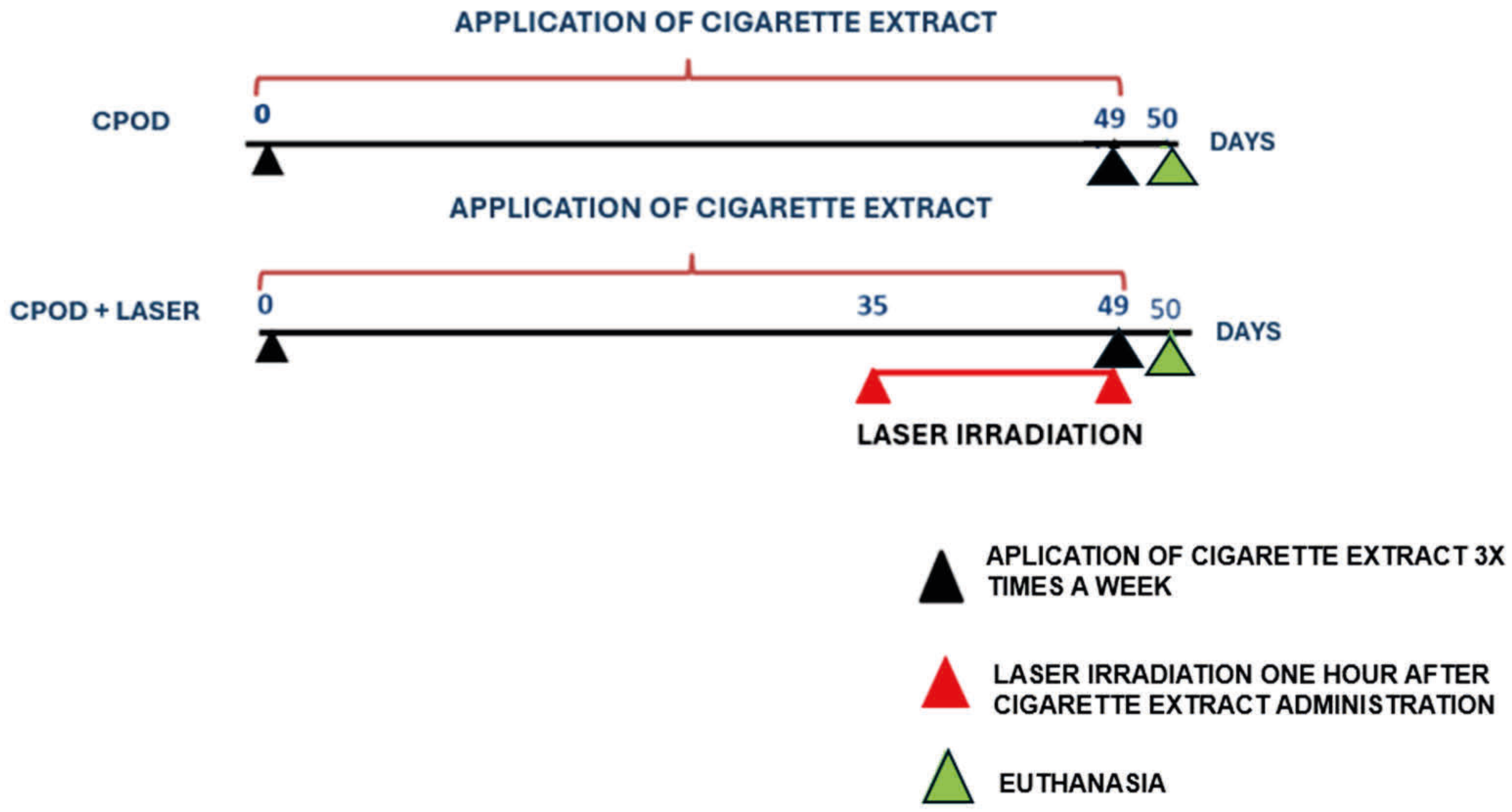
To induce the disease using cigarette extract, the animals were anesthetized intramuscularly injection of 2% xylazine (0.06 mL/100 g) 10% ketamine (0.08 mL/100 g), 4 µL per animal. The animals were positioned vertically in a way that allowed proper access to the administration route. 30 µL of the cigarette extract was administered via orotracheal route, three times per week for 7 weeks (49 days). The experimental design of COPD induction and photobiomodulation therapy is shown in (Figure 2)

3.6.2. Irradiation

After 35 days of disease induction, the COPD+LASER group received a punctual application of a diode laser, wavelength of 660 nm, irradiated area of 0.785 cm2, in three regions: one below the trachea, and the other two in each lung lobe (right and left). The time and power varied for each group as shown in the following diagram. The MMO Optics laser/Model: TF Premier PLUS was used.
The animals were immobilized and irradiated at the points illustrated in the following (Table 1).

3.7. Euthanasia

After 45 days of experiment, the animals were euthanized with a high dosage of anesthesia, with intramuscular (i.m.) injection of xylazine 2% (0.06 mL/100 g) ketamine 10% (0.08 mL/100 g). The substance was administered minutes before exsanguination, using a 1 mL syringe and a 25 × 5 mm hypodermic needle. After blood collection, for smear slides and differential cell count in circulation, the animals were tracheostomized and cannulated, and the lungs were lavage with 3 × 0.5 mL of phosphate-buffered saline (PBS).

3.8. Assessment of Lung Inflammation in Bronchoalveolar Lavage (BAL)

The recovered lavage volume was centrifuged at 1600 rpm at 4 °C in 5 min. The supernatant was stored at −70 °C for cytokine analysis by ELISA. The cell button was resuspended in 1 mL of phosphate-buffered saline (PBS) and used to determine the total number of cells in the BAL performed by counting in the Neubauer Chamber. The remaining resuspended material was used to prepare cytospin slides. A 200 µL sample from each animal was used for differential cell counting, centrifuged for 10 min, 450 rpm in the Cytospin-2 model equipment, Shandon Instruments Sewickley, PA. The slides were stained using the Instant Prov staining technique: (1) place the slides in the Instant Prov I staining tank, leave for 10 s, remove and let drain for 5 s. (2). Place the slides in the Instant Prov II staining tank, leave for 10 s, remove and let drain for 5 s. (3). Place the slides in the Instant Prov III staining tank, leave for 20 s, remove, let drain for 5 s and wash the slides in running water. After staining, 100 cells were counted to determine the differential count.

3.9. Flow Cytometry

After lung extractions, the tissue was fragmented and incubated for 30 min at 37 °C with constant agitation in 2 mg/mL collagenase IV and 1 mg/mL deoxyribonuclease I (DNAse), (Sigma-Aldrich, St. Louis, MO, USA) After this period, we introduced Hank’s balanced solution (HBSS) together with EDTA to slow down the digestion of the material. After crushing and filtering the lung fragments through a 40 mm sieve, the contents were centrifuged for 10 min at 1,500 rpm, and the pellets were then resuspended in PBS buffer. After the incubation period, the samples were resuspended in 200 μL of the same buffer after being cleaned with PBS containing 0.01% BSA and sodium azide. Samples were acquired on a BD Accuri flow cytometer and analyzed using CSampler software (version 227.4, Becton Dickinson—BD ®, East Rutherford, NJ, USA).

4. Results

4.1. Effects of PBMT on the Number of Cells Present in BAL in a COPD Model

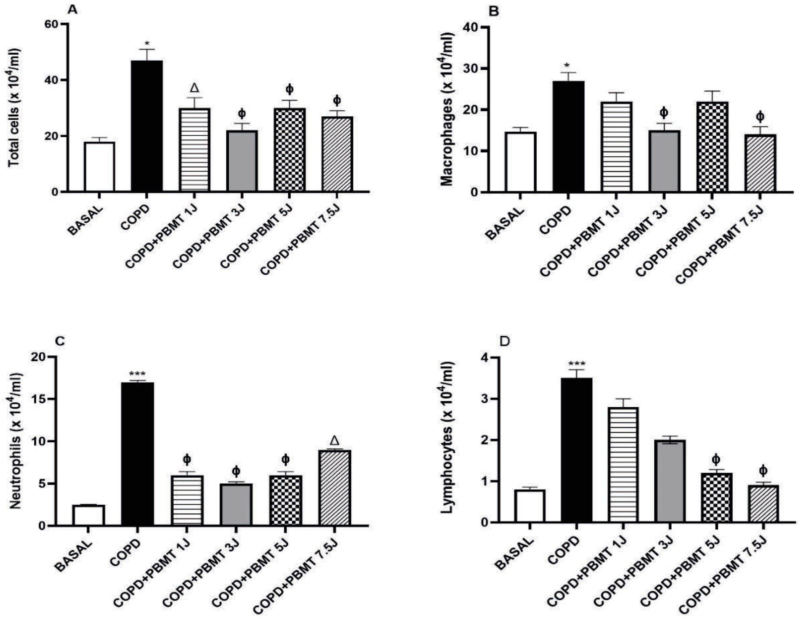
The data on the effects of PBMT on the quantification of cells present in bronchoalveolar lavage (BAL) are presented in (Figure 3). We found a significant increase in the total number of cells (A), macrophages (B), neutrophils (C) and lymphocytes (D) in the COPD group induced by cigarette smoke extract when compared to basal group (Figure 3A–D). On the other hand, regarding the total number of cells and neutrophils, we observed a decrease in all groups submitted to PBMT when compared to the COPD group (Figure 3A,C). In addition, we verified a reduction in the number of macrophages in the COPD+PBMT groups (3 J and 5 J) in relation to the COPD group (Figure 3B). In Figure 3D, we observed a reduction in the COPD+PBMT groups (5 J and 7.5 J) when compared to the COPD group.

4.2. Effects of PBMT on the Quantification of Cells Present in the Lung in a COPD Model

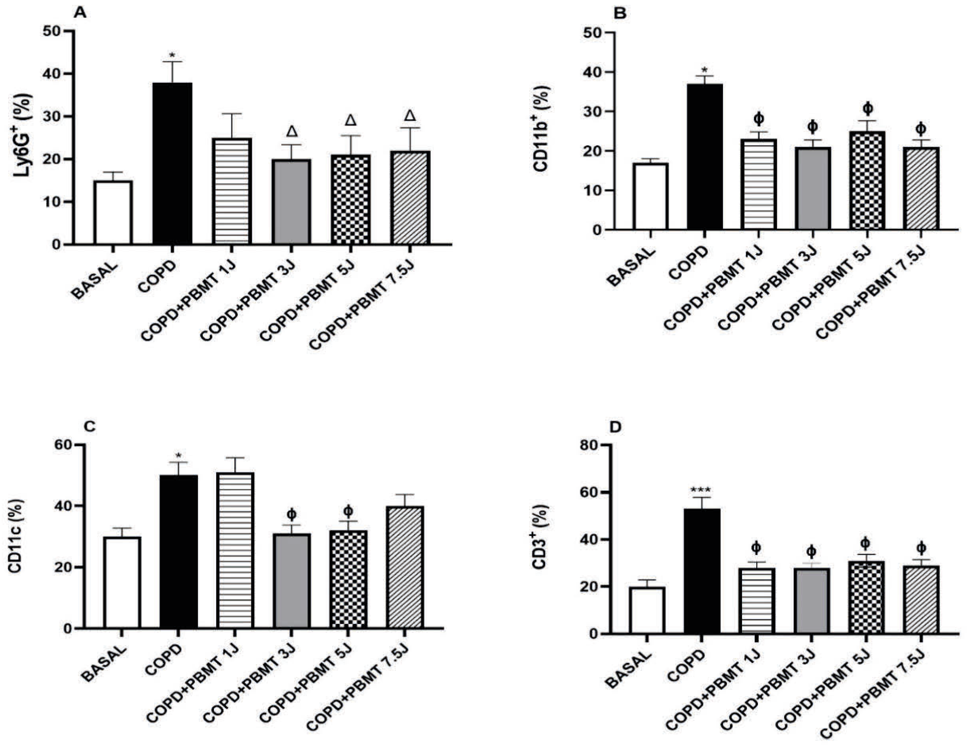
Data on the effects of PBMT on the quantification of cells present in the lung are presented in (Figure 4) We observed an increase in the number of neutrophils (Ly6G+), macrophages (CD11b+), dendritic cells (CD11c+), and total lymphocytes (CD3+) in the lungs of animals in the COPD group compared to the baseline group (Figure 4A–D). Regarding PBMT, we observed that all radiant energy levels used were able to reduce the number of macrophages and lymphocytes compared to the COPD group (Figure 4B,D). Additionally, only the COPD+PBMT groups (3 J, 5 J, and 7.5 J) reduced the number of neutrophils (Figure 4A) when compared to the COPD group. Furthermore, we observed a reduction in the number of dendritic cells in the COPD+PBMT groups (3 J and 5 J) compared to the COPD group (Figure 4C).

4.3. Effects of PBMT on Necrosis, Apoptosis and Reactive Oxygen Species Production in Leukocytes in the Lung

We observed that the application of cigarette extract in the COPD group led to an increase in leukocyte necrosis, apoptosis, and the production of reactive oxygen species (ROS) compared to the baseline group (Figure 5A–C). On the other hand, all COPD+PBMT groups reduced leukocyte necrosis compared to the COPD group (Figure 5A). Furthermore, as observed in Figure 5B,C, there was a reduction in apoptosis and ROS production in the COPD+PBMT (7.5 J) group compared to the COPD group.

4.4. Effect of PBMT Therapy on the Percentage of CD3+TGF- β + (A) and CD3+IL-1 β+ (B) Lymphocytes in BAL in an Experimental Model of COPD

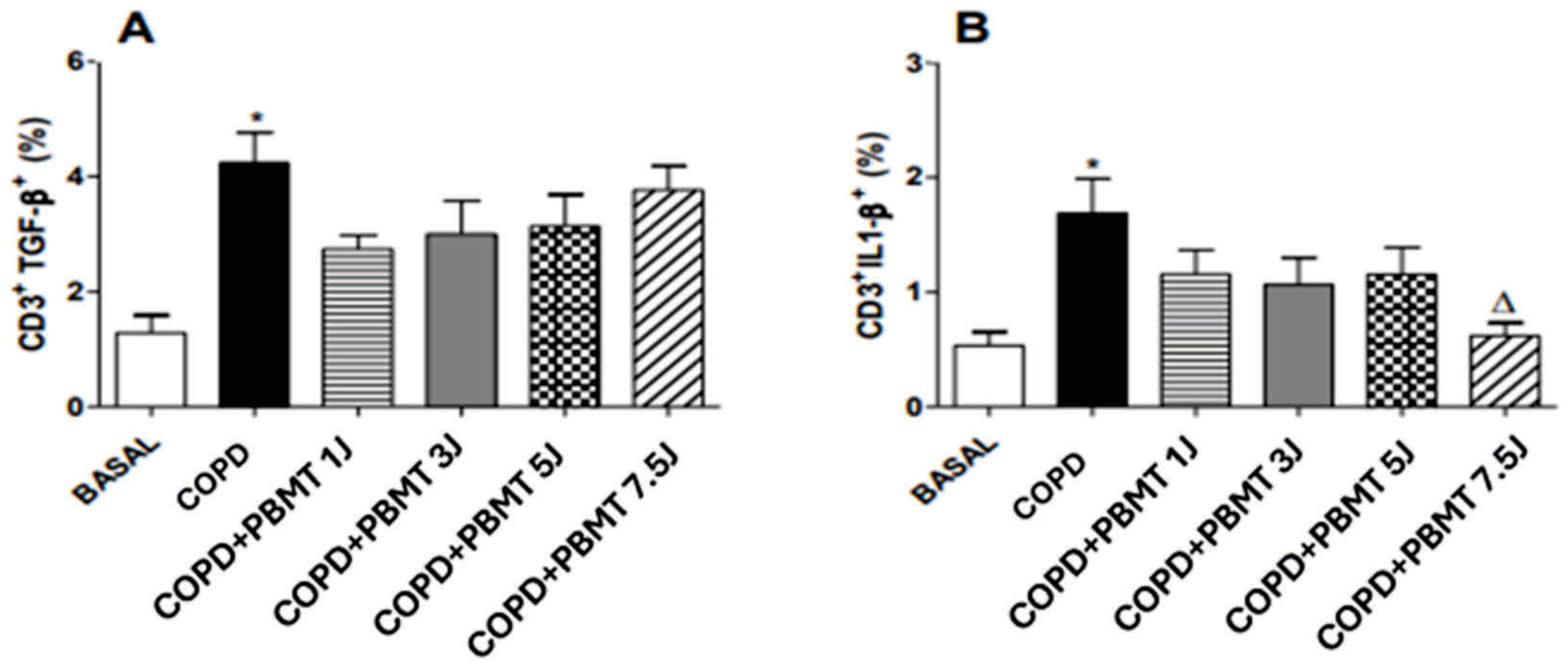
We observed that the induction of chronic obstructive pulmonary disease (COPD) using cigarette extract resulted in a significant increase in the percentage of TGF-β-producing lymphocytes (Figure 6A) and IL-1β (Figure 6B) in the bronchoalveolar lavage (BAL) of animals in the COPD group compared to the baseline group. While the levels of TGF-β-producing lymphocytes showed no significant differences in any of the COPD+PBMT groups, the COPD+PBMT (7.5 J) group presented a significantly reduced percentage of IL-1β-producing lymphocytes compared to the COPD group (Figure 6B).

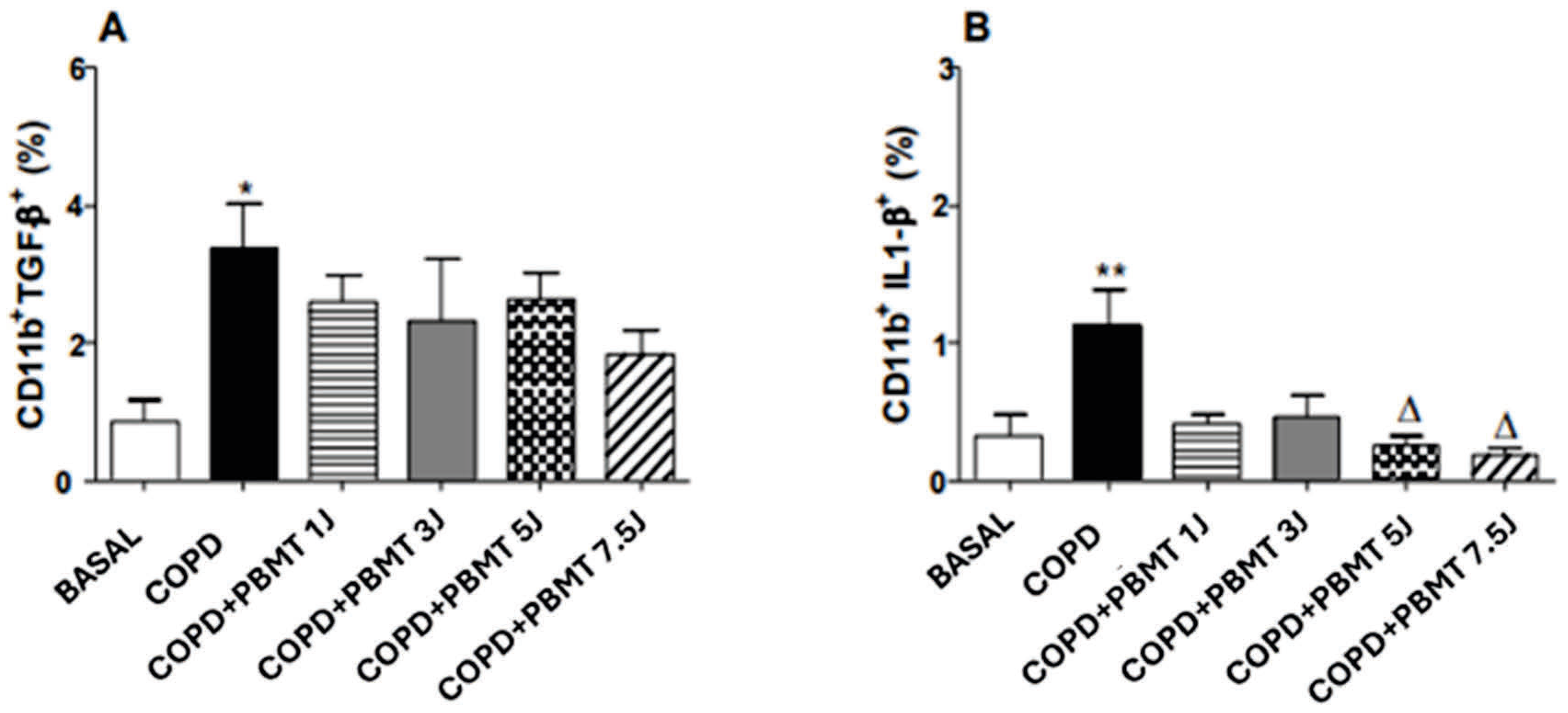
4.5. Effect of PBMT Therapy on the Quantification of CD11b+TGF- β+ (A) and CD11b+IL-1 β+ (B) Macrophages in BAL in an Experimental Model COPD

We observed that the induction of chronic obstructive pulmonary disease (COPD) by cigarette extract resulted in a significant increase in the percentage of TGF-β-producing macrophages and CD11b+ cells in the bronchoalveolar lavage (BAL) of animals in the COPD group compared to the baseline group (Figure 7A,B). While a reduction in TGF-β-producing macrophages was observed in the COPD+PBMT (5 J and 7.5 J) groups compared to the COPD group (Figure 7B).

5. Discussion

In this study, we demonstrated that photobiomodulation therapy (PBMT), applied at different radiant energy doses, produced significant anti-inflammatory effects in an experimental model of chronic obstructive pulmonary disease (COPD). A reduction in macrophage and neutrophil infiltration was observed—cells involved in the release of reactive oxygen species (ROS), which cause membrane rupture, receptor inactivation, and damage to transcription factors and enzymes [27]. Additionally, there was a decrease in pro-inflammatory cytokines, an increase in IL-10, modulation of immune subpopulations, and a lower incidence of necrosis. The doses of 3 J and 5 J were more effective, indicating the potential of PBMT as a therapeutic or adjuvant approach for COPD treatment.
Smoking-related diseases such as COPD pose a substantial social and economic burden and severely compromise quality of life [28]. Conventional treatments like bronchodilators and corticosteroids are limited in efficacy and may cause adverse effects, particularly in frail, polymedicated patients or those intolerant to systemic therapies [29,30].
PBMT, on the other hand, does not induce cellular apoptosis or impair mitochondrial function when used at appropriate doses, making it a promising alternative. A previous study showed reduced levels of cardiac markers such as CK-MB and troponins in a model of cardiac injury treated with PBMT [31]. Another study indicated that the combination of carvedilol and PBMT promoted cellular and cardiac protection by reducing oxidized proteins and increasing the activity of the antioxidant enzyme catalase [32]. These findings suggest beneficial effects of PBMT not only in COPD but also in associated comorbidities [33].
Previous studies have shown that PBMT inhibits the expression of the P2X7 purinergic receptor, involved in the inflammatory response and regulation of cell death [3]. However, insufficient energy doses may be ineffective, while excessive doses can cause adverse effects [34,35]. This motivated the present investigation, which aims to determine the ideal radiant energy dose to promote anti-inflammatory and reparative effects in lung tissue.
Studies in COPD and asthma models have shown that red laser irradiation (660 nm) attenuates inflammatory markers, reduces P2X7 receptor expression, and minimizes airway remodeling such as mucus hyperproduction and collagen accumulation [21,22,23,24,31].
Flow cytometric analysis in this study revealed an increase in neutrophils, macrophages, dendritic cells, and lymphocytes in the COPD group, indicating an exacerbated inflammatory response induced by cigarette extract [34]. PBMT significantly reduced these populations, with the best response at doses of 3 J and 5 J, consistent with findings from house dust mite (HDM)-induced asthma models [24].
COPD is characterized by an imbalance between Th17 and regulatory T cells, contributing to chronic inflammation and lung destruction [31]. Studies indicate that PBMT may increase IL-10–producing regulatory T cells, which could suppress effector T cells and restore immune homeostasis [36].
PBMT also reduced leukocyte necrosis, indicating a protective effect against inflammation-induced damage. These findings are consistent with data showing PBMT efficacy in emphysema models, with a reduction in inflammatory cells in BALF and CK-MB levels [37].
Despite the absence of significant differences in apoptosis between groups, the relationship between apoptosis and inflammation in COPD is complex. Conflicting results in the literature, such as increased BAX without evidence of apoptosis in osteosarcoma cells treated with PBMT, suggest that experimental variables may influence this process [38].
Although COPD induction did not increase ROS levels, PBMT at 7.5 J did lead to their elevation, which may reflect an immunomodulatory effect. The same dose also reduced IL-1β expression in lymphocytes, suggesting an anti-inflammatory action. However, excessive ROS production may be associated with oxidative stress [39]. Other studies have shown that PBMT at doses of 1 J/cm2 and 5 J/cm2 reduced ROS and ATP levels in BALF, indicating that inflammatory regulation may occur via ROS control [40].
Intracellular cytokine analysis in BAL revealed alterations in lymphocyte and macrophage subpopulations, aligning with previous studies that showed increased IL-10 and reduced pro-inflammatory cytokines following PBMT in pleurisy models [41].
COPD induction led to increased CD3+ lymphocytes producing TGF-β and IL-1β, markers of tissue remodeling and chronic inflammation [39]. The absence of TGF-β variation among treated groups may indicate that PBMT does not directly influence its production in this specific model. However, a study using a bleomycin-induced fibrosis model showed that PBMT reduced TGF-β levels [42].
In the 7.5 J treatment group, IL-1β expression in lymphocytes was reduced, consistent with previous findings in COPD [34,40]. Moreover, CD11b+ macrophages producing TGF-β and IL-1β increased following COPD induction. PBMT modulated this response in a dose-dependent manner, reducing these subpopulations. Although specific studies on CD11b+ macrophages are scarce, evidence suggests that PBMT favors an anti-inflammatory phenotype [43,44].
In interpreting the results of this study, it is important to consider the data presented by Balbi et al. (2025) [45]. who proposed a biological mechanism mediated by exposure to red light (635 nm), emphasizing alterations in mitochondrial energy metabolism and increased nitric oxide (NO) release. Despite methodological differences among experimental models, these findings support the notion that PBMT exerts significant cellular effects via the activation of mitochondrial photoacceptors, such as cytochromes, influencing bioenergetic and redox processes.
In addition to mitochondrial energy metabolism modulation, nitric oxide (NO) plays a central role in the biological effects of PBMT, particularly under red light (635–660 nm) irradiation. The literature consistently describes the ability of this spectrum to induce vasodilation through NO release, promoting increased local blood flow, tissue oxygenation, and nutrient delivery—critical factors for repair and inflammatory modulation. Notably, NO’s effects are not limited to vasodilation: more complex molecular mechanisms such as protein S-nitrosylation are also involved and may exert biphasic effects on cellular signaling pathways, as discussed by Ricciardolo FL et al. (2006) [23]. At physiological concentrations, NO acts as a regulator of mitochondrial function and redox signaling, whereas at high levels, it can cause nitrosative stress and metabolic dysfunction. This duality may help explain, at least in part, the different responses observed under various dosimetric conditions, reinforcing the need for precise optimization of parameters such as dose, power, and exposure time to achieve desired therapeutic effects without triggering adverse outcomes.
Thus, after identifying the most effective anti-inflammatory and reparative doses, PBMT emerges as a promising and safe therapeutic strategy for the treatment of COPD and its comorbidities. Taken together, after identifying the most effective anti-inflammatory and reparative doses, PBMT emerges as a promising and safe therapeutic strategy for COPD and its associated comorbidities.

Author Contributions

Conceptualization, C.E.A. and A.P.L.d.O.; Methodology, C.E.A.; Software, C.E.A.; Validation, A.P.L.d.O. and F.A.; Formal analysis, C.E.A. and T.G.S.; Investigation, C.E.A., T.G.S. and L.B.V.; Resources, A.P.L.d.O.; Data curation, C.E.A.; Writing—original draft preparation, C.E.A.; Writing—review and editing, A.P.L.d.O.; Visualization, C.E.A. and L.B.V.; Supervision, A.P.L.d.O., M.C.C., J.A.S.J., C.C.G.D., S.Z., R.L. and R.K.d.P.; Project administration, C.E.A. and A.P.L.d.O.; Funding acquisition, A.P.L.d.O. and C.E.A. All authors have read and agreed to the published version of the manuscript.

Funding

This research was funded by the São Paulo Research Foundation (FAPESP), grant number 2015/11459-0. The scholarship was valid from 15 June 2015 to 14 June 2016, under the funding line: BCO—Scientific Initiation/Continuous Flow.

Institutional Review Board Statement

The clinical study protocol was submitted and approved by the Ethics and Research Committee of University Nove de Julho—nº AN006.2013 (Approved on 24 June 2014).

Conflicts of Interest

There are no conflicts of interest.

References

  1. Boers, E.; Barrett, M.; Su, J.G.; Benjafield, A.V.; Sinha, S.; Kaye, L.; Zar, H.J.; Vuong, V.; Tellez, D.; Gondalia, R.; et al. Global Burden of Chronic Obstructive Pulmonary Disease Through 2050. JAMA Netw. Open. 2023, 6, e2346598. [Google Scholar] [PubMed]
  2. Devereux, G. ABC of chronic obstructive pulmonary disease. Definition, epidemiology, and risk factors. BMJ 2006, 332, 1142–1144. [Google Scholar] [PubMed]
  3. Da Cunha Moraes, G.; Vitoretti, L.B.; de Brito, A.A.; Alves, C.E.; de Oliveira, N.C.R.; Dos Santos Dias, A.; Matos, Y.S.T.; Oliveira-Junior, M.C.; Oliveira, L.V.F.; da Palma, R.K.; et al. Low-Level Laser Therapy Reduces Lung Inflammation in an Experimental Model of Chronic Obstructive Pulmonary Disease Involving P2X7 Receptor. Oxid. Med. Cell Longev. 2018, 2018, 6798238. [Google Scholar]
  4. Lomas, D.A.; Silverman, E.K. The genetics of chronic obstructive pulmonary disease. Respir Res. 2001, 2, 20–26. [Google Scholar]
  5. Barnes, P.J. Oxidative Stress in Chronic Obstructive Pulmonary Disease. Antioxidants 2022, 11, 965. [Google Scholar] [CrossRef]
  6. Dekhuijzen, P.N.; Aben, K.K.; Dekker, I.; Aarts, L.P.; Wielders, P.L.; van Herwaarden, C.L.; Bast, A. Increased exhalation of hydrogen peroxide in patients with stable and unstable chronic obstructive pulmonary disease. Am. J. Respir. Crit. Care Med. 1996, 154 Pt 1, 813–816. [Google Scholar] [CrossRef]
  7. Rahman, I.; Adcock, I.M. Oxidative stress and redox regulation of lung inflammation in COPD. Eur. Respir. J. 2006, 28, 219–242. [Google Scholar] [CrossRef]
  8. Noguera, A.; Batle, S.; Miralles, C.; Iglesias, J.; Busquets, X.; Macnee, W. Agusti AG Resposta aumentada de neutrófilos na doença pulmonar obstrutiva crônica. Thorax 2001, 56, 432–437. [Google Scholar]
  9. Barnes, P.J. Cellular and molecular mechanisms of chronic obstructive pulmonary disease. Clin. Chest Med. 2014, 35, 71–86. [Google Scholar] [CrossRef]
  10. Chung, K.F. Cytokines in chronic obstructive pulmonary disease. Eur. Respir. J. 2001, 18 (Suppl. 34), 50s–59s. [Google Scholar] [CrossRef]
  11. Farrell, L.A.; O’Rourke, M.B.; Padula, M.P.; Souza-Fonseca-Guimaraes, F.; Caramori, G.; Wark, P.A.B.; Dharmage, S.C.; Hansbro, P.M. The Current Molecular and Cellular Landscape of Chronic Obstructive Pulmonary Disease (COPD): A Review of Therapies and Efforts towards Personalized Treatment. Proteomes 2024, 12, 23. [Google Scholar] [CrossRef] [PubMed]
  12. Barnes, P.J.; Celli, B.R. Systemic manifestations and comorbidities of COPD. Eur. Respir. J. 2009, 33, 1165–1185. [Google Scholar] [CrossRef]
  13. Ritchie, A.I.; Wedzicha, J.A. Definition, Causes, Pathogenesis, and Consequences of Chronic Obstructive Pulmonary Disease Exacerbations. Clin. Chest Med. 2020, 41, 421–438. [Google Scholar] [CrossRef]
  14. Dourado, V.Z.; Tanni, S.E.; Vale, S.A.; Faganello, M.M.; Sanchez, F.F.; Godoy, I. Manifestações sistêmicas na doença pulmonar obstrutiva crônica. J. Bras. Pneumol. 2006, 32, 161–171. [Google Scholar]
  15. MacIntyre, N.; Huang, Y.C. Acute exacerbations and respiratory failure in chronic obstructive pulmonary disease. Proc Am Thorac Soc. 2008, 5, 530–535. [Google Scholar] [CrossRef] [PubMed] [PubMed Central]
  16. Drummond, M.B.; Dasenbrook, E.C.; Pitz, M.W.; Murphy, D.J.; Fan, E. Inhaled corticosteroids in patients with stable chronic obstructive pulmonary disease: A systematic review and meta-analysis. JAMA 2008, 300, 2407–2416. [Google Scholar]
  17. Singh, S.; Loke, Y.K.; Furberg, C.D. Inhaled anticholinergics and risk of major adverse cardiovascular events in patients with chronic obstructive pulmonary disease: A systematic review and meta-analysis. JAMA 2008, 300, 1439–1450. [Google Scholar] [CrossRef]
  18. Dompe, C.; Moncrieff, L.; Matys, J.; Grzech-Leśniak, K.; Kocherova, I.; Bryja, A.; Bruska, M.; Dominiak, M.; Mozdziak, P.; Skiba, T.H.I.; et al. Photobiomodulation-Underlying Mechanism and Clinical Applications. J. Clin. Med. 2020, 9, 1724. [Google Scholar] [CrossRef]
  19. Amaroli, A.; Clemente Vargas, M.R.; Pasquale, C.; Raffetto, M.; Ravera, S. Photobiomodulation on isolated mitochondria at 810 nm: First results on the efficiency of the energy conversion process. Sci Rep. 2024, 14, 11060. [Google Scholar] [PubMed]
  20. Hamblin, M.R. Mechanisms and Mitochondrial Redox Signaling in Photobiomodulation. Photochem. Photobiol. 2018, 94, 199–212. [Google Scholar] [CrossRef] [PubMed]
  21. Bisht, D.; Gupta, S.C.; Misra, V.; Mital, V.P.; Sharma, P. Effect of low intensity laser radiation on healing of open skin wounds in rats. Indian J. Med. Res. 1994, 100, 43–46. [Google Scholar] [PubMed]
  22. Cios, A.; Cieplak, M.; Szymański, Ł.; Lewicka, A.; Cierniak, S.; Stankiewicz, W.; Mendrycka, M.; Lewicki, S. Effect of Different Wavelengths of Laser Irradiation on the Skin Cells. Int. J. Mol. Sci. 2021, 22, 2437. [Google Scholar] [CrossRef]
  23. Ricciardolo, F.L.; Nijkamp, F.P.; Folkerts, G. Nitric oxide synthase (NOS) as therapeutic target for asthma and chronic obstructive pulmonary disease. Curr. Drug Targets 2006, 7, 721–735. [Google Scholar] [CrossRef]
  24. Li, C.L.; Liu, J.F.; Liu, S.F. Mitochondrial Dysfunction in Chronic Obstructive Pulmonary Disease: Unraveling the Molecular Nexus. Biomedicines 2024, 12, 814. [Google Scholar] [CrossRef]
  25. Wang, X.Y.; Ma, W.J.; Liu, C.S.; Li, Y.X. Effect of low-level laser therapy on allergic asthma in rats. Lasers Med. Sci. 2014, 29, 1043–1050. [Google Scholar] [CrossRef]
  26. Oliveira, N.C.R.; Soares, J.M.; Cordeiro, J.M.; Paiva, M.S.; Pinheiro, A.L.B. Effect of low-level laser therapy in pulmonary inflammation in asthma induced by house dust mite: A dosimetry study. Int. J. Inflamm. 2018, 2018, 1–10. [Google Scholar]
  27. Rahman, I. Oxidative stress in pathogenesis of chronic obstructive pulmonary disease: Cellular and molecular mechanisms. Cell. Biochem. Biophys. 2005, 43, 167–188. [Google Scholar] [CrossRef]
  28. Marciniuk, D.; Nana, A.; Rabe, K.; Zar, H.; Ferkol, T.; MontesdeOca, M. Doenças respiratórias no mundo: Realidades de hoje, oportunidades para amanhã. Afr. J. Resp. Dis. 2014, 6, 4–13. [Google Scholar]
  29. Pauwels, R.A.; Löfdahl, C.-G.; Laitinen, L.A.; Schouten, J.P.; Postma, D.S.; Pride, N.B.; Ohlsson, S.V. Tratamento de longo prazo com budesonida inalada em pessoas com doença pulmonar obstrutiva crônica leve que continuam fumando. Estudo da Sociedade Respiratória Europeia sobre Doença Pulmonar Obstrutiva Crônica. N. Engl. J. Med. 1999, 340, 1948–1953. [Google Scholar] [CrossRef] [PubMed]
  30. Vestbo, J.; Sorensen, T.; Lange, P.; Brix, A.; Torre, P.; Viskum, K. Efeito de longo prazo da budesonida inalada na doença pulmonar obstrutiva crônica leve e moderada: Um ensaio clínico randomizado. Lancet 1999, 353, 1819–1823. [Google Scholar] [CrossRef] [PubMed]
  31. Feliciano, R.D.S.; Atum, A.L.B.; Ruiz, É.G.D.S.; Serra, A.J.; Antônio, E.L.; Manchini, M.T.; Silva, J.M.A.; Tucci, P.J.F.; Nathanson, L.; Morris, M.; et al. Photobiomodulation Therapy on Myocardial Infarction in Rats: Transcriptional and Posttranscriptional Implications to Cardiac Remodeling. Lasers Surg. Med. 2021, 53, 1247–1257. [Google Scholar] [CrossRef]
  32. Grandinetti, V.; Carlos, F.P.; Antonio, E.L.; de Oliveira, H.A.; Dos Santos, L.F.N.; Yoshizaki, A.; Mansano, B.S.D.M.; Silva, F.A.; Porte, L.A.; Albuquerque-Pontes, G.M.; et al. Photobiomodulation therapy combined with carvedilol attenuates post-infarction heart failure by suppressing excessive inflammation and oxidative stress in rats. Sci. Rep. 2019, 9, 9425. [Google Scholar] [CrossRef]
  33. Matsunaga, K.; Harada, M.; Suizu, J.; Oishi, K.; Asami-Noyama, M.; Hirano, T. Comorbid Conditions in Chronic Obstructive Pulmonary Disease: Potential Therapeutic Targets for Unmet Needs. J. Clin. Med. 2020, 9, 3078. [Google Scholar] [CrossRef]
  34. Huang, Y.Y.; Sharma, S.K.; Carroll, J.; Hamblin, M.R. Biphasic dose response in low level light therapy—An update. Dose Response 2011, 9, 602–618. [Google Scholar] [CrossRef]
  35. Bjordal, J.M.; Couppe, C.; Ljunggren, A.E. Low level laser therapy for tendinopathy: Evidence of a dose-response pattern. In Database of Abstracts of Reviews of Effects (DARE): Quality-Assessed Reviews [Internet]; Centre for Reviews and Dissemination (UK): York, UK, 2001. Available online: https://www.ncbi.nlm.nih.gov/books/NBK68932/ (accessed on 26 May 2025).
  36. De Brito, A.A.; Herculano, K.Z.; de Alvarenga-Nascimento, C.R.; Estefano-Alves, C.; Duran, C.C.G.; Marcos, R.L.; Silva Junior, J.A.; Chavantes, M.C.; Zamuner, S.R.; Aimbire, F.; et al. Effect of photobiomodulation in the balance between effector and regulatory T cells in an experimental model of COPD. Front. Med. 2024, 11, 1347517. [Google Scholar] [CrossRef]
  37. Fonseca, K.M.; de Melo Rodrigues, L.; Ferreira, D.C.L.; Vilaça, M.M.O.; Galvão, S.M.P.; dos Santos Alves, W. Efeitos da fotobiomodulação nas alterações extrapulmonares ocasionadas pelo enfisema experimental em rattus norvegicus. Saúde Pesquisa 2016, 9, 143–152. [Google Scholar] [CrossRef]
  38. Secchi, T.M.; Oliveira, T.L.P.; Pereira, T.I.M.; Araújo, P.F.; Sganzerla, J.T. Photobiomodulation for the prevention and treatment of bisphosphonate-induced osteonecrosis: A pilot study in an animal model. Rev. Cereus. 2023, 15, 120–132. [Google Scholar]
  39. Cosio, M.G.; Saetta, M.; Agusti, A. Immunologic aspects of chronic obstructive pulmonary disease. N. Engl. J. Med. 2009, 360, 2445–2454. [Google Scholar] [CrossRef] [PubMed]
  40. Soares, S.S. Terapia de Fotobiomodulação Regula a Produção de Espécies Reativas de Oxigênio (ROS) em Modelo Experimental de Doença Pulmonar Obstrutiva Crônica (DPOC). Ph.D. Thesis, Universidade Nove de Julho, São Paulo, Brazil, 2021. [Google Scholar]
  41. Boschi, E.S. Efeito Antiinflamatório da Terapia Laser de Baixa Potência (660 nm) na Pleurisia em Ratos. Ph.D. Thesis, Pontifícia Universidade Católica do Rio Grande do Sul, Porto Alegre, Brazil, 2007. [Google Scholar]
  42. De Brito, A.A.; da Silveira, E.C.; Ligeiro de Oliveira, A.P. Low-level laser therapy attenuates lung inflammation and airway remodeling in a murine model of idiopathic pulmonary fibrosis: Relevance to cytokines secretion from lung structural cells. J. Photochem. Photobiol. B Biol. 2020, 203, 111731. [Google Scholar] [CrossRef] [PubMed]
  43. Asnaashari, M.; Shojaeian, S.; Mesgharani, A.; Mehrabinia, P. The effect of low-level laser therapy on the viability of human dental pulp stem cells. J. Lasers Med. Sci. 2022, 13, e60. [Google Scholar] [CrossRef] [PubMed]
  44. Bonvicini, J.F.S.; de Souza, G.L.; Basso, F.G.; de Souza Costa, C.A.; Moura, C.C.G.; Silva, M.J.B.; Turrioni, A.P. The effect of infrared LED irradiation on the oxidative stress induced in human dental pulp cells by lipopolysaccharide (LPS). Int. Endod. J. 2014, 47, 747–755. [Google Scholar]
  45. Balbi, M.; Lai, R.; Stigliani, S.; Massarotti, C.; Bozzo, M.; Scaruffi, P.; Ravera, S.; Amaroli, A. Efficacy and Safety of Visible and Near-Infrared Photobiomodulation Therapy on Astenospermic Human Sperm: Wavelength-Dependent Regulation of Nitric Oxide Levels and Mitochondrial Energetics. Biology 2025, 14, 491. [Google Scholar] [CrossRef] [PubMed]
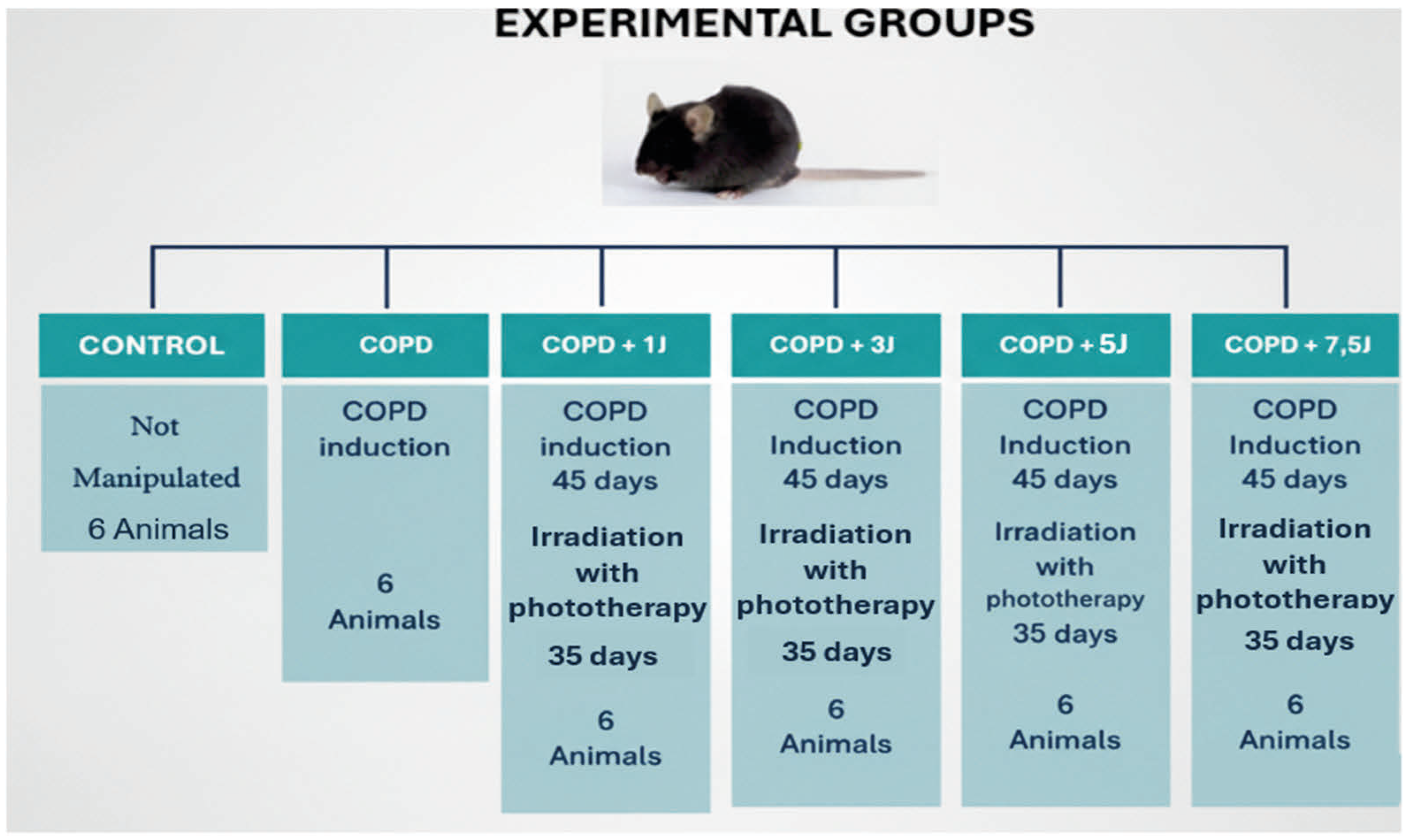
Figure 1. Flowchart with experimental groups. Illustrates the experimental groups used in the study. The project includes an unmanipulated control group and five groups that underwent a 45-day process of COPD (Chronic Obstructive Pulmonary Disease) induction. After COPD induction, four of these groups were subjected to phototherapy irradiation for 35 days, with different energy levels: 1 J, 3 J, 5 J, and 7.5 J. Each experimental group consists of 6 animals.
Allergies 05 00033 g001
Figure 2. Illustrative diagram of the experiment. Experimental timeline for COPD induction by cigarette extract and treatment with photobiomodulation therapy (PBMT). The COPD group received intranasal instillations of cigarette extract three times per week, from day 0 to day 49, followed by euthanasia on day 50. The COPD + PBMT group received the same cigarette extract exposure protocol, combined with photobiomodulation therapy, which was initiated on day 35 and applied one hour after each cigarette extract administration until day 49. Euthanasia was performed on day 50.
Allergies 05 00033 g002
Figure 3. Effect of photobiomodulation (PBMT) on the influx of inflammatory cells into the lungs in an experimental model of COPD. (A) Total cell count, (B) macrophages, (C) neutrophils, and (D) lymphocytes, recovered from the bronchoalveolar lavage (BAL) 24 h after the last orotracheal application. Data are presented as mean ± standard error of the mean (S.E.M.). * p < 0.05; *** p < 0.001 compared to the basal group; Δ p <0.05; ϕ p < 0.001 compared to the COPD group.
Allergies 05 00033 g003
Figure 4. Effect of photobiomodulation therapy (PBMT) on the number of inflammatory cells in the lung in an experimental model of COPD. In (A) the number of neutrophils, (B) macrophages, (C) dendritic cells, (D) total lymphocytes, present in the lung. Data represent the mean ± standard error of the mean (S.E.M.). * p < 0.05; *** p < 0.001 compared to the basal group; Δ p < 0.05; ϕ p < 0.001 compared to the COPD group.
Allergies 05 00033 g004
Figure 5. Effect of photobiomodulation (PBMT) on necrosis, apoptosis and production of reactive oxygen species in pulmonary leukocytes in an experimental model of COPD. Results represented in CD 45+ cell gate, being (A) necrosis values, (B) apoptosis and (C) production of reactive oxygen species. Data represent the mean ± standard error of the mean (S.E.M.). * p < 0.05 compared to the basal group; Δ p < 0.05; ϕ p < 0.001 compared to the COPD group.
Allergies 05 00033 g005
Figure 6. Effect of PBMT therapy on the percentage of CD3+TGF- β+ (A) and CD3+IL-1 β+ (B) lymphocytes in BAL in an experimental model of COPD. In (A) the values of CD3+TGF-β+ lymphocytes and in (B), CD3+IL-1β+ lymphocytes in the bronchoalveolar lavage. Values expressed as mean and standard deviation. * p < 0.05 when compared to the basal group; ∆ p < 0.01 when compared to the COPD group.
Allergies 05 00033 g006
Figure 7. Effect of PBMT therapy on the quantification of CD11b+TGF- β+ (A) and CD11b+IL-1 β+ (B) macrophages in BAL in an experimental model of chronic obstructive pulmonary disease. In (A) values of CD11b+TGF-β+ macrophages in (B) CD11b+IL-1β+ macrophages in bronchoalveolar lavage. Values expressed as mean and standard deviation. * p < 0.05; ** p < 0.01 when compared to the basal group; Δ p < 0.05 when compared to the COPD group.
Allergies 05 00033 g007
Table 1. Dosimetric parameters.
ParametersGroup 1Group 3Group 5Group 7,5
Radiant Energy (J)1 J3 J5 J7.5 J
Wavelength (nm)660660660660
Spectral width (nm)±20±20±20±20
Operating modeContinuousContinuousContinuousContinuous
Average radiant power (mW)100100100100
Radiant exposure (J/cm2)2266111116
Beam area on target (cm2)0.0450.0450.0450.045
Exposure time (s)10305075
Frequency (Hz)UniqueUniqueUniqueUnique
Application points3 points3 points3 points3 points
Application techniqueContactContactContactContact
Disclaimer/Publisher’s Note: The statements, opinions and data contained in all publications are solely those of the individual author(s) and contributor(s) and not of MDPI and/or the editor(s). MDPI and/or the editor(s) disclaim responsibility for any injury to people or property resulting from any ideas, methods, instructions or products referred to in the content.