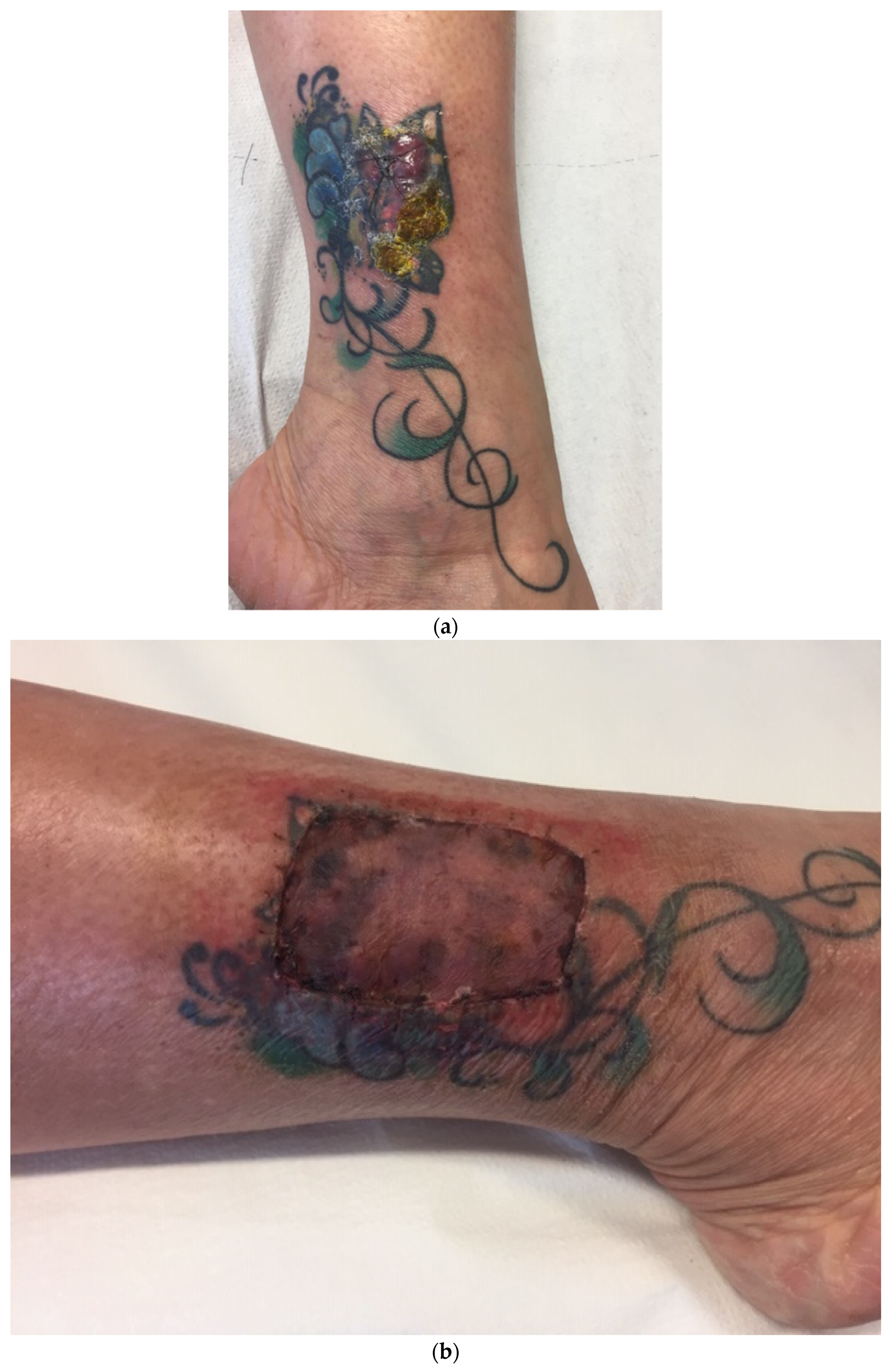
Abstract
This case report describes a 50-year-old woman who developed an allergic granulomatous reaction after a tattoo touch-up.
Tattooing has become increasingly popular in recent years. Adverse cutaneous reactions against tattoo ink compounds have been largely reported and they include eczematous, lichenoid and granulomatous lesions, pseudolymphoma and pseudo-epitheliomatous hyperplasia [1]. Notably, red tattoo pigments are the ones which are associated to adverse events the most [2].
A 50-year-old female patient presented to our Department complaining of a cutaneous lesion localized on her tattoo. The patient reported that her tattoo had been placed on her skin using black inks 15 years before and that these body modifications had not been associated with any adverse skin reaction. However, when she decided to have her tattoo touched-up with colored inks (including red) she developed a cutaneous reaction a week later.
On physical examination, thick erythematous nodular lesions were present in the areas in which the red pigment was used (Figure 1). Pain and itching were present.
Figure 1.
Allergic granulomatous reaction on the tattoo of a 50 year-old woman before and after surgical treatment: (a) Thick erythematous nodular lesions at the red-pigment areas of tattoo; (b) Surgically treated tattoo.
Before seeking a dermatological consultation, the patient had tried several local therapies, such as topical steroids and serial intra-lesional corticosteroid injections, with no clinical benefits. Furthermore, she had tried systemic therapies with steroids, antibiotics and antihistamine, which were associated with temporary beneficial effects.
We decided to perform a cutaneous biopsy which revealed acanthosis, papillomatosis and focal ulceration of the epidermis, with spongiosis and basal vacuolization; an intense chronic dermal inflammatory reaction with eosinophils was evident. The histological pattern was compatible with an allergic granulomatous reaction which was presumptively attributable to one of the colored pigments that were added to the black tattoo.
We also performed SIDAPA and tattoo series patch tests which highlighted a positivity to nickel sulphate at 48 h (+) and 72 h (+++).
Considering the severe local pain and because of the inefficacy of topical, systemic and intra-lesional therapies, a surgical excision of the affected areas followed by skin grafting with autograft was made.
As mentioned before, tattoo adverse reactions are becoming increasingly common, probably because of the growing popularity of this practice among young people. Allergic reactions are among the most frequent tattoo-related complications and they are usually caused by the chemicals found in tattoo inks: interestingly, the published literature points to the red pigment as the main culprit of allergic adverse reactions on the skin [3,4]. Nevertheless, other pigments like yellow, violet and blue have been reported to be able to cause adverse local reactions as well [4,5].
Nickel sulphate is frequently present in red pigments and it can cause various allergic complications in sensitized people [4]. Indeed, the allergic granulomatous reaction described in this report was localized in the red areas of the tattoo, which makes the red ink the most probable one to have triggered the cutaneous side-effects. Moreover, this statement was supported by the result of the patch test that was performed.
Chromium, cadmium, iron and titanium have been identified inside of colored tattoo inks and their presence has been linked to environmental factors like contamination [4]. Moreover, there is no strong evidence that supports a role of these metals in the allergic events associated with the application of colored tattoos [4].
Unfortunately, we were not able to perform a pigment analysis in our patient.
In conclusion, we presented this clinical case to highlight that tattoos can present several adverse effects, which are expected to be increasingly observed in the future due to their growing popularity. Additionally, tattoo ink composition is frequently unknown and it can lead to allergic granulomatous reactions. Further studies to elucidate the composition of tattoo inks are warranted to avoid the occurrence of unwanted adverse reactions. In addition, strict legislation should be developed to regulate the compounds which are used to make tattoo colors [1]. It is advisable for people to seek dermatological advice before changing the color of a pre-existing tattoo, to avoid the onset of adverse reactions. Lastly, we would like to emphasize that typical local and systemic therapies are not always sufficient for the complete resolution of extended cutaneous lesions, especially when they are associated with symptoms like persistent pain and pruritus. In these cases, it is necessary to perform surgical treatments, with results which are not always aesthetically pleasing.
Author Contributions
Conceptualization: A.T., F.L., G.D.M.; Methodology: A.T., F.L.; Investigation: A.T., A.C., C.I., C.C.; Writing-original draft preparation: G.A.R.A., F.M., F.R.P.; Writing-review and editing: F.M., G.A.R.A.; supervision: A.T., G.D.M. All authors have read and agreed to the published version of the manuscript.
Funding
This research received no external funding.
Institutional Review Board Statement
Not applicable.
Informed Consent Statement
Informed consent was obtained from the patient to publish this paper.
Data Availability Statement
No new data were created or analyzed in this study. Data sharing is not applicable to this article.
Conflicts of Interest
The authors declare no conflict of interest.
References
- Tammaro, A.; Raffa, S.; Petrigliano, N.; Zollo, V.; Gelormini, E.; Moliterni, E.; Magri, F.; Parisella, F.R.; Persechino, S. Marked pseudoepitheliomatous hyperplasia secondary to a red-pigmented tattoo: A case report. J. Eur. Acad. Dermatol. Venereol. 2018, 32, e272–e273. [Google Scholar] [CrossRef] [PubMed]
- Høgsberg, T.; Thomsen, B.M.; Serup, J. Histopathology and immune histochemistry of red tattoo reactions. Interface dermatitis is the lead pathology, with increase in T-lymphocytes and Langerhans cells suggesting an allergic pathomechanism. Skin Res. Technol. 2015, 21, 449–458. [Google Scholar] [CrossRef] [PubMed]
- Tammaro, A.; Toniolo, C.; Giulianelli, V.; Serafini, M.; Persechino, S. Chemical research on red pigments after adverse reactions to tattoo. Eur. Ann. Allergy Clin. Immunol. 2016, 48, 46–48. [Google Scholar] [PubMed]
- Serup, J.; Hutton Carlsen, K.; Dommershausen, N.; Sepehri, M.; Hesse, B.; Seim, C.; Luch, A.; Schreiver, I. Identification of pigments related to allergic tattoo reactions in 104 human skin biopsies. Contact Dermat. 2020, 82, 73–82. [Google Scholar] [CrossRef] [PubMed] [Green Version]
- Serup, J.; Sepehri, M.; Hutton Carlsen, K. Classification of Tattoo Complications in a Hospital Material of 493 Adverse Events. Dermatology 2016, 232, 668–678. [Google Scholar] [CrossRef] [PubMed]
Publisher’s Note: MDPI stays neutral with regard to jurisdictional claims in published maps and institutional affiliations. |
© 2021 by the authors. Licensee MDPI, Basel, Switzerland. This article is an open access article distributed under the terms and conditions of the Creative Commons Attribution (CC BY) license (https://creativecommons.org/licenses/by/4.0/).