
Segmentation of 4D Flow MRI: Comparison between 3D Deep Learning and Velocity-Based Level Sets
 , , , ,
Abstract
1. Introduction
1.1. Objective
1.2. Related Work
2. Materials and Methods
2.1. Study Population
2.2. 4D Flow MRI Acquisition Settings
2.3. Ground Truth
2.4. Pre-Processing and Post-Processing
2.5. Segmentation with Level Sets
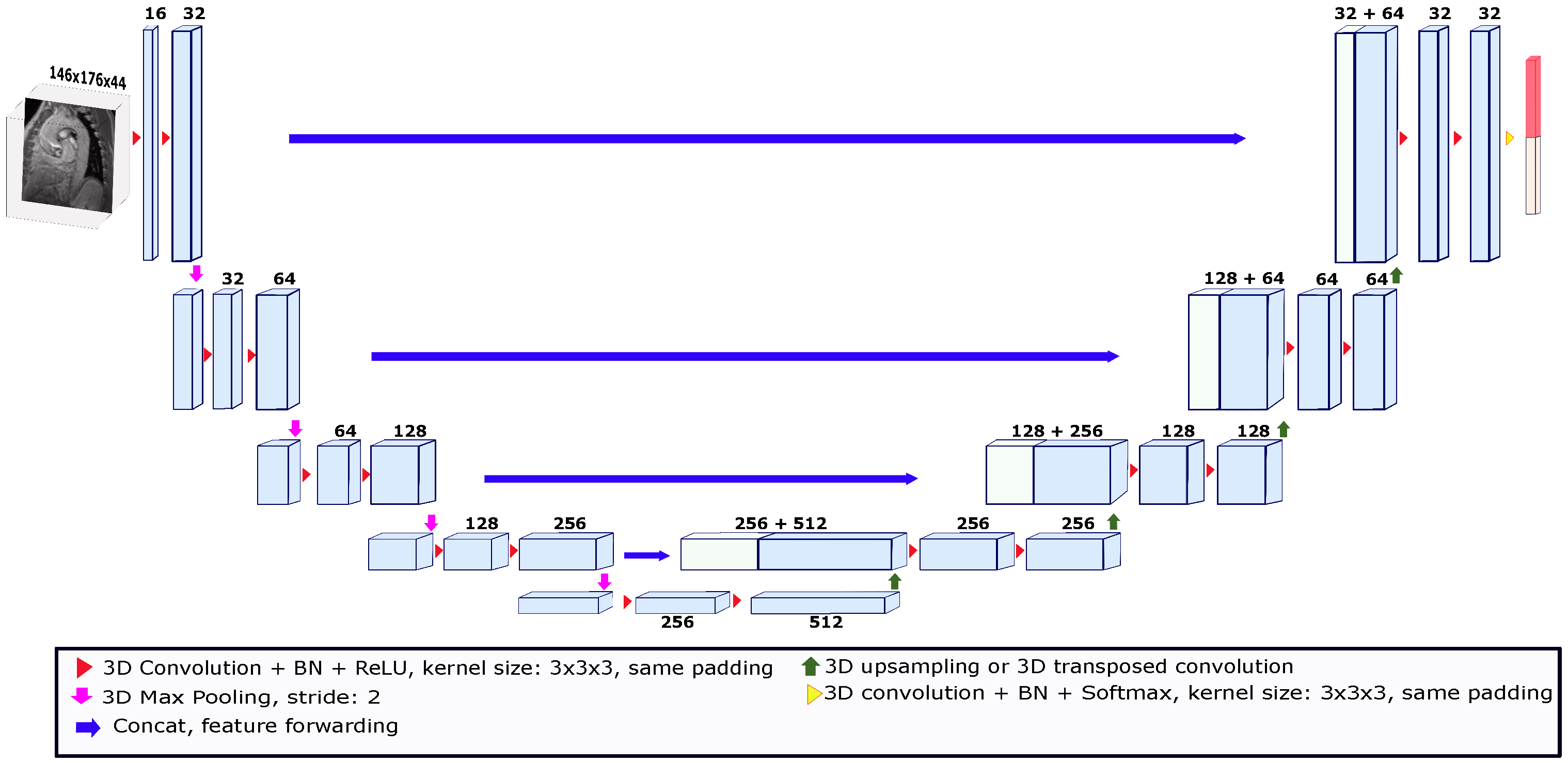
2.6. Segmentation Using Deep Learning
2.7. Evaluation
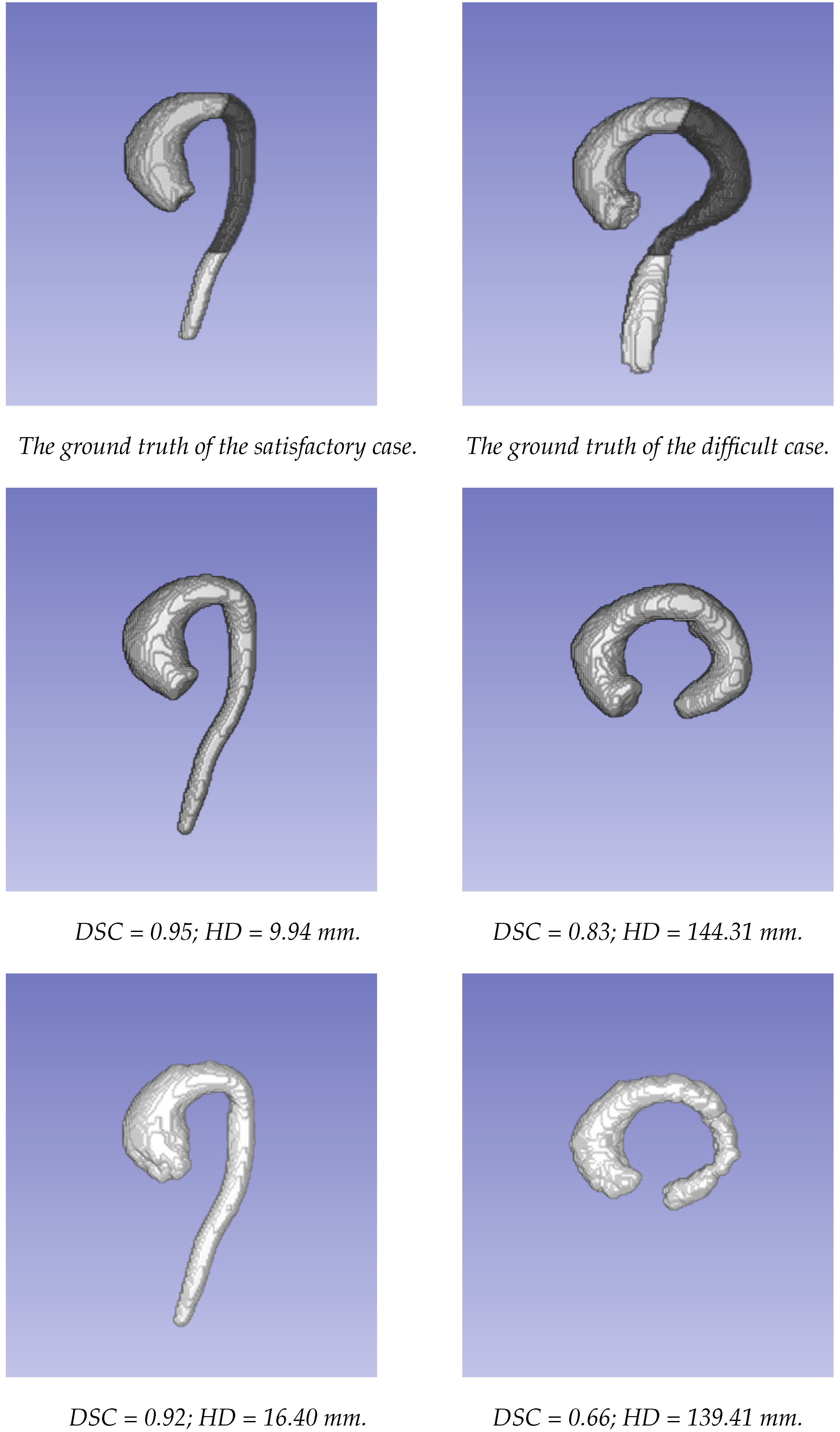
3. Results
4. Discussion
5. Conclusions
Author Contributions
Funding
Institutional Review Board Statement
Informed Consent Statement
Data Availability Statement
Conflicts of Interest
References
- Davis, F.M.; Daugherty, A.; Lu, H.S. Updates of Recent Aortic Aneurysm Research. Arterioscler. Thromb. Vasc. Biol. 2019, 39, e83–e90. [Google Scholar] [CrossRef] [PubMed]
- Aggarwal, S.; Qamar, A.; Sharma, V.; Sharma, A. Abdominal AORTIC ANEURYSM: A comprehensive review. Exp. Clin. Cardiol. 2011, 16, 11–15. [Google Scholar] [PubMed]
- Kim, E.K.; Choi, S.H.; Sung, K.; Kim, W.S.; Choe, Y.H.; Oh, J.K.; Kim, D.K. Aortic diameter predicts acute type A aortic dissection in patients with Marfan syndrome but not in patients without Marfan syndrome. J. Thorac. Cardiovasc. Surg. 2014, 147, 1505–1510. [Google Scholar] [CrossRef] [PubMed]
- Pape, L.A.; Tsai, T.T.; Isselbacher, E.M.; Oh, J.K.; O’Gara, P.T.; Evangelista, A.; Fattori, R.; Meinhardt, G.; Trimarchi, S.; Bossone, E.; et al. Aortic diameter ≥5.5 cm is not a good predictor of type A aortic dissection: Observations from the International Registry of Acute Aortic Dissection (IRAD). Circulation 2007, 116, 1120–1127. [Google Scholar] [CrossRef] [PubMed]
- Zhuang, B.; Sirajuddin, A.; Zhao, S.; Lu, M. The role of 4D flow MRI for clinical applications in cardiovascular disease: Current status and future perspectives. Quant. Imaging Med. Surg. 2021, 11, 4193–4210. [Google Scholar] [CrossRef]
- Manzo, M.; Pellino, S. FastGCN + ARSRGemb: A novel framework for object recognition. J. Electron. Imaging 2021, 30, 033011. [Google Scholar] [CrossRef]
- Sato, Y.; Nakajima, S.; Shiraga, N.; Atsumi, H.; Yoshida, S.; Koller, T.; Gerig, G.; Kikinis, R. Three-dimensional multi-scale line filter for segmentation and visualization of curvilinear structures in medical images. Med. Image Anal. 1998, 2, 143–168. [Google Scholar] [CrossRef]
- Frangi, A.F.; Niessen, W.J.; Vincken, K.L.; Viergever, M.A. Multiscale vessel enhancement filtering. In Medical Image Computing and Computer-Assisted Intervention— MICCAI’98; Springer: Berlin/Heidelberg, Germany, 1998; pp. 130–137. [Google Scholar] [CrossRef]
- Ngo, M.T.; Lee, U.Y.; Ha, H.; Jung, J.; Lee, D.H.; Kwak, H.S. Improving Blood Flow Visualization of Recirculation Regions at Carotid Bulb in 4D Flow MRI Using Semi-Automatic Segmentation with ITK-SNAP. Diagnostics 2021, 11, 1890. [Google Scholar] [CrossRef]
- Bustamante, M.; Gupta, V.; Forsberg, D.; Carlhäll, C.J.; Engvall, J.; Ebbers, T. Automated multi-atlas segmentation of cardiac 4D flow MRI. Med Image Anal. 2018, 49, 128–140. [Google Scholar] [CrossRef]
- Peper, E.S.; van Ooij, P.; Jung, B.; Huber, A.; Gräni, C.; Bastiaansen, J.A.M. Advances in machine learning applications for cardiovascular 4D flow MRI. Front. Cardiovasc. Med. 2022, 9, 1052068. [Google Scholar] [CrossRef]
- Bratt, A.; Kim, J.; Pollie, M.; Beecy, A.N.; Tehrani, N.H.; Codella, N.; Perez-Johnston, R.; Palumbo, M.C.; Alakbarli, J.; Colizza, W.; et al. Machine learning derived segmentation of phase velocity encoded cardiovascular magnetic resonance for fully automated aortic flow quantification. J. Cardiovasc. Magn. Reson. 2019, 21, 1. [Google Scholar] [CrossRef]
- Garrido-Oliver, J.; Aviles, J.; Córdova, M.M.; Dux-Santoy, L.; Ruiz-Muñoz, A.; Teixido-Tura, G.; Talou, G.D.M.; Ferez, X.M.; Jiménez, G.; Evangelista, A.; et al. Machine learning for the automatic assessment of aortic rotational flow and wall shear stress from 4D flow cardiac magnetic resonance imaging. Eur. Radiol. 2022, 32, 7117–7127. [Google Scholar] [CrossRef] [PubMed]
- Berhane, H.; Scott, M.; Elbaz, M.; Jarvis, K.; McCarthy, P.; Carr, J.; Malaisrie, C.; Avery, R.; Barker, A.J.; Robinson, J.D.; et al. Fully automated 3D aortic segmentation of 4D flow MRI for hemodynamic analysis using deep learning. Magn. Reson. Med. 2020, 84, 2204–2218. [Google Scholar] [CrossRef] [PubMed]
- Bustamante, M.; Viola, F.; Engvall, J.; Carlhäll, C.J.; Ebbers, T. Automatic Time-Resolved Cardiovascular Segmentation of 4D Flow MRI Using Deep Learning. J. Magn. Reson. Imaging 2022, 57, 191–203. [Google Scholar] [CrossRef] [PubMed]
- Marin-Castrillon, D.M.; Lalande, A.; Leclerc, S.; Ambarki, K.; Morgant, M.C.; Cochet, A.; Lin, S.; Bouchot, O.; Boucher, A.; Presles, B. 4D segmentation of the thoracic aorta from 4D flow MRI using deep learning. Magnetic Reson. Imaging 2023, 99, 20–25. [Google Scholar] [CrossRef]
- Köhler, B.; Preim, U.; Grothoff, M.; Gutberlet, M.; Fischbach, K.; Preim, B. Guided Analysis of Cardiac 4D PC-MRI Blood Flow Data. Eurographics 2015, 2015, 2–5. [Google Scholar]
- van Pelt, R.; Nguyen, H.; ter Haar Romeny, B.; Vilanova, A. Automated segmentation of blood-flow regions in large thoracic arteries using 3D-cine PC-MRI measurements. Int. J. Comput. Assist. Radiol. Surg. 2012, 7, 217–224. [Google Scholar] [CrossRef]
- Fujiwara, T.; Berhane, H.; Scott, M.B.; Englund, E.K.; Schäfer, M.; Fonseca, B.; Berthusen, A.; Robinson, J.D.; Rigsby, C.K.; Browne, L.P.; et al. Segmentation of the aorta and pulmonary arteries based on 4D flow MRI in the pediatric setting using fully automated multi-site, multi-vendor, and multi-label dense U-Net. J. Magn. Reson. Imaging 2022, 55, 1666–1680. [Google Scholar] [CrossRef]
- Rothenberger, S.M.; Patel, N.M.; Zhang, J.; Schnell, S.; Craig, B.A.; Ansari, S.A.; Markl, M.; Vlachos, P.P.; Rayz, V.L. Automatic 4D flow MRI Segmentation Using the Standardized Difference of Means Velocity. IEEE Trans. Med. Imaging 2023. [Google Scholar] [CrossRef]
- Warfield, S.; Zou, K.; Wells, W. Simultaneous Truth and Performance Level Estimation (STAPLE): An Algorithm for the Validation of Image Segmentation. IEEE Trans. Med Imaging 2004, 23, 903–921. [Google Scholar] [CrossRef]
- Craiem, D.; Pascaner, A.F.; Casciaro, M.E.; Gencer, U.; Alcibar, J.; Soulat, G.; Mousseaux, E. Automatic correction of background phase offset in 4D-flow of great vessels and of the heart in MRI using a third-order surface model. Magn. Reson. Mater. Phys. Biol. Med. 2019, 32, 629–642. [Google Scholar] [CrossRef]
- Solem, J.E.; Persson, M.; Heyden, A. Velocity Based Segmentation in Phase Contrast MRI Images. In Medical Image Computing and Computer-Assisted Intervention—MICCAI 2004; Springer: Berlin/Heidelberg, Germany, 2004; pp. 459–466. [Google Scholar] [CrossRef]
- Sethian, J. Level Set Methods; Cambridge University Press: Cambridge, UK, 1996. [Google Scholar]
- Bustamante, M.; Gupta, V.; Carlhäll, C.J.; Ebbers, T. Improving visualization of 4D flow cardiovascular magnetic resonance with four-dimensional angiographic data: Generation of a 4D phase-contrast magnetic resonance CardioAngiography (4D PC-MRCA). J. Cardiovasc. Magn. Reson. 2017, 19, 47. [Google Scholar] [CrossRef]
- Yamashita, R.; Nishio, M.; Do, R.K.G.; Togashi, K. Convolutional neural networks: An overview and application in radiology. Insights Imaging 2018, 9, 611–629. [Google Scholar] [CrossRef]
- Ronneberger, O.; Fischer, P.; Brox, T. U-net: Convolutional networks for biomedical image segmentation. In Proceedings of the 18th International Conference on Medical Image Computing and Computer-Assisted Intervention–MICCAI 2015, Munich, Germany, 5–9 October 2015; Springer: Berlin/Heidelberg, Germany, 2015; pp. 234–241. [Google Scholar]
- Çiçek, Ö.; Abdulkadir, A.; Lienkamp, S.S.; Brox, T.; Ronneberger, O. 3D U-Net: Learning dense volumetric segmentation from sparse annotation. In Proceedings of the 19th International Conference on Medical Image Computing and Computer-Assisted Intervention–MICCAI 2016, Athens, Greece, 17–21 October 2016; Springer: Berlin/Heidelberg, Germany, 2016; pp. 424–432. [Google Scholar]
- Janssens, R.; Zeng, G.; Zheng, G. Fully automatic segmentation of lumbar vertebrae from CT images using cascaded 3D fully convolutional networks. In Proceedings of the 2018 IEEE 15th International Symposium on Biomedical Imaging (ISBI 2018), Washington, DC, USA, 4–7 April 2018; pp. 893–897. [Google Scholar]
- Huttenlocher, D.P.; Klanderman, G.A.; Rucklidge, W.J. Comparing images using the Hausdorff distance. IEEE Trans. Pattern Anal. Mach. Intell. 1993, 15, 850–863. [Google Scholar] [CrossRef]
- Osinnski, J.N.; Ku, D.N.; Mukundan, S.; Loth, F.; Pettigrew, R.I. Determination of wall shear stress in the aorta with the use of MR phase velocity mapping. J. Magn. Reson. Imaging 1995, 5, 640–647. [Google Scholar] [CrossRef]
- Lantz, J.; Renner, J.; Karlsson, M. Wall shear stress in a subject specific human aorta—Influence of fluid-structure interaction. Int. J. Appl. Mech. 2011, 03, 759–778. [Google Scholar] [CrossRef]
- Myronenko, A.; Yang, D.; Buch, V.; Xu, D.; Ihsani, A.; Doyle, S.; Michalski, M.; Tenenholtz, N.; Roth, H. 4D CNN for semantic segmentation of cardiac volumetric sequences. In Proceedings of the International Workshop on Statistical Atlases, and Computational Models of the Heart. Multi-Sequence CMR Segmentation, CRT-EPiggy and LV Full Quantification Challenges, Shenzhen, China, 13 October 2019; Springer: Berlin/Heidelberg, Germany, 2019; pp. 72–80. [Google Scholar]
- Stalder, A.; Russe, M.; Frydrychowicz, A.; Bock, J.; Hennig, J.; Markl, M. Quantitative 2D and 3D phase contrast MRI: Optimized analysis of blood flow and vessel wall parameters. Magn. Reson. Med. 2008, 60, 1218–1231. [Google Scholar] [CrossRef]


| Metric | Region | U-Net | Level Set | p-Value |
|---|---|---|---|---|
| DSC | Whole Aorta | 0.92 ± 0.02 | 0.86 ± 0.05 | < |
| Ascending Aorta | 0.93 ± 0.02 | 0.88 ± 0.02 | < | |
| Descending Aorta | 0.93 ± 0.02 | 0.85 ± 0.08 | < | |
| Abdominal Aorta | 0.84 ± 0.09 | 0.72 ± 0.22 | 0.005 | |
| HD (mm) | Whole Aorta | 21.49 ± 24.8 | 35.79 ± 31.33 | 0.002 |
| Ascending Aorta | 9.63 ± 3.4 | 12.39 ± 4.84 | 0.013 | |
| Descending Aorta | 5.97 ± 6.39 | 7.11 ± 3.99 | 0.063 | |
| Abdominal Aorta | 16.38 ± 13.6 | 30.98 ± 27.31 | 0.002 | |
| Max WSS Absolute Difference (Pa) | Whole Aorta | 0.754 ± 1.07 | 0.737 ± 0.79 | 0.907 |
| Ascending Aorta | 0.743 ± 1.08 | 0.742 ± 0.79 | 0.992 | |
| Descending Aorta | 0.116 ± 0.14 | 0.133 ± 0.11 | 0.526 | |
| Abdominal Aorta | 0.155 ± 0.26 | 0.112 ± 0.11 | 0.354 |
Disclaimer/Publisher’s Note: The statements, opinions and data contained in all publications are solely those of the individual author(s) and contributor(s) and not of MDPI and/or the editor(s). MDPI and/or the editor(s) disclaim responsibility for any injury to people or property resulting from any ideas, methods, instructions or products referred to in the content. |
© 2023 by the authors. Licensee MDPI, Basel, Switzerland. This article is an open access article distributed under the terms and conditions of the Creative Commons Attribution (CC BY) license (https://creativecommons.org/licenses/by/4.0/).
Share and Cite
Barrera-Naranjo, A.; Marin-Castrillon, D.M.; Decourselle, T.; Lin, S.; Leclerc, S.; Morgant, M.-C.; Bernard, C.; De Oliveira, S.; Boucher, A.; Presles, B.; et al. Segmentation of 4D Flow MRI: Comparison between 3D Deep Learning and Velocity-Based Level Sets. J. Imaging 2023, 9, 123. https://doi.org/10.3390/jimaging9060123
Barrera-Naranjo A, Marin-Castrillon DM, Decourselle T, Lin S, Leclerc S, Morgant M-C, Bernard C, De Oliveira S, Boucher A, Presles B, et al. Segmentation of 4D Flow MRI: Comparison between 3D Deep Learning and Velocity-Based Level Sets. Journal of Imaging. 2023; 9(6):123. https://doi.org/10.3390/jimaging9060123
Chicago/Turabian StyleBarrera-Naranjo, Armando, Diana M. Marin-Castrillon, Thomas Decourselle, Siyu Lin, Sarah Leclerc, Marie-Catherine Morgant, Chloé Bernard, Shirley De Oliveira, Arnaud Boucher, Benoit Presles, and et al. 2023. "Segmentation of 4D Flow MRI: Comparison between 3D Deep Learning and Velocity-Based Level Sets" Journal of Imaging 9, no. 6: 123. https://doi.org/10.3390/jimaging9060123
APA StyleBarrera-Naranjo, A., Marin-Castrillon, D. M., Decourselle, T., Lin, S., Leclerc, S., Morgant, M.-C., Bernard, C., De Oliveira, S., Boucher, A., Presles, B., Bouchot, O., Christophe, J.-J., & Lalande, A. (2023). Segmentation of 4D Flow MRI: Comparison between 3D Deep Learning and Velocity-Based Level Sets. Journal of Imaging, 9(6), 123. https://doi.org/10.3390/jimaging9060123