Article

Detection by Infrared Thermography of the Effect of Local Cryotherapy Exposure on Thermal Spreadin Skin

Biomechanics Team/Research Group in Engineering Sciences (GRESPI EA4694), University of Reims, Moulin de la Housse, Reims 51100, France
*
Author to whom correspondence should be addressed.
J. Imaging 2016, 2(2), 20; https://doi.org/10.3390/jimaging2020020
Submission received: 12 March 2016 / Revised: 25 May 2016 / Accepted: 31 May 2016 / Published: 13 June 2016
(This article belongs to the Special Issue The World in Infrared Imaging)

Abstract

:
The aim of the study is to evaluate the impact of the exposure duration of local cryotherapy on the skin temperature of the thigh and of the knee. Ten subjects performed a low-intensity exercise, rested for 20 min without ice, and then rested for 5 min and 10 min with ice under the right knee. The skin temperatures were measured by infrared thermography to assess the thermal spread. The results of the statistical analysis reveal an increase of skin temperature of the knee after an exposure of 5 min to the cryotherapy (p < 0.05). There are also differences inthermal regulation between the 10-min exposure and the absence of ice pack. The exposure time variation of local cryotherapy gives different physiological responses which vary in intensity and in location.

Graphical Abstract

1. Introduction

Nowadays, cryotherapy is commonly used as a therapeutic method in various fields [1,2,3]. Indeed, its application covers abroad scope of domains including sporting recovery, physiotherapy, and simply well-being. It also finds some use in cancer treatment [4,5]. Indeed, cryotherapy has been used for the treatment of malignancies. Aza et al. [4] studied the role of cryotherapy as a method of treatment and the risks of its use in the management of prostate and renal malignancy. Even if this type of therapy has been known for a while in the physiotherapy field, its use in sports has started to become more common [1,2,6]. In fact, Banfi et al. [7] showed that stimulation by cold temperatures has positive effects on the muscular enzymes creatine kinase and lactate dehydrogenase, and considered a procedure that facilitates athletes’ recovery from injury. They show that cryotherapy is not harmful and does not induce general or specific negative effects in athletes. The treatment does not induce modifications of biochemical and hematological parameters.
There are different ways to use local cryotherapy, such as cold water baths and whole-body cryotherapy (WBC). Local cryotherapy is an application type commonly utilized by health professionals such as physiotherapists. Indeed, its ease of use helped to democratize it. Scientists have noted beneficial effects such as the decrease in muscular metabolism and ATP catabolism, blood flow decrease, the reduction of edema, and markers of inflammation and have even found an analgesic effect via a reduction of nerve transmission speed. These effects are extensive but rarely proven.
Several studies have evaluated the spread of cold in lower limbs. Mars et al. [8] compared the cooling of skin, subcutaneous fat, and muscle produced by an icepack while at rest and after short-duration exhaustive exercise. They have shown that the reductions in temperature were significant in the control limbs and the exercised limbs. Thanks to a thermocouple, they proved that cold continues to spread for 15 min after installing a cold pack. They also found a significant decrease in skin temperatures. For certain authors, this decrease is dependent on the subcutaneous fat rate and on the delay of the sympathetic nervous system activation [9]. Cryotherapy also reduces blood flow and muscle metabolism, even after the removal of the cryogenic source, as shown by Ho et al. [10]. Thorsson et al. [11] studied the effect of local cold application on intramuscular blood flow at rest and after running in eight male distance runners, where one leg was cooled for 20 min by applying two instant cold packs on the quadriceps muscle. They measured a reduction in blood flow compared to the control leg by up to 49%.
The measure of the skin temperature via infrared thermography is a reliable, non-invasive method. For years, infrared thermography has become a powerful investigation tool for in section in many applications, namely, mechanical, building, sports, and medical applications [12,13,14]. Due to its non-intrusive feature [15,16], infrared thermography (IRT) can be defined as the science of analysis of data from non-contact thermal imaging devices.
Many studies using infrared thermography have been devoted to a sport’s performance and diagnostic of pathologies in the medical field. Several authors evaluated this tool to validate and to set up measurement protocols [17,18,19]. In particular, Jones and Plassmann [19] discussed different ways of using infrared thermography as a diagnostic tool by capturing either a single static thermal image or a sequence of images and using the region of interest (ROI) method to calculate the temperature as well. Later, Ludwig et al. [20] analyzed three different methods: Troi, Tmax, and Tot, all of which are based on the evaluation of mean (Troi and Ttot) or maximum (Tmax) temperature values in a region of interest, and concluded that the three methods may be considered to be equivalent depending on the specific application case. Costello et al. [21] established a body map with the maximal temperature losses under various types of cryotherapy. It determined the most relevant ROIs to assess with infrared thermography.
This study is mainly aimed at evaluating the impact of the exposure duration of local cryotherapy on the skin temperature of the thigh and of the knee after exercise.

2. Materials and Methods

2.1. Methodology

The study was conducted on 10 volunteers, 8 men and 2 women (Table 1).
Prior to the study, all participants received a full explanation concerning the nature and purpose of the study. They signed informed consent forms conforming to Helsinki’s declaration. The cooling type each participant underwent was randomly determined.
This study was designed with healthy subjects without surgical antecedent to the lower limbs and without contraindication to cryotherapy application [9,22,23]. It is monocentric and was conducted in cross-over meaning that each individual was both in the control group (without cryotherapy) and in the test groups (cryotherapy for 5 min and for 10 min). The study was conducted in accordance with ethical guidelines [24].
The participants were placed supinely for 5 min to homogenize their blood pressures (controlled by an arm blood pressure monitor) and to allow its recording.
After rest, we measured the skin temperature of the lower limbs via IRT method.
The exercise was a race of 5 min at 10 km∙h−1 on a treadmill, followed by 20 min of rest.
At the end of the physical exercise, the subjects lay supinely for 20 min with the cooling type drawn under one knee only. Meanwhile, the thermal camera recorded temperatures of the skin of the lower limbs every 30 s.
Each subject performed this exercise three times with at least 48 h apart. Each time, we set up, in a randomized manner, a certain type of skin cooling:
-
passive: the subject undergoes the environment (22°C, H = 60%) cooling only (control group);
-
an application of a cold pack for 5 min behind the popliteal fossa (group ice 5 min);
-
an application of a cold pack for 10 min behind the popliteal fossa (group ice 10 min);

2.2. Material

The skin temperature was measured with the VarioCAM® hr head and connected to the IRBIS3® Infratec software.
The infrared thermal camera is a non-invasive tool for measuring temperature, providing the surface temperature of a body. On this camera, we installed a type 1 lens with a focal length of 50 mm [25]. This allows us to retain 99% of the information 1.4m away. This distance has been chosen in order to view the body between the shins and the hips.
The range of temperature measurement of the VarioCAM® hr is between −40 °C and 1200 °C with an accuracy of ±2% in the measuring range. The radiometric thermographic system is based on an uncooled microbolometer FPA detector at 640 × 480 IR pixels. The camera spectral range is between 7.5 and 14 μm.
In this work, the reflected temperature barely affected results and can be neglected.
The IR camera was placed on a 2.2-m tripod. Pre-settings were:
-
emissivity of the body: 0.976 [26];
-
environment temperature: 22 °C;
-
object distance: 1.4 m.
The cryogenic source used was a reusable refrigerant gel pack of 21 × 14 cm. It was placed in a freezer for at least 2 h before its application, as recommended by the manufacturer.
In order to obtain reliable repeated recordings of the cutaneous temperature, we placed rectangular markers on each thermal image taken. They define the ROI. These markers were placed in the following locations (Figure 1).
One marker is in the middle of the right thigh (R7), another on the anteromedial part of the right thigh (R8). Following the same schema, a third marker was placed in the middle of the left thigh (R9), and a fourth one on the anteromedial part of the left thigh (R10).
The mid-thigh is identified by the intersection of a straight line passing through the anterosuperior iliac spine then the top of the patella (L1 on the right and L4 on the left) and another straight line passing through the proximal insertion of the adductor and the lateral femoral condyle (L2 on the right and L6 on the left).
Anteromedial parts are identified by the lower part of the lateral femoral condyle and a line parallel to the axis of the basin through the center of the patella (L3 on the right and L5 on the left).
Data were analyzed using the Student’s t-test for paired samples. The significance level was p = 0.05. The normality of the variables was investigated by the Shapiro-Wilk test. These tests showed that all variables followed normal distributions (p < 0.05).

3. Results and Discussion

3.1. Results

For each ROI, we studied the evolution of the skin temperatures over time. Figure 2 and Figure 3 show an example of the evolution of the skin temperature ofa subject. In all cases, the temperature increased during the application of cryotherapy for all zones.
For thighs areas (Figure 2), the temperature increase was more than 2 °C. During the first 200 s, the skin heating rate seems to be the same on both legs with or without ice. We observed a similarity between both evolutions of temperatures on the left and right thigh. Indeed, the minimal standard deviation in percentage is 0.02%, and the maximal standard deviation in percentage is 0.4%. One may notice that infrared images and distribution of average cutaneous temperatures in left- and right-thigh areas did not revealthermal asymmetry.
For temperature recorded on the anteromedial parts of knees (Figure 3), one can note that the heating rate is less important during the first 200 s without ice. Consequently, the skin temperature is slightly higher without the ice than with the ice, but the changes are similar.
One may averaged temperature over 400 s for each experiment to compare it. There are significant differences between measurements taken without ice and with ice for 10 min near the anteromedial part of the right knee during the first 800 s (Figure 4).
Temperatures are significantly higher around the left knee when the cold pack was applied for 5 min, compared to when no ice was applied (Figure 5) (p < 0.001).
However, if we consider the total ice application time (5 or 10 min) and we compare the variation in temperature before and after the ice removal, we can observe significant differences between measurements taken without ice, with ice for 5 min, and with ice for 10 min near the anteromedial part of the right knee before and after ice removal (Figure 6).
These results demonstrate significant differences between the three groups:
-
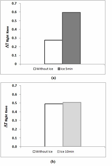
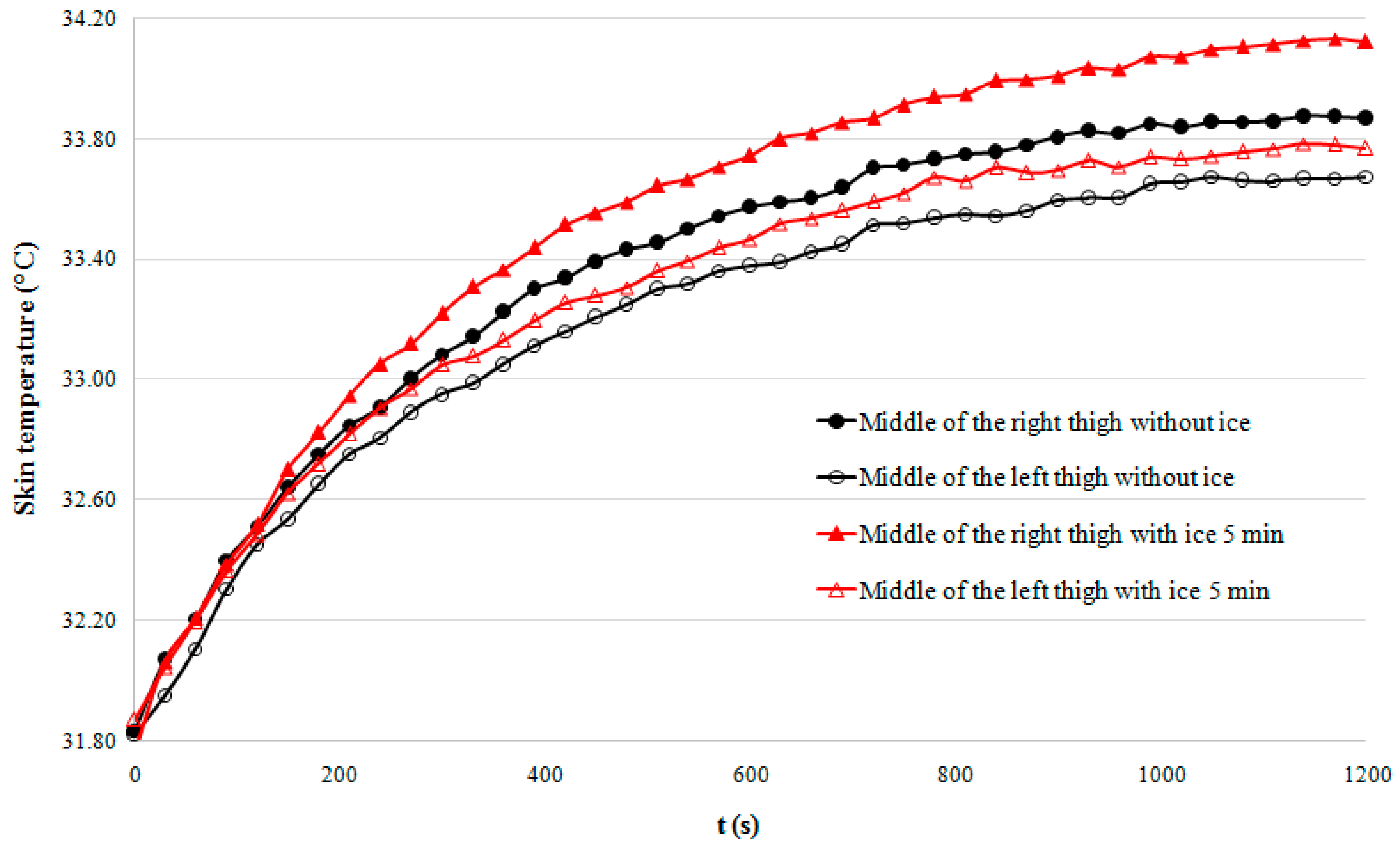
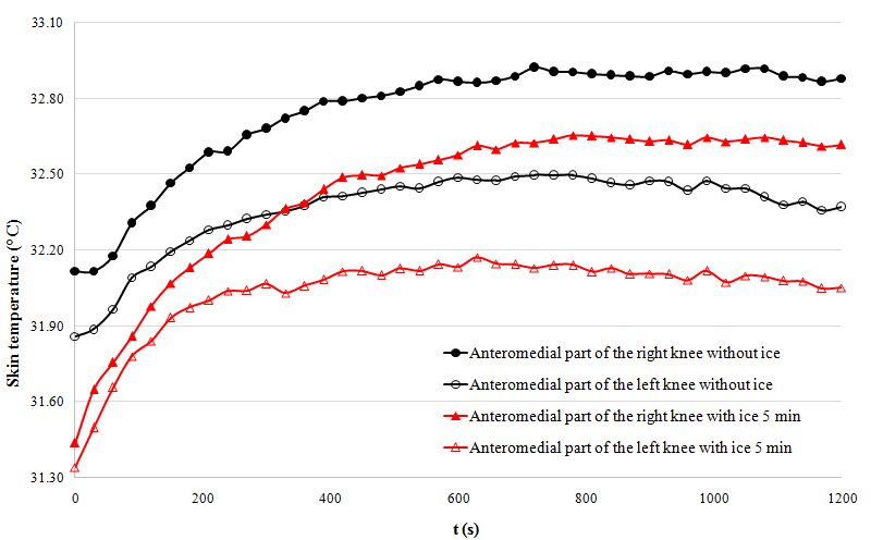
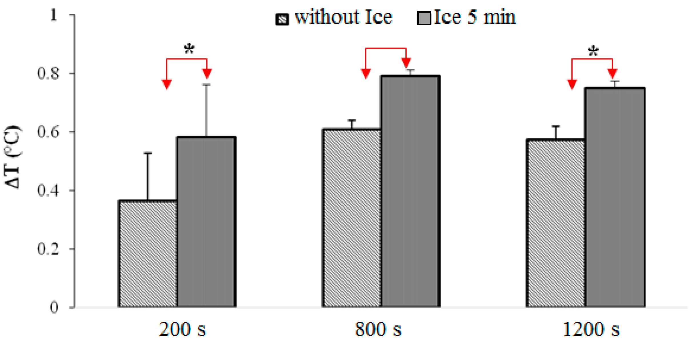
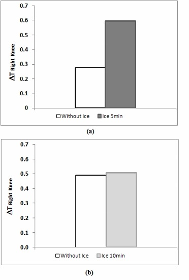
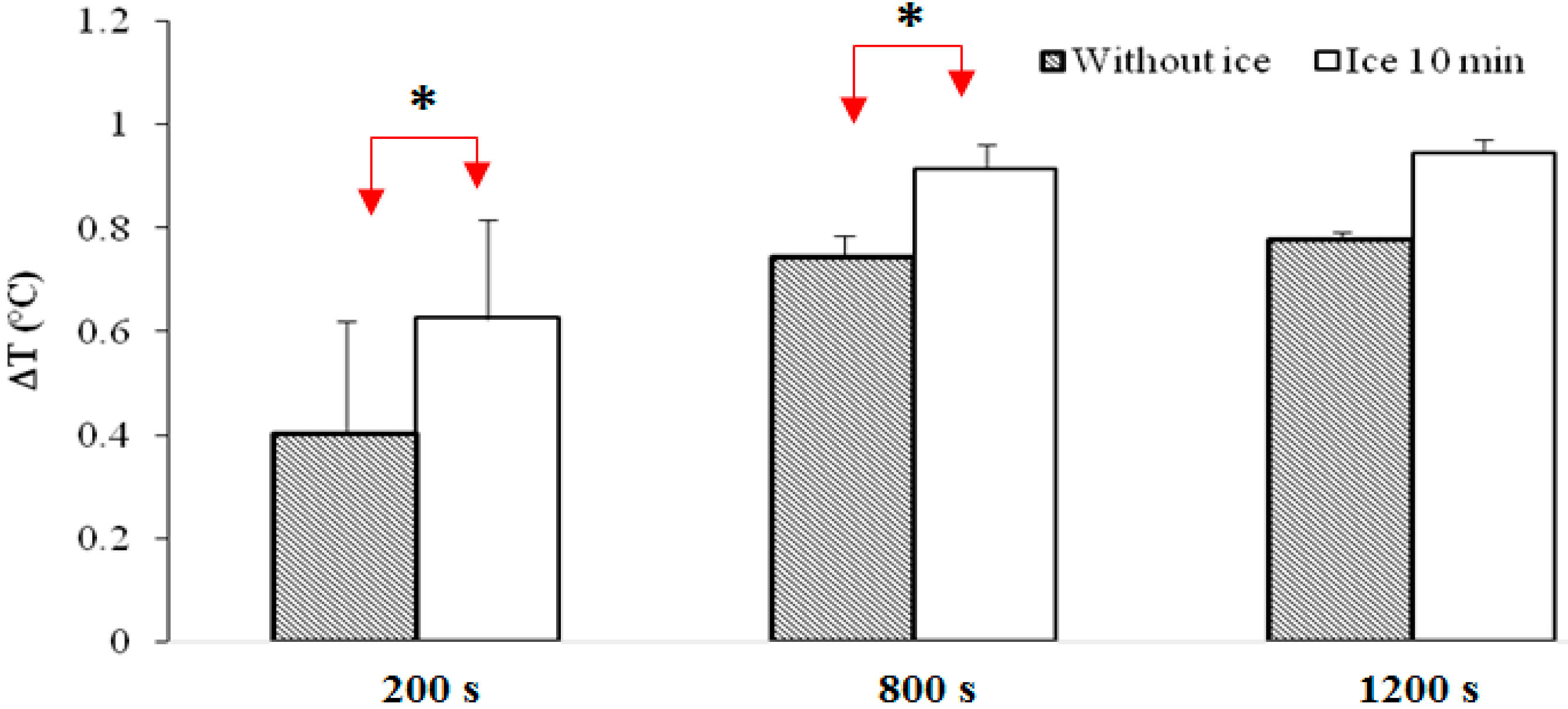
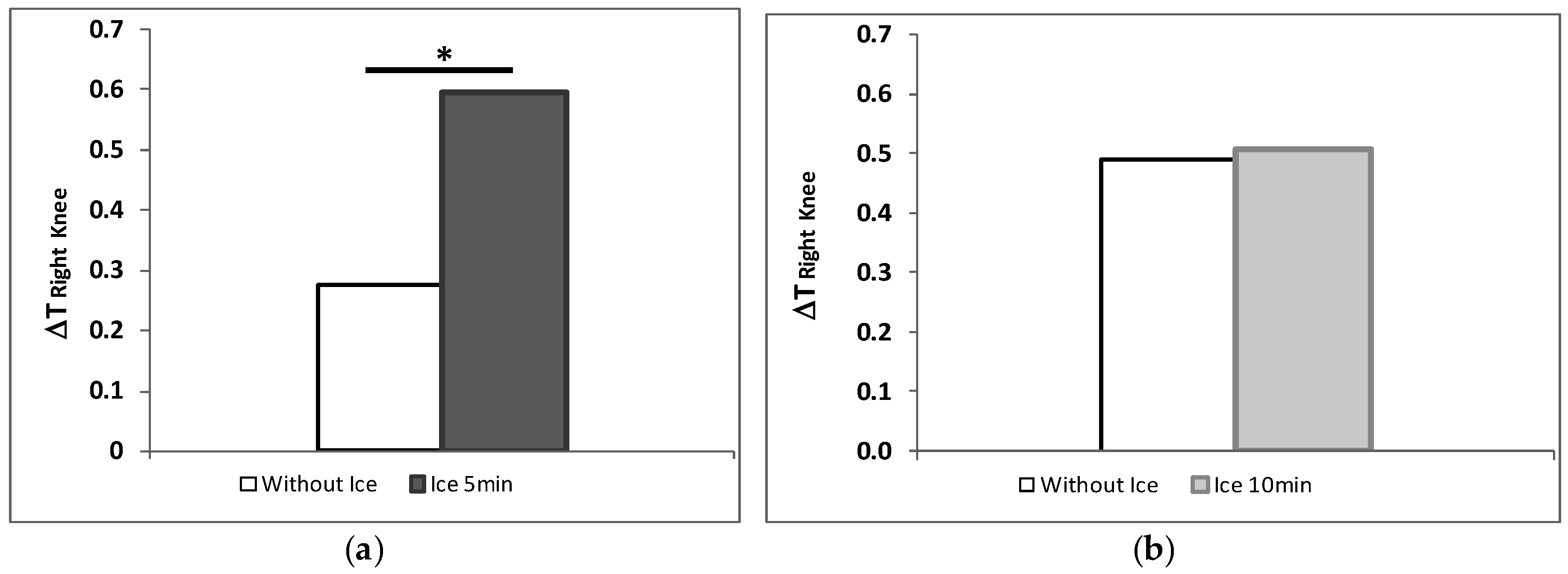
The 5-min cooling group shows significantly higher temperatures than the group that did not undergo cryotherapy (p < 0.001).
-
Putting an ice pack for 5 min during the post-exercise rest seems to provide significantly higher temperatures compared to the other experiments.

3.2. Discussion

The results obtained show surface temperature variations overtime after the moderate exertion of effort (5 min of running 10 km∙h−1). All studied areas presented an increase in the temperature (with or without cryotherapy).
We also demonstrated that exposure time plays a role in the cutaneous temperature evolution. Indeed, significant differences were found between the diverse experiments.
Cryotherapy applied in the popliteal significantly increased the speed of warming in a close area (the anteromedial part of the knee), regardless of exposure time. The measured temperatures still remain inferior to those obtained without the installation of ice.
Statistical analyses show that the rate of warming was no different with or without ice on the contralateral limb (in our study, left). Similarly, temperatures do not differ significantly according to the thigh environments.
Furthermore, 5 min of exposure to cryotherapy reaches surface temperatures significantly higher than those found with 10 min of exposure and those without cryotherapy near the right knee (directly in contact with the cold pack). This is the case for the entire cryotherapy exposure time.
These results indicate
-
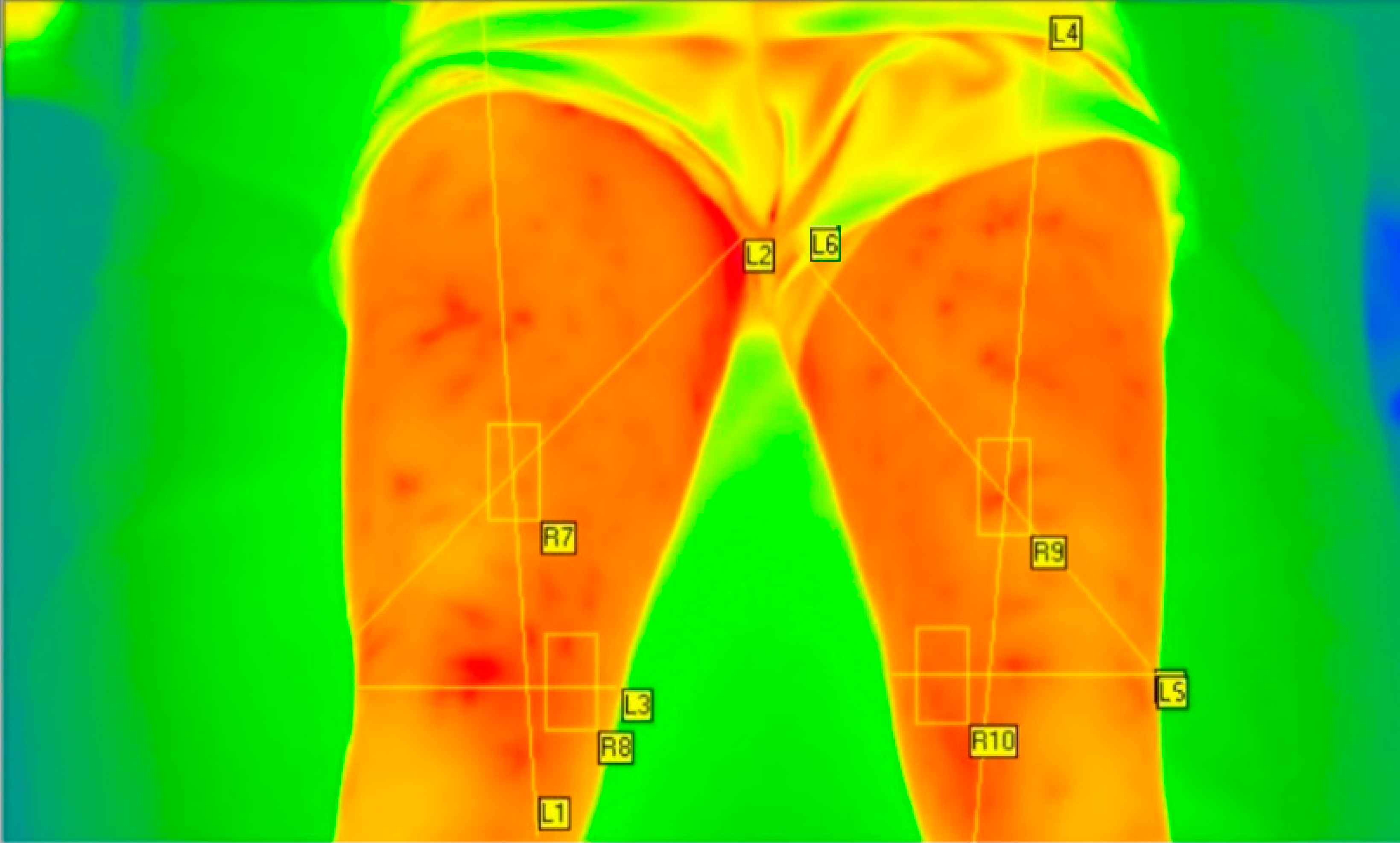
that cryotherapy application increases skin temperature in specific areas;
-
that the phenomenon does not only have a local impact.
It appears that the effects of the local cryotherapy are directed by many factors that are involved in the thermoregulation. Few of them are likely central [27].
Our results seem to contradict results that can be found in the literature.
Indeed, Mars [8] and his staff have conducted experiments that look like ours, but their results were different. They have shown that local cryotherapy applications decrease the cutaneous and the subcutaneous temperatures for the duration of the cryogenic application. The measured decreases were about 20 °C.
Nevertheless, the studied areas differ between the two works. Mars et al. had assessed areas much closer to the cryogenic source. Moreover, we did not measure temperatures under the pack like they did with thermocouples.
Indeed, the skin temperatures next to the cryogenic source were higher for the 10 min application than they were without ice. This increase of skin temperature can be explained by the decreased blood flow due to the vasoconstriction and the possible stagnation of hot blood near the recorded area.
The temperature increases we observed can also be explained by the subsequent vasoconstriction of the small capillaries, useful for the heat evacuation of the skin. Thorsson et al. [11] revealed that the large arteries are only slightly affected by the application of cold, contrary to the capillaries. Thus, the heat removal by transfer to the central bodies was reduced, and the discharge of thermal energy is mainly that of the skin.

4. Conclusions

To conclude, this study highlights the importance of the application of local cryotherapy on the thermal body reactions.
Indeed, the results showed that there are significant differences between cutaneous temperatures measured for 5 min compared to 10 min of exposure or even to cooling under an environment effect only.
These findings could have an impact on a therapist’s practice because the exposure time might have a significant impact on the desired effect of the cryotherapy.
The limitations of this study lie mainly in three points: the lack of a double blind, the small number of included subjects, and the exercise duration in the experiments before the local cryotherapy. The reduced effort leads perhaps to a simple early activation of the thermoregulation process and not a complete activation. In this study, one may desire to reach a partial activation of these phenomena in order to study the effects of cryotherapy on limited thermoregulation. Indeed, a limited effort was required to analyze the effect of cryotherapy on thermoregulation.
Moreover, the inclusion of 10 subjects suggests that the sample is not representative of the general population. In addition, all subjects lived in the same town. This might have generated geographic selection or ethnic bias [28].
One question remains: the long-term interest of cryotherapy. Indeed, this study was focused on short-term cryotherapy, influencing only momentarily thermoregulatory mechanisms. Some athletes use this kind of therapy daily. One may can wonder: is it relevant to inhibit these thermoregulatory mechanisms over the long term? Is there a risk of disruption of these mechanisms?

Acknowledgments

The authors thank the subjects for their enthusiastic participation and their availability.

Author Contributions

M.V. and A.A. developed the experiments; M.V. performed the experiments and analyzed the data; M.V. and A.A. wrote the paper.

Conflicts of Interest

The authors declare no conflict of interest.

References

  1. Galliera, E.; Dogliotti, G.; Melegati, G.; MCorsi, M.; Cabitza, P.; Banfi, G. Bone remodelling biomarkers after whole body cryotherapy (WBC) in elite rugby players. Injury 2013, 44, 1117–1121. [Google Scholar] [CrossRef] [PubMed]
  2. Banfi, G.; Melegati, G.; Barassi, A.; Dogliotti, G.; Melzid’Eril, G.; Dugue, B.; MCorsi, M. Effects of whole-body cryotherapy on serum mediators of inflammation and serum muscle enzymes in athletes. J. Therm. Biol. 2009, 34, 55–59. [Google Scholar] [CrossRef]
  3. Pinho Júnior, E.A.; Brito, C.J.; Costa Santos, W.O.; Nardelli Valido, C.; Lacerda Mendes, E.; Franchini, E. Influence of cryotherapy on muscle damage markers in jiujitsu fighters after competition: Acrossover study. Rev. Andal. Med. Deporte 2014, 7, 7–12. [Google Scholar] [CrossRef]
  4. Aza, M.; Miller, S.; Douglas-Moore, J.; Miller, M. Cryotherapy and its applications in the management of urologic malignancies: A review of its use in prostate and renal cancers. Urol. Oncol. Semin. Orig. Investig. 2014, 32, e19–e27. [Google Scholar]
  5. Chuan-Hang, Y.; Hung-Pin, L.; Shih-Jung, C.; Andy, S.; Hsin-Ming, C. Cryotherapy for oral precancers and cancers. J. Formos. Med. Assoc. 2014, 113, 272–277. [Google Scholar]
  6. Bleakley, C.M.; Bieuzen, F.; Davison, G.W.; Costello, J.T. Whole-body cryotherapy: Empirical evidence and theoretical perspectives. J. Sports Med. 2014, 5, 25–36. [Google Scholar] [CrossRef] [PubMed]
  7. Banfi, G.; Lombardi, G.; Colombini, A.; Melegati, G. Whole-Body Cryotherapy in Athletes. Sports Med. 2010, 40, 509–517. [Google Scholar] [CrossRef] [PubMed]
  8. Mars, M.; Hadebe, B.; Tufts, M. The effect of icepack cooling on skin and muscle temperature at rest and after exercise. S. Afr. J. Sports Med. 2006, 18. [Google Scholar] [CrossRef]
  9. Jeri, E.; Zemke, J.C.; Andersen, W.; Guion, K.; McMillan, J.; Joyner, A.B. Intramuscular Temperature Responses in the Human Leg to Two Forms of cryotherapy: Ice Massage and Ice Bag. J. Orthop. Sports Phys. Ther. 1998, 27, 301–307. [Google Scholar]
  10. Ho, S.S.; Illgen, R.L.; Meyer, R.W.; Torok, P.J.; Cooper, M.D.; Reider, B. Comparison of Various Icing Times in Decreasing Bone Metabolism and Blood Flowin the Knee. Am. J. Sports Med. 1995, 23, 74–76. [Google Scholar] [CrossRef] [PubMed]
  11. Thorsson, O.; Lilja, B.; Ahlgren, L.; Hemdal, B.; Westlin, N. The effect of local cold application on intramuscular blood flow at rest and after running. Med. Sci. Sports Exerc. 1985, 17, 710–713. [Google Scholar] [CrossRef] [PubMed]
  12. Ring, E.F.J.; Ammer, K. The technique of infrared imaging in medicine. Thermol. Int. 2000, 10, 7–14. [Google Scholar]
  13. Hoover, K.C.; Burlingame, S.E.; Lautz, C.H. Opportunities and challenges in concrete with thermal imaging. Concr. Int. 2004, 26, 23–27. [Google Scholar]
  14. Hildebrandt, C.; Raschner, C.; Ammer, K. An overview of Recent Application of Medical Infrared Thermography in Sports Medicine in Austria. Sensors 2010, 10, 4700–4715. [Google Scholar] [CrossRef] [PubMed]
  15. Bouzida, N.; Bendada, A.; Maldague, X. Visualization of body thermoregulation by infrared imaging. J. Therm. Biol. 2009, 34, 120–126. [Google Scholar] [CrossRef]
  16. Barcelos, E.Z.; Caminhas, W.M.; Ribeiro, E.; Pimenta, E.M.; Palhares, R.M. A Combined Method for Segmentation and Registration for an Advanced and Progressive Evaluation of Thermal Images. Sensors 2014, 14, 21950–21967. [Google Scholar] [CrossRef] [PubMed]
  17. Wu, C.L.; Yu, K.L.; Chuang, H.Y.; Huang, M.H.; Chen, T.W.; Chen, C.H. The application of Infrared Thermography in the assessment of patients with Coccygodynia before and after manual therapy combined with Diathermy. J. Manip. Physiol. Ther. 2009, 32, 281–293. [Google Scholar] [CrossRef] [PubMed]
  18. Ammer, K. The Glamorgan Protocol for recording and evaluation of thermal images of the human body. Thermol. Int. 2008, 18, 125–144. [Google Scholar]
  19. Jones, B.F.; Plassmann, P. Digital infrared thermal imaging of human skin. IEEE Eng. Med. Biol. 2002, 21, 41–48. [Google Scholar] [CrossRef]
  20. Ludwig, N.; Formenti, D.; Gargano, M.; Alberti, G. Skin temperature evaluation by infrared thermography: Comparison of image analysis methods. Infrared Phys. Technol. 2014, 62, 1–6. [Google Scholar] [CrossRef]
  21. Costello, J.T.; McInerney, C.D.; Bleakley, C.M.; Selfe, J.; Donnelly, A.E. The Use of Thermal Imaging in Assessing Skin Temperature Following Cryotherapy: A Review. J. Therm. Biol. 2012, 37, 103–110. [Google Scholar] [CrossRef] [Green Version]
  22. Monod, H.; Flandrois, R.; Vandewalle, H. Bases physiologiques des activités physiques et sportives. In Physiologie du Sport, 6th ed.; Elsevier Masson: Paris, France, 2007. [Google Scholar]
  23. Schwartz, R.G. Guidelines for Neuromusculoskeletal Thermography. Thermol. Int. 2006, 16, 5–9. [Google Scholar]
  24. Harriss, D.J.; Atkinson, G. Ethical Standards in Sport and Exercise Science Research. Int. J. Sports Med. 2009, 30, 701–702. [Google Scholar] [CrossRef] [PubMed]
  25. Infratec Infrared. Available online: http://www.infratec-infrared.com/fileadmin/downloads/pdf/VC_hr/VC_hr_headen.pdf (assessed on 1 September 2015).
  26. Villaseñor-Mora, C.; Sánchez-Marin, F.J.; Calixto-Carrera, S. An indirect skin emissivity measurement in the infrared thermal range through reflection of a CO2 laser beam. Rev. Mex. Fis. 2009, 55, 387–392. [Google Scholar]
  27. Foda, E.; Almesri, I.; Awbi, H.B.; Sirén, K. Models of human thermoregulation and the prediction of local and overall thermal sensations. Build. Environ. 2011, 46, 2023–2032. [Google Scholar] [CrossRef]
  28. Laporte, S.; Cucherat, M.; Mismetti, P. Méthodologie des essais cliniques: Une perspective critique. Méd. Thér. 2006, 12, 19–28. [Google Scholar]
Figure 1. Location of the four ROIs with the IRBIS® 3 software.
Jimaging 02 00020 g001
Figure 2. Variation of skin temperature of middle thighs with ice over a5-min period and without ice.
Jimaging 02 00020 g002
Figure 3. Skin temperature of the anteromedial part of the knees with ice over a 5-min period and without ice.
Jimaging 02 00020 g003
Figure 4. Variation in average temperatures of the skin of the right knee, comparing exposure to ice over a 10-m period and no exposure to ice. *: Significant difference p < 0.001.
Jimaging 02 00020 g004
Figure 5. Variation in average temperatures of the skin of the left knee, comparing exposure to ice over a 5-min period and no exposure to ice.
Jimaging 02 00020 g005
Figure 6. Variation in average temperatures in the skin of the left knee, comparing (a) exposure to ice for 5 min and no exposure to ice and (b) exposure to ice for10 min and no exposure to ice. *: Significant difference p < 0.001.
Jimaging 02 00020 g006
Table 1. Anthropometric data subjects.
Age (years)Height (m)Weight (kg)BMI (kg∙m−2)% of Body Fat
Averages23.201.75568.3022.1318.65
SD1.690.0777.101.334.65

