NS-GUSL: Green U-Shaped Learning for Nuclei Segmentation from Histopathology Images

 , , and
Abstract
1. Introduction
- We propose a 2D Green U-Shaped Learning framework for nuclei segmentation (NS-GUSL), featuring a small model size and low computational complexity.
- A low-confidence sample binarization (LCSB) technique is implemented to improve the predictions of hard samples, such as outlier nuclei or boundary regions.
- This work demonstrates competitive quantitative and qualitative performance in various metrics.
- We present a cross-dataset validation study to demonstrate the generalizability of our model across datasets compiled under different conditions.
2. Related Work
2.1. Traditional and Machine Learning Methods
2.2. Deep Learning Methods
2.3. Green Learning Methods
3. Methodology
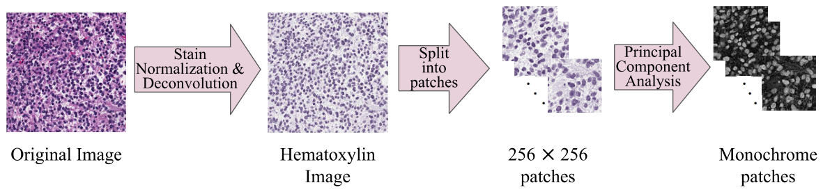
3.1. Preprocessing
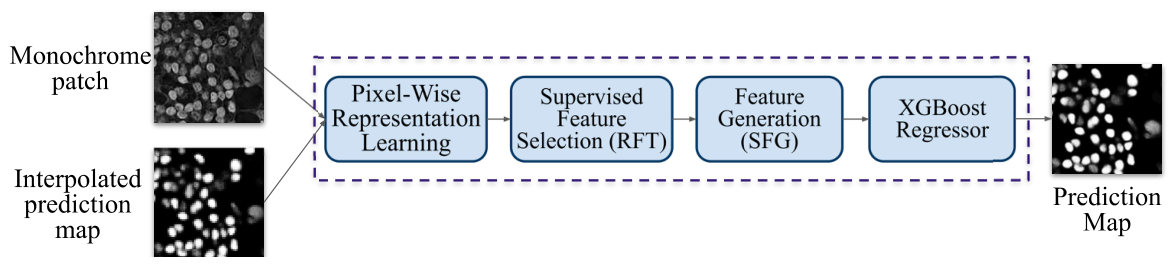
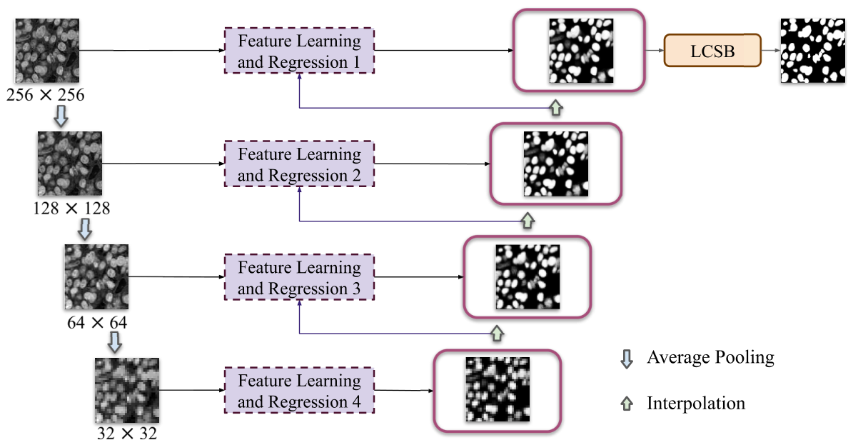
3.2. NS-GUSL
3.3. Representation Learning
3.3.1. Saab Features
3.3.2. Spatial Features
3.3.3. Laws Features
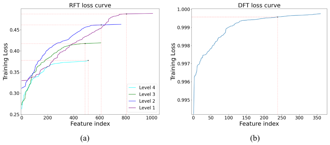
3.4. Feature Selection and Learning
3.5. Regression
3.6. Hard Sample-Aware Training
3.7. Low-Confidence Sample Binarization (LCSB)
3.8. Post-Processing
4. Experimental Setup
4.1. Datasets
4.1.1. MoNuSeg
4.1.2. CPM-17
4.1.3. CryoNuSeg
4.1.4. TNBC
4.2. Data Augmentation
4.3. Evaluation Metrics
5. Experimental Results
5.1. Quantitative Results
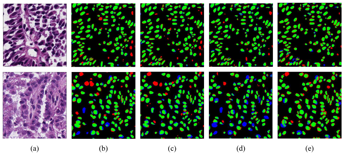
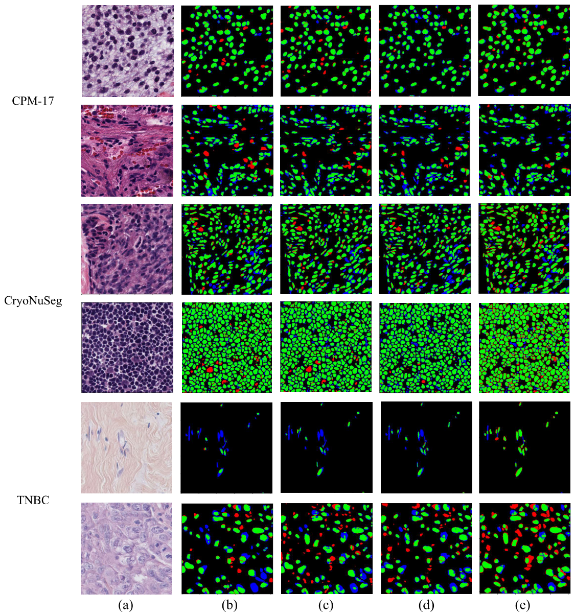
5.2. Qualitative Results
5.3. Ablation Study
5.4. Model Size, Complexity, and Energy Consumption
6. Discussion
7. Conclusions and Future Work
Author Contributions
Funding
Institutional Review Board Statement
Informed Consent Statement
Data Availability Statement
Conflicts of Interest
References
- Fischer, E.G. Nuclear morphology and the biology of cancer cells. Acta Cytol. 2020, 64, 511–519. [Google Scholar] [CrossRef] [Scilit]
- Masood, S. The changing role of pathologists from morphologists to molecular pathologists in the era of precision medicine. Breast J. 2020, 26, 27–34. [Google Scholar] [CrossRef] [Scilit]
- Basu, A.; Senapati, P.; Deb, M.; Rai, R.; Dhal, K.G. A survey on recent trends in deep learning for nucleus segmentation from histopathology images. Evol. Syst. 2024, 15, 203–248. [Google Scholar]
- Salvi, M.; Acharya, U.R.; Molinari, F.; Meiburger, K.M. The impact of pre-and post-image processing techniques on deep learning frameworks: A comprehensive review for digital pathology image analysis. Comput. Biol. Med. 2021, 128, 104129. [Google Scholar] [CrossRef] [Scilit]
- Gutiérrez Pérez, J.C.; Otero Baguer, D.; Maass, P. StainCUT: Stain Normalization with Contrastive Learning. J. Imaging 2022, 8, 202. [Google Scholar] [CrossRef] [Scilit]
- Magoulianitis, V.; Alexander, C.A.; Kuo, C.C.J. A Comprehensive Overview of Computational Nuclei Segmentation Methods in Digital Pathology. arXiv 2023, arXiv:2308.08112. [Google Scholar] [CrossRef] [Scilit]
- Kumar, N.; Verma, R.; Sharma, S.; Bhargava, S.; Vahadane, A.; Sethi, A. A dataset and a technique for generalized nuclear segmentation for computational pathology. IEEE Trans. Med. Imaging 2017, 36, 1550–1560. [Google Scholar] [CrossRef] [Scilit]
- Xing, F.; Xie, Y.; Yang, L. An Automatic Learning-Based Framework for Robust Nucleus Segmentation. IEEE Trans. Med. Imaging 2016, 35, 550–566. [Google Scholar] [CrossRef] [Scilit]
- Shelhamer, E.; Long, J.; Darrell, T. Fully Convolutional Networks for Semantic Segmentation. IEEE Trans. Pattern Anal. Mach. Intell. 2017, 39, 640–651. [Google Scholar] [CrossRef] [Scilit]
- Girshick, R.; Donahue, J.; Darrell, T.; Malik, J. Rich feature hierarchies for accurate object detection and semantic segmentation. In Proceedings of the IEEE Conference on Computer Vision and Pattern Recognition, Columbus, OH, USA, 23–28 June 2014; pp. 580–587. [Google Scholar]
- Ren, S.; He, K.; Girshick, R.; Sun, J. Faster R-CNN: Towards Real-Time Object Detection with Region Proposal Networks. In Proceedings of the Advances in Neural Information Processing Systems (NIPS), Montreal, QC, Canada, 7–12 December 2015. [Google Scholar]
- Ronneberger, O.; Fischer, P.; Brox, T. U-net: Convolutional networks for biomedical image segmentation. In Proceedings of the International Conference on Medical Image Computing and Computer-Assisted Intervention; Springer: Cham, Switzerland, 2015; pp. 234–241. [Google Scholar]
- Mahbod, A.; Schaefer, G.; Dorffner, G.; Hatamikia, S.; Ecker, R.; Ellinger, I. A dual decoder U-Net-based model for nuclei instance segmentation in hematoxylin and eosin-stained histological images. Front. Med. 2022, 9, 978146. [Google Scholar] [CrossRef] [Scilit]
- Qin, J.; He, Y.; Zhou, Y.; Zhao, J.; Ding, B. REU-Net: Region-enhanced nuclei segmentation network. Comput. Biol. Med. 2022, 146, 105546. [Google Scholar] [CrossRef] [Scilit]
- Chowdary, G.J.; G, S.; M, P.; Yogarajah, P. Nucleus segmentation and classification using residual SE-UNet and feature concatenation approach incervical cytopathology cell images. Technol. Cancer Res. Treat. 2023, 22, 15330338221134833. [Google Scholar] [CrossRef] [Scilit]
- Hancer, E.; Traore, M.; Samet, R.; Yıldırım, Z.; Nemati, N. An imbalance-aware nuclei segmentation methodology for H&E stained histopathology images. Biomed. Signal Process. Control 2023, 83, 104720. [Google Scholar] [CrossRef] [Scilit]
- Qu, H.; Wu, P.; Huang, Q.; Yi, J.; Yan, Z.; Li, K.; Riedlinger, G.M.; De, S.; Zhang, S.; Metaxas, D.N. Weakly Supervised Deep Nuclei Segmentation Using Partial Points Annotation in Histopathology Images. IEEE Trans. Med. Imaging 2020, 39, 3655–3666. [Google Scholar] [CrossRef] [Scilit]
- Verghese, G.; Lennerz, J.K.; Ruta, D.; Ng, W.; Thavaraj, S.; Siziopikou, K.P.; Naidoo, T.; Rane, S.; Salgado, R.; Pinder, S.E.; et al. Computational pathology in cancer diagnosis, prognosis, and prediction—Present day and prospects. J. Pathol. 2023, 260, 551–563. [Google Scholar] [CrossRef] [Scilit]
- Jahanifar, M.; Raza, M.; Xu, K.; Vuong, T.T.L.; Jewsbury, R.; Shephard, A.; Zamanitajeddin, N.; Kwak, J.T.; Raza, S.E.A.; Minhas, F.; et al. Domain generalization in computational pathology: Survey and guidelines. ACM Comput. Surv. 2025, 57, 285. [Google Scholar] [CrossRef] [Scilit]
- Adadi, A.; Berrada, M. Explainable AI for Healthcare: From Black Box to Interpretable Models. In Proceedings of the Embedded Systems and Artificial Intelligence; Bhateja, V., Satapathy, S.C., Satori, H., Eds.; Springer: Singapore, 2020; pp. 327–337. [Google Scholar]
- Cai, H.; Yang, Z.; Cao, X.; Xia, W.; Xu, X. A new iterative triclass thresholding technique in image segmentation. IEEE Trans. Image Process. 2014, 23, 1038–1046. [Google Scholar] [CrossRef] [Scilit]
- Magoulianitis, V.; Yang, Y.; Kuo, C.C.J. HUNIS: High-Performance Unsupervised Nuclei Instance Segmentation. In Proceedings of the 2022 IEEE 14th Image, Video, and Multidimensional Signal Processing Workshop (IVMSP); IEEE: Nafplio, Greece, 2022; pp. 1–5. [Google Scholar]
- Phoulady, H.A.; Goldgof, D.B.; Hall, L.O.; Mouton, P.R. Nucleus segmentation in histology images with hierarchical multilevel thresholding. In Proceedings of the Medical Imaging 2016: Digital Pathology; SPIE: Bellingham, WA, USA, 2016; Volume 9791, pp. 280–285. [Google Scholar]
- Hu, M.; Ping, X.; Ding, Y. Automated cell nucleus segmentation using improved snake. In Proceedings of the 2004 International Conference on Image Processing; ICIP’04; IEEE: Piscataway, NJ, USA, 2004; Volume 4, pp. 2737–2740. [Google Scholar]
- Faridi, P.; Danyali, H.; Helfroush, M.S.; Jahromi, M.A. An automatic system for cell nuclei pleomorphism segmentation in histopathological images of breast cancer. In Proceedings of the 2016 IEEE Signal Processing in Medicine and Biology Symposium (SPMB); IEEE: Piscataway, NJ, USA, 2016; pp. 1–5. [Google Scholar]
- Vahadane, A.; Sethi, A. Towards generalized nuclear segmentation in histological images. In Proceedings of the 13th IEEE International Conference on Bioinformatics and Bioengineering; IEEE: Piscataway, NJ, USA, 2013; pp. 1–4. [Google Scholar]
- Shu, J.; Fu, H.; Qiu, G.; Kaye, P.; Ilyas, M. Segmenting overlapping cell nuclei in digital histopathology images. In Proceedings of the 2013 35th Annual International Conference of the IEEE Engineering in Medicine and Biology Society (EMBC); IEEE: Piscataway, NJ, USA, 2013; pp. 5445–5448. [Google Scholar]
- Zhang, M.; Wu, T.; Bennett, K.M. Small Blob Identification in Medical Images Using Regional Features from Optimum Scale. IEEE Trans. Biomed. Eng. 2015, 62, 1051–1062. [Google Scholar] [CrossRef] [Scilit]
- Kong, H.; Gurcan, M.; Belkacem-Boussaid, K. Partitioning Histopathological Images: An Integrated Framework for Supervised Color-Texture Segmentation and Cell Splitting. IEEE Trans. Med. Imaging 2011, 30, 1661–1677. [Google Scholar] [CrossRef] [Scilit]
- Sheikh, T.S.; Cho, M. Segmentation of Variants of Nuclei on Whole Slide Images by Using Radiomic Features. Bioengineering 2024, 11, 252. [Google Scholar] [CrossRef] [Scilit]
- Graham, S.; Vu, Q.D.; Raza, S.E.A.; Azam, A.; Tsang, Y.W.; Kwak, J.T.; Rajpoot, N. Hover-net: Simultaneous segmentation and classification of nuclei in multi-tissue histology images. Med. Image Anal. 2019, 58, 101563. [Google Scholar] [CrossRef] [Scilit]
- Zhou, Z.; Siddiquee, M.M.R.; Tajbakhsh, N.; Liang, J. UNet++: Redesigning Skip Connections to Exploit Multiscale Features in Image Segmentation. arXiv 2020, arXiv:1912.05074. [Google Scholar] [CrossRef] [Scilit]
- Cao, H.; Wang, Y.; Chen, J.; Jiang, D.; Zhang, X.; Tian, Q.; Wang, M. Swin-unet: Unet-like pure transformer for medical image segmentation. In Proceedings of the European Conference on Computer Vision; Springer: Cham, Switzerland, 2022; pp. 205–218. [Google Scholar]
- Oda, H.; Roth, H.R.; Chiba, K.; Sokolić, J.; Kitasaka, T.; Oda, M.; Hinoki, A.; Uchida, H.; Schnabel, J.A.; Mori, K. BESNet: Boundary-enhanced segmentation of cells in histopathological images. In Proceedings of the International Conference on Medical Image Computing and Computer-Assisted Intervention; Springer: Cham, Switzerland, 2018; pp. 228–236. [Google Scholar]
- Zhou, Y.; Onder, O.F.; Dou, Q.; Tsougenis, E.; Chen, H.; Heng, P.A. CIA-Net: Robust Nuclei Instance Segmentation with Contour-Aware Information Aggregation. In Proceedings of the Information Processing in Medical Imaging; Chung, A.C.S., Gee, J.C., Yushkevich, P.A., Bao, S., Eds.; Springer: Cham, Switzerland, 2019; pp. 682–693. [Google Scholar]
- He, Z.; Unberath, M.; Ke, J.; Shen, Y. Transnuseg: A lightweight multi-task transformer for nuclei segmentation. In Proceedings of the International Conference on Medical Image Computing and Computer-Assisted Intervention; Springer: Cham, Switzerland, 2023; pp. 206–215. [Google Scholar]
- Du, Y.; Chen, X.; Fu, Y. Multiscale transformers and multi-attention mechanism networks for pathological nuclei segmentation. Sci. Rep. 2025, 15, 12549. [Google Scholar] [CrossRef] [Scilit]
- Ivanov, D.; Barbano, C.A.; Grangetto, M. Multi-target Stain Normalization for Histology Slides. In Proceedings of the Medical Optical Imaging and Virtual Microscopy Image Analysis; Huo, Y., Millis, B.A., Zhou, Y., Younis, K., Wang, X., Tang, Y., Eds.; Springer: Cham, Switzerland, 2025; pp. 36–44. [Google Scholar]
- Mahbod, A.; Dorffner, G.; Ellinger, I.; Woitek, R.; Hatamikia, S. Improving generalization capability of deep learning-based nuclei instance segmentation by non-deterministic train time and deterministic test time stain normalization. Comput. Struct. Biotechnol. J. 2024, 23, 669–678. [Google Scholar] [CrossRef] [Scilit]
- Kuo, C.C.J.; Madni, A.M. Green learning: Introduction, examples and outlook. J. Vis. Commun. Image Represent. 2023, 90, 103685. [Google Scholar] [CrossRef] [Scilit]
- Kuo, C.C.J.; Zhang, M.; Li, S.; Duan, J.; Chen, Y. Interpretable convolutional neural networks via feedforward design. J. Vis. Commun. Image Represent. 2019, 60, 346–359. [Google Scholar] [CrossRef] [Scilit]
- Magoulianitis, V.; Yang, J.; Yang, Y.; Xue, J.; Kaneko, M.; Cacciamani, G.; Abreu, A.; Duddalwar, V.; Kuo, C.C.J.; Gill, I.S.; et al. PCa-RadHop: A transparent and lightweight feed-forward method for clinically significant prostate cancer segmentation. Comput. Med. Imaging Graph. 2024, 116, 102408. [Google Scholar] [CrossRef] [Scilit]
- Yang, J.; Magoulianitis, V.; Alexander, C.A.C.; Xue, J.; Kaneko, M.; Cacciamani, G.; Abreu, A.; Duddalwar, V.; Kuo, C.C.J.; Gill, I.S.; et al. GUSL: A Novel and Efficient Machine Learning Model for Prostate Segmentation on MRI. arXiv 2025, arXiv:2506.23688. [Google Scholar]
- Magoulianitis, V.; Alexander, C.A.; Yang, J.; Kuo, C.C.J. LG-NuSegHop: A Local-to-Global Self-Supervised Pipeline for Nuclei Instance Segmentation. arXiv 2025, arXiv:2511.04892. [Google Scholar] [CrossRef] [Scilit]
- Macenko, M.; Niethammer, M.; Marron, J.S.; Borland, D.; Woosley, J.T.; Guan, X.; Schmitt, C.; Thomas, N.E. A method for normalizing histology slides for quantitative analysis. In Proceedings of the 2009 IEEE International Symposium on Biomedical Imaging: From Nano to Macro, Boston, MA, USA, 28 June–1 July 2009; pp. 1107–1110. [Google Scholar] [CrossRef] [Scilit]
- Yang, Y.; Wang, W.; Fu, H.; Kuo, C.C.J. On supervised feature selection from high dimensional feature spaces. APSIPA Trans. Signal Inf. Process. 2022, 11, 1–23. [Google Scholar] [CrossRef] [Scilit]
- Wang, X.; Wu, Y.; Li, H.; Mishra, V.K.; Kuo, C.C.J. A Statistics-based Feature Generation (SFG) Method: Theory and Applications. In Proceedings of the 2024 IEEE International Conference on Big Data (BigData), Washington DC, USA, 15–18 December 2024; pp. 5731–5738. [Google Scholar] [CrossRef] [Scilit]
- Wang, X.; Mishra, V.K.; Jay Kuo, C.C. Enhancing Edge Intelligence with Highly Discriminant LNT Features. In Proceedings of the 2023 IEEE International Conference on Big Data (BigData), Sorrento, Italy, 15–18 December 2023; pp. 3880–3887. [Google Scholar] [CrossRef] [Scilit]
- Coelho, L.P. Mahotas: Open source software for scriptable computer vision. J. Open Res. Softw. 2013, 1, e3. [Google Scholar] [CrossRef] [Scilit]
- Vu, Q.D.; Graham, S.; Kurc, T.; To, M.N.N.; Shaban, M.; Qaiser, T.; Koohbanani, N.A.; Khurram, S.A.; Kalpathy-Cramer, J.; Zhao, T.; et al. Methods for Segmentation and Classification of Digital Microscopy Tissue Images. Front. Bioeng. Biotechnol. 2019, 7, 53. [Google Scholar] [CrossRef] [Scilit]
- Mahbod, A.; Schaefer, G.; Bancher, B.; Löw, C.; Dorffner, G.; Ecker, R.; Ellinger, I. CryoNuSeg: A dataset for nuclei instance segmentation of cryosectioned H&E-stained histological images. Comput. Biol. Med. 2021, 132, 104349. [Google Scholar] [CrossRef] [Scilit]
- Naylor, P.; Laé, M.; Reyal, F.; Walter, T. Segmentation of Nuclei in Histopathology Images by Deep Regression of the Distance Map. IEEE Trans. Med. Imaging 2019, 38, 448–459. [Google Scholar] [CrossRef] [Scilit]
- Iakubovskii, P. Segmentation Models Pytorch. 2019. Available online: https://github.com/qubvel/segmentation_models.pytorch (accessed on 5 May 2026).
- Xue, X.; Kamata, S.I. Contextual Mixing Feature Unet for Multi-Organ Nuclei Segmentation. Front. Signal Process. 2022, 2, 833433. [Google Scholar] [CrossRef] [Scilit]
- Wang, H.; Cao, P.; Wang, J.; Zaiane, O.R. UCTransNet: Rethinking the Skip Connections in U-Net from a Channel-Wise Perspective with Transformer. Proc. AAAI Conf. Artif. Intell. 2022, 36, 2441–2449. [Google Scholar] [CrossRef] [Scilit]
- Choi, J.W.; Bedard, D.; Fowler, R.; Vuduc, R. A Roofline Model of Energy. In Proceedings of the 2013 IEEE 27th International Symposium on Parallel and Distributed Processing, Cambridge, MA, USA, 20–24 May 2013; pp. 661–672. [Google Scholar] [CrossRef] [Scilit]




| Level | Neighborhood Size | Saab Kernel Size |
|---|---|---|
| Level 1 | ||
| Level 2 | ||
| Level 3 | ||
| Level 4 |
| Level/Module | Raw Features | RFT/DFT Selected Features | LNT Features | XGBoost Features |
|---|---|---|---|---|
| Level 4 | 375 | 375 | 137 | 512 |
| Level 3 | 552 | 441 | 168 | 488 |
| Level 2 | 648 | 518 | 247 | 612 |
| Level 1 | 768 | 614 | 394 | 807 |
| LCSB | 535 | 155 | 205 | 238 |
| Method | AJI | F1 | Dice | PQ |
|---|---|---|---|---|
| U-Net [12] | 0.5668 | 0.8463 | 0.7838 | 0.7364 |
| SwinU-Net [33] | 0.5812 | 0.8162 | 0.7862 | 0.6937 |
| U-Net++ [32] | 0.5877 | 0.8339 | 0.7947 | 0.7419 |
| CMF-UNet [54] | 0.6153 | 0.8226 | - | - |
| UCTransNet [55] | 0.4652 | 0.8709 | 0.7986 | 0.6470 |
| ON-DDU-Net [39] | 0.6620 | - | 0.8320 | 0.6357 |
| NS-GUSL (Ours) | 0.6060 | 0.8849 | 0.7948 | 0.7727 |
| Method | AJI | F1 | Dice | PQ |
|---|---|---|---|---|
| U-Net [12] | 0.4778 | 0.8221 | 0.7779 | 0.5876 |
| SwinU-Net [33] | 0.4825 | 0.7840 | 0.7634 | 0.5510 |
| U-Net++ [32] | 0.4676 | 0.8061 | 0.7607 | 0.5931 |
| ON-DDU-Net [39] | 0.5040 | - | 0.7890 | 0.4710 |
| NS-GUSL (Ours) | 0.4500 | 0.8129 | 0.7538 | 0.6177 |
| Method | AJI | F1 | Dice | PQ |
|---|---|---|---|---|
| U-Net [12] | 0.5662 | 0.8415 | 0.7883 | 0.5659 |
| SwinU-Net [33] | 0.5419 | 0.8223 | 0.7697 | 0.5009 |
| U-Net++ [32] | 0.5661 | 0.8390 | 0.7949 | 0.5758 |
| ON-DDU-Net [39] | 0.6380 | - | 0.8200 | 0.5890 |
| NS-GUSL (Ours) | 0.5479 | 0.8723 | 0.8016 | 0.6106 |
| Method | AJI | F1 | Dice | PQ |
|---|---|---|---|---|
| U-Net [12] | 0.4561 | 0.7180 | 0.6734 | 0.4713 |
| SwinU-Net [33] | 0.5052 | 0.7569 | 0.7107 | 0.4969 |
| U-Net++ [32] | 0.4973 | 0.7409 | 0.6975 | 0.5130 |
| ON-DDU-Net [39] | 0.6080 | - | 0.7840 | 0.5810 |
| NS-GUSL (Ours) | 0.4602 | 0.7806 | 0.6965 | 0.7383 |
| Representation Learning Method | AJI | F1 | Dice | PQ | Parameters |
|---|---|---|---|---|---|
| ResNet-50 | 0.5265 | 0.7164 | 0.7857 | 0.6719 | 1.75M |
| DenseNet-121 | 0.5617 | 0.6028 | 0.7871 | 0.6040 | 2.71M |
| Saab Transform | 0.5840 | 0.8587 | 0.7942 | 0.6841 | 319 |
| LCS Probability Thresholds | AJI | F1 | Dice | PQ |
|---|---|---|---|---|
| (0.1, 0.9) | 0.4950 | 0.6996 | 0.7760 | 0.6200 |
| (0.2, 0.8) | 0.5320 | 0.7602 | 0.7885 | 0.6852 |
| (0.3, 0.7) | 0.5550 | 0.7319 | 0.7932 | 0.6541 |
| (0.4, 0.6) | 0.5840 | 0.8587 | 0.7942 | 0.6841 |
| Thresholding (0.5) | LCSB | Post-Processing | AJI | F1 | Dice | PQ |
|---|---|---|---|---|---|---|
| ✓ | 0.5818 | 0.8852 | 0.8010 | 0.7227 | ||
| ✓ | ✓ | 0.5994 | 0.8911 | 0.7929 | 0.7978 | |
| ✓ | 0.5840 | 0.8587 | 0.7942 | 0.6841 | ||
| ✓ | ✓ | 0.6060 | 0.8849 | 0.7948 | 0.7727 |
| Method | Model Size | FLOPs/Pixel | Energy ( kWh) | Carbon Footprint ( g CO2e) |
|---|---|---|---|---|
| SwinU-Net [33] | 27 M () | 118 K () | 8.29 () | 24.9 () |
| U-Net++ [32] | 26 M () | 281 K () | 25.7 () | 77.3 () |
| U-Net [12] | 24 M () | 120 K () | 10.9 () | 33 () |
| NS-GUSL (with LCSB) | 0.8 M () | 61.50 K () | 5.60 (×4) | 16.8 (×4) |
| NS-GUSL | 0.76 M () | 17.26 K () | 1.57 (×1) | 4.73 (×1) |
Disclaimer/Publisher’s Note: The statements, opinions and data contained in all publications are solely those of the individual author(s) and contributor(s) and not of MDPI and/or the editor(s). MDPI and/or the editor(s) disclaim responsibility for any injury to people or property resulting from any ideas, methods, instructions or products referred to in the content. |
© 2026 by the authors. Licensee MDPI, Basel, Switzerland. This article is an open access article distributed under the terms and conditions of the Creative Commons Attribution (CC BY) license.
Share and Cite
Christie Alexander, C.A.; Magoulianitis, V.; Yang, J.; Kuo, C.-C.J. NS-GUSL: Green U-Shaped Learning for Nuclei Segmentation from Histopathology Images. J. Imaging 2026, 12, 316. https://doi.org/10.3390/jimaging12070316
Christie Alexander CA, Magoulianitis V, Yang J, Kuo C-CJ. NS-GUSL: Green U-Shaped Learning for Nuclei Segmentation from Histopathology Images. Journal of Imaging. 2026; 12(7):316. https://doi.org/10.3390/jimaging12070316
Chicago/Turabian StyleChristie Alexander, Catherine Aurelia, Vasileios Magoulianitis, Jiaxin Yang, and C.-C. Jay Kuo. 2026. "NS-GUSL: Green U-Shaped Learning for Nuclei Segmentation from Histopathology Images" Journal of Imaging 12, no. 7: 316. https://doi.org/10.3390/jimaging12070316
APA StyleChristie Alexander, C. A., Magoulianitis, V., Yang, J., & Kuo, C.-C. J. (2026). NS-GUSL: Green U-Shaped Learning for Nuclei Segmentation from Histopathology Images. Journal of Imaging, 12(7), 316. https://doi.org/10.3390/jimaging12070316