Human–AI Interaction in Interventional Radiology: A Narrative Review of Current Applications, Challenges, and Future Directions
 , , , ,
  and
and
Abstract
1. Introduction
2. Conceptual Framework
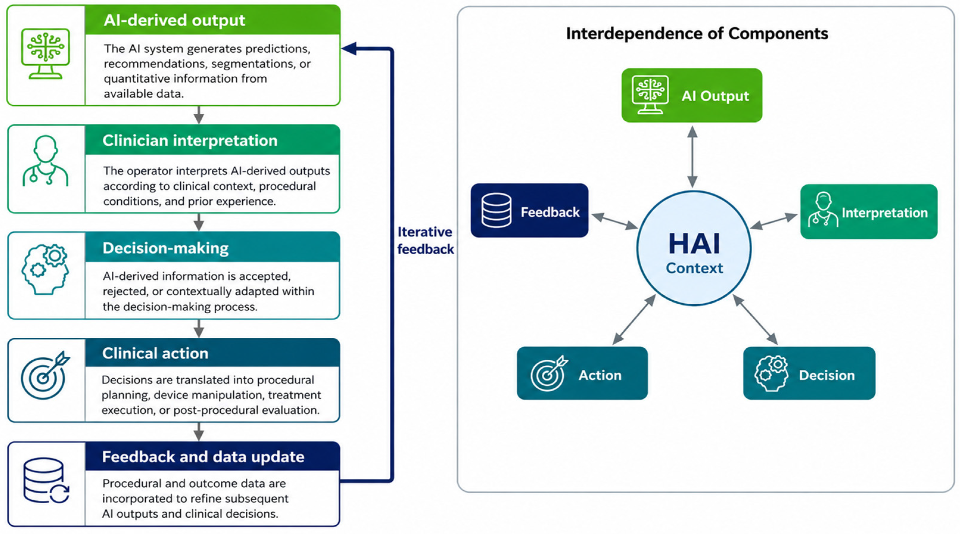
2.1. Human–AI Interaction as an Overarching Framework
2.2. Interaction Dynamics in Clinical Practice
2.3. Determinants of Effective Human–AI Interaction
3. AI for Decision Support
4. AI for Procedural Verification
5. Immersive Interfaces in Interventional Radiology: A Transversal Layer of Human–AI Interaction
6. Limitations and Challenges
7. Future Directions
8. Conclusions
Author Contributions
Funding
Institutional Review Board Statement
Informed Consent Statement
Data Availability Statement
Conflicts of Interest
Abbreviations
| BEVAR | Branched Endovascular Aneurysm Repair |
| CNR | Contrast-to-Noise Ratio |
| CTA | Computed Tomography Angiography |
| DECT | Dual-Energy Computed Tomography |
| DECTA | Dual-Energy Computed Tomography Angiography |
| DSA | Digital Subtraction Angiography |
References
- Hawkins, C.M.; Duszak, R., Jr.; Hughes, D.R.; Liu, R.; Resnick, A.S.; Kooby, D.A.; Durack, J.C.; Kaufman, J.A.; Dubeck, F.; Canada, M.B.; et al. Defining the Value of Interventional Radiology to Healthcare Stakeholders: Proceedings from a Society of Interventional Radiology Research Consensus Panel. J. Vasc. Interv. Radiol. 2021, 32, 1088.e1–1088.e8. [Google Scholar] [CrossRef] [PubMed]
- Huth, H.; Saccenti, L.; Anibal, J.; Ukeh, I.; Kassin, M.; Gichoya, J.; Varble, N.; Li, M.; Karanian, J.; Turkbey, B.; et al. Artificial Intelligence and Intelligent Interventional Radiology: Fusion of Human and Machine. J. Vasc. Interv. Radiol. 2025, 36, 1929–1936.e1. [Google Scholar] [CrossRef] [PubMed]
- Ferrari, R.; Trinci, M.; Casinelli, A.; Treballi, F.; Leone, E.; Caruso, D.; Polici, M.; Faggioni, L.; Neri, E.; Galluzzo, M. Radiomics in Radiology: What the Radiologist Needs to Know about Technical Aspects and Clinical Impact. Radiol. Med. 2024, 129, 1751–1765. [Google Scholar] [CrossRef] [PubMed]
- Lesaunier, A.; Khlaut, J.; Dancette, C.; Tselikas, L.; Bonnet, B.; Boeken, T. Artificial Intelligence in Interventional Radiology: Current Concepts and Future Trends. Diagn. Interv. Imaging 2025, 106, 5–10. [Google Scholar] [CrossRef] [PubMed]
- Campbell, W.A.; Chick, J.F.B.; Shin, D.S.; Makary, M.S. Value of Interventional Radiology and Their Contributions to Modern Medical Systems. Front. Radiol. 2024, 4, 1403761. [Google Scholar] [CrossRef] [PubMed]
- Kocak, B.; Cuocolo, R. Human–AI Interaction and Collaboration in Radiology: From Conceptual Frameworks to Responsible Implementation. Diagn. Interv. Radiol. 2026. [Google Scholar] [CrossRef] [PubMed]
- Maniaci, A.; Lavalle, S.; Gagliano, C.; Lentini, M.; Masiello, E.; Parisi, F.; Iannella, G.; Cilia, N.D.; Salerno, V.; Cusumano, G.; et al. The Integration of Radiomics and Artificial Intelligence in Modern Medicine. Life 2024, 14, 1248. [Google Scholar] [CrossRef] [PubMed]
- Petrillo, A.; Fusco, R.; Petrosino, T.; Vallone, P.; Granata, V.; Rubulotta, M.R.; Pariante, P.; Raiano, N.; Scognamiglio, G.; Fanizzi, A.; et al. A Multicentric Study of Radiomics and Artificial Intelligence Analysis on Contrast-Enhanced Mammography to Identify Different Histotypes of Breast Cancer. Radiol. Med. 2024, 129, 864–878. [Google Scholar] [CrossRef] [PubMed]
- Fahim, Y.A.; Hasani, I.W.; Kabba, S.; Ragab, W.M. Artificial Intelligence in Healthcare and Medicine: Clinical Applications, Therapeutic Advances, and Future Perspectives. Eur. J. Med. Res. 2025, 30, 848. [Google Scholar] [CrossRef] [PubMed]
- Mohseni, A.; Ghotbi, E.; Kazemi, F.; Shababi, A.; Jahan, S.C.; Mohseni, A.; Shababi, N. Artificial Intelligence in Radiology: What Is Its True Role at Present, and Where Is the Evidence? Radiol. Clin. N. Am. 2024, 62, 935–947. [Google Scholar] [CrossRef] [PubMed]
- Jung, H.K.; Kim, K.; Park, J.E.; Kim, N. Image-Based Generative Artificial Intelligence in Radiology: Comprehensive Updates. Korean J. Radiol. 2024, 25, 959–981. [Google Scholar] [CrossRef] [PubMed]
- Park, S.H.; Han, K.; Lee, J.G. Conceptual Review of Outcome Metrics and Measures Used in Clinical Evaluation of Artificial Intelligence in Radiology. Radiol. Med. 2024, 129, 1644–1655. [Google Scholar] [CrossRef] [PubMed]
- Mennella, C.; Maniscalco, U.; Di Pietro, G.; Esposito, M. Ethical and Regulatory Challenges of AI Technologies in Healthcare: A Narrative Review. Heliyon 2024, 10, e26297. [Google Scholar] [CrossRef] [PubMed]
- Lastrucci, A.; Iosca, N.; Wandael, Y.; Barra, A.; Lepri, G.; Forini, N.; Ricci, R.; Miele, V.; Giansanti, D. AI and Interventional Radiology: A Narrative Review of Reviews on Opportunities, Challenges, and Future Directions. Diagnostics 2025, 15, 893. [Google Scholar] [CrossRef] [PubMed]
- Lanza, C.; Angileri, S.A.; Carriero, S.; Triggiani, S.; Ascenti, V.; Mortellaro, S.R.; Ginolfi, M.; Leo, A.; Arnone, F.; Torcia, P.; et al. Artificial Intelligence Applications in Interventional Radiology. J. Pers. Med. 2025, 15, 569. [Google Scholar] [CrossRef] [PubMed]
- Javan, H.; Rokni, M.; Warrior, S.; Ajam, M.; Lam, K.; Ghazaryan, S.; Morshedi, M. Abstract No. 523 The Role of Artificial Intelligence in Image-Guided Procedures: Revolutionizing Interventional Radiology. J. Vasc. Interv. Radiol. 2025, 36, S179. [Google Scholar] [CrossRef]
- Topol, E.J. High-Performance Medicine: The Convergence of Human and Artificial Intelligence. Nat. Med. 2019, 25, 44–56. [Google Scholar] [CrossRef] [PubMed]
- Colarieti, A.; Sardanelli, F. The Radiologist as an Independent “Third Party” to the Patient and Clinicians in the Era of Generative AI. Radiol. Med. 2025, 130, 281–283. [Google Scholar] [CrossRef] [PubMed]
- Shirangi, A.; Pittinsky, T.; Gichoya, J. Rethinking Human-AI Collaboration in Radiology. Radiology 2026, 318, e252760. [Google Scholar] [CrossRef] [PubMed]
- Kim, S.H.; Wihl, J.; Schramm, S.; Berberich, C.; Rosenkranz, E.; Schmitzer, L.; Serguen, K.; Klenk, C.; Lenhart, N.; Zimmer, C.; et al. Human-AI Collaboration in Large Language Model-Assisted Brain MRI Differential Diagnosis: A Usability Study. Eur. Radiol. 2025, 35, 5252–5263. [Google Scholar] [CrossRef] [PubMed]
- Hua, D.; Petrina, N.; Sacks, A.J.; Young, N.; Cho, J.G.; Smith, R.; Poon, S.K. Towards Human-AI Collaboration in Radiology: A Multi-Dimensional Evaluation of the Acceptability of AI for Chest Radiograph Analysis in Supporting Pulmonary Tuberculosis Diagnosis. JAMIA Open 2025, 8, ooae151. [Google Scholar] [CrossRef] [PubMed]
- Amann, J.; Blasimme, A.; Vayena, E.; Frey, D.; Madai, V.I. Precise4Q Consortium. Explainability for Artificial Intelligence in Healthcare: A Multidisciplinary Perspective. BMC Med. Inform. Decis. Mak. 2020, 20, 310. [Google Scholar] [CrossRef] [PubMed]
- Cellina, M.; Cacioppa, L.M.; Cè, M.; Chiarpenello, V.; Costa, M.; Vincenzo, Z.; Pais, D.; Bausano, M.V.; Rossini, N.; Bruno, A.; et al. Artificial Intelligence in Lung Cancer Screening: The Future Is Now. Cancers 2023, 15, 4344. [Google Scholar] [CrossRef] [PubMed]
- Zhang, X.; He, Y.; Rao, T.; Qiu, X.; Lai, Q.; Zhang, Y.; Zhang, G. Mining Tumor and Surrounding Tissue Information Using Artificial Intelligence to Predict Responses to EGFR-Targeted Therapies and Immunotherapy in Lung Cancer: A Multicenter Attribution Analysis. Radiol. Med. 2025, 130, 1959–1972. [Google Scholar] [CrossRef] [PubMed]
- Recht, M.P.; Dewey, M.; Dreyer, K.; Langlotz, C.; Niessen, W.; Prainsack, B.; Smith, J.J. Integrating Artificial Intelligence into the Clinical Practice of Radiology: Challenges and Recommendations. Eur. Radiol. 2020, 30, 3576–3584. [Google Scholar] [CrossRef] [PubMed]
- Sendak, M.P.; D’Arcy, J.; Kashyap, S.; Gao, M.; Nichols, M.; Corey, K.; Ratliff, W.; Balu, S. A Path for Translation of Machine Learning Products into Healthcare Delivery. EMJ Innov. 2020. [Google Scholar] [CrossRef]
- Cellina, M.; Irmici, G.; Pepa, G.D.; Ce, M.; Chiarpenello, V.; Alì, M.; Papa, S.; Carrafiello, G. Radiomics and Artificial Intelligence in Renal Lesion Assessment. Crit. Rev. Oncol. 2024, 29, 65–75. [Google Scholar] [CrossRef] [PubMed]
- Russe, M.F.; Reisert, M.; Fink, A.; Hohenhaus, M.; Nakagawa, J.M.; Wilpert, C.; Simon, C.P.; Kotter, E.; Urbach, H.; Rau, A. In-Context Learning Enables Large Language Models to Achieve Human-Level Performance in Spinal Instability Neoplastic Score Classification from Synthetic CT and MRI Reports. Radiol. Med. 2025, 130, 2073–2080. [Google Scholar] [CrossRef] [PubMed]
- Holzinger, A.; Langs, G.; Denk, H.; Zatloukal, K.; Müller, H. Causability and Explainability of Artificial Intelligence in Medicine. Wiley Interdiscip. Rev. Data Min. Knowl. Discov. 2019, 9, e1312. [Google Scholar] [CrossRef] [PubMed]
- Steinhauser, S.; Welsch, S. Large Language Models in Radiology Workflows: An Exploratory Study of Generative AI for Non-Visual Tasks in the German Healthcare System. Health Policy 2025, 161, 105444. [Google Scholar] [CrossRef] [PubMed]
- Buijs, E.; Maggioni, E.; Mazziotta, F.; Lega, F.; Carrafiello, G. Clinical Impact of AI in Radiology Department Management: A Systematic Review. Radiol. Med. 2024, 129, 1656–1666. [Google Scholar] [CrossRef] [PubMed]
- Friebe, M. AI in Radiology and Interventions: A Structured Narrative Review of Workflow Automation, Accuracy, and Efficiency Gains of Today and What’s Coming. Int. J. Comput. Assist. Radiol. Surg. 2026, 21, 1–10. [Google Scholar] [CrossRef] [PubMed]
- Sheu, R.K.; Pardeshi, M.S. A Survey on Medical Explainable AI (XAI): Recent Progress, Explainability Approach, Human Interaction and Scoring System. Sensors 2022, 22, 8068. [Google Scholar] [CrossRef] [PubMed]
- Zhang, Y.; Weng, Y.; Lund, J. Applications of Explainable Artificial Intelligence in Diagnosis and Surgery. Diagnostics 2022, 12, 237. [Google Scholar] [CrossRef] [PubMed]
- Zhang, J.; Fang, J.; Xu, Y.; Si, G. How AI and Robotics Will Advance Interventional Radiology: Narrative Review and Future Perspectives. Diagnostics 2024, 14, 1393. [Google Scholar] [CrossRef] [PubMed]
- Getzmann, J.M.; Zantonelli, G.; Messina, C.; Albano, D.; Serpi, F.; Gitto, S.; Sconfienza, L.M. The Use of Artificial Intelligence in Musculoskeletal Ultrasound: A Systematic Review of the Literature. Radiol. Med. 2024, 129, 1405–1411. [Google Scholar] [CrossRef] [PubMed]
- Matsui, Y.; Ueda, D.; Fujita, S.; Fushimi, Y.; Tsuboyama, T.; Kamagata, K.; Ito, R.; Yanagawa, M.; Yamada, A.; Kawamura, M.; et al. Applications of Artificial Intelligence in Interventional Oncology: An Up-to-Date Review of the Literature. Jpn. J. Radiol. 2025, 43, 164–176. [Google Scholar] [CrossRef] [PubMed]
- Urso, L.; Manco, L.; Cittanti, C.; Adamantiadis, S.; Szilagyi, K.E.; Scribano, G.; Mindicini, N.; Carnevale, A.; Bartolomei, M.; Giganti, M. 18F-FDG PET/CT Radiomic Analysis and Artificial Intelligence to Predict Pathological Complete Response after Neoadjuvant Chemotherapy in Breast Cancer Patients. Radiol. Med. 2025, 130, 543–554. [Google Scholar] [CrossRef] [PubMed]
- Langlotz, C.P.; Allen, B.; Erickson, B.J.; Kalpathy-Cramer, J.; Bigelow, K.; Cook, T.S.; Flanders, A.E.; Lungren, M.P.; Mendelson, D.S.; Rudie, J.D.; et al. A Roadmap for Foundational Research on Artificial Intelligence in Medical Imaging: From the 2018 NIH/RSNA/ACR/The Academy Workshop. Radiology 2019, 291, 781–791. [Google Scholar] [CrossRef] [PubMed]
- European Society of Radiology (ESR). What the radiologist should know about artificial intelligence—An ESR white paper. Insights Imaging 2019, 10, 44. [Google Scholar] [CrossRef] [PubMed]
- Nozaki, T.; Hashimoto, M.; Ueda, D.; Fujita, S.; Fushimi, Y.; Kamagata, K.; Matsui, Y.; Ito, R.; Tsuboyama, T.; Tatsugami, F.; et al. Recent Topics in Musculoskeletal Imaging Focused on Clinical Applications of AI: How Should Radiologists Approach and Use AI? Radiol. Med. 2025, 130, 587–597. [Google Scholar] [CrossRef] [PubMed]
- Sutton, R.T.; Pincock, D.; Baumgart, D.C.; Sadowski, D.C.; Fedorak, R.N.; Kroeker, K.I. An Overview of Clinical Decision Support Systems: Benefits, Risks, and Strategies for Success. npj Digit. Med. 2020, 3, 17. [Google Scholar] [CrossRef] [PubMed]
- Cabitza, F.; Rasoini, R.; Gensini, G.F. Unintended Consequences of Machine Learning in Medicine. JAMA 2017, 318, 517. [Google Scholar] [CrossRef] [PubMed]
- Scicolone, R.; Vacca, S.; Pisu, F.; Benson, J.C.; Nardi, V.; Lanzino, G.; Suri, J.S.; Saba, L. Radiomics and Artificial Intelligence: General Notions and Applications in the Carotid Vulnerable Plaque. Eur. J. Radiol. 2024, 176, 111497. [Google Scholar] [CrossRef] [PubMed]
- Yazdani, E.; Geramifar, P.; Karamzade-Ziarati, N.; Sadeghi, M.; Amini, P.; Rahmim, A. Radiomics and Artificial Intelligence in Radiotheranostics: A Review of Applications for Radioligands Targeting Somatostatin Receptors and Prostate-Specific Membrane Antigens. Diagnostics 2024, 14, 181. [Google Scholar] [CrossRef] [PubMed]
- Lecointre, L.; Dana, J.; Lodi, M.; Akladios, C.; Gallix, B. Artificial Intelligence-Based Radiomics Models in Endometrial Cancer: A Systematic Review. Eur. J. Surg. Oncol. 2021, 47, 2734–2741. [Google Scholar] [CrossRef] [PubMed]
- Tschandl, P.; Rinner, C.; Apalla, Z.; Argenziano, G.; Codella, N.; Halpern, A.; Janda, M.; Lallas, A.; Longo, C.; Malvehy, J.; et al. Human–Computer Collaboration for Skin Cancer Recognition. Nat. Med. 2020, 26, 1229–1234. [Google Scholar] [CrossRef] [PubMed]
- Gaube, S.; Suresh, H.; Raue, M.; Merritt, A.; Berkowitz, S.J.; Lermer, E.; Coughlin, J.F.; Guttag, J.V.; Colak, E.; Ghassemi, M. Do as AI Say: Susceptibility in Deployment of Clinical Decision-Aids. npj Digit. Med. 2021, 4, 31. [Google Scholar] [CrossRef] [PubMed]
- Knop, M.; Weber, S.; Mueller, M.; Niehaves, B. Human Factors and Technological Characteristics Influencing the Interaction of Medical Professionals with Artificial Intelligence-Enabled Clinical Decision Support Systems: Literature Review. JMIR Hum. Factors 2022, 9, e28639. [Google Scholar] [CrossRef] [PubMed]
- Scipion, C.E.A.; Manchester, M.A.; Federman, A.; Wang, Y.; Arias, J.J. Barriers to and Facilitators of Clinician Acceptance and Use of Artificial Intelligence in Healthcare Settings: A Scoping Review. BMJ Open 2025, 15, e092624. [Google Scholar] [CrossRef] [PubMed]
- Onetiu, F.; Bratu, M.L.; Bratosin, F.; Bratu, T. Physicians’ Perceptions of AI and Extended Reality in Telemedicine: A Multi-Specialty Cross-Sectional Survey in Romania. Healthcare 2025, 13, 2675. [Google Scholar] [CrossRef] [PubMed]
- Sani, H.S.; Mobaraki, M.; Sholibor, A.; Fallah, S.; Abdi, F.; Hasannezhad, M.; Jandaghian-Bidgoli, M. Adoption of Artificial Intelligence in Primary Health Care: Systematic Synthesis of Stakeholder Perspectives. BMC Prim. Care 2026, 27, 50. [Google Scholar] [CrossRef] [PubMed]
- Wang, Y.; Lu, D.; Fan, J.; Wang, D.; Chen, X. Augmented Reality Surgical Navigation: Clinical Applications, Key Technologies, and Future Directions. Virtual Real. Intell. Hardw. 2026, 8, 1–27. [Google Scholar] [CrossRef]
- Brock, K.K.; Chen, S.R.; Sheth, R.A.; Siewerdsen, J.H. Imaging in Interventional Radiology: 2043 and Beyond. Radiology 2023, 308, e230146. [Google Scholar] [CrossRef] [PubMed]
- Glielmo, P.; Fusco, S.; Gitto, S.; Zantonelli, G.; Albano, D.; Messina, C.; Sconfienza, L.M.; Mauri, G. Artificial Intelligence in Interventional Radiology: State of the Art. Eur. Radiol. Exp. 2024, 8, 62. [Google Scholar] [CrossRef] [PubMed]
- Steeg, K.; Krombach, G.A.; Friebe, M.H. A Review of Needle Navigation Technologies in Minimally Invasive Cardiovascular Surgeries—Toward a More Effective and Easy-to-Apply Process. Diagnostics 2025, 15, 197. [Google Scholar] [CrossRef] [PubMed]
- Sokol, K.; Fackler, J.; Vogt, J.E. Artificial Intelligence Should Genuinely Support Clinical Reasoning and Decision Making to Bridge the Translational Gap. npj Digit. Med. 2025, 8, 345. [Google Scholar] [CrossRef] [PubMed]
- Esnaashari, S.; Hashem, Y.; Francis, J.; Morgan, D.; Poletaev, A.; Bright, J. Exploring Doctors’ Perspectives on Generative-AI and Diagnostic-Decision-Support Systems. BMJ Health Care Inform. 2025, 32, e101371. [Google Scholar] [CrossRef] [PubMed]
- Eisbach, S.; Langer, M.; Hertel, G. Optimizing Human-AI Collaboration: Effects of Motivation and Accuracy Information in AI-Supported Decision-Making. Comput. Hum. Behav. Artif. Hum. 2023, 1, 100015. [Google Scholar] [CrossRef]
- Kwon, M.R.; Kim, S.H.; Park, G.E.; Mun, H.S.; Kang, B.J.; Kim, Y.T.; Youn, I. Artificial Intelligence-Based Tumor Size Measurement on Mammography: Agreement with Pathology and Comparison with Human Readers’ Assessments across Multiple Imaging Modalities. Radiol. Med. 2025, 130, 1339–1351. [Google Scholar] [CrossRef] [PubMed]
- Shalu Verma, N.; Dev, K.; Bhardwaj, A.B.; Kumar, K. The Cognitive Cost of AI: How AI Anxiety and Attitudes Influence Decision Fatigue in Daily Technology Use. Ann. Neurosci. 2025, 33, 73–84. [Google Scholar] [CrossRef] [PubMed]
- Kauttonen, J.; Rousi, R.; Alamäki, A. Trust and Acceptance Challenges in the Adoption of AI Applications in Health Care: Quantitative Survey Analysis. J. Med. Internet Res. 2025, 27, e65567. [Google Scholar] [CrossRef] [PubMed]
- Tun, H.M.; Rahman, H.A.; Naing, L.; Malik, O.A. Trust in Artificial Intelligence-Based Clinical Decision Support Systems Among Health Care Workers: Systematic Review. J. Med. Internet Res. 2025, 27, e69678. [Google Scholar] [CrossRef] [PubMed]
- Oschinsky, F.M.; Nguyen, T.P.T. Cognitive Processes While Using Artificial Intelligence at Work: A Research Agenda on Challenges and Opportunities. Form@re 2025, 25, 203–222. [Google Scholar] [CrossRef]
- Abgrall, G.; Holder, A.L.; Dagdia, Z.C.; Zeitouni, K.; Monnet, X. Should AI Models Be Explainable to Clinicians? Crit. Care 2024, 28, 301. [Google Scholar] [CrossRef] [PubMed]
- Hildt, E. What Is the Role of Explainability in Medical Artificial Intelligence? A Case-Based Approach. Bioengineering 2025, 12, 375. [Google Scholar] [CrossRef] [PubMed]
- Kassem, K.; Sperti, M.; Cavallo, A.; Vergani, A.M.; Fassino, D.; Moz, M.; Liscio, A.; Banali, R.; Dahlweid, M.; Benetti, L.; et al. An Innovative Artificial Intelligence-Based Method to Compress Complex Models into Explainable, Model-Agnostic and Reduced Decision Support Systems with Application to Healthcare (NEAR). Artif. Intell. Med. 2024, 151, 102841. [Google Scholar] [CrossRef] [PubMed]
- Amann, J.; Vetter, D.; Blomberg, S.N.; Christensen, H.C.; Coffee, M.; Gerke, S.; Gilbert, T.K.; Hagendorff, T.; Holm, S.; Livne, M.; et al. To Explain or Not to Explain?—Artificial Intelligence Explainability in Clinical Decision Support Systems. PLoS Digit. Health 2022, 1, e0000016. [Google Scholar] [CrossRef] [PubMed]
- Wong, K.K.L.; Han, Y.; Cai, Y.; Ouyang, W.; Du, H.; Liu, C. From Trust in Automation to Trust in AI in Healthcare: A 30-Year Longitudinal Review and an Interdisciplinary Framework. Bioengineering 2025, 12, 1070. [Google Scholar] [CrossRef] [PubMed]
- Seah, J.; Boeken, T.; Sapoval, M.; Goh, G.S. Prime Time for Artificial Intelligence in Interventional Radiology. Cardiovasc. Interv. Radiol. 2022, 45, 283–289. [Google Scholar] [CrossRef] [PubMed]
- Chitoran, E.; Rotaru, V.; Gelal, A.; Ionescu, S.O.; Gullo, G.; Stefan, D.C.; Simion, L. Using Artificial Intelligence to Develop Clinical Decision Support Systems—The Evolving Road of Personalized Oncologic Therapy. Diagnostics 2025, 15, 2391. [Google Scholar] [CrossRef] [PubMed]
- Hassan, R.; Nguyen, N.; Finserås, S.R.; Adde, L.; Strumke, I.; Stoen, R. Unlocking the Black Box: Enhancing Human-AI Collaboration in High-Stakes Healthcare Scenarios through Explainable AI. Technol. Forecast. Soc. Change 2025, 219, 124265. [Google Scholar] [CrossRef]
- Zhang, Z.; Xu, A.; Zhang, Y.; Jiang, N.; Hao, Y.; Zhou, R.; Mu, K.; Yin, X. Clinical MRI-Radiomics Model Based on Support Vector Machine to Predict the Efficacy of First Transarterial Chemoembolization for Unresectable Intrahepatic Cholangiocarcinoma. Radiol. Med. 2026, 131, 363–373. [Google Scholar] [CrossRef] [PubMed]
- Shojaei, S.; Mousavi, A.; Kazemian, S.; Armani, S.; Maleki, S.; Fallahtafti, P.; Arashlow, F.T.; Daryabari, Y.; Naderian, M.; Alkhouli, M. Artificial Intelligence in Risk Stratification and Outcome Prediction for Transcatheter Aortic Valve Replacement: A Systematic Review and Meta-Analysis. J. Pers. Med. 2025, 15, 302. [Google Scholar] [CrossRef] [PubMed]
- Garin, D.; Arroyo, D.; Skalidis, I.; Di Cicco, P.; Ferry, C.; Bennar, W.; Puricel, S.; Meier, P.; Togni, M.; Cook, S. AI-Automated Risk Operative Stratification for Severe Aortic Stenosis: A Proof-of-Concept Study. J. Clin. Med. 2025, 14, 8304. [Google Scholar] [CrossRef] [PubMed]
- Mariotti, F.; Agostini, A.; Borgheresi, A.; Marchegiani, M.; Zannotti, A.; Giacomelli, G.; Pierpaoli, L.; Tola, E.; Galiffa, E.; Giovagnoni, A. Insights into Radiomics: A Comprehensive Review for Beginners. Clin. Transl. Oncol. 2025, 27, 4091–4102. [Google Scholar] [CrossRef] [PubMed]
- Yao, L.; Adwan, H.; Bernatz, S.; Li, H.; Vogl, T.J. Artificial Intelligence for Multi-Time-Point Arterial Phase Contrast-Enhanced MRI Profiling to Predict Prognosis after Transarterial Chemoembolization in Hepatocellular Carcinoma. Radiol. Med. 2025, 130, 1517–1539. [Google Scholar] [CrossRef] [PubMed]
- An, H.; Bhatia, I.; Cao, F.; Huang, Z.; Xie, C. CT Texture Analysis in Predicting Treatment Response and Survival in Patients with Hepatocellular Carcinoma Treated with Transarterial Chemoembolization Using Random Forest Models. BMC Cancer 2023, 23, 201. [Google Scholar] [CrossRef] [PubMed]
- Li, H.; Fan, Y.; Li, Y.; Ren, W. Artificial Intelligence-Driven CT Radiomics Model Predicts Prognosis in TACE-Refractory Hepatocellular Carcinoma. Abdom. Radiol. 2025, 51, 3165–3175. [Google Scholar] [CrossRef]
- Bo, Z.; Song, J.; He, Q.; Chen, B.; Chen, Z.; Xie, X.; Shu, D.; Chen, K.; Wang, Y.; Chen, G. Application of Artificial Intelligence Radiomics in the Diagnosis, Treatment, and Prognosis of Hepatocellular Carcinoma. Comput. Biol. Med. 2024, 173, 108337. [Google Scholar] [CrossRef] [PubMed]
- Kiani, I.; Razeghian, I.; Valizadeh, P.; Esmaeilian, Y.; Jannatdoust, P.; Khosravi, B. Performance of Artificial Intelligence Models in Predicting Responsiveness of Hepatocellular Carcinoma to Transarterial Chemoembolization (TACE): A Systematic Review and Meta-Analysis. J. Am. Coll. Radiol. 2026, 23, 76–88. [Google Scholar] [CrossRef] [PubMed]
- Soni, L.; Soopramanien, J.; Acharya, A.; Ashrafian, H.; Giannarou, S.; Fotiadis, N.; Darzi, A. The Use of Machine Learning in Transarterial Chemoembolisation/Transarterial Embolisation for Patients with Intermediate-Stage Hepatocellular Carcinoma: A Systematic Review. Radiol. Med. 2025, 130, 1124–1138. [Google Scholar] [CrossRef] [PubMed]
- Vithayathil, M.; Koku, D.; Campani, C.; Nault, J.C.; Sutter, O.; Ganne-Carrié, N.; Aboagye, E.O.; Sharma, R. Machine Learning Based Radiomic Models Outperform Clinical Biomarkers in Predicting Outcomes after Immunotherapy for Hepatocellular Carcinoma. J. Hepatol. 2025, 83, 959–970. [Google Scholar] [CrossRef] [PubMed]
- Ding, F.; Ota, T.; Cai, S.; Ma, H.; Yanagawa, M.; Nakamoto, A.; Tomiyama, N.; Chen, Y.; Song, B.; Zhao, X. Predictive Model Development and Validation of Functional Liver Imaging Score for Prognosis of Patients with Hepatocellular Carcinoma after Surgical Resection: A Multicenter Study. Radiol. Med. 2026, 131, 201–215. [Google Scholar] [CrossRef] [PubMed]
- Keshavamurthy, K.N.; Eickhoff, C.; Ziv, E. Pre-Operative Lung Ablation Prediction Using Deep Learning. Eur. Radiol. 2024, 34, 7161–7172. [Google Scholar] [CrossRef] [PubMed]
- Floridi, C.; Cellina, M.; Irmici, G.; Bruno, A.; Rossini, N.; Borgheresi, A.; Agostini, A.; Bruno, F.; Arrigoni, F.; Arrichiello, A.; et al. Precision Imaging Guidance in the Era of Precision Oncology: An Update of Imaging Tools for Interventional Procedures. J. Clin. Med. 2022, 11, 4028. [Google Scholar] [CrossRef] [PubMed]
- Aslan, S.; Al-Smadi, M.W.; Kozma, I.; Viola, Á. Enhanced Precision and Safety in Thermal Ablation: O-Arm Cone Beam Computed Tomography with Magnetic Resonance Imaging Fusion for Spinal Column Tumor Targeting. Cancers 2023, 15, 5744. [Google Scholar] [CrossRef] [PubMed]
- Floridi, C.; Cacioppa, L.M.; Rossini, N.; Macchini, M.; Bruno, A.; Agostini, A.; Consoli, V.; Inchingolo, R.; Acquafredda, F.; Nicolini, D.; et al. Microwave Ablation Followed by cTACE in 5-cm HCC Lesions: Does a Single-Session Approach Affect Liver Function? Radiol. Med. 2024, 129, 1252–1264. [Google Scholar] [CrossRef] [PubMed]
- Cox, M.; Panagides, J.C.; Capua, J.D.; Dua, A.; Kalva, S.; Kalpathy-Cramer, J.; Daye, D. An Interpretable Machine Learning Model for the Prevention of Contrast-Induced Nephropathy in Patients Undergoing Lower Extremity Endovascular Interventions for Peripheral Arterial Disease. Clin. Imaging 2023, 101, 1–7. [Google Scholar] [CrossRef] [PubMed]
- Choi, H.; Choi, B.; Han, S.; Lee, M.; Shin, G.T.; Kim, H.; Son, M.; Kim, K.H.; Kwon, J.M.; Park, R.W.; et al. Applicable Machine Learning Model for Predicting Contrast-Induced Nephropathy Based on Pre-Catheterization Variables. Intern. Med. 2024, 63, 773–780. [Google Scholar] [CrossRef]
- Li, M.; Han, S.; Liang, F.; Hu, C.; Zhang, B.; Hou, Q.; Zhao, S. Machine Learning for Predicting Risk and Prognosis of Acute Kidney Disease in Critically Ill Elderly Patients During Hospitalization: Internet-Based and Interpretable Model Study. J. Med. Internet Res. 2024, 26, e51354. [Google Scholar] [CrossRef]
- Mazzei, M.A.; Cerase, A.; Bagnacci, G.; Perrella, A.; Di Meglio, N.; Mazzei, F.G.; Volterrani, L.; Zamboni, G.A.; Cellina, M.; Guerrini, S.; et al. Italian Radiologists and Dual-Energy CT: A State of the Art from a Shared Document by the Computed Tomography Subspecialty Section of the Italian Society of Radiology. Radiol. Med. 2025, 130, 1396–1408. [Google Scholar] [CrossRef] [PubMed]
- Guerrini, S.; Zanoni, M.; Sica, C.; Bagnacci, G.; Mancianti, N.; Galzerano, G.; Garosi, G.; Cacioppa, L.M.; Cellina, M.; Zamboni, G.A.; et al. Dual-Energy CT as a Well-Established CT Modality to Reduce Contrast Media Amount: A Systematic Review from the Computed Tomography Subspecialty Section of the Italian Society of Radiology. J. Clin. Med. 2024, 13, 6345. [Google Scholar] [CrossRef] [PubMed]
- Harish, V.; Morgado, F.; Stern, A.D.; Das, S. Artificial Intelligence and Clinical Decision Making: The New Nature of Medical Uncertainty. Acad. Med. 2020, 96, 31–36. [Google Scholar] [CrossRef] [PubMed]
- Lambert, B.; Forbes, F.; Doyle, S.; Dehaene, H.; Dojat, M. Trustworthy Clinical AI Solutions: A Unified Review of Uncertainty Quantification in Deep Learning Models for Medical Image Analysis. Artif. Intell. Med. 2024, 150, 102830. [Google Scholar] [CrossRef] [PubMed]
- Sato, T.; Kawai, T.; Shimohira, M.; Ohta, K.; Suzuki, K.; Nakayama, K.; Takikawa, J.; Kawaguchi, T.; Urano, M.; Ng, K.W.; et al. Robot-Assisted CT-Guided Biopsy with an Artificial Intelligence-Based Needle-Path Generator: An Experimental Evaluation Using a Phantom Model. J. Vasc. Interv. Radiol. 2025, 36, 869–876. [Google Scholar] [CrossRef] [PubMed]
- Floridi, C.; Radaelli, A.; Pesapane, F.; Fumarola, E.M.; Lecchi, M.; Agostini, A.; Giovagnoni, A.; Carrafiello, G.; Wood, B. Clinical Impact of Cone Beam Computed Tomography on Iterative Treatment Planning during Ultrasound-Guided Percutaneous Ablation of Liver Malignancies. Med. Oncol. 2017, 34, 113. [Google Scholar] [CrossRef] [PubMed]
- Arrichiello, A.; Ierardi, A.M.; Caruso, A.; Grillo, P.; Di Meglio, L.; Biondetti, P.; Iavarone, M.; Sangiovanni, A.; Angileri, S.A.; Floridi, C.; et al. Virtual Treatment Zone From Cone Beam CT Commonly Alters Treatment Plan and Identifies Tumor at Risk for Under-Treatment in US or US Fusion-Guided Microwave Ablation of Liver Tumors. Technol. Cancer Res. Treat. 2023, 22, 15330338231181284. [Google Scholar] [CrossRef] [PubMed]
- He, H.; Banerjee, A.; Choudhury, R.P.; Grau, V. Deep Learning Based Coronary Vessels Segmentation in X-ray Angiography Using Temporal Information. Med. Image Anal. 2025, 102, 103496. [Google Scholar] [CrossRef] [PubMed]
- Park, J.; Kweon, J.; Kim, Y.I.; Back, I.; Chae, J.; Roh, J.H.; Kang, D.Y.; Lee, P.H.; Ahn, J.M.; Kang, S.J.; et al. Selective Ensemble Methods for Deep Learning Segmentation of Major Vessels in Invasive Coronary Angiography. Med. Phys. 2023, 50, 7822–7839. [Google Scholar] [CrossRef] [PubMed]
- Butova, X.; Shayakhmetov, S.; Fedin, M.; Zolotukhin, I.; Gianesini, S. Artificial Intelligence Evidence-Based Current Status and Potential for Lower Limb Vascular Management. J. Pers. Med. 2021, 11, 1280. [Google Scholar] [CrossRef] [PubMed]
- Yu, L.; Yu, Y.; Li, M.; Ling, R.; Li, Y.; Wang, A.; Wang, X.; Song, Y.; Zhang, X.; Dong, P.; et al. Deep Learning Reconstruction for Coronary CT Angiography in Patients with Origin Anomaly, Stent or Bypass Graft. Radiol. Med. 2024, 129, 1173–1183. [Google Scholar] [CrossRef] [PubMed]
- Chau, K.-K.; Zhu, M.; AlHadidi, A.; Wang, C.; Hung, K.; Wohlgemuth, P.; Lam, W.Y.H.; Liu, W.; Yuan, Y.; Chen, H. A Novel AI Model for Detecting Periapical Lesion on CBCT: CBCT-SAM. J. Dent. 2025, 153, 105526. [Google Scholar] [CrossRef] [PubMed]
- Xiang, B.; Lu, J.; Yu, J. Evaluating Tooth Segmentation Accuracy and Time Efficiency in CBCT Images Using Artificial Intelligence: A Systematic Review and Meta-Analysis. J. Dent. 2024, 146, 105064. [Google Scholar] [CrossRef] [PubMed]
- Fontenele, R.C.; Jacobs, R. Unveiling the Power of Artificial Intelligence for Image-Based Diagnosis and Treatment in Endodontics: An Ally or Adversary? Int. Endod. J. 2025, 58, 155–170. [Google Scholar] [CrossRef]
- Ravi, A.; Bernhardt, P.; Hoffmann, M.; Kordon, F.; Bayer, S.; Achenbach, S.; Maier, A. Deep Learning-Based Classification of Coronary Arteries and Left Ventricle Using Multimodal Data for Autonomous Protocol Selection or Adjustment in Angiography. Sci. Rep. 2025, 15, 15186. [Google Scholar] [CrossRef] [PubMed]
- Gao, Z.; Wang, L.; Soroushmehr, R.; Wood, A.; Gryak, J.; Nallamothu, B.; Najarian, K. Vessel Segmentation for X-ray Coronary Angiography Using Ensemble Methods with Deep Learning and Filter-Based Features. BMC Med. Imaging 2022, 22, 10. [Google Scholar] [CrossRef] [PubMed]
- Li, A.; Javidan, A.P.; Namazi, B.; Madani, A.; Forbes, T.L. Development of an Artificial Intelligence Tool for Intraoperative Guidance during Endovascular Abdominal Aortic Aneurysm Repair. Ann. Vasc. Surg. 2024, 99, 96–104. [Google Scholar] [PubMed]
- Keschenau, P.R.; Döring, M.; Elshafei, S.; Palacios, D.; Stark, M.; Ghazal, M.; Albertini, J.N.; Kalder, J. Early Clinical Experiences with AI-Based EVAR Planning Using the Endoleak Risk Index Support Its Value for Individualized Decision-Making and Education. J. Vasc. Surg. Cases Innov. Tech. 2025, 12, 102037. [Google Scholar] [CrossRef] [PubMed]
- Singh, S.P.; Ramprasad, A.; Makary, M.S. Clinical Utility of Artificial Intelligence Models in Radiology: A Systemic Scoping Review of Diagnostic and Endovascular Applications. CVIR Endovasc. 2025, 8, 97. [Google Scholar] [CrossRef] [PubMed]
- Campbell, W.A., 4th; Chick, J.F.B.; Shin, D.; Makary, M.S. Understanding ChatGPT for Evidence-Based Utilization in Interventional Radiology. Clin. Imaging 2024, 108, 110098. [Google Scholar] [CrossRef] [PubMed]
- Rockwell, H.D.; Cyphers, E.D.; Makary, M.S.; Keller, E.J. Ethical Considerations for Artificial Intelligence in Interventional Radiology: Balancing Innovation and Patient Care. Semin. Interv. Radiol. 2023, 40, 323–326. [Google Scholar] [CrossRef] [PubMed]
- De Simone, B.; Kasongo, L.; Gumbs, A.A.; Vecchio, F.; De Franceschi, A.; DèAngelis, N.; Kirkpatrick, A.W.; Wachs, J.P.; Loftus, T.J.; Abu-Zidan, F.M.; et al. Artificial Intelligence in Emergency Surgery: A Scoping Review within the Artificial Intelligence in Emergency and Trauma Surgery (ARIES) Project. World J. Emerg. Surg. 2026, 21, 12. [Google Scholar] [CrossRef] [PubMed]
- Rosa, G.D.; Giuggia, M.; Peyracchia, M.; Peddis, M.; Di Summa, R.; Pelissero, E.; Trapani, G.; De Los Rios, D.; Ugliano, F.; Cirillo, P.; et al. Artificial Intelligence-Based Evaluation of Post-Procedural Electrocardiographic Parameters to Identify Patients at Risk of Atrial Fibrillation Recurrence After Transcatheter Ablation. J. Clin. Med. 2025, 14, 8248. [Google Scholar] [CrossRef] [PubMed]
- Ma, S.; Li, J.; Chen, Y.; Zhang, Z.; Hu, L.; Li, C.; Jia, H. Machine Learning Based on Clinical Information and Integrated CT Radiomics to Predict Local Recurrence of Stage Ia Lung Adenocarcinoma after Microwave Ablation. J. Vasc. Interv. Radiol. 2024, 35, 1823–1832.e3. [Google Scholar] [CrossRef] [PubMed]
- Song, X.; Duan, X.; He, X.; Wang, Y.; Li, K.; Deng, B.; Chen, X.; Wang, Y.; Li, M.; Shan, H. Computer-Aided Diagnosis of Distal Metastasis in Non-Small Cell Lung Cancer by Low-Dose CT Based Radiomics and Deep Learning Signatures. Radiol. Med. 2024, 129, 239–251. [Google Scholar] [CrossRef] [PubMed]
- Mehtali, J.; Verde, J.; Essert, C. Heat: High-Efficiency Simulation for Thermal Ablation Therapy. Int. J. Comput. Assist. Radiol. Surg. 2025, 20, 1135–1143. [Google Scholar] [CrossRef] [PubMed]
- Alpers, J.; Rötzer, M.; Gutberlet, M.; Wacker, F.; Hensen, B.; Hansen, C. Adaptive Simulation of 3D Thermometry Maps for Interventional MR-Guided Tumor Ablation Using Pennes’ Bioheat Equation and Isotherms. Sci. Rep. 2022, 12, 20356. [Google Scholar] [CrossRef] [PubMed]
- Zimmermann, C.; Michelmann, A.; Daniel, Y.; Enderle, M.D.; Salkic, N.; Linzenbold, W. Application of Deep Learning for Real-Time Ablation Zone Measurement in Ultrasound Imaging. Cancers 2024, 16, 1700. [Google Scholar] [CrossRef] [PubMed]
- Westby, K.; Westby, D.; McKevitt, K.; Moloney, B.M. Artificial Intelligence in Thermal Ablation: Current Applications and Future Directions in Microwave Technologies. Biomimetics 2025, 10, 818. [Google Scholar] [CrossRef] [PubMed]
- Lucatelli, P.; Rocco, B.; Argirò, R.; Semeraro, V.; Lai, Q.; Bozzi, E.; Crociati, S.; Barone, M.; Posa, A.; Catalano, C.; et al. Percutaneous Thermal Segmentectomy for Liver Malignancies over 3 cm: Mid-Term Oncological Performance and Predictors of Sustained Complete Response from a Multicentric Italian Retrospective Study. Radiol. Med. 2024, 129, 1543–1554. [Google Scholar] [CrossRef] [PubMed]
- Keshavarz, P.; Nezami, N.; Yazdanpanah, F.; Khojaste-Sarakhs, M.; Mohammadigoldar, Z.; Azami, M.; Hajati, A.; Ebrahimian Sadabad, F.; Chiang, J.; McWilliams, J.P.; et al. Prediction of Treatment Response and Outcome of Transarterial Chemoembolization in Patients with Hepatocellular Carcinoma Using Artificial Intelligence: A Systematic Review of Efficacy. Eur. J. Radiol. 2025, 184, 111948. [Google Scholar] [CrossRef] [PubMed]
- Huang, K.; Liu, H.; Wu, Y.; Fan, W.; Zhao, Y.; Xue, M.; Tang, Y.; Feng, S.T.; Li, J. Development and Validation of Survival Prediction Models for Patients with Hepatocellular Carcinoma Treated with Transcatheter Arterial Chemoembolization plus Tyrosine Kinase Inhibitors. Radiol. Med. 2024, 129, 1597–1610. [Google Scholar] [CrossRef] [PubMed]
- Cho, E.E.L.; Law, M.; Yu, Z.; Yong, J.N.; Tan, C.S.; Tan, E.Y.; Takahashi, H.; Danpanichkul, P.; Nah, B.; Soon, G.S.T.; et al. Artificial Intelligence and Machine Learning Predicting Transarterial Chemoembolization Outcomes: A Systematic Review. Dig. Dis. Sci. 2025, 70, 533–542. [Google Scholar] [CrossRef] [PubMed]
- Shi, Z.X.; Li, C.F.; Zhao, L.F.; Sun, Z.Q.; Cui, L.M.; Xin, Y.J.; Wang, D.Q.; Kang, T.R.; Jiang, H.J. Computed Tomography Radiomic Features and Clinical Factors Predicting the Response to First Transarterial Chemoembolization in Intermediate-Stage Hepatocellular Carcinoma. Hepatobiliary Pancreat. Dis. Int. 2024, 23, 361–369. [Google Scholar] [CrossRef] [PubMed]
- Lorenzon, A.; Ekbote, P.; Gowda, P. Image-Based Metrics of Arterial Embolization Endpoints Using an In Vitro Model. In Proceedings of the Medical Imaging 2025: Image-Guided Procedures, Robotic Interventions, and Modeling, San Diego, CA, USA, 16–21 February 2025. [Google Scholar] [CrossRef]
- Liao, J.; Misaki, K.; Sakamoto, J. Impact Exploration of Spatiotemporal Feature Derivation and Selection on Machine Learning-Based Predictive Models for Post-Embolization Cerebral Aneurysm Recanalization. Cardiovasc. Eng. Technol. 2024, 15, 394–404. [Google Scholar] [CrossRef] [PubMed]
- Peng, Y.; Wang, Y.; Wen, Z.; Xiang, H.; Guo, L.; Su, L.; He, Y.; Pang, H.; Zhou, P.; Zhan, X. Deep Learning and Machine Learning Predictive Models for Neurological Function after Interventional Embolization of Intracranial Aneurysms. Front. Neurol. 2024, 15, 1321923. [Google Scholar] [CrossRef] [PubMed]
- Granata, V.; Fusco, R.; Coluccino, S.; Russo, C.; Grassi, F.; Tortora, F.; Conforti, R.; Caranci, F. Preliminary Data on Artificial Intelligence Tool in Magnetic Resonance Imaging Assessment of Degenerative Pathologies of Lumbar Spine. Radiol. Med. 2024, 129, 623–630. [Google Scholar] [CrossRef] [PubMed]
- Liao, S.C.; Shao, S.C.; Gao, S.Y.; Lai, E.C. Augmented Reality Visualization for Ultrasound-Guided Interventions: A Pilot Randomized Crossover Trial to Assess Trainee Performance and Cognitive Load. BMC Med. Educ. 2024, 24, 1058. [Google Scholar] [CrossRef] [PubMed]
- Dagnaw, G.H.; Zhu, Y.; Maqsood, M.H.; Yang, W.; Dong, X.; Yin, X.; Liew, A.W.C. Explainable Artificial Intelligence in Biomedical Image Analysis: A Comprehensive Survey. arXiv 2025, arXiv:2507.07148. [Google Scholar] [CrossRef]
- Boeken, T.; Feydy, J.; Lecler, A.; Soyer, P.; Feydy, A.; Barat, M.; Duron, L. Artificial Intelligence in Diagnostic and Interventional Radiology: Where Are We Now? Diagn. Interv. Imaging 2023, 104, 1–5. [Google Scholar] [CrossRef] [PubMed]
- Daye, D.; Staziaki, P.V.; Furtado, V.F.; Tabari, A.; Fintelmann, F.J.; Frenk, N.E.; Shyn, P.; Tuncali, K.; Silverman, S.; Arellano, R.; et al. CT Texture Analysis and Machine Learning Improve Post-Ablation Prognostication in Patients with Adrenal Metastases: A Proof of Concept. Cardiovasc. Interv. Radiol. 2019, 42, 1771–1776. [Google Scholar] [CrossRef] [PubMed]
- Chaer, R.A.; DeRubertis, B.G.; Lin, S.C.; Bush, H.L.; Karwowski, J.K.; Birk, D.; Morrissey, N.J.; Faries, P.L.; McKinsey, J.F.; Kent, K.C. Simulation Improves Resident Performance in Catheter-Based Intervention. Trans. Meet. Am. Surg. Assoc. 2006, 124, 9–18. [Google Scholar] [CrossRef]
- Lezcano, D.A.; Zhetpissov, Y.; Bernardes, M.C.; Moreira, P.; Tokuda, J.; Kim, J.S.; Iordachita, I.I. Hybrid Deep Learning and Model-Based Needle Shape Prediction. IEEE Sens. J. 2024, 24, 18359–18371. [Google Scholar] [CrossRef] [PubMed]
- Robertshaw, H.; Karstensen, L.; Jackson, B.; Sadati, H.; Rhode, K.; Ourselin, S.; Granados, A.; Booth, T.C. Artificial Intelligence in the Autonomous Navigation of Endovascular Interventions: A Systematic Review. Front. Hum. Neurosci. 2023, 17, 1239374. [Google Scholar] [CrossRef] [PubMed]
- Robertshaw, H.; Jackson, B.; Wang, J.; Sadati, H.; Karstensen, L.; Granados, A.; Booth, T.C. Reinforcement Learning for Safe Autonomous Two-Device Navigation of Cerebral Vessels in Mechanical Thrombectomy. Int. J. Comput. Assist. Radiol. Surg. 2025, 20, 1077–1086. [Google Scholar] [CrossRef] [PubMed]
- Abajian, A.; Murali, N.; Savic, L.J.; Laage-Gaupp, F.M.; Nezami, N.; Duncan, J.S.; Schlachter, T.; Lin, M.; Geschwind, J.F.; Chapiro, J. Predicting Treatment Response to Intra-Arterial Therapies for Hepatocellular Carcinoma with the Use of Supervised Machine Learning—An Artificial Intelligence Concept. J. Vasc. Interv. Radiol. 2018, 29, 850–857.e1. [Google Scholar] [CrossRef] [PubMed]
- Zhang, L.; Jin, Z.; Li, C.; He, Z.; Zhang, B.; Chen, Q.; You, J.; Ma, X.; Shen, H.; Wang, F.; et al. An Interpretable Machine Learning Model Based on Contrast-Enhanced CT Parameters for Predicting Treatment Response to Conventional Transarterial Chemoembolization in Patients with Hepatocellular Carcinoma. Radiol. Med. 2024, 129, 353–367. [Google Scholar] [CrossRef] [PubMed]
- Ren, D.; Li, X.; Guo, S.; Tian, Y.; Guo, B.; Zhou, B. Quantitative Determination of Embolization Endpoints Based on Local Arterial Pressure. Bioeng. Transl. Med. 2026, 11, e70078. [Google Scholar] [CrossRef] [PubMed]
- Deng, K.; Chen, T.; Leng, Z.; Yang, F.; Lu, T.; Cao, J.; Pan, W.; Zheng, Y. Radiomics as a Tool for Prognostic Prediction in Transarterial Chemoembolization for Hepatocellular Carcinoma: A Systematic Review and Meta-Analysis. Radiol. Med. 2024, 129, 1099–1117. [Google Scholar] [CrossRef] [PubMed]
- Yannakula, V.K.; Alluri, A.A.; Samuel, D.; Popoola, S.A.; Barake, B.A.; Alabbasi, A.; Ahmed, A.S.; Cortes Bandy, D.A.; Jesi, N.J. The Role of Artificial Intelligence in Providing Real-Time Guidance During Interventional Cardiology Procedures: A Narrative Review. Cureus 2025, 17, e83464. [Google Scholar] [CrossRef] [PubMed]
- Arapi, V.; Hardt-Stremayr, A.; Weiss, S.; Steinbrener, J. Bridging the Simulation-to-Real Gap for AI-Based Needle and Target Detection in Robot-Assisted Ultrasound-Guided Interventions. Eur. Radiol. Exp. 2023, 7, 30. [Google Scholar] [CrossRef] [PubMed]
- Barash, Y.; Livne, A.; Klang, E.; Sorin, V.; Cohen, I.; Khaitovich, B.; Raskin, D. Artificial Intelligence for Identification of Images with Active Bleeding in Mesenteric and Celiac Arteries Angiography. Cardiovasc. Interv. Radiol. 2024, 47, 785–792. [Google Scholar] [CrossRef] [PubMed]
- Tiralongo, F.; Perini, D.; Crimi, L.; Taninokuchi Tomassoni, M.; Braccischi, L.; Castiglione, D.G.; Modestino, F.; Vacirca, F.; Falsaperla, D.; Libra, F.M.R.; et al. Transarterial Embolization for Acute Lower Gastrointestinal Bleeding: A Retrospective Bicentric Study. Radiol. Med. 2025, 130, 1050–1057. [Google Scholar] [CrossRef] [PubMed]
- Loper, M.R.; Makary, M.S. Evolving and Novel Applications of Artificial Intelligence in Abdominal Imaging. Tomography 2024, 10, 1814–1831. [Google Scholar] [CrossRef] [PubMed]
- Gupta, A.; Rajamohan, N.; Bansal, B.; Chaudhri, S.; Chandarana, H.; Bagga, B. Applications of Artificial Intelligence in Abdominal Imaging. Abdom. Radiol. 2025, 50, 6172–6191. [Google Scholar] [CrossRef] [PubMed]
- Cacioppa, L.M.; Floridi, C.; Bruno, A.; Rossini, N.; Valeri, T.; Borgheresi, A.; Inchingolo, R.; Cortese, F.; Novelli, G.; Felicioli, A.; et al. Extravasated Contrast Volumetric Assessment on Computed Tomography Angiography in Gastrointestinal Bleeding: A Useful Predictor of Positive Angiographic Findings. World J. Radiol. 2024, 16, 115–127. [Google Scholar] [CrossRef] [PubMed]
- Taghavi, R.M.; Shah, A.; Filkov, V.; Goldman, R.E. Deep Learning Models for Automatic Classification of Anatomic Location in Abdominopelvic Digital Subtraction Angiography. J. Imaging Inform. Med. 2025, 38, 2831–2840. [Google Scholar] [CrossRef] [PubMed]
- Grube, S.; Latus, S.; Behrendt, F.; Riabova, O.; Neidhardt, M.; Schlaefer, A. Needle Tracking in Low-Resolution Ultrasound Volumes Using Deep Learning. Int. J. Comput. Assist. Radiol. Surg. 2024, 19, 1975–1981. [Google Scholar] [CrossRef] [PubMed]
- Hui, X.; Rajendran, P.; Ling, T.; Dai, X.; Xing, L.; Pramanik, M. Ultrasound-Guided Needle Tracking with Deep Learning: A Novel Approach with Photoacoustic Ground Truth. Photoacoustics 2023, 34, 100575. [Google Scholar] [CrossRef] [PubMed]
- Finos, K.; Datta, S.; Sedrakyan, A.; Milsom, J.W.; Pua, B.B. Mixed Reality in Interventional Radiology: A Focus on First Clinical Use of XR90 Augmented Reality-Based Visualization and Navigation Platform. Expert Rev. Med. Devices 2024, 21, 679–688. [Google Scholar] [CrossRef] [PubMed]
- Park, B.J.; Hunt, S.J.; Martin, C., 3rd; Nadolski, G.J.; Wood, B.J.; Gade, T.P. Augmented and Mixed Reality: Technologies for Enhancing the Future of IR. J. Vasc. Interv. Radiol. 2020, 31, 1074–1082. [Google Scholar] [CrossRef] [PubMed]
- Ghosn, Y.; Schefflein, J.; Jabbour, Y.; Sarkar, D.; Cornelis, F.H. Augmented Reality in Interventional Radiology: A Review of Systems, Ergonomics, Applications, and Outcomes. J. Med. Ext. Real. 2025, 2, 273–289. [Google Scholar] [CrossRef] [PubMed]
- Christie, M. Augmented Reality and Radiology: Visual Enhancement or Monopolized Mirage. BJR Open 2024, 6, tzae021. [Google Scholar] [CrossRef] [PubMed]
- Rohmer, K.; Becker, M.; Georgiades, M.; March, C.; Melekh, B.; Sperka, P.; Spinczyk, D.; Wolińska-Sołtys, A.; Pech, M. Acceptance and Feasibility of an Augmented Reality-Based Navigation System with Optical Tracking for Percutaneous Procedures in Interventional Radiology—A Simulation-Based Phantom Study. Fortschr. Röntgenstr. 2024, 197, 936–944. [Google Scholar] [CrossRef]
- Elsakka, A.; Park, B.J.; Marinelli, B.; Swinburne, N.C.; Schefflein, J. Virtual and Augmented Reality in Interventional Radiology: Current Applications, Challenges, and Future Directions. Tech. Vasc. Interv. Radiol. 2023, 26, 100919. [Google Scholar] [CrossRef] [PubMed]
- Lee, K.; Muniyandi, P.; Li, M.; Saccenti, L.; Christou, A.; Xu, S.; Wood, B.J. Smartphone Technology for Applications in Image-Guided Minimally Invasive Interventional Procedures. Cardiovasc. Interv. Radiol. 2025, 48, 142–156. [Google Scholar] [CrossRef]
- Long, D.J.; Li, M.; De Ruiter, Q.M.B.; Hecht, R.; Li, X.; Varble, N.; Blain, M.; Kassin, M.T.; Sharma, K.V.; Sarin, S.; et al. Comparison of Smartphone Augmented Reality, Smartglasses Augmented Reality, and 3D CBCT-Guided Fluoroscopy Navigation for Percutaneous Needle Insertion: A Phantom Study. Cardiovasc. Interv. Radiol. 2021, 44, 774–781. [Google Scholar] [CrossRef] [PubMed]
- Boul-Atarass, I.L.; Franco, C.C.; Sierra, J.D.S.; Castell Monsalve, J.; Padillo Ruiz, J. Virtual 3D Models, Augmented Reality Systems and Virtual Laparoscopic Simulations in Complicated Pancreatic Surgeries: State of Art, Future Perspectives, and Challenges. Int. J. Surg. 2025, 111, 2613–2623. [Google Scholar] [CrossRef] [PubMed]
- Portnoy, Y.; Koren, J.; Khoury, A.; Factor, S.; Dadia, S.; Ran, Y.; Benady, A. Three-Dimensional Technologies in Presurgical Planning of Bone Surgeries: Current Evidence and Future Perspectives. Int. J. Surg. 2023, 109, 3–10. [Google Scholar] [CrossRef] [PubMed]
- von Ende, E.; Ryan, S.; Crain, M.A.; Makary, M.S. Artificial Intelligence, Augmented Reality, and Virtual Reality Advances and Applications in Interventional Radiology. Diagnostics 2023, 13, 892. [Google Scholar] [CrossRef] [PubMed]
- D’Amore, B.; Smolinski-Zhao, S.; Daye, D.; Uppot, R.N. Role of Machine Learning and Artificial Intelligence in Interventional Oncology. Curr. Oncol. Rep. 2021, 23, 70. [Google Scholar] [CrossRef] [PubMed]
- Lee, K.H.; Li, M.; Varble, N.; Negussie, A.H.; Kassin, M.T.; Arrichiello, A.; Carrafiello, G.; Hazen, L.A.; Wakim, P.G.; Li, X.; et al. Smartphone Augmented Reality Outperforms Conventional CT Guidance for Composite Ablation Margins in Phantom Models. J. Vasc. Interv. Radiol. 2024, 35, 452–461.e3. [Google Scholar] [CrossRef] [PubMed]
- Li, H.; Yan, W.; Zhao, J.; Ji, Y.; Qian, L.; Ding, H.; Zhao, Z.; Wang, G. Navigate Biopsy with Ultrasound under Augmented Reality Device: Towards Higher System Performance. arXiv 2024. [Google Scholar] [CrossRef]
- Dost, A.; Alaraj, R.; Mayet, R.; Agrawal, D.K. Reshaping Anesthesia with Artificial Intelligence: From Concept to Reality. Anesth. Crit. Care 2025, 7, 77–90. [Google Scholar] [CrossRef]
- Ayhan, C.; Mekhaeil, M.; Channawi, R.; Ozcan, A.E.; Akargul, E.; Deger, A.; Cayan, I.; Abdalla, A.; Chan, C.; Mahon, R.; et al. Applications of Artificial Intelligence as a Prognostic Tool in the Management of Acute Aortic Syndrome and Aneurysm: A Comprehensive Review. J. Clin. Med. 2025, 14, 8420. [Google Scholar] [CrossRef] [PubMed]
- Domanin, M.; Mandigers, T.J.; Quarteroni, A.M.; Trimarchi, S. Artificial Intelligence Techniques May Innovate Patient Selection for Thoracic Endovascular Aortic Repair. Eur. J. Cardiothorac. Surg. 2022, 62, ezac526. [Google Scholar] [CrossRef] [PubMed]
- Cacioppa, L.M.; Boscarato, P.; Valeri, T.; Mariotti, F.; Bruno, A.; Rossini, N.; Francavilla, G.; Aste, A.; Di Lello, O.; Rosati, M.; et al. Endovascular Treatment of True Visceral Artery Aneurysms: A Decade of Experience and Key Outcomes from a High-Volume Single Center. CVIR Endovasc. 2026, 9, 21. [Google Scholar] [CrossRef] [PubMed]
- Ng, K.W.; Gao, Y.; Furqan, S.M.; Yeo, Z.; Lau, J.; Ngiam, K.Y.; Khoo, T.E. HoloPOCUS: Portable Mixed-Reality 3D Ultrasound Tracking, Reconstruction and Overlay. arXiv 2023, arXiv:2308.13823. [Google Scholar] [CrossRef]
- Chen, Z.; Cruciani, L.; Fan, K.; Fontana, M.; Lievore, E.; De Cobelli, O.; Musi, G.; Ferrigno, G.; De Momi, E. Towards Safer Robot-Assisted Surgery: A Markerless Augmented Reality Framework. Neural Netw. 2024, 178, 106469. [Google Scholar] [CrossRef] [PubMed]
- Gholizadeh, M.; Bakhshali, M.A.; Mazlooman, S.R.; Aliakbarian, M.; Gholizadeh, F.; Eslami, S.; Modrzejewski, A. Minimally Invasive and Invasive Liver Surgery Based on Augmented Reality Training: A Review of the Literature. J. Robot. Surg. 2023, 17, 753–763. [Google Scholar] [CrossRef] [PubMed]
- Oh, M.Y.; Yoon, K.C.; Kong, H.J.; Jang, T.; Choi, Y.; Kim, J.; Kim, J.Y.; Choi, Y.; Chai, Y.J. Leveraging Augmented Reality for Dynamic Guidance in 3-Dimensional Laparoscopic and Robotic Liver Surgery: A Prospective Case Series Study. Ann. Surg. Treat. Res. 2025, 109, 44–52. [Google Scholar] [CrossRef] [PubMed]
- Hosny, A.; Parmar, C.; Quackenbush, J.; Schwarts, L.H.; Aerts, H.J.W.L. Artificial Intelligence in Radiology. Nat. Rev. Cancer 2018, 18, 500–510. [Google Scholar] [CrossRef] [PubMed]
- van Ooijen, P.M.A. Artificial Intelligence in Medical Imaging, Opportunities, Applications and Risks; Springer: Berlin/Heidelberg, Germany, 2019; pp. 247–255. [Google Scholar] [CrossRef]
- Willemink, M.J.; Koszek, W.A.; Hardell, C.; Wu, J.; Fleischmann, D.; Harvey, H.; Folio, L.R.; Summers, R.M.; Rubin, D.L.; Lungren, M.P. Preparing Medical Imaging Data for Machine Learning. Radiology 2020, 295, 4–15. [Google Scholar] [CrossRef] [PubMed]
- Cacioppa, L.M.; Mariotti, F.; Mari, A.; Bruno, A.; Rossini, N.; Francavilla, G.; Macchini, M.; Rosati, M.; Matteagi, D.; Veccia, P.; et al. A Step toward Diagnostic Reference Levels for Male Varicocele Embolization: Dosimetric Comparison across Two Generations of Angiographic Systems. Radiol. Med. 2026, 131, 697–708. [Google Scholar] [CrossRef] [PubMed]
- Ghassemi, M.; Oakden-Rayner, L.; Beam, A.L. The False Hope of Current Approaches to Explainable Artificial Intelligence in Health Care. Lancet Digit. Health 2021, 3, e745–e750. [Google Scholar] [CrossRef] [PubMed]
- Rajpurkar, P.; Chen, E.; Banerjee, O.; Topol, E.J. AI in Health and Medicine. Nat. Med. 2022, 28, 31–38. [Google Scholar] [CrossRef] [PubMed]
- Cacioppa, L.M.; Cellina, M.; Agliata, G.; Mariotti, F.; Rossini, N.; Valeri, T.; Francavilla, G.; Felicioli, A.; Bruno, A.; Rosati, M.; et al. Spectral Computed Tomography Angiography in Visceral Artery Aneurysms: Technical Principles and Clinical Applications. Tomography 2026, 12, 22. [Google Scholar] [CrossRef] [PubMed]
- Park, S.H.; Han, K. Methodologic Guide for Evaluating Clinical Performance and Effect of Artificial Intelligence Technology for Medical Diagnosis and Prediction. Radiology 2018, 286, 800–809. [Google Scholar] [CrossRef] [PubMed]
- Kelly, C.J.; Karthikesalingam, A.; Suleyman, M.; Corrado, G.; King, D. Key Challenges for Delivering Clinical Impact with Artificial Intelligence. BMC Med. 2019, 17, 195. [Google Scholar] [CrossRef] [PubMed]
- Hoppe, B.F.; Rueckel, J.; Rudolph, J.; Fink, N.; Weidert, S.; Hohlbein, W.; Cavalcanti-Kußmaul, A.; Trappmann, L.; Munawwar, B.; Ricke, J.; et al. Automated Spinopelvic Measurements on Radiographs with Artificial Intelligence: A Multi-Reader Study. Radiol. Med. 2025, 130, 359–367. [Google Scholar] [CrossRef] [PubMed]
- Sounderajah, V.; Ashrafian, H.; Golub, R.M.; Shetty, S.; De Fauw, J.; Hooft, L.; Moons, K.; Collins, G.; Moher, D.; Bossuyt, P.M.; et al. Developing a Reporting Guideline for Artificial Intelligence-Centred Diagnostic Test Accuracy Studies: The STARD-AI Protocol. BMJ Open 2021, 11, e047709. [Google Scholar] [CrossRef] [PubMed]
- Jacobs, M.; Pradier, M.F.; McCoy, T.H., Jr.; Perlis, R.H.; Doshi-Velez, F.; Gajos, K.Z. How Machine-Learning Recommendations Influence Clinician Treatment Selections: The Example of Antidepressant Selection. Transl. Psychiatry 2021, 11, 108. [Google Scholar] [CrossRef] [PubMed]
- Niraula, D.; Cuneo, K.C.; Dinov, I.D.; Gonzalez, B.D.; Jamaluddin, J.B.; Jin, J.J.; Luo, Y.; Matuszak, M.M.; Ten Haken, R.K.; Bryant, A.K. Intricacies of Human–AI Interaction in Dynamic Decision-Making for Precision Oncology. Nat. Commun. 2025, 16, 1138. [Google Scholar] [CrossRef] [PubMed]
- Nair, M.; Svedberg, P.; Larsson, I.; Nygren, J.M. A Comprehensive Overview of Barriers and Strategies for AI Implementation in Healthcare: Mixed-Method Design. PLoS ONE 2024, 19, e0305949. [Google Scholar] [CrossRef] [PubMed]
- Weiner, E.B.; Dankwa-Mullan, I.; Nelson, W.A.; Hassanpour, S. Ethical Challenges and Evolving Strategies in the Integration of Artificial Intelligence into Clinical Practice. PLoS Digit. Health 2025, 4, e0000810. [Google Scholar] [CrossRef] [PubMed]
- Mohsin Khan, M.; Shah, N.; Shaikh, N.; Thabet, A.; Alrabayah, T.; Belkhair, S. Towards Secure and Trusted AI in Healthcare: A Systematic Review of Emerging Innovations and Ethical Challenges. Int. J. Med. Inform. 2025, 195, 105780. [Google Scholar] [CrossRef] [PubMed]
- Ueda, D.; Kakinuma, T.; Fujita, S.; Kamagata, K.; Fushimi, Y.; Ito, R.; Matsui, Y.; Nozaki, T.; Nakaura, T.; Fujima, N.; et al. Fairness of Artificial Intelligence in Healthcare: Review and Recommendations. Jpn. J. Radiol. 2024, 42, 3–15. [Google Scholar] [CrossRef] [PubMed]
- Mamone, G.; Comelli, A.; Porrello, G.; Milazzo, M.; Di Piazza, A.; Stefano, A.; Benfante, V.; Tuttolomondo, A.; Sparacia, G.; Maruzzelli, L.; et al. Radiomics Analysis of Preprocedural CT Imaging for Outcome Prediction after Transjugular Intrahepatic Portosystemic Shunt Creation. Life 2024, 14, 726. [Google Scholar] [CrossRef] [PubMed]
- Abdelsalam, H.; Emara, D.M.; Hassouna, E.M. The efficacy of TACE; how can automated feeder software help? Egypt. J. Radiol. Nucl. Med. 2022, 53, 43. [Google Scholar] [CrossRef]
- Bartnik, K.; Krzyziński, M.; Bartczak, T.; Korzeniowski, K.; Lamparski, K.; Wróblewski, T.; Grąt, M.; Hołówko, W.; Mech, K.; Lisowska, J.; et al. A novel radiomics approach for predicting TACE outcomes in hepatocellular carcinoma patients using deep learning for multi-organ segmentation. Sci. Rep. 2024, 14, 14779. [Google Scholar] [CrossRef] [PubMed]
- Li, Z. Enhancing Human-AI Collaboration through Adaptive Interaction and Explainability. Proc. AAAI/ACM Conf. AI Ethics Soc. 2025, 7, 26–27. [Google Scholar] [CrossRef]
- Najafi, A.; Cazzato, R.L.; Meyer, B.C.; Pereira, P.L.; Alberich, A.; López, A.; Ronot, M.; Fritz, J.; Maas, M.; Benson, S.; et al. CIRSE Position Paper on Artificial Intelligence in Interventional Radiology. Cardiovasc. Interv. Radiol. 2023, 46, 1303–1307. [Google Scholar] [CrossRef] [PubMed]
- Chapiro, J.; Allen, B.; Abajian, A.; Wood, B.; Kothary, N.; Daye, D.; Bai, H.; Sedrakyan, A.; Diamond, M.; Simonyan, V.; et al. Proceedings from the Society of Interventional Radiology Foundation Research Consensus Panel on Artificial Intelligence in Interventional Radiology: From Code to Bedside. J. Vasc. Interv. Radiol. 2022, 33, 1113–1120. [Google Scholar] [CrossRef] [PubMed]
- Vlastaris, K.; Alrez, A.; Friedland, S.; Randazzo, A.; Abboud, R.; Martin, C. The Transformative Impact of AI, Extended Reality, and Robotics in Interventional Radiology: Current Trends and Applications. Tech. Vasc. Interv. Radiol. 2024, 27, 101003. [Google Scholar] [CrossRef] [PubMed]
- Brenner, J.L.; Anibal, J.T.; Hazen, L.A.; Song, M.J.; Huth, H.B.; Xu, D.; Xu, S.; Wood, B.J. IR-GPT: AI Foundation Models to Optimize Interventional Radiology. Cardiovasc. Interv. Radiol. 2025, 48, 585–592. [Google Scholar] [CrossRef] [PubMed]
- Nimri, R.; Phillip, M.; Clements, M.A.; Kovatchev, B. Closed-Loop, Artificial Intelligence-Based Decision Support Systems, and Data Science. Diabetes Technol. Ther. 2025, 27, S64–S78. [Google Scholar] [CrossRef] [PubMed]
- Demrozi, F.; Farmanbar, M.; Engan, K. Multimodal AI (MMAI) for Next-Generation Healthcare: Data Domains, Algorithms, Challenges, and Future Perspectives. Curr. Opin. Biomed. Eng. 2026, 37, 100632. [Google Scholar] [CrossRef]
- Reschke, P.; Gruenewald, L.D.; Koch, V.; Gotta, J.; Höhne, E.; Nica, A.I.; Yel, I.; D’Angelo, T.; Ascenti, G.; Bucolo, G.M.; et al. AI Assistance Improves Radiology Resident Reader Performance in CT Diagnosis of Intracranial Hemorrhage. Radiol. Med. 2026, 131, 280–291. [Google Scholar] [CrossRef] [PubMed]
- Al-Naser, Y.; Halka, F.; Alshadeedi, F.; Albahhar, M.; Athreya, S. The Applications of Augmented Reality in Image-Guided Tumor Ablations: A Scoping Review. J. Med. Imaging Radiat. Sci. 2024, 55, 125–133. [Google Scholar] [CrossRef] [PubMed]
- Fehrenbach, U. Augmented Reality Applications in Interventional Radiology: Possibilities and Challenges. Cardiovasc. Interv. Radiol. 2023, 46, 680–681. [Google Scholar] [CrossRef] [PubMed]
- Blezek, D.J.; Olson-Williams, L.; Missert, A.; Korfiatis, P. AI Integration in the Clinical Workflow. J. Digit. Imaging 2021, 34, 1435–1446. [Google Scholar] [CrossRef] [PubMed]
- Nitrosi, A.; Giorgi Rossi, P.; Verzellesi, L.; Creola, M.; Campari, C.; Vacondio, R.; Coriani, C.; Iotti, V.; Pattacini, P.; Besutti, G.; et al. Adding Artificial Intelligence Case Malignancy Scoring to Reduce Screen-Reading Workload in Breast Screening Program: Results of the Retrospective REAI Program. Radiol. Med. 2026, 131, 406–415. [Google Scholar] [CrossRef] [PubMed]
- Hartsock, I.; Koutsoubis, N.; Ahmed, S.; Parker, N.; Schabath, M.B.; Araujo, C.; Qayyum, A.; Lam, C.; Gatenby, R.A.; Rasool, G. Clinical AI in Radiology: Foundations, Trends, Applications, and Emerging Directions. Cancers 2026, 18, 942. [Google Scholar] [CrossRef] [PubMed]
- Abd-Alrazaq, A.; Solaiman, B.; Mekki, Y.M.; Al-Thani, D.; Farooq, F.; Alkubeyyer, M.; Abubacker, M.Z.; AlSaad, R.; Aziz, S.; Serag, A.; et al. Hype vs. Reality in the Integration of Artificial Intelligence in Clinical Workflows. JMIR Form. Res. 2025, 9, e70921. [Google Scholar] [CrossRef] [PubMed]
- Tortora, M.; Luppi, A.; Pacchiano, F.; Marisei, M.; Grassi, F.; Werner, H.; Kitamura, F.C.; Tortora, F.; Caranci, F.; Ferraciolli, S.F. Current Applications and Future Perspectives of Extended Reality in Radiology. Radiol. Med. 2025, 130, 905–920. [Google Scholar] [CrossRef] [PubMed]


| Procedural Phase | AI Application | Functional Role | Interaction Modality | Clinical Added Value |
|---|---|---|---|---|
| Pre-procedural | Risk stratification, patient selection, procedural planning | Decision support | Probabilistic prediction integrated into clinical reasoning | Reduction in uncertainty, improved patient selection and planning consistency |
| Intra-procedural | Image guidance, target segmentation, needle trajectory planning | Real-time decision support | Continuous human–AI interaction with dynamic feedback | Increased targeting accuracy, reduced cognitive load, improved procedural efficiency |
| Device tracking, treatment monitoring, endpoint prediction | Procedural verification | Real-time feedback and deviation detection | Early error detection, improved treatment completeness | |
| Post-procedural | Ablation zone assessment, outcome prediction | Outcome verification and evaluation | Retrospective interpretation of AI outputs | Objective assessment of treatment success and margins |
| Transversal (all phases) | Augmented reality (AR), virtual reality (VR), extended reality (XR) | Immersive interaction layer | Spatial integration of AI outputs into operator field | Enhanced spatial understanding, reduced cognitive load, improved workflow integration |
| Study | AI Task | IR Procedure/ Application | Study Type | Sample Size | Validation Type | Primary Metric | Clinical Endpoint | HAI/Usability Assessment | Clinical Maturity Level | Main Limitations |
|---|---|---|---|---|---|---|---|---|---|---|
| Abajian et al., 2018 [138] | Supervised ML prediction of treatment response | Intra-arterial therapy/TACE for HCC | Retrospective proof-of-concept/methodological study | 36 patients | Leave-one-out internal cross-validation | AUC/accuracy | Imaging-based tumor response according to qEASL | - | Proof of concept/technical validation | Small cohort; no external validation; limited assessment of clinical utility or workflow integration |
| Mamone et al., 2024 [191] | CT radiomics/ML outcome prediction | TIPS creation | Retrospective single-center cohort | 76 patients | Internal model evaluation | AUROC: 0.767 for clinical response; 0.757 for 6-month survival; 0.744 for grade ≥ 2 HE | Hepatic encephalopathy, clinical response, and 6-month survival after TIPS | - | Technical validation | Single-center retrospective study; small cohort; no external validation; no prospective decision-impact analysis |
| Ueda et al., 2025 [190] | Automated tumor-feeder detection | Selective TACE for HCC using angio-CT | Retrospective clinical workflow study | 74 patients; 107 HCC tumors; 114 feeding arteries | Comparison with radiologist interpretation and procedural reference standard | Sensitivity 90.4%; PPV 90.4% | Feeding artery identification; technical success defined by complete lipiodol uptake | Partial: software integrated into workflow; analysis generally completed in <5 min | Clinical feasibility/early clinical validation | Retrospective; single-center; false-positive and missed feeders; failures more frequent in complex/repeated TACE cases |
| Abdelsalam et al., 2022 [192] | Automated feeder detection/procedural guidance | CBCT-guided TACE with EmboGuide for HCC | Prospective comparative non-randomized study | Study group: 44 patients/57 lesions; control group: 41 patients/55 lesions | Clinical comparison with control group lacking AFD software | Feeder agreement rate: 91.2% | Residual non-embolized tumor area at 1 month; radiation exposure; procedural efficiency | Partial: applied during real TACE workflow by interventional radiologist | Clinical feasibility/comparative clinical evaluation | Non-randomized design; limited sample size; single procedural context; operator and institutional effects possible |
| Bartnik et al., 2024 [193] | Automated DL segmentation + radiomics/survival prediction | Pre-TACE assessment in unresectable HCC | Retrospective single-center cohort | 252 patients; 734 TACE procedures | Repeated cross-validation: 10 × eightfold for OS; 10 × fivefold for PFS | C-index: 0.640 for OS; 0.713 for PFS | Overall survival and progression-free survival after TACE | Indirect: no radiologist input required for multi-organ segmentation | Technical validation/translational research | Single-center retrospective study; no prospective validation; clinical decision impact not tested |
| Grube et al., 2024 [150] | 3D needle-tip localization | Ultrasound-guided needle navigation | Experimental preclinical study | Large dataset of low-resolution US volumes acquired in water and chicken liver tissue | Internal experimental validation; comparison with conventional needle segmentation | Mean position error | Needle-tip localization accuracy for real-time navigation | - | Preclinical technical validation | Phantom/ex vivo setting; no patients; no procedural clinical endpoints; no usability or operator-impact assessment |
Disclaimer/Publisher’s Note: The statements, opinions and data contained in all publications are solely those of the individual author(s) and contributor(s) and not of MDPI and/or the editor(s). MDPI and/or the editor(s) disclaim responsibility for any injury to people or property resulting from any ideas, methods, instructions or products referred to in the content. |
© 2026 by the authors. Licensee MDPI, Basel, Switzerland. This article is an open access article distributed under the terms and conditions of the Creative Commons Attribution (CC BY) license.
Share and Cite
Mariotti, F.; Cacioppa, L.M.; Rossini, N.; Bruno, A.; Francavilla, G.; Felicioli, A.; Macchini, M.; Coppola, A.; Cellina, M.; Floridi, C. Human–AI Interaction in Interventional Radiology: A Narrative Review of Current Applications, Challenges, and Future Directions. J. Imaging 2026, 12, 274. https://doi.org/10.3390/jimaging12060274
Mariotti F, Cacioppa LM, Rossini N, Bruno A, Francavilla G, Felicioli A, Macchini M, Coppola A, Cellina M, Floridi C. Human–AI Interaction in Interventional Radiology: A Narrative Review of Current Applications, Challenges, and Future Directions. Journal of Imaging. 2026; 12(6):274. https://doi.org/10.3390/jimaging12060274
Chicago/Turabian StyleMariotti, Francesco, Laura Maria Cacioppa, Nicolo’ Rossini, Alessandra Bruno, Giangabriele Francavilla, Alessandro Felicioli, Marco Macchini, Andrea Coppola, Michaela Cellina, and Chiara Floridi. 2026. "Human–AI Interaction in Interventional Radiology: A Narrative Review of Current Applications, Challenges, and Future Directions" Journal of Imaging 12, no. 6: 274. https://doi.org/10.3390/jimaging12060274
APA StyleMariotti, F., Cacioppa, L. M., Rossini, N., Bruno, A., Francavilla, G., Felicioli, A., Macchini, M., Coppola, A., Cellina, M., & Floridi, C. (2026). Human–AI Interaction in Interventional Radiology: A Narrative Review of Current Applications, Challenges, and Future Directions. Journal of Imaging, 12(6), 274. https://doi.org/10.3390/jimaging12060274