Spherical Aberration and Scattering Compensation in Microscopy Images through a Blind Deconvolution Method
Abstract
1. Introduction
2. Materials and Methods
2.1. Spherical Aberration Point Spread Function
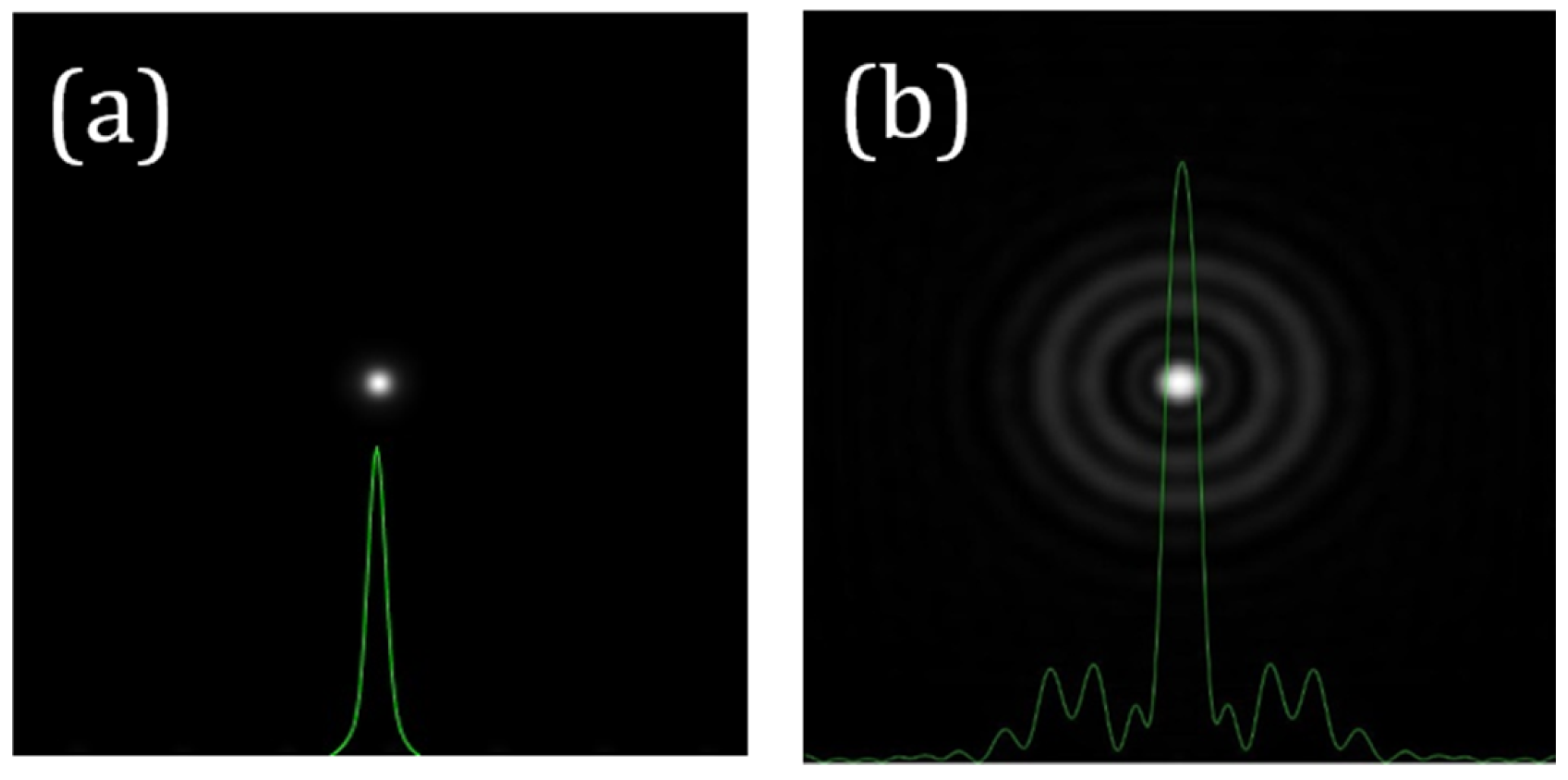
2.2. Wide-Angle Scattering PSF
2.3. Deconvolution Procedure
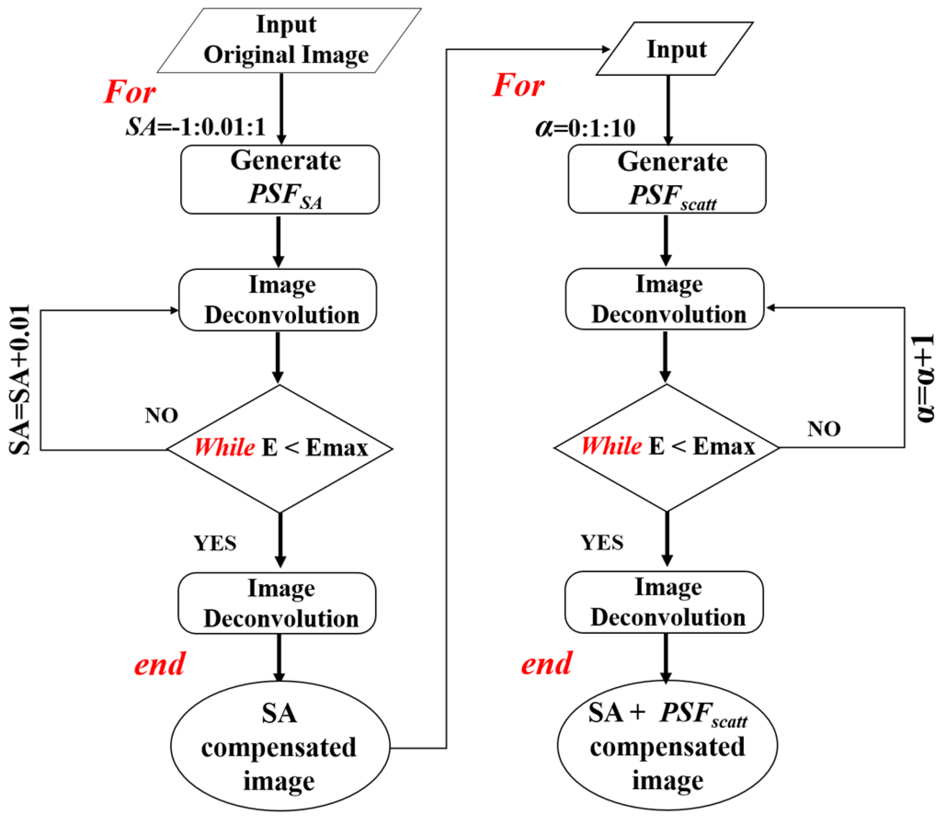
2.4. Algorithm Description
2.4.1. Step #1: Sampling PSF Optimization
2.4.2. Step #2: Deconvolution through PSFSA and PSFscatt
2.5. Samples and Images
3. Results
3.1. Validation of the Algorithm with Degradation-Controlled Artificial Images: A Proof of Concept
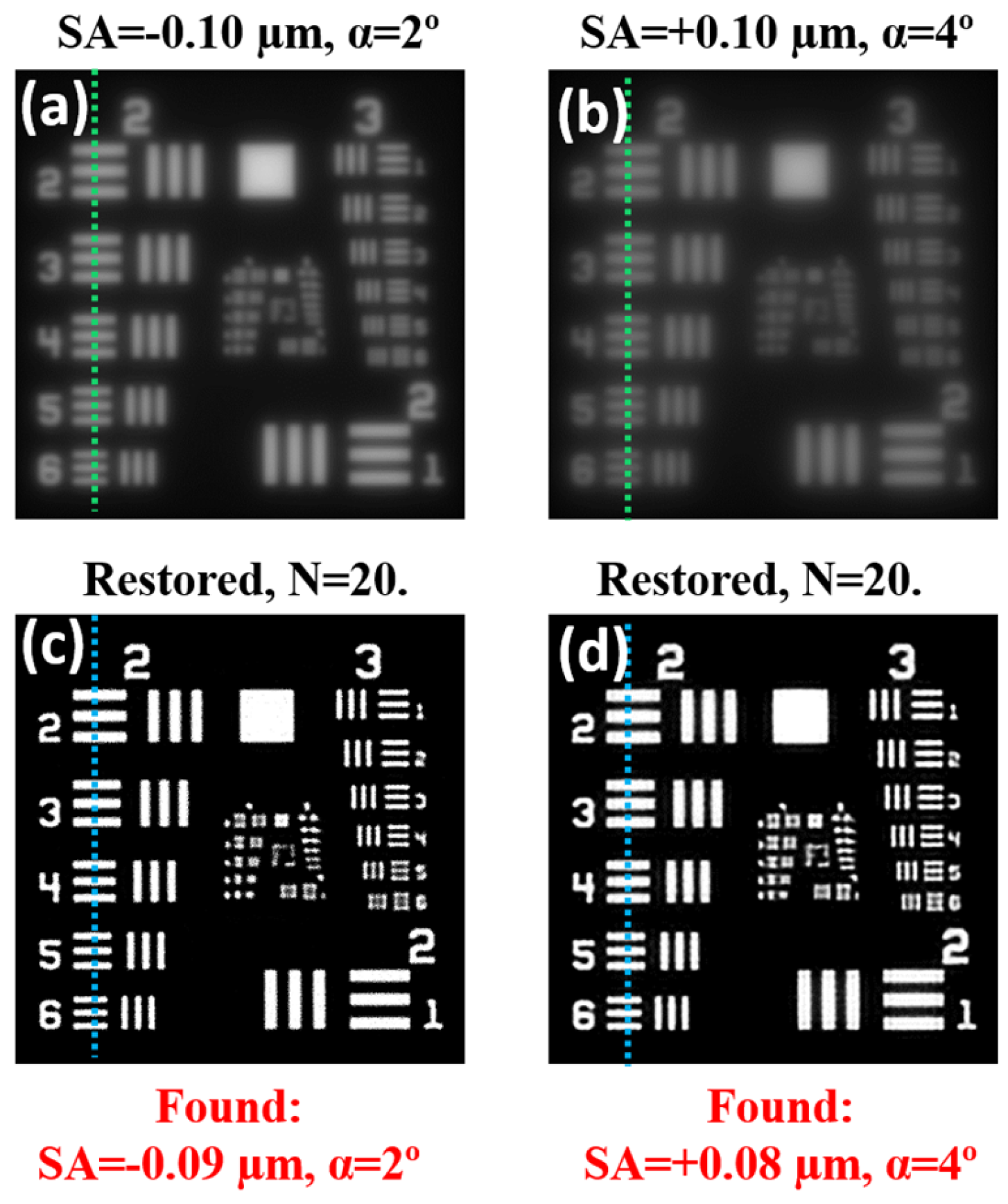


3.2. Deconvolution of Human Retinal Images with Induced Scattering
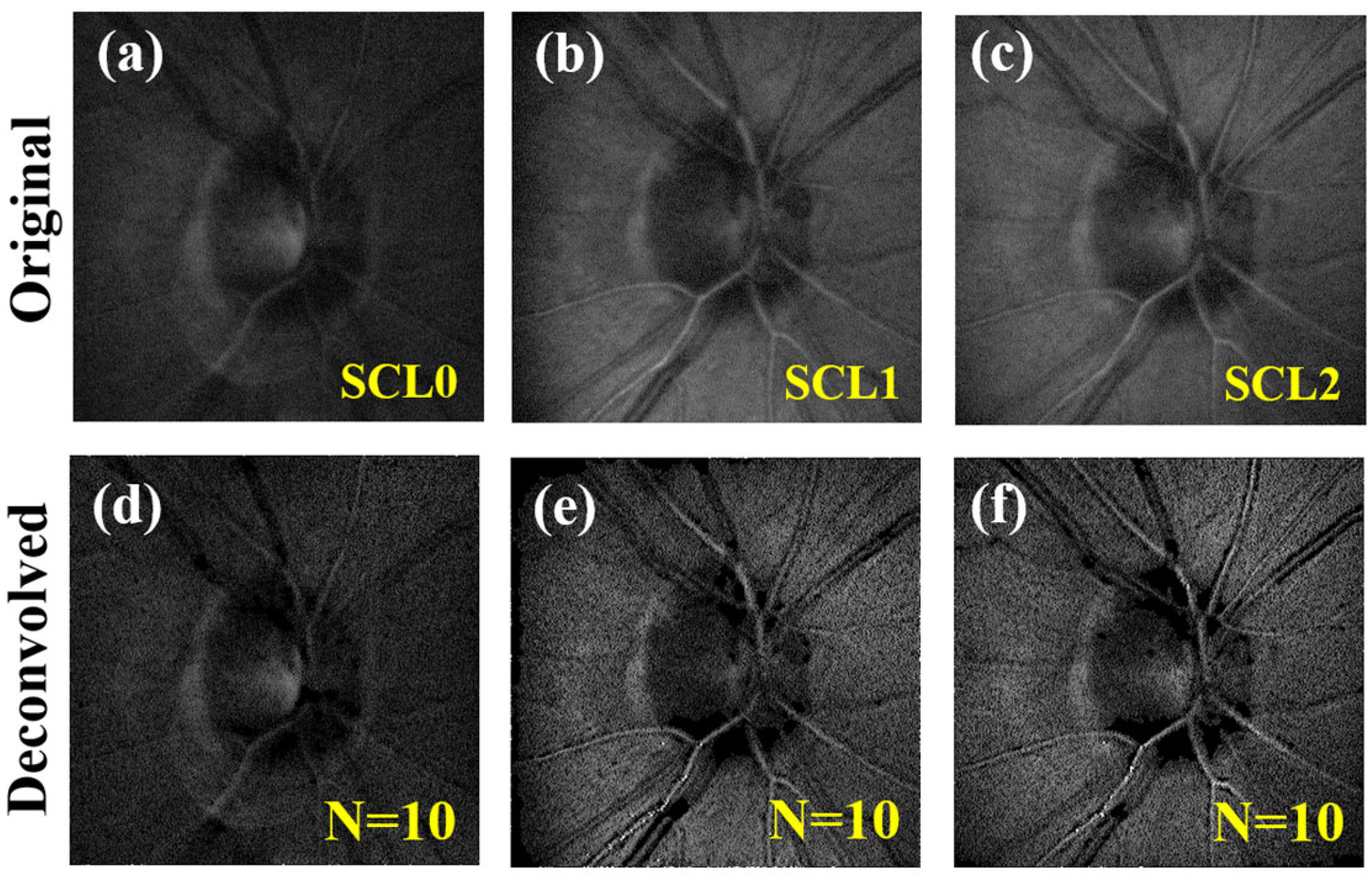
3.3. Deconvolution of Multiphoton Microscopy Images
3.4. Contribution of SA and Scattering to Image Quality Degradation
4. Discussion and Conclusions
Author Contributions
Funding
Institutional Review Board Statement
Informed Consent Statement
Data Availability Statement
Acknowledgments
Conflicts of Interest
References
- Jonkman, J.; Brown, C.M.; Wright, G.D.; Anderson, K.I.; North, A.J. Tutorial: Guidance for quantitative confocal microscopy. Nat. Protoc. 2020, 15, 1585–1611. [Google Scholar] [CrossRef]
- Rodríguez, C.; Ji, N. Adaptive optical microscopy for neurobiology. Curr. Opin. Neurobiol. 2018, 50, 83–91. [Google Scholar] [CrossRef] [PubMed]
- Burns, S.A.; Elsner, A.E.; Sapoznik, K.A.; Warner, R.L.; Gast, T.J. Adaptive optics imaging of the human retina. Prog. Retin. Eye Res. 2019, 68, 1–30. [Google Scholar] [CrossRef]
- Ávila, F.J.; Gambín, A.; Artal, P.; Bueno, J.M. In vivo two photon microscopy of the human eye. Sci. Rep. 2019, 9, 10121. [Google Scholar] [CrossRef]
- Bohren, C.F.; Huffman, D.R. Absorption and Scattering of Light by Small Particles; Wiley: Weinheim, Germany, 1998. [Google Scholar]
- Sanderson, J. Optical aberrations of the microscope. In Understanding Light Microscopy; John Wiley & Sons: Hoboken, NJ, USA, 2019. [Google Scholar]
- Lo, W.; Sun, Y.; Lin, S.-J.; Jee, S.-H.; Dong, C.-Y. Spherical aberration correction in multiphoton fluorescence imaging using objective correction collar. J. Biomed. Opt. 2005, 10, 034006. [Google Scholar] [CrossRef] [PubMed]
- Débarre, D.; Botcherby, E.J.; Watanabe, T.; Srinivas, S.; Booth, M.J.; Wilson, T. Image-based adaptive optics for two-photon microscopy. Opt. Lett. 2009, 34, 2495–2497. [Google Scholar] [CrossRef]
- Bueno, J.M.; Skorsetz, M.; Palacios, R.; Gualda, E.J.; Artal, P. Multiphoton imaging microscopy at deeper layers with adaptive optics control of spherical aberration. J. Biomed. Opt. 2014, 19, 011007. [Google Scholar] [CrossRef]
- Skorsetz, M.; Artal, P.; Bueno, J.M. Performance evaluation of a sensorless adaptive optics multiphoton microscope. J. Microsc. 2016, 261, 249–258. [Google Scholar] [CrossRef] [PubMed]
- Park, J.-H.; Sun, W.; Cui, M. High-resolution in vivo imaging of mouse brain through the intact skull. Proc. Natl. Acad. Sci. USA 2015, 112, 9236–9241. [Google Scholar] [CrossRef]
- Chaigneau, E.; Wright, A.J.; Poland, S.P.; Girkin, J.M.; Silver, R.A. Silver, Impact of wavefront distortion and scattering on 2-photon microscopy in mammalian brain tissue. Opt. Express 2011, 19, 22755–22774. [Google Scholar] [CrossRef]
- Sinefeld, D.; Paudel, H.P.; Ouzounov, D.G.; Bifano, T.G.; Xu, C. Adaptive optics in multiphoton microscopy: Comparison of two, three and four photon fluorescence. Opt. Express 2015, 23, 31472–31483. [Google Scholar] [CrossRef]
- Papadopoulos, I.N.; Jouhanneau, J.-S.; Poulet, J.F.A.; Judkewitz, B. Scattering compensation by focus scanning holographic aberration probing (F-SHARP). Nat. Photonics 2017, 11, 116–123. [Google Scholar] [CrossRef]
- Pozzi, P.; Gandolfi, D.; Porro, C.A.; Bigiani, A.; Mapelli, J. Scattering compensation for deep brain microscopy: The long road to get proper images. Front. Phys. 2020, 8, 26. [Google Scholar] [CrossRef]
- DBurke, D.; Patton, B.; Huang, F.; Bewersdorf, J.; Booth, M.J. Adaptive optics correction of specimen-induced aberrations in single-molecule switching microscopy. Optica 2015, 2, 177–185. [Google Scholar]
- Starck, J.L.; Pantin, E.; Murtagh, F. Deconvolution in astronomy: A review. Publ. Astron. Soc. Pac. 2002, 114, 1051–1069. [Google Scholar] [CrossRef]
- Levin, A.; Weiss, Y.; Durand, F.; Freeman, W.T. Understanding and evaluating blind deconvolution algorithms. In Proceedings of the 2009 IEEE Conference on Computer Vision and Pattern Recognition, Miami, FL, USA, 20–25 June 2009; pp. 1964–1971. [Google Scholar]
- Michailovich, O.; Tannenbaum, A. Blind deconvolution of medical ultrasound images: A parametric inverse filtering approach. IEEE Trans. Image Process. 2007, 16, 3005–3019. [Google Scholar] [CrossRef] [PubMed]
- Al Ameen, Z.; Bin Sulong, G.; Johar, G.M. Computer forensics and image deblurring: An inclusive investigation. Int. J. Mod. Educ. Comput. Sci. 2013, 5, 42–48. [Google Scholar] [CrossRef][Green Version]
- Thibos, L.N.; A Applegate, R.; Schwiegerling, J.T.; Webb, R. Standards for reporting the optical aberrations of eyes. J. Refract. Surg. 2002, 18, S652–S660. [Google Scholar] [CrossRef]
- van den Berg, T.J.; IJspeert, J.K.; de Waard, P.W. Dependence of intraocular straylight on pigmentation and light transmission through the ocular wall. Vis. Res. 1991, 31, 1361–1367. [Google Scholar] [CrossRef] [PubMed]
- Vos, J.; van den Berg, T.J. Report on Disability Glare. CIE Collect. 1999, 135, 1–9. [Google Scholar]
- Sibarita, J. Deconvolution Microscopy. Adv. Biochem. Eng. Biotechnol. 2005, 95, 201–243. [Google Scholar] [PubMed]
- Gonzalez, R.C.; Woods, R.E.; Eddins, S.L. Digital Image Processing Using MATLAB; Prentice Hall: Hoboken, NJ, USA, 2003; Chapter 11. [Google Scholar]
- Narayan, R.; Nityananda, R. Maximum entropy image restoration in astronomy. Annu. Rev. Astron. Astrophys. 1986, 24, 127–170. [Google Scholar] [CrossRef]
- Skilling, J.; Bryan, R.K. Maximum entropy image reconstruction: General algorithm. Mon. Not. R. Astron. Soc. 1984, 211, 111–124. [Google Scholar] [CrossRef]
- Russell, S.; Norvig, P. Artificial Intelligence: A Modern Approach, 2nd ed.; Prentice Hall: Hoboken, NJ, USA, 2003; pp. 111–114. [Google Scholar]
- Hunter, J.J.; Cookson, C.J.; Kisilak, M.; Bueno, J.M.; Campell, M.C.W. Characterizing image quality in a scanning laser ophthalmoscope with different pinholes and induced scattered light. J. Opt. Soc. Am. A 2007, 24, 1284–1295. [Google Scholar] [CrossRef]
- Swedlow, J.R. Quantitative fluorescence microscopy and image deconvolution. Methods Cell Biol. 2013, 114, 407–426. [Google Scholar] [PubMed]
- Doi, A.; Oketani, R.; Nawa, Y.; Fujita, K. High-resolution imaging in two-photon excitation microscopy using in situ estimations of the point spread function. Biomed. Opt. Express 2018, 9, 202–213. [Google Scholar] [CrossRef]
- Martínez-Ojeda, R.M.; Mugnier, L.M.; Artal, P.; Bueno, J.M. Blind deconvolution of second harmonic microscopy images of the living human eye. Biomed. Opt. Express 2023, 14, 2117–2128. [Google Scholar] [CrossRef]
- Blanco, L.; Mugnier, L.M. Marginal blind deconvolution of adaptive optics retinal images. Opt. Express 2011, 19, 23227–23239. [Google Scholar] [CrossRef]
- Fétick, R.J.-L.; Mugnier, L.M.; Fusco, T.; Neichel, B. Blind deconvolution in astronomy with adaptive optics: The parametric marginal approach. Mon. Not. R. Astron. Soc. 2020, 496, 4209–4220. [Google Scholar] [CrossRef]
- Benno, K.-S.; Lars, O.; Tobias, S.-M.; Timo, M.; Vasilis, N. Scattering correction through a space-variant blind deconvolution algorithm. J. Biomed. Opt. 2016, 21, 096005. [Google Scholar] [CrossRef]
- Seibert, J.A.; Nalcioglu, O.; Roeck, W. Removal of image intensifier veiling glare by mathematical deconvolution techniques. Med. Phys. 1985, 12, 281–288. [Google Scholar] [CrossRef] [PubMed]
- Karabal, E.; Duc, P.-A.; Kuntschner, H.; Chanial, P.; Cuillandre, J.-C.; Gwyn, S. A deconvolution technique to correct deep images of galaxies from instrumental scattered light. Astron. Astrophys. 2017, 601, A86. [Google Scholar] [CrossRef]
- Shajkofci, A.; Liebling, M. Spatially-variant CNN-based point spread function estimation for blind deconvolution and depth estimation in optical microscopy. IEEE Trans. Image Process. 2020, 29, 5848–5861. [Google Scholar] [CrossRef] [PubMed]
- Fahmy, M.F.; Raheem, G.M.A.; Mohammed, U.S.; Fahmy, O.F. A fast iterative blind image restoration algorithm. In Proceedings of the 2011 28th National Radio Science Conference (NRSC), Cairo, Egypt, 26–28 April 2011; pp. 1–8. [Google Scholar]


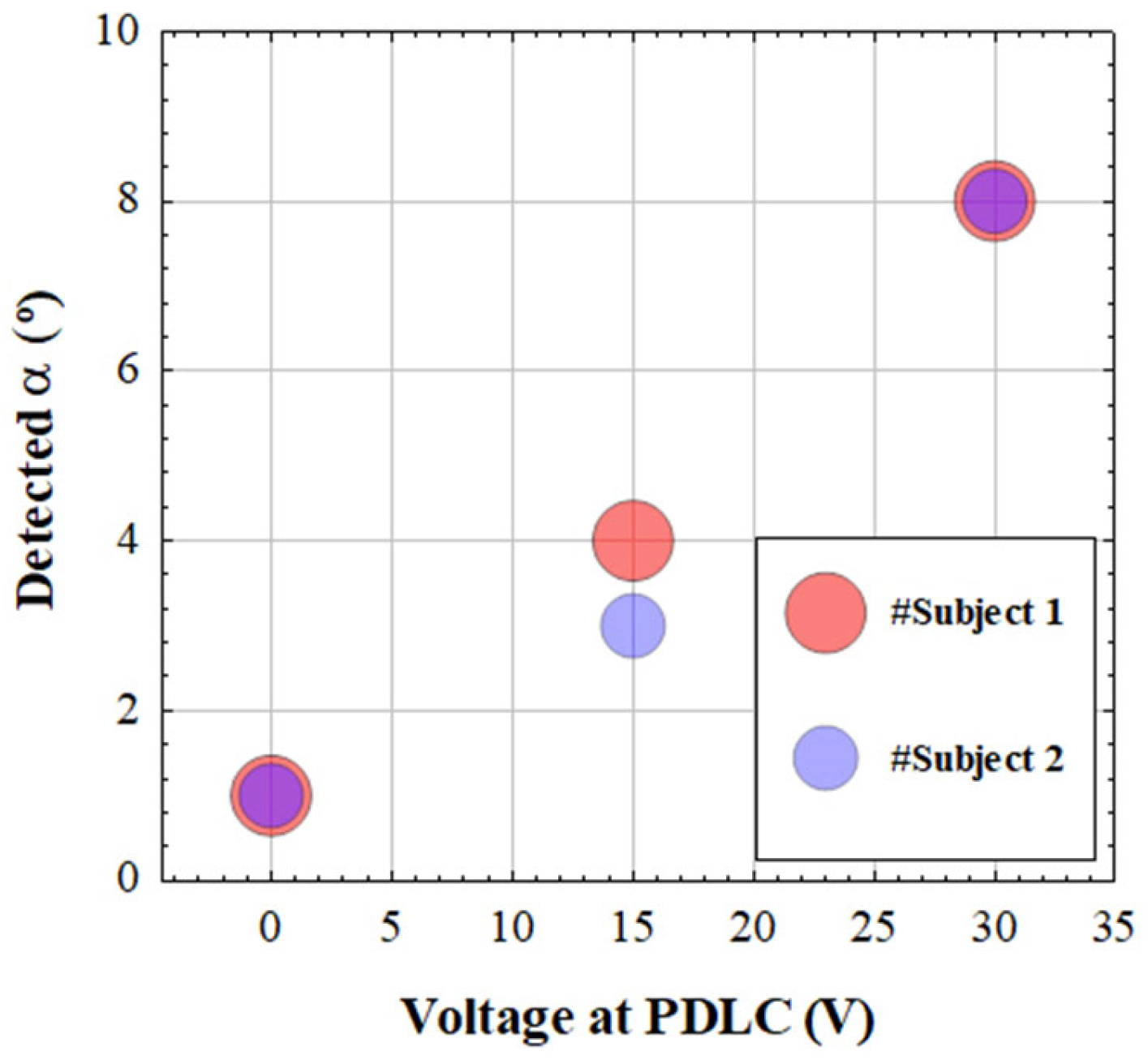
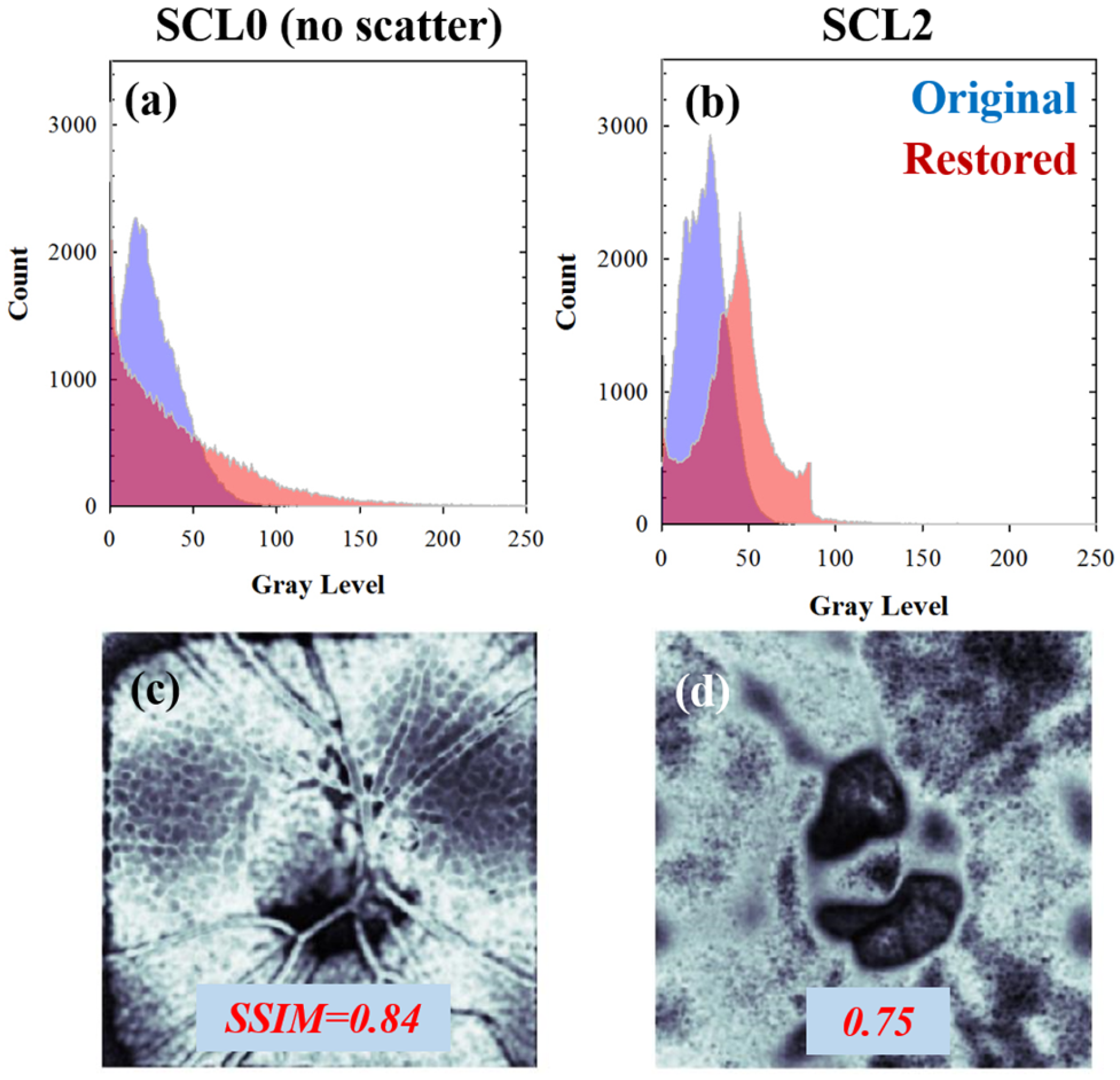

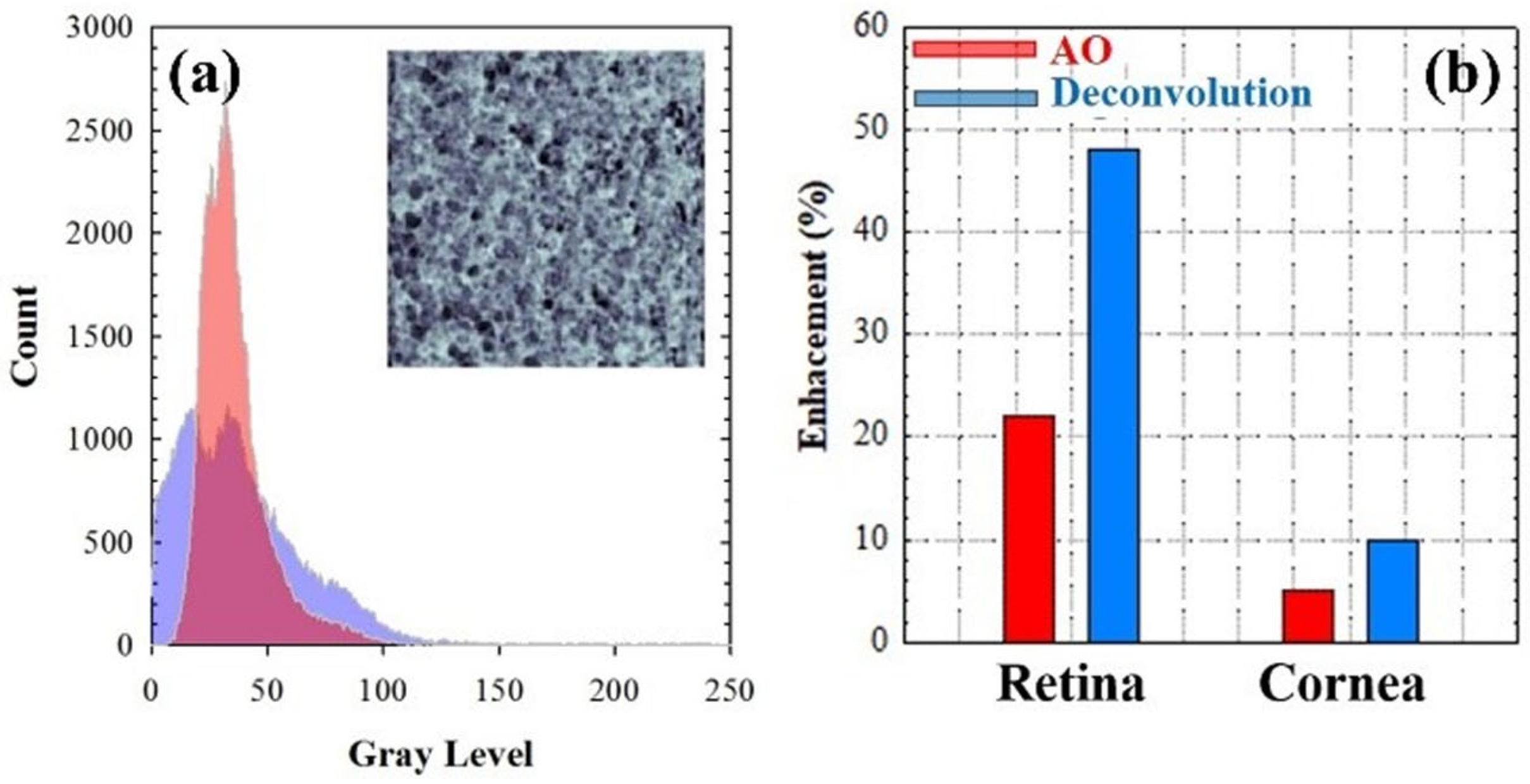
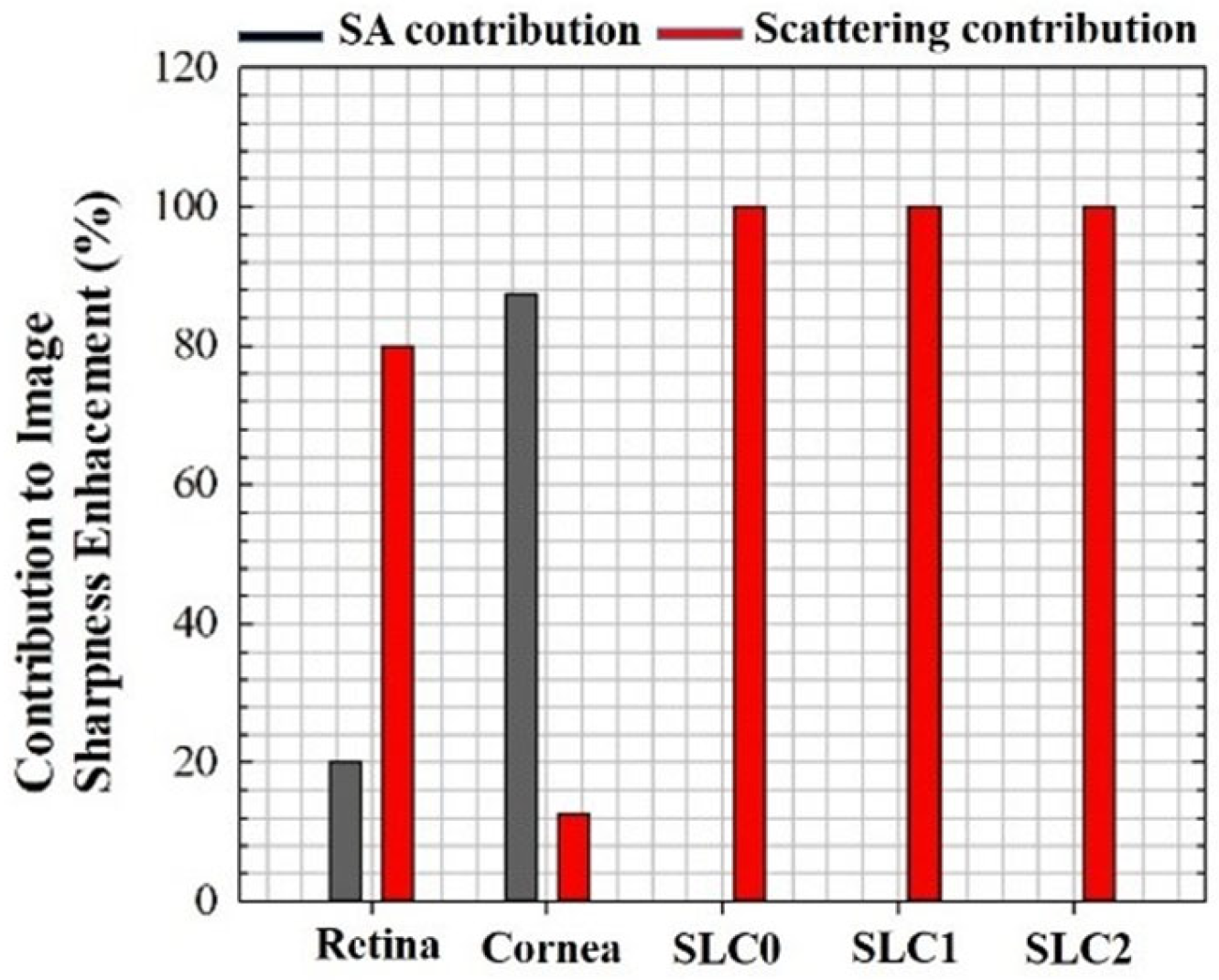
| SA (µm) [AO] | SA (µm) [Deconvolution] | α (°) [Deconvolution] | N |
|---|---|---|---|
| −0.25 | −0.36 | 4 | 20 |
| −0.10 | −0.18 | 7 | 30 |
Disclaimer/Publisher’s Note: The statements, opinions and data contained in all publications are solely those of the individual author(s) and contributor(s) and not of MDPI and/or the editor(s). MDPI and/or the editor(s) disclaim responsibility for any injury to people or property resulting from any ideas, methods, instructions or products referred to in the content. |
© 2024 by the authors. Licensee MDPI, Basel, Switzerland. This article is an open access article distributed under the terms and conditions of the Creative Commons Attribution (CC BY) license (https://creativecommons.org/licenses/by/4.0/).
Share and Cite
Ávila, F.J.; Bueno, J.M. Spherical Aberration and Scattering Compensation in Microscopy Images through a Blind Deconvolution Method. J. Imaging 2024, 10, 43. https://doi.org/10.3390/jimaging10020043
Ávila FJ, Bueno JM. Spherical Aberration and Scattering Compensation in Microscopy Images through a Blind Deconvolution Method. Journal of Imaging. 2024; 10(2):43. https://doi.org/10.3390/jimaging10020043
Chicago/Turabian StyleÁvila, Francisco J., and Juan M. Bueno. 2024. "Spherical Aberration and Scattering Compensation in Microscopy Images through a Blind Deconvolution Method" Journal of Imaging 10, no. 2: 43. https://doi.org/10.3390/jimaging10020043
APA StyleÁvila, F. J., & Bueno, J. M. (2024). Spherical Aberration and Scattering Compensation in Microscopy Images through a Blind Deconvolution Method. Journal of Imaging, 10(2), 43. https://doi.org/10.3390/jimaging10020043


