Abstract
Lithium-ion batteries have been commonly employed as power sources in portable devices and are of great interest for large-scale energy storage. To further enhance the fundamental understanding of the electrode structure, we report on the use of multi-wavelength Raman spectroscopy for the detailed characterization of layered cathode materials for Li-ion batteries (LiCoO2, LiNixCo1−xO2, LiNi1/3Mn1/3Co1/3O2). Varying the laser excitation from the UV to the visible (257, 385, 515, 633 nm) reveals wavelength-dependent changes in the vibrational profile and overtone/combination bands, originating from resonance effects in LiCoO2. In mixed oxides, the influence of resonance effects on the vibrational profile is preserved but mitigated by the presence of Ni and/or Mn, highlighting the influence of resonance Raman spectroscopy on electronic structure changes. The use of UV laser excitation (257, 385 nm) is shown to lead to a higher scattering efficiency towards Ni in LiNi1/3Mn1/3Co1/3O2 compared to visible wavelengths, while deep UV excitation at 257 nm allows for the sensitive detection of surface species and/or precursor species reminiscent of the synthesis. Our results demonstrate the potential of multi-wavelength Raman spectroscopy for the detailed characterization of cathode materials for lithium-ion batteries, including phase/impurity identification and quantification, as well as electronic structure analysis.
1. Introduction
Li-ion batteries already play a dominant role as power sources for portable devices. They are also of great importance regarding the development of larger scale devices used in more demanding applications, such as electric vehicles, allowing us to shift the power source in ground transportation from internal combustion engines to electrical propulsion [1]. Starting from LiCoO2, alternative cathode materials have been developed, including olivine LiFePO4, spinel LiM2O4 (for instance, M = Ni and Mn), layered oxides of LiMO2 (for instance, M = Ni and Co, Mn), and lithium-rich layered oxides xLi2MnO3(1 − x)LiMO2 (for instance, M = Ni and Co, Mn) [2,3] An important aspect regarding optimization, concerns the (partial) replacement of cobalt by other 3d transition metals, i.e., nickel and/or manganese, due to availability and costs. While LiNiO2 shows a higher specific capacity than LiCoO2, it possesses a low stability for storage; additionally, it is difficult to correctly adjust its stoichiometry. For example, Liu et al. reported a substantial capacity reduction from 215 mAh/g to 165 mAh/g after LiNiO2 storage for a month, while storage for a year resulted in a completely inactive material [4]. On the other hand, NMC oxides with the composition LiNi1/3Mn1/3Co1/3O2 can achieve a reversible specific capacity of 200 mAh/g [5], whereas for lithium-rich oxides xLi2MnO3(1 − x)LiMO2 (such as, M = Ni, Co and Mn), even higher capacities of over 250 mAh/g were demonstrated [6,7]. While mixtures of the three transition metals, Ni, Mn, and Co, are already used commercially, a detailed understanding of their functioning and degradation will be important for developing the next generation of cathode materials [8,9].
Raman spectroscopy proved to be a powerful method for the characterization of electrode materials for lithium-ion batteries, providing important structural information, complementary to XRD, and enabling microscopy and in situ/operando measurements [10,11,12,13,14,15,16,17,18]. In addition, its application is straightforward, requiring no specific sample preparation. The scattering intensity of a solid material in normal Raman spectroscopy depends on a variety of factors, including the concentration and the Raman cross-section, and typically shows a weak dependence on the wavelength of the exciting laser. In contrast, in resonance Raman spectroscopy, which is based on the excitation of an electronic transition, a strong dependence on the laser wavelength is observed, providing access to enhanced Raman cross-sections and unique Raman signatures of different compounds or components within a material [19]. This may be of particular importance for the characterization of materials with a structural complexity, such as cathode materials for Li-ion batteries, which are known to exhibit complex vibrational signatures. In addition to sensitive phase identification, the resonance Raman spectra may be used for structural characterization under working conditions of the battery, as previously demonstrated for LiCoO2-based composite cathodes by using visible excitation wavelengths [20].
As a result of the variations in electronic structure, the exploitation of resonance enhancements for materials characterization requires, in principal, a wavelength tunable excitation laser to meet the resonance conditions for the electronic transitions of the material under study, but more frequently, lasers with fixed excitation wavelengths are employed. While Raman spectroscopy has been increasingly applied to cathode materials for Li-ion batteries [8,10,12,13], only few studies exploited the resonance Raman effects of layered cathode materials, i.e., LiCoO2 [20,21], LiNiO2 [21], LiMn2O4 [22], LiNi0.8Co0.15Al0.05O2 [23], LiNixMn2-xO4 [24] and LiNi1/3Co1/3Mn1/3O2 [25].
In this contribution, we present a multi-wavelength Raman spectroscopic study on layered cathode materials (LiCoO2, LiNixCo1−xO2, LiNi1/3Mn1/3Co1/3O2) for Li-ion batteries. By using excitation ranging from the deep UV to the visible (257, 385, 515, 633 nm), we investigate for the first time the wavelength-dependent structural behavior of common cathode materials, thereby exploring the influence of resonance Raman effects on the spectral behavior. We discuss the potential of multi-wavelength spectroscopy, including UV Raman excitation for (electronic) structure analysis, quantification, phase and impurity identification, in battery materials.
2. Experimental Section
2.1. Preparation of Cathode Materials
For the preparation of the LiNixMnyCozO2 materials, we applied the Pechini process to ensure a statistical distribution of the cations [26]. Briefly, as precursors, LiNO3 (Merck KGaA, ≥98%), Co(NO3)2·6H2O (Merck KGaA, ≥99.0%), Ni(NO3)2·6H2O (Puratronic, >99,99%), Mn(NO3)2·4H2O (Sigma Aldrich, 97%) and citric acid (AppliChem, ≥98%) were employed. After their dissolution in water, we added dropwise a concentrated ammonia solution (25%) until a pH value of 5 was reached. We then added ethylene glycol into the suspension and set the temperature to 180 °C for 6 h. As product, a black solid was obtained, which was first ground and then pre-calcined at 450 °C for 6 h (heating rate: 1.5 °C/min), yielding a brown powder, which was ground. LiCoO2 was calcined at 800 °C for 20 h (heating rate: 20 °C/min), LiNixCO1−xO2 at 775 °C for 10 h (heating rate: 20 °C/min) and LiNi1/3Mn1/3Co1/3O2 (NMC) at 900 °C for 15 h (heating rate: 20 °C/min). LiNiO2 (Sigma-Aldrich, ≥98%) and other reference compounds were commercially available.
2.2. Characterization
Raman Spectroscopy. UV Raman spectra were recorded on a triple-stage spectrograph (Princeton Instruments, TriVista 555), equipped with a charge-coupled device (CCD, 2048 × 512 pixels) and by using 256.7 and 385.1 nm radiation generated by a Ti:sapphire laser and frequency conversion crystals (Indigo Coherent). The spectral resolution of the spectrometer was approximately 1 cm−1. Great care was taken of the potential UV-induced effects, by using a UV Raman setup with a large spot size (0.6 mm2). The setup was based on a spherical mirror for laser excitation and parabolic mirrors for the collection of the scattered radiation. Based on this setup, we can avoid sample damage when using a low laser power of 3 mW. The analysis of the UV Raman data included cosmic ray removal and background subtraction. Prior to each measurement, the focus conditions were optimized using boron nitride (BN, 99%).
The Raman spectra at 514.5 nm excitation were recorded with an argon ion laser (Melles Griot), whereas for 632.8 nm spectra, a diode laser (Ondax) was employed as the light source. The Raman spectrometer (Kaiser Optical, HL5R) was equipped with an electronically cooled CCD detector (256 × 1024 pixels), and characterized by a spectral resolution of 5 cm−1 and a wavelength stability of better than 0.5 cm−1. We adjusted the laser power to 0.8 mW as measured at the sample position. The Raman analysis included a cosmic ray removal and an auto new-dark correction. The acquisition time for a single spectrum was 600 s, including the application of a cosmic ray filter and subtraction of the dark spectrum (laser off).
In the following, the Raman excitation wavelengths 256.7, 385.1, 514.5, and 632.8 nm will be referred to as 257, 385, 515, and 633 nm, respectively.
Diffuse Reflectance UV–Vis Spectroscopy. For recording the diffuse reflectance (DR) UV-Vis spectra, we employed a UV-Vis spectrometer (Jasco V-770) and used D2 and halogen light sources. For analysis, a reaction cell (HVC-MRA-5, Harrick Scientific) was employed. MgO powder was used as a white standard in the same geometry as the sample.
X-ray Diffraction. For X-ray powder diffraction experiments, we employed an X-ray powder diffractometer (StadiP, Stoe & Cie GmbH) in transmission geometry, using Cu Kα1 (λ = 1.540598 Å; Ge[111]-monochromator) radiation and a Mythen 1K (Dectris) detector, and Mo Kα1 (λ = 0.70930 Å; Ge[111]-monochromator) radiation and a position-dependent (Stoe) detector. For analysis, the program WinXPOW (Stoe & Cie GmbH) and the program package GSAS-II was employed [27].
3. Results and Discussion
Prior analysis using multi-wavelength Raman spectroscopy allowed us to characterized the layered cathode materials (LiCoO2, LiNi0.2Co0.8O2, LiNi0.8Co0.2O2 and LiNi1/3Mn1/3Co1/3O2) by using XRD. The XRD results confirm the phase purity and high crystallinity of the materials (see Supplementary Materials, Figures S1–S3).
3.1. LiCoO2
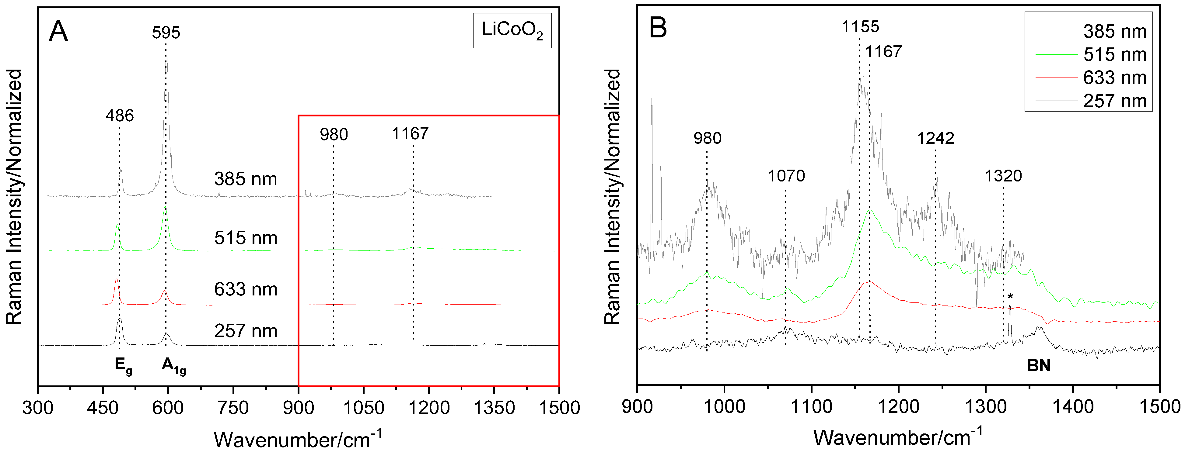
Figure 1 depicts the Raman spectra of LiCoO2 prepared by the Pechini process using 257, 385, 515, and 633 nm excitation. The two Raman active modes (Eg: 486 cm−1; A1g: 595 cm−1) [28] are clearly visible in all spectra, but show variations in the intensity ratio with the excitation wavelength. The Eg mode involves mainly O–Co–O bending, and the A1g mode Co–O stretching. As suggested by the corresponding UV-Vis spectrum (see Figure S4) and previously discussed by Gross et al., [20]. Raman spectra with an increased A1g/Eg ratio are recorded under resonance conditions, thus leading to the appearance of overtone and combination bands. This is further supported by the Raman spectra obtained after the (partial) exchange of cobalt by nickel, as will be discussed below. LiCoO2 electronic transitions, which may be related to resonance Raman effects, are located at around 2.1 eV (591 nm), attributed to the d–d transition from Cot2g to Coeg bands, and above 3.3 eV (375 nm), originating from Li1s to O2p or O2p to Co3d transitions [29].
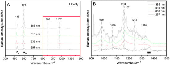
Figure 1.
Raman spectra of LiCoO2 recorded with 257, 385, 515 and 633 nm laser excitations, normalized to the Eg signal and offset for clarity. (A) Survey spectra. (B) Detailed view of the overtone/combination band region (marked red in (A)). Note that the spectrum recorded at 257 nm excitation contains a cosmic ray spike (*) and a signal from residual BN used for focus optimization.
Based on the Raman spectra in Figure 1, the A1g/Eg intensity ratios were determined for all laser wavelengths by using the ratios of the peak heights. For the 633 and 515 nm excitations, the A1g/Eg values of 0.5 and 1.65 were obtained, in agreement with our previous study [20]. A further decrease in the excitation wavelength to 385 nm leads to an increase in the intensity ratio to 5.3, while for deep UV excitation at 257 nm, a ratio drop to 0.4 is observed. Hence, the resonance enhancement of the A1g band at ~595 cm−1 is most pronounced at the 385 nm excitation, and leads to the appearance of a strong overtone signal at ~1170 cm−1. The right panel of Figure 1 gives an enlarged view of the overtone region. Interestingly, in addition to the A1g overtone signal, additional smaller features are detected within 950–1400 cm−1, i.e., at 980, 1070, 1167, 1242, 1320 and 1360 cm−1. In the case of the 257 nm excitation, no overtone signals are detected, strongly suggesting the absence of resonance conditions. This is also supported by the lower absorption in the UV as compared to the visible region (see Figure S4). Accordingly, the Raman band observed at around 1070 cm−1 for 257 nm excitation is attributed to a surface species (see discussion below), whereas the feature at 1360 cm−1 originates from residual BN used for focus optimization.
In the following, we will address the origin of the Raman features in the 950–1400 cm−1 region. First, it is noteworthy that there is an enhancement of the signals at 980 and ~1160 cm−1, which increases with the decreasing wavelength of the excitation laser, i.e., 633 nm < 515 nm < 385 nm. The appearance of overtones at 633 nm excitation, despite the lower A1g/Eg ratio, suggests the presence of a pre-resonance. The application of group theory to LiCoO2 (point group D3d) facilitates the assignment of the observed Raman features (see Table S1). In addition to the fundamental Eg and A1g modes, the band assignments include overtones of the fundamental Raman active phonon modes (A1g, Eg), the IR active modes (A2u, Eu) and combination bands (A1g × Eg, A2u × Eu). Accordingly, the signal at 980 cm−1 is attributed to the overtone of the Eg mode of LiCoO2, while the weak feature at 1070 cm−1 detected for 385, 515 and 633 nm excitations under (pre-)resonance conditions can be assigned to a combination band of the A1g and Eg modes, with a possible contribution from an Eu overtone. According to Rao et al. [30] and Julien [31], the IR spectrum of LiCoO2 is characterized by an intense signal at 595 cm−1 with a shoulder towards higher wavenumbers, which are assigned to the ν(MO6)-mode of a CoO6 unit (A2u). It was postulated that the signal at 595 cm−1 originates from the cobalt ions occupying its regular site and the shoulder at 653 cm−1 from cobalt ions in an octahedral void in the lithium layer; the signal at 526 cm−1 was attributed to a δ(O-C-O)-mode (Eu). Based on these assignments, the signal at 1167 cm−1 (see Figure 1B) may draw a contribution from the A2u overtone, in addition to the overtone of the A1g phonon. The position of the broad Raman band at ~1320 cm−1 is consistent with the overtone of the IR active A2u mode. This shows that the defect structure of cobalt ions in LiCoO2 may become accessible under resonance conditions. The feature at 1155 cm−1 is located very close to that at 1167 cm−1, and may draw a contribution from a combination band of the A2u and Eu modes. Finally, the Raman signal at 1242 cm−1, only observed for excitation at 385 nm (see Figure 1B), may result from the overtone of the inactive fundamental A2u mode. The assignments of the observed Raman features for LiCoO2 are summarized in Table 1.

Table 1.
Assignment of the observed Raman bands for LiCoO2.
3.2. LiNiyCo1−yO2
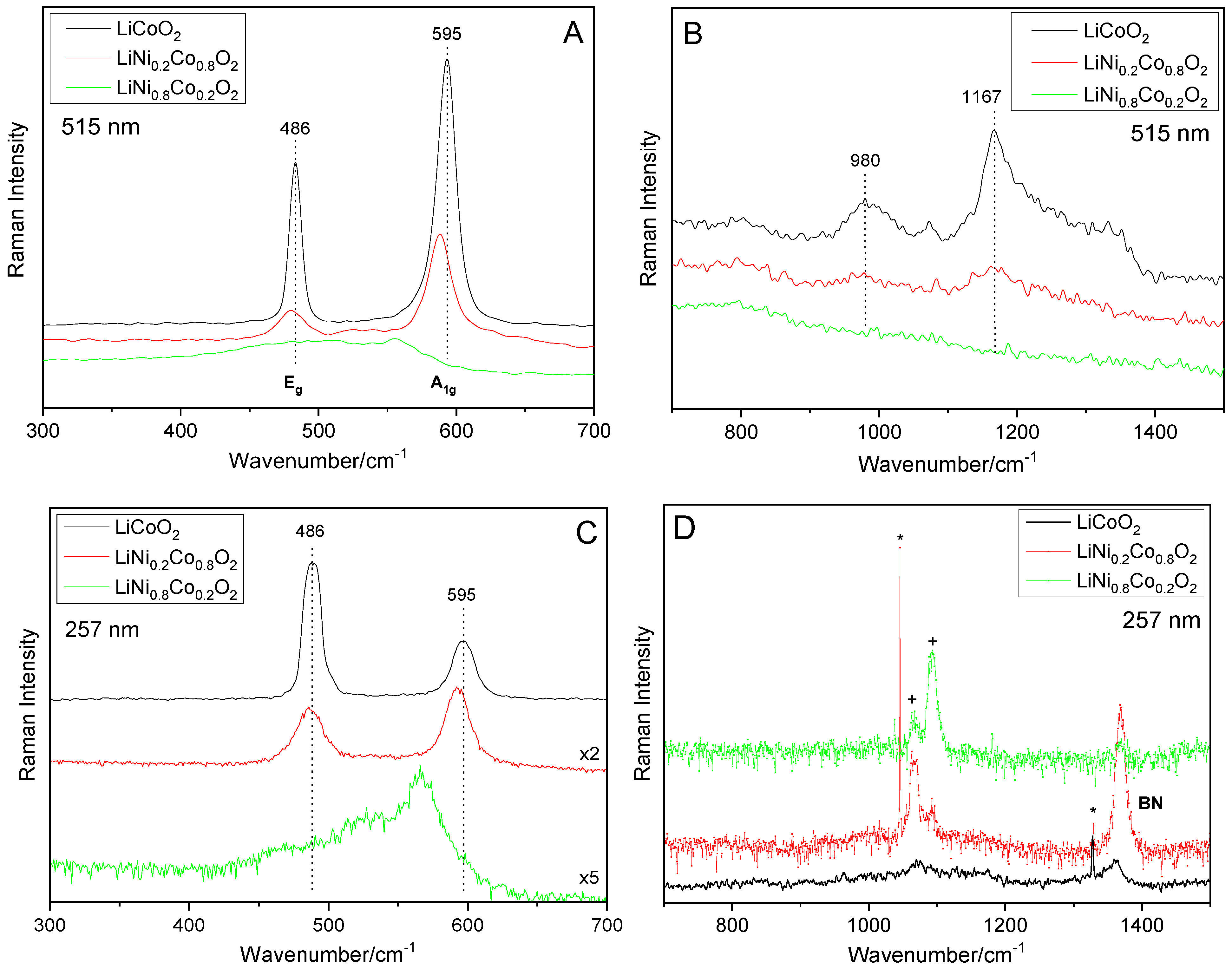
Figure 2 depicts the Raman spectra of the fundamental (left panel) and overtone/combination (right panel) band region of LiCoO2 and LiNixCo1−xO2 recorded at 515 nm (A, B) and 257 nm (C, D) excitations. As can be seen in the left panels, the A1g and Eg Raman signals show a strong decrease in the intensity and a red-shift with an increasing amount of nickel, whereas the red-shift is more pronounced for the A1g mode. At first sight, a similar behavior is observed for visible and UV excitation, as well as for the other excitation wavelengths (not shown). The presence of the two Raman bands (A1g, Eg) for LiNi0.2Co0.8O2 is consistent with the formation of mixed nickel–cobalt layers, i.e., a solid solution isomorphic with the pure phases. The observed red-shift can be explained by the expansion of the unit cell upon nickel substitution [10]. The increasing Ni content leads to a broadening of the Raman bands, especially the Eg mode, which is attributed to cation mixing in the crystal layers of the Ni-rich compounds [21]. The broadening of the phonon modes in LiNi0.8Co0.2O2 makes a more detailed (quantitative) analysis challenging. Interestingly, in case of 257 nm excitation, an additional Raman feature is detected at around 530 cm−1. Upon closer inspection, such a contribution may also be identified in the visible spectra of LiNi1−xCoxO2 compounds in this work (see Figure 2A), as well as in the literature spectra [31]. To the best of our knowledge, the origin of this feature has not been addressed yet. For the 257 nm excitation, the Raman spectrum of bare LiNiO2 exhibits a broad feature with a maximum at ~520–530 cm−1 (see Figure 5). Hence, the 530 cm−1 Raman feature observed for LiNi0.8Co0.2O2 may indicate the presence of LiNiO2 domains. However, as UV excitation leads to highest surface sensitivity among the excitation wavelengths used in this study (see also below), we cannot exclude a contribution from surface phonons.
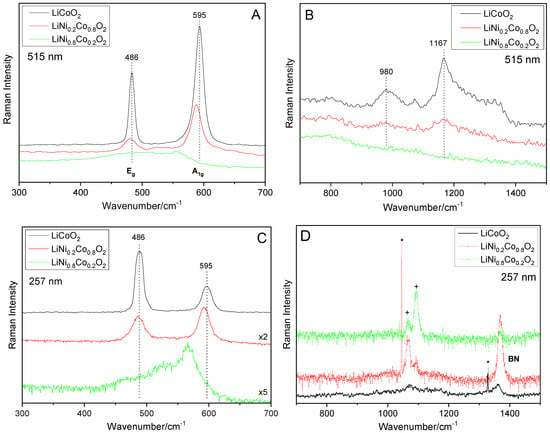
Figure 2.
Raman spectra of LiCoO2 and LiNiyCo1−yO2 recorded at 515 nm (A,B) and 257 nm (C,D) laser excitations, showing the fundamental modes (left) and the overtone/combination band region (right). Spectra are offset for clarity. Note that the spectrum recorded at 257 nm excitation contains cosmic ray spikes (*), surface signals (+) and a signal from residual BN used for focus optimization.
Upon substitution of cobalt by nickel, the Raman intensity of the fundamental modes decreases dramatically, independent of the excitation wavelength, as depicted in Figure S5 for the A1g mode. Furthermore, as can be seen in Figure 2B, the overtone/combination bands strongly decline and have completely disappeared for LiNi0.8Co0.2O2. On the other hand, with the increasing nickel content, we do not detect any new overtone features. Please note that the features detected in the overtone region for the 257 nm excitation (see Figure 2D) can be fully accounted for by cosmic ray spikes, surface signals as well as residual BN used for focus optimization in the UV.
The observed intensity changes in the fundamental and overtone region of the Raman spectra clearly show the influence of nickel on the electronic structure of the mixed lithium nickel–cobalt oxides. Previously, in the context of lithium deintercalation from LiCoO2, the strong decrease in Raman intensity has been associated with a reduction of the rhombohedral distortion by increasing the Ni content [31] and/or a skin depth effect [32]. To this end, the literature results suggested a higher electrical conductivity of LiNiO2 compared to LiCoO2 [33,34,35]. From this behavior of the bare oxides, we may expect an increased electrical conductivity, which decreases the optical skin depth of the excitation laser, thus resulting in a reduced scattering efficiency for the higher nickel content in LiNi1−xCoxO2 [32]. As can be seen in Figure 2 and Figure S5, such a scenario is fully consistent with our Raman results. Note that the Raman spectra show a broadening of the Eg mode with an increasing nickel content, which is associated with the non-stoichiometry induced by the presence of extra nickel [36].
The disappearance of the overtone/combination features in Figure 2B demonstrates that the presence of nickel significantly changes the conditions for resonance Raman spectroscopy, and thus the electronic structure in LiNi1−xCoxO2. The substitution of cobalt by nickel leads to a gradual disappearance of the overtone/combination bands, but at the same time, we do not detect any new features in the overtone region for any of the excitation wavelengths (257, 385, 515, 633 nm), indicative of the presence of new resonance effects. While our results highlight the sensitivity of resonance Raman spectroscopy for the electronic structure changes in LiNi1−xCoxO2, a more detailed description would require the input from theoretical calculations.
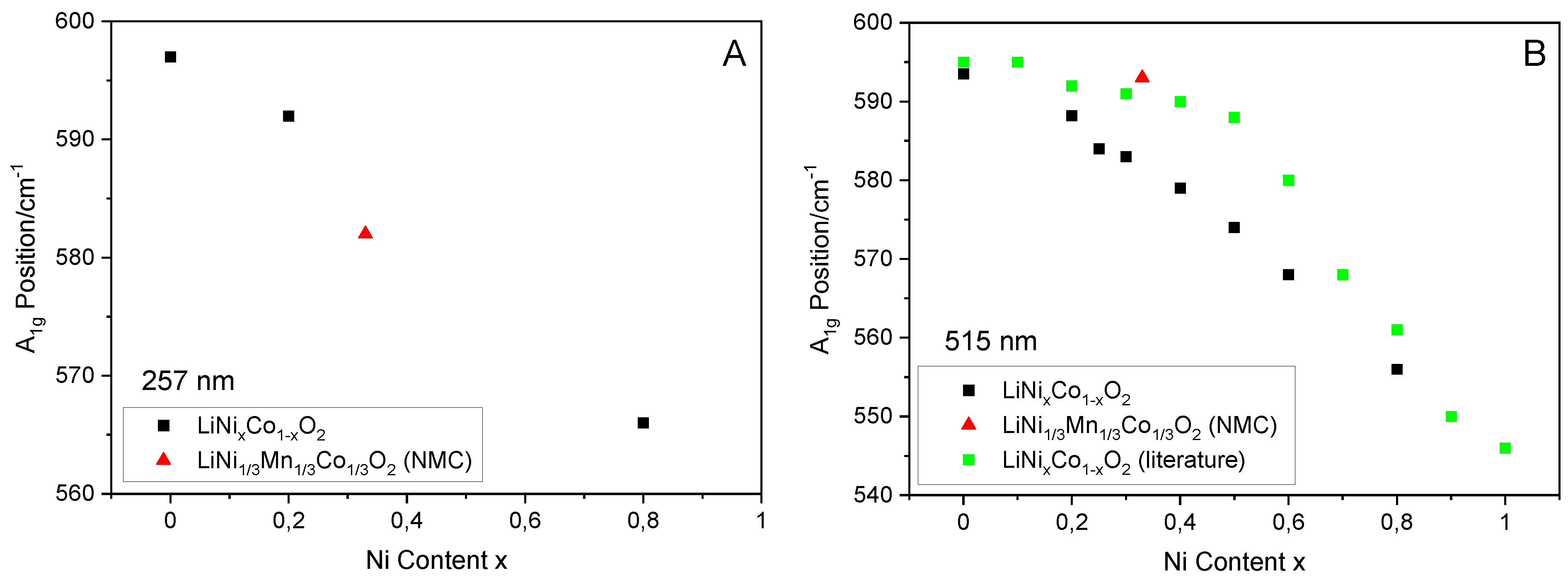
Figure 3 depicts the observed red-shift in A1g position with an increasing amount of nickel in LiNixCo1−xO2 for 257 nm (A) and 515 nm (B) laser excitations. We observe a linear dependence of the A1g position on the nickel content for all the excitation wavelengths, which give spectra of an acceptable quality, i.e., 257, 517 and 633 nm. From a linear fit to the data, the slopes of −39.8 ± 3.7 cm−1/x (257 nm), −47.3 ± 2.8 cm−1/x (515 nm) and −47.8 ± 2.0 cm−1/x (633 nm) were obtained, where x represents the nickel content. Thus, within the error of the experiment, we find the red-shift of the A1g mode to be independent of the excitation wavelength. The right panel also contains literature data from Julien recorded at 515 nm excitation for comparison [29]. While the dependence of the A1g position on the nickel content shows the same tendency, a deviation from the linear behavior was observed, which may be associated with the cation disorder in the mixed oxide, resulting from the extensive calcination of 15 h at 750 °C (twofold), in contrast to the 10 h calcination at 775 °C applied in this work.
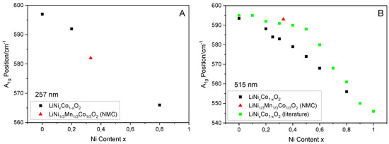
Figure 3.
Position of the A1g Raman mode for LiNixCo1−xO2 as a function of the Ni content × for 257 nm (A) and 515 nm (B) laser excitations. For comparison, our data for LiNi1/3Mn1/3Co1/3O2 (red) as well as 515 nm literature data [29] for LiNixCo1−xO2 (green) was added.
For comparison, Figure 3 contains data for LiNi1/3Mn1/3Co1/3O2 (NMC), which suggests that the nickel content may be determined quantitatively at 257 nm excitation, while the use of 515 nm excitation leads to significant deviations from the linear behavior. Accordingly, the A1g intensity of NMC is consistent with the exponential decay curve obtained for LiNiyCo1−yO2 at 257 nm excitation, in contrast to the 515 nm behavior. Compared to the mixed lithium nickel cobalt oxides, the NMC material was calcined under harsher conditions, i.e., 15 h at 900 °C. It thus appears that a 515 nm laser excitation is more sensitive to the detailed state of the material. A discussion of the Raman analysis of NMC, including the role of manganese, will be presented in the next section.
To summarize, the above results demonstrate the potential of Raman spectroscopy for the quantification of the nickel content in LiNixCo1−xO2 materials. The results suggest that if an appropriate excitation wavelength is chosen, NMC materials and possibly also other nickel containing layered materials can be analyzed. This is a very promising perspective, considering the importance of nickel in the development of novel cathode materials.
3.3. LiNi1/3Mn1/3Co1/3O2 (NMC)
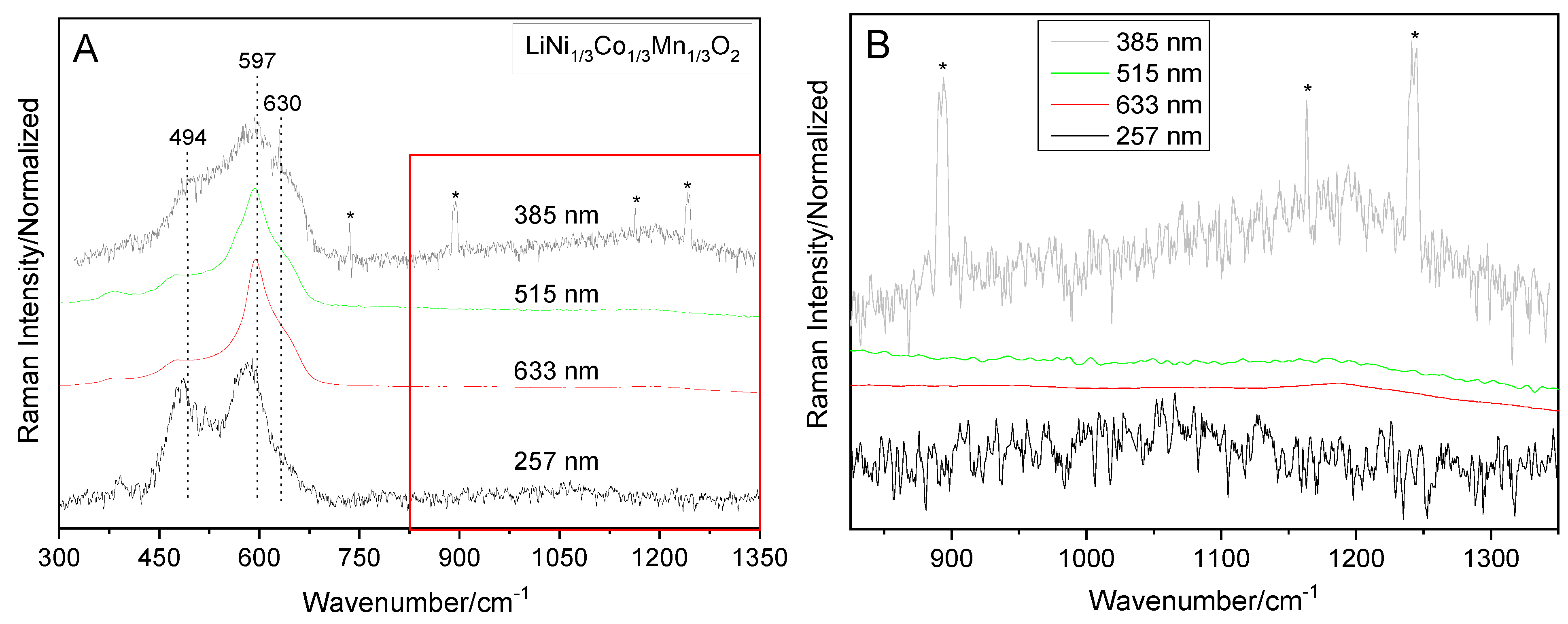
The Raman spectra of NMC are characterized by a strong dependence on the excitation wavelength. As shown in Figure 4, pronounced differences in the Raman spectra are observed for the 257, 385 and 515/633 nm excitations. NMC has been proposed to represent a LiMO2 mixed oxide derived from the compounds LiCoO2, LiNiO2 and LiMnO2 (M = Ni + Co + Mn). Considering the first coordination shell only, the Raman spectra of NMC may be expected to consist of a combination of six signals, with each of the three oxides, LiCoO2, LiNiO2 and LiMnO2, contributing an Eg and A1g mode [37,38,39]. However, as can be seen from Figure 4, due to the complexity of the recorded Raman spectra, an unambiguous fit analysis aiming at a quantitative description is not feasible. This is in agreement with the results of previous studies, showing the challenge of Raman band assignments in NMC mixed oxides [12]. In this context, it has been pointed that the NMC Raman profile represents more than the individual contributions (LiCoO2, LiNiO2, LiMnO2) summed up according to their proportion, and, in particular, the presence of multiple interactions between metal ions changing their local environments, but also the sample preparation, the experimental conditions as well as Raman spectroscopic aspects. In the following, we will therefore discuss the wavelength-dependent behavior of the NMC Raman spectra on a qualitative basis, which still provides a valuable insight into structural properties.
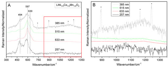
Figure 4.
Raman spectra of LiNi1/3Mn1/3Co1/3O2 (NMC) recorded with 257, 385, 515 and 633 nm laser excitations, normalized to the A1g signal and offset for clarity. (A) Survey spectra. (B) Detailed view of the overtone/combination band region (marked red in (A)). Spectra recorded at 257 and 385 nm excitations contain cosmic ray spikes (*).
Apparently, the Raman spectra of NMC are more complex than those of LiCoO2. However, an indication for the presence of Co in NMC is readily provided by the overtone/combination band region measured by the 385 nm excitation (see Figure 4B), which shows a Raman signal at 1190 cm−1. As expected, this signal is significantly weaker than that observed for LiCoO2 (see Figure 1B). Nevertheless, its detection at 385 nm excitation strongly suggests the presence of resonance Raman effects operating in a similar manner, as in the case of LiCoO2. Consistent with the wavelength-dependent behavior observed for LiCoO2, an overtone feature is also observed for the 515 and 633 nm excitations, in contrast to the deep UV Raman spectrum (see Figure 4B).
The Raman spectra of NMC are characterized by major contributions located at around 494, 597 and 630 cm−1. While these contributions can be clearly observed for all excitation wavelengths, different intensity ratios are detected for 257, 385 and 515/633 nm excitations. Upon closer inspection, additional Raman features can be identified as a separate band at around 380 cm−1 (257, 515, 633 nm) and as a shoulder at around 560 cm−1 (515 nm). Although not directly detectable (as a band or shoulder), the latter feature may also contribute to the 257 and 385 nm spectra. In addition to the Co-related Eg and A1g features (~494 cm−1, ~597 cm−1), previous Raman studies on NMC materials have associated features at around 470 cm−1 with the presence of Ni, features at around 530 cm−1 with the presence of Ni (and Mn) and features > 600 cm−1 with the presence of Mn [25,37,38,39]. A smaller contribution (shoulder) < 425 cm−1 was previously observed for NMC, but also for LiNi0.8Co0.15Al0.05O2 (NCA) materials [12,25,39], thus showing no ion specificity.
Turning to the wavelength-dependent spectral behavior, most noticeable are the different vibrational profiles in the fundamental region in addition to the overtone properties discussed above. Considering the intensity ratios detected for LiCoO2 (see Figure 1A), we can first of all conclude that the presence of Ni and Mn has a pronounced effect on all spectral profiles. Interestingly, there are major differences in the intensity ratio of the NMC features around 494 and 597 cm−1 for visible (515, 633 nm), near UV (385 nm) and deep UV (257 nm) excitations. Compared to LiCoO2, for the visible excitation, the 494 cm−1/597 cm−1 intensity ratio (I) is strongly reduced, whereas for near UV excitation a slight and for deep UV excitation a strong increase in I is observed. Furthermore, in case of UV excitation, the spectral profile contains additional scattering intensity at around 530 cm−1. These differences are more pronounced than those detected for the shoulder at around 630 cm−1, which is slightly reduced in intensity for 257 nm excitation, compared to the laser wavelengths. Hence, we can state that UV excitation emphasizes the Raman scattering in NMC within 450–550 cm−1. Based on the previous literature assignments discussed above, phonons in this region may be associated with the presence of Ni and/or Mn. However, due the absence of major wavelength-dependent differences of the shoulder feature at round 600 cm−1 (and considering its univocal association with Mn), the observed intensity increase can be related to the presence of Ni. We therefore conclude that the use of UV laser excitation (257, 385 nm) leads to a higher scattering efficiency towards Ni in NMC, compared to the visible wavelengths (515, 633 nm).
3.4. Detection of Surface and Precursor Species
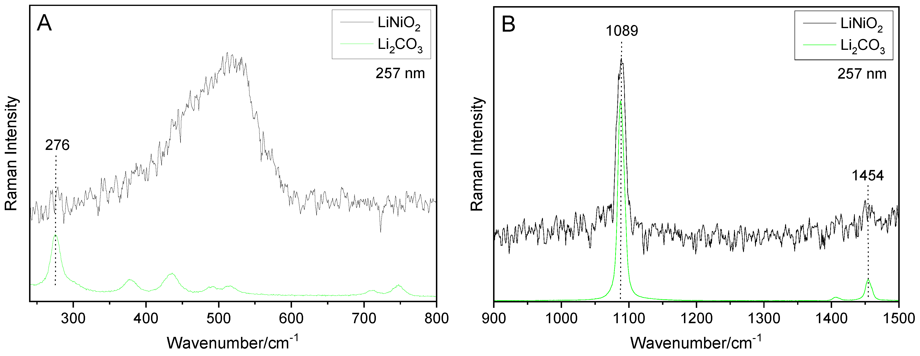
Our wavelength-dependent analysis revealed that Raman spectroscopy can be employed for the detection of surface and/or precursor species in electrode materials. As we will discuss in the following section, UV Raman spectra, in particular, shows a high sensitivity towards surface species, such as Li2CO3. Figure 5 depicts the Raman spectra of LiNiO2 at 257 nm excitation, in comparison to those of reference compounds. The spectrum of LiNiO2 is characterized by a broad Ni-related feature at around 530 cm−1, which exhibits a shoulder at around 460 cm−1, as well as additional features at 276, 1065 (shoulder), 1089 and 1454 cm−1. To the best of our knowledge, only the visible Raman spectra of LiNiO2 were reported, to date, showing more pronounced peaks at around 540 and 470 cm−1, attributed to the Eg and A1g mode, respectively [12,32,40]. The additional signals we detected at 276, 1089 and 1454 cm−1, can readily be assigned by comparison with a Li2CO3 reference (see Figure 5). In particular, the signals at 1089 and 1454 cm−1 were attributed to symmetric and asymmetric carbonate stretching, respectively, whereas the small feature at 276 cm−1 was not covered by calculations [41,42,43], and the small shoulder feature at around 1065 cm−1 will be discussed below.
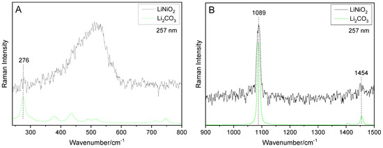
Figure 5.
Raman spectra of LiNiO2 and Li2CO3 (reference) at 257 nm excitation. (A) Low-wavenumber region. (B) Region within 900–1500 cm−1. Spectra were offset for clarity.
The presence of additional (non-oxidic) Raman features was also observed for the LiNixCo1−xO2 mixed oxides. In fact, as can be seen in Figure 2D, at 257 nm excitation, two signals are detected at 1067 and 1093 cm−1. Based on the above discussion, the signal at 1093 cm−1 can be attributed to carbonate. Interestingly, with higher nickel content, the 1093 cm−1 signal increases in intensity (see Figure 2D), suggesting an increased affinity of Ni-rich mixed oxides towards Li2CO3 formation under ambient conditions. On the other hand, the position of the signal at 1067 cm−1 is consistent with the symmetric stretching vibration of nitrate, as evidenced by a comparison with the spectrum of a LiNO3 reference (see Figure S6) [44]. The presence of nitrate may originate from precursors used for the synthesis, which were not completely decomposed during the calcination treatment. As discussed in the context of Figure 1, the Raman spectrum of LiCoO2 at 257 excitation shows a small signal at ~1070 cm−1, which does not originate from resonance effects. In light of the above discussion, an assignment to nitrate stretching seems likely. On the other hand, the Raman spectrum of NMC (see Figure 4) shows no additional signal indicating the presence of nitrate. The different behavior may result from the different calcination temperatures for LiNixCo1−xO2 and NMC. In fact, LiNixCo1−xO2 was calcined at 775 °C (10 h) and NMC at 900 °C (15 h), strongly suggesting an incomplete decomposition of LiNO3 in the case of LiNixCo1−xO2. In this context, it should be mentioned that for LiNixCo1−xO2 mixed oxides, a calcination temperature of 775 °C was chosen to minimize the cation disorder [45]. To summarize, the above studies show that deep UV excitation at 257 nm allows for the sensitive detection of surface species and/or precursor species reminiscent of the synthesis.
4. Conclusions
The further development of lithium-ion batteries will strongly depend on a detailed understanding of the structure–stability–function relationships of the cathodes, which largely limit the energy density and dominate the battery cost. Raman spectroscopy was shown to be a powerful tool to enhance the structural insight into layered cathode materials also under electrochemical conditions.
For the further development of the technique towards cathode characterization, we systematically applied multi-wavelength Raman spectroscopy to common layered cathode materials (LiCoO2, LiNixCo1−xO2, NMC) for the first time. The variation of the laser excitation from the deep UV to the visible (257, 385, 515, 633 nm) allowed us gain new insight into the (electronic) structure, by analyzing the wavelength-dependent spectral profiles. For LiCoO2, a resonance effect was observed, which was most pronounced for 385 nm excitation and lead to the appearance of overtone/combination bands, in addition to the intensity changes of the fundamental modes. In LiNixCo1−xO2 mixed oxides and NMC, the overtone features persisted but were mitigated by the presence of Ni and/or Mn, highlighting the sensitivity of the resonance effect on electronic structure changes.
The use of UV excitation was demonstrated to further enhance the possibilities of the Raman technique in the context of cathode characterization. First, laser excitation in the near UV (385 nm) and deep UV (257 nm) was shown to increase the sensitivity towards Ni in NMC, as compared to the employed visible wavelengths (515, 633 nm). Moreover, using 257 nm laser excitation, we demonstrated that the nickel content in LiNixCo1−xO2 (and NMC) materials can be quantified, which is of great interest also for other nickel containing layered materials. Future studies may be directed towards a quantitative analysis of spectral profiles by combining experiment with theoretical calculations. Secondly, deep UV excitation at 257 nm was shown to provide a sensitive detection of surface species and/or precursor species reminiscent of the synthesis. In particular, we identified carbonate and nitrate species by comparison with reference compounds. To this end, future work may specify the sensitivity for surface species/impurity detection by a comparison with dedicated surface and bulk analysis.
Our results demonstrate the potential of multi-wavelength Raman spectroscopy for the characterization of layered cathode materials for lithium-ion batteries, allowing for phase identification and quantification, insight into the electronic structure as well as surface species/impurity detection.
Supplementary Materials
The following supporting information can be downloaded at: https://www.mdpi.com/article/10.3390/batteries8020010/s1. Results from XRD diffraction, UV-Vis spectroscopy, UV Raman spectroscopy and visible Raman spectroscopy of layered oxide materials as well as reference compounds.
Author Contributions
Conceptualization, C.H.; Investigation, M.H. and K.H.; Data Analysis, M.H. and K.H.; Writing—Original Draft Preparation, M.H. and C.H.; Writing—Review and Editing, M.H., K.H. and C.H.; Supervision, Project Administration, Funding Acquisition, C.H. All authors have read and agreed to the published version of the manuscript.
Funding
This work was supported by the Deutsche Forschungsgemeinschaft (DFG, HE 4515/8-1).
Institutional Review Board Statement
Not applicable.
Informed Consent Statement
Not applicable.
Data Availability Statement
Not applicable.
Acknowledgments
The authors thank Philipp Waleska and Simone Rogg for performing the UV Raman measurements.
Conflicts of Interest
The authors declare no competing financial interest.
References
- Etacheri, V.; Marom, R.; Elazari, R.; Salitra, G.; Aurbach, D. Challenges in the development of advanced Li-ion batteries: A review. Energy Environ. Sci. 2011, 4, 3243–3262. [Google Scholar] [CrossRef]
- Nitta, N.; Wu, F.; Lee, J.T.; Yushin, G. Li-ion battery materials: Present and future. Mater. Today 2015, 18, 252–264. [Google Scholar] [CrossRef]
- Manthiram, A. A reflection on lithium-ion battery cathode chemistry. Nat. Commun. 2020, 11, 1550. [Google Scholar] [CrossRef] [PubMed]
- Liu, H.S.; Zhang, Z.R.; Gong, Z.L.; Yang, Y. Origin of deterioration for LiNiO2 cathode material during storage in air. Electrochem. Solid-State Lett. 2004, 7, A190–A193. [Google Scholar] [CrossRef]
- Yabuuchi, N.; Ohzuku, T. Novel lithium insertion material of LiCo1/3Ni1/3Mn1/3O2 for advanced lithium-ion batteries. Power Sources 2003, 171, 119–121. [Google Scholar] [CrossRef]
- Liu, W.; Oh, P.; Liu, X.; Lee, M.J.; Cho, W.; Chae, S.; Kim, Y.; Cho, J. Nickel-rich layered lithium transition-metal oxide for high-energy lithium-ion batteries. Angew. Chem. Int. Ed. 2015, 54, 4440–4457. [Google Scholar] [CrossRef]
- Guo, B.; Zhao, J.H.; Fan, X.M.; Zhang, W.; Li, S.; Yang, Z.H.; Chen, Z.X.; Zhang, W.X. Aluminum and fluorine co-doping for promotion of stability and safety of lithium-rich layered cathode material. Electrochim. Acta 2017, 236, 171–179. [Google Scholar] [CrossRef]
- Hausbrand, R.; Cherkashinin, G.; Ehrenberg, H.; Gröting, M.; Albe, K.; Hess, C.; Jaegermann, W. Fundamental degradation mechanisms of layered oxide Li-ion battery cathode materials: Methodology, insights and novel approaches. Mater. Sci. Eng. B. 2015, 192, 3–25. [Google Scholar] [CrossRef]
- Pender, J.P.; Jha, G.; Youn, D.H.; Ziegler, J.M.; Andoni, I.; Choi, E.J.; Heller, A.; Dunn, B.S.; Weiss, P.S.; Penner, R.M.; et al. Electrode degradation in lithium-ion batteries. ACS Nano 2020, 14, 1243–1295. [Google Scholar] [CrossRef] [Green Version]
- Baddour-Hadjean, R.; Pereira-Ramos, J.P. Raman microspectrometry applied to the study of electrode materials for lithium batteries. Chem. Rev. 2010, 110, 1278–1319. [Google Scholar] [CrossRef]
- Tripathi, A.M.; Su, W.N.; Hwang, B.J. In situ analytical techniques for battery interface analysis. Chem. Soc. Rev. 2018, 47, 736–851. [Google Scholar] [CrossRef] [PubMed]
- Flores, E.; Novák, P.; Berg, E.J. In situ and operando Raman spectroscopy of layered transition metal oxides for Li-ion battery cathodes. Front. Energy Res. 2018, 6, 82. [Google Scholar] [CrossRef]
- Julien, C.M.; Mauger, A. In situ Raman analyses of electrode materials for Li-ion batteries. AIMS Mater. Sci. 2018, 5, 650. [Google Scholar] [CrossRef]
- Flores, E.; Novák, P.; Aschauer, U.; Berg, E.J. Cation Ordering and Redox Chemistry of Layered Ni-Rich LixNi1–2yCoyMnyO2: An Operando Raman Spectroscopy Study. Chem. Mater. 2020, 32, 186–194. [Google Scholar] [CrossRef]
- Jehnichen, P.; Korte, C. Operando Raman Spectroscopy Measurements of a High-Voltage Cathode Material for Lithium-Ion Batteries. Anal. Chem. 2019, 91, 8054–8061. [Google Scholar] [CrossRef]
- Matsuda, Y.; Kuwata, N.; Okawa, T.; Dorai, A.; Kamishima, O.; Kawamura, J. In situ Raman spectroscopy of LixCoO2 cathode in Li/Li3PO4/LiCoO2 all-solid-state thin-film lithium battery. Solid State Ionics 2019, 335, 7–14. [Google Scholar] [CrossRef]
- Otoyama, M.; Ito, Y.; Sakuda, A.; Tatsumisago, M.; Hayashi, A. Reaction uniformity visualized by Raman imaging in the composite electrode layers of all-solid-state lithium batteries. Phys. Chem. Chem. Phys. 2020, 22, 13271–13276. [Google Scholar] [CrossRef]
- Flores, E.; Mozhzhukhina, N.; Aschauer, U.; Berg, E.J. Operando Monitoring the Insulator–Metal Transition of LiCoO2. ACS Appl. Mater. Interfaces 2021, 13, 22540–22548. [Google Scholar] [CrossRef]
- Yellampalle, B.; Sluch, M.; Asher, S.; Lemoff, B. Multiple-excitation-wavelength resonance-Raman explosives detection. Proc. SPIE 2011, 8018, 801819. [Google Scholar]
- Gross, T.; Hess, C. Raman diagnostics of LiCoO2 electrodes for lithium-ion batteries. J. Power Sources 2014, 256, 220–225. [Google Scholar] [CrossRef]
- Julien, C.M.; Massot, M.; Ramana, C.V. Structure of electrode materials for Li-ion batteries: The Raman spectroscopy investigations. In Portable and Emergency Energy Sources; Stoynov, Z., Vladikova, D., Eds.; Martin Drinov Academic Publishing House: Sofia, Bulgaria, 2006. [Google Scholar]
- Ammundsen, B.; Burns, G.R.; Islam, M.S.; Kanoh, H.; Rozière, J. Lattice dynamics and vibrational spectra of lithium manganese oxides: A computer simulation and spectroscopic study. J. Phys. Chem. B 1999, 103, 5175–5180. [Google Scholar] [CrossRef]
- Lei, J.; McLarnon, F.; Kostecki, R. In situ raman microscopy of individual LiNi0.8Co0.15Al0.05O2 particles in a Li-ion battery composite cathode. J. Phys. Chem. B 2005, 109, 952–957. [Google Scholar] [CrossRef] [PubMed] [Green Version]
- Dokko, K.; Mohamedi, M.; Anzue, N.; Itoh, T.; Uchida, I. In situ Raman spectroscopic studies of LiNixMn2−xO4 thin film cathode materials for lithium ion secondary batteries. J. Mater. Chem. 2002, 12, 3688–3693. [Google Scholar] [CrossRef]
- Kerlau, M.; Marcinek, M.; Srinivasan, V.; Kostecki, R.M. Studies of local degradation phenomena in composite cathodes for lithium-ion batteries. Electrochim. Acta 2007, 53, 1385–1392. [Google Scholar] [CrossRef]
- Pechini, P.M. Method of Preparing Lead and Alkaline Earth Titanates and Niobates and Coating Method Using the Same to form a Capacitor. U.S. Patent Nr. 3.330.697, 11 July 1967. [Google Scholar]
- Toby, B.H.; Von Dreele, R.B. GSAS-II: The genesis of a modern open-source all purpose crystallography software package. J. Appl. Cryst. 2013, 46, 544–549. [Google Scholar] [CrossRef]
- Inaba, M.; Iriyama, Y.; Ogumi, Z.; Todzuka, Y.; Tasaka, A. Raman study of layered rock-salt LiCoO2 and its electrochemical lithium deintercalation. J. Raman Spectrosc. 1997, 28, 613–617. [Google Scholar] [CrossRef]
- Ghosh, P.; Mahanty, S.; Raja, M.W.; Basu, R.N.; Maiti, H.S. Structure and optical absorption of combustion-synthesized nanocrystalline LiCoO2. J. Mater. Res. 2006, 22, 1162–1167. [Google Scholar] [CrossRef] [Green Version]
- Rao, K.J.; Benqlilou-Moudden, H.; Desbat, B.; Vinatier, P.; Levasseur, A. Infrared spectroscopic study of LiCoO2 thin films. J. Solid State Chem. 2002, 165, 42–47. [Google Scholar] [CrossRef]
- Julien, C. Local cationic environment in lithium nickel–cobalt oxides used as cathode materials for lithium batteries. Solid State Ionics 2000, 136–137, 887–896. [Google Scholar] [CrossRef]
- Inaba, M.; Todzuka, Y.; Yoshida, H.; Grincourt, Y.; Tasaka, A.; Tomida, Y.; Ogumi, Z. Raman spectra of LiCo1-yNiyO2. Chem. Lett. 1995, 24, 889–890. [Google Scholar] [CrossRef]
- Kalyani, P.; Kalaiselvi, N. Various aspects of LiNiO2 chemistry: A review. Sci. Technol. Adv. Mater. 2005, 6, 689–703. [Google Scholar] [CrossRef] [Green Version]
- Chakraborty, A.; Dixit, M.; Aurbach, D.; Major, D.T. Predicting accurate cathode properties of layered oxide materials using the SCAN meta-GGA density functional. NPJ Comput. Mater. 2018, 4, 60. [Google Scholar] [CrossRef]
- Laubach, S.; Laubach, S.; Schmidt, P.C.; Ensling, D.; Schmid, S.; Jaegermann, W.; Thißen, A.; Nikolowski, K.; Ehrenberg, H. Changes in the crystal and electronic structure of LiCoO2 and LiNiO2 upon Li intercalation and de-intercalation. Phys. Chem. Chem. Phys. 2009, 11, 3278–3289. [Google Scholar] [CrossRef] [PubMed]
- Dahn, J.R.; von Sacken, U.; Michal, C.A. Structure and electrochemistry of Li1±yNiO2 and a new Li2NiO2 phase with the Ni(OH)2 structure. Solid State Ionics 1990, 44, 87–97. [Google Scholar] [CrossRef]
- Zhang, X.; Mauger, A.; Lu, Q.; Groult, H.; Perrigaud, L.; Gendron, F.; Julien, C.M. Synthesis and characterization of LiNi1/3Mn1/3Co1/3O2 by wet-chemical method. Electrochim. Acta 2010, 55, 6440–6449. [Google Scholar] [CrossRef]
- Ruther, R.E.; Callender, A.F.; Zhou, H.; Martha, S.K.; Nanda, J. Raman microscopy of lithium-manganese-rich transition metal oxide cathodes. Electrochem. Soc. 2015, 162, A98. [Google Scholar] [CrossRef] [Green Version]
- Ghanty, C.; Markovsky, B.; Erickson, E.M.; Talianker, M.; Haik, O.; Tal-Yossef, Y.; Mor, A.; Aurbach, D.; Lampert, J.; Volkov, A.; et al. Li+-ion extraction/insertion of Ni-rich Li1+ x(NiyCozMnz)wO2 (0.005 < x < 0.03; y:z = 8:1, w ≈ 1) electrodes: In situ XRD and Raman spectroscopy study. ChemElectroChem 2015, 2, 1479–1486. [Google Scholar]
- Julien, C.M.; Massot, M. Raman scattering of LiNi1−yAlyO2. Solid State Ionics 2002, 148, 53–59. [Google Scholar] [CrossRef]
- Brooker, M.H.; Bates, J.B. Raman and infrared spectral studies of anhydrous Li2CO3 and Na2CO3. J. Chem. Phys. 1971, 54, 4788–4796. [Google Scholar] [CrossRef]
- Koura, N.; Kohara, S.; Takeuchi, K.; Takahashi, S.; Curtiss, L.A. Alkali carbonates: Raman spectroscopy, ab initio calculations, and structure. J. Mol. Struct. 1996, 382, 163–169. [Google Scholar] [CrossRef]
- Brooker, M.H.; Wang, J. Raman and infrared studies of lithium and cesium carbonates. Spectrochim. Acta 1992, 48A, 999–1008. [Google Scholar] [CrossRef]
- James, D.W.; Leong, W.H. Structure of molten nitrates. III. Vibrational spectra of LiNO3, NaNO3, and AgNO3. J. Chem. Phys. 1969, 51, 640–646. [Google Scholar] [CrossRef] [Green Version]
- Gross, T.; Buhrmester, T.; Bramnik, K.G.; Bramnik, N.N.; Nikolowski, K.; Baehtz, C.; Ehrenberg, H.; Fuess, H. Structure–intercalation relationships in LiNiyCo1-yO2. Solid State Ionics 2005, 176, 1193–1199. [Google Scholar] [CrossRef]
Publisher’s Note: MDPI stays neutral with regard to jurisdictional claims in published maps and institutional affiliations. |
© 2022 by the authors. Licensee MDPI, Basel, Switzerland. This article is an open access article distributed under the terms and conditions of the Creative Commons Attribution (CC BY) license (https://creativecommons.org/licenses/by/4.0/).

