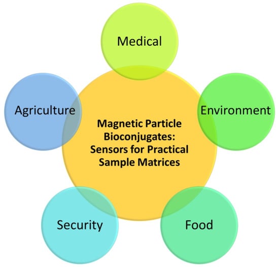
Magnetic Particle Bioconjugates: A Versatile Sensor Approach


1
Department of Chemistry, Oklahoma State University, Stillwater, OK 74078, USA
2
Department of Chemistry, National Institute of Technology Warangal, Telangana 506004, India
*
Author to whom correspondence should be addressed.
†
Equal contribution.
Magnetochemistry 2019, 5(4), 64; https://doi.org/10.3390/magnetochemistry5040064
Submission received: 16 October 2019
/
Revised: 14 November 2019
/
Accepted: 14 November 2019
/
Published: 19 November 2019
(This article belongs to the Special Issue Magnetic Nanoparticles)
Abstract
:Nanomaterial biosensors have revolutionized the entire scientific, technology, biomedical, materials science, and engineering fields. Among all nanomaterials, magnetic nanoparticles, microparticles, and beads are unique in offering facile conjugation of biorecognition probes for selective capturing of any desired analytes from complex real sample matrices (e.g., biofluids such as whole blood, serum, urine and saliva, tissues, food, and environmental samples). In addition, rapid separation of the particle-captured analytes by the simple use of a magnet for subsequent detection on a sensor unit makes the magnetic particle sensor approach very attractive. The easy magnetic isolation feature of target analytes is not possible with other inorganic particles, both metallic (e.g., gold) and non-metallic (e.g., silica), which require difficult centrifugation and separation steps. Magnetic particle biosensors have thus enabled ultra-low detection with ultra-high sensitivity that has traditionally been achieved only by radioactive assays and other tedious optical sources. Moreover, when traditional approaches failed to selectively detect low-concentration analytes in complex matrices (e.g., colorimetric, electrochemistry, and optical methods), magnetic particle-incorporated sensing strategies enabled sample concentration into a defined microvolume of large surface area particles for a straightforward detection. The objective of this article is to highlight the ever-growing applications of magnetic materials for the detection of analytes present in various real sample matrices. The central idea of this paper was to show the versatility and advantages of using magnetic particles for a variety of sample matrices and analyte types and the adaptability of different transducers with the magnetic particle approaches.
1. Introduction
Most chemical and biological detection labels are limited to one transducer type or, sometimes, a few at best. In contrast, nanomaterials are not limited to a particular type of transducer, and they can be employed to a wide range of transducers. Moreover, nanomaterials can be used as labels for signal amplification and detection in sensors and assays. Because of these astonishing properties, nanomaterial sensors can be used with various detection methods (e.g., sensors based on optical, electrical, electrochemical, mass, mechanical, luminescent, and other physical properties). Thus, the unusual versatility of nanomaterial sensors supersedes traditional approaches and enables the analysis of complex sample matrices [1,2]. The magnetic separability of nanomaterials allows selective capturing of desired target analytes from complex real sample matrices through the analyte-specific probes. The bioconjugation is made feasible through viable chemical functionalization of magnetic materials to link desired molecular probes for analyte detection in sensors. Chemical and bio sensors constitute one common theme of involving magnetic particle applications [1,3,4,5,6,7,8,9], and other equally important areas include biomedical imaging [10,11,12,13,14,15,16,17], food science [18,19,20], drug delivery [21,22,23], disease treatment [24,25], nanomedicine [26], diagnostics [27,28,29,30,31,32], catalysis [33,34,35,36,37,38,39,40], separation and purification [41,42], health [43], and environmental applications [44,45,46,47]. Thus, a plethora of studies have been reviewed for the unusually broad research fields in which magnetic materials play crucial roles. In addition to several characteristics such as optical, electrochemical, and catalytic properties of magnetic particles, their biocompatibility and tunable chemical functionalities and geometries (i.e., size and shape) pave the way for their in vivo applications. A literature search by the authors conveys that the research progress, number of publications, and reviews on magnetic particles and their applications in a vast range of topics have gained never-ceasing attention. This article specifically focused on magnetic particle (both nano and micro) and magnetic bead-based biosensors for the detection of biomarkers (for the most part) and other classes of analytes.
Xianyu et al. [4] recently reviewed the advances in magnetic particle-based biosensors for point-of-care testing and mechanisms of assaying biomarkers. They additionally outlined the milestones that limit the realization of real-world applications of certain magnetic materials. They emphasized the need to synthesize high-quality magnetic particles with control over their size distribution and magnetization and the need of portable and miniaturized magnetic signal readout systems. Farka et al. [48] recently published a very informative and comprehensive review of nanomaterials, including magnetic materials for immunochemical biosensors and assays. They discussed several types of transducers (e.g., optical, electrochemical, surface-sensitive methods, and transistors) and covered a range of target analytes including proteins, small molecules, cancer cells, toxic substances, and pathogens.
With regard to the specific needs of successfully utilizing magnetic materials for ultra-low analyte detection with high sensitivity, several strategies are being explored. Magnetic preconcentration of desired analytes, (basically, capturing low concentrations of target analytes from real samples onto a large surface area of magnetic particles; as a result, the analyte molecules are concentrated in small geometric areas to enable sensitive detection) to minimize the effect of interference from coexisting irrelevant molecules in sample matrices, has been explored. In addition, the use of an external magnetic field to selectively attract desired target analytes attached to magnetic conjugates onto the sensor surface is another strategy. Furthermore, sensitive detection and imaging of magnetically tagged analytes (or their complexes with biorecognition molecules) in solutions have also been explored in sensors [48]. Another review discussed the transduction mechanisms and applications of complementary metal-oxide-semiconductor biosensor designs based on magnetic and other particles for in vitro diagnosis [49].
In a typical heterogeneous biosensor design, the analyte molecules in the magnetic conjugates diffuse to the sensor surface to generate signals. In contrast, magnetic particle sensors in which the analytes are detected in the whole solution volume of the sample as a homogeneous sensing approach have also been developed [50]. Sample preparation steps are shown to be reduced in the homogeneous sensor assay, and the combined diffusion of analyte molecules and capture probes is shown to reduce the total assay time compared with a heterogeneous assay. In this article, we will mainly focus on the heterogeneous sensor systems that are shown to offer high sensitivity, reduced nonspecific or false positive signals, ultra-low detection limits, and tunability to attain a wide dynamic range of detection.
In addition to the synthesis and sensor application of iron oxide-based magnetic particles, considerable focus has been also placed on making hybrid magnetic materials (e.g., core/shell, bimetallic, and alloys) and composites with other materials (e.g., polymers, graphene, chitosan, and carbon nanostructures) to obtain a multifactorial advantage from a single end material [51,52,53,54,55,56]. We also present here several examples on core/shell and nanocomposite magnetic materials. This article is not a comprehensive review of magnetic nanoparticles or biosensors.
2. Rationale for the Selection of Transducer
On deciding the type of sensor signal transducers, major factors, such as the molecular properties of the analytes, detection limits required, and the design of suitable biorecognition process for selective detection, are considered. For a redox-active analyte or reagents that can offer redox-active products, electrochemical methods are suitable and offer sensitive detection (e.g., amperometric, potentiometric, and impedance sensors) (Table 1). When the analyte concentration is large enough (e.g., micromolar to millimolar range) and can be selectively visualized by the naked eye by a color reaction, the colorimetric detection is advantageous. For ultra-low detection limits, fluorescence, luminescence, and surface-sensitive optical methods (e.g., surface-enhanced Raman scattering and surface plasmon resonance) have been developed using magnetic nanomaterials (Table 2). Thus, each transducer has its significance depending on the target analyte nature and the associated extractable molecular properties in the sensor design.
Molecular Recognition Probes
Antibodies and aptamers for proteins and small molecules, complementary nucleotides for deoxyribonucleic acid (DNA) and ribonucleic acid (RNA) hybridization, enzymes for specific substrates by either direct or indirect catalytic conversions, receptors for ligands, and non-enzymatic molecular probes (e.g., molecularly imprinted polymers) for analytes are commonly used approaches in magnetic biosensors. For example, a magnetic force-aided electrochemical sensor was developed using sandwich biorecognitions from an aptamer and antibody combination for the detection of thrombin in serum samples [57]. Similarly, a magnetic field-induced self-assembly of Fe3O4(magnetite)@polyaniline nanoparticles incorporated with a molecularly imprinted polymer biorecognition element-delivered electrochemical sensing of creatinine in human plasma and urine samples [58]. Electrochemical glucose biosensing by glucose oxidase, on an electrode consisting of gold nanoparticles/bovine serum albumin/Fe3O4 nanocomposite [59], is an example for enzymatic biorecognition event (Figure 1-I–III).
An antibody biorecognition probe immobilized on a magnetic (CoFe2O4) metal–organic framework (MOF)@Au tetrapods (AuTPs) surface, with toluidine blue as the surface-enhanced Raman spectroscopy (SERS) tag, was used for detection of N-terminal pro-brain natriuretic peptides [54]. This strategy offered a large dynamic range with ultra-low detection limits. Effective magnetic bead-based separation and highly selective aptamer recognition of G-quadruplex formation of the anterior gradient homolog 2 (AGR2) protein, cancer biomarker, offered picomolar detection limits. This system also utilized gold nanoparticles. The detection was based on UV–vis spectrometry [60]. Another antibody-conjugated magnetic-bead sensor detected chloramphenicol antibiotic based on a competitive surface-enhanced Raman scattering method [61] (Figure 2-I,II).
Magnetic particle sensors for detection of various classes of analytes in a variety of complex sample matrices. In the subsequent sections, we extend our discussion to selective classes of analytes and various sample matrices, biorecognition materials, and transducers that best addressed the sensor purpose and analytical performance.
3. Biomarkers
Biomarkers indicate normal versus abnormal conditions based on the levels of their concentrations in organs, tissues, and circulating body fluids. Hence, sensors that allow selective and required sensitive detection of the biomarkers are significant in diagnosis and treatment. We present here a few examples of biomarker sensors based on magnetic materials which use various transducers. Recently, Gooding et al. [62] devised a novel microRNA detection probe consisting of a DNA sequence attached to gold-coated magnetic nanoparticles. By electrically reconfiguring the nanoparticle probe, they achieved electrochemical (square wave voltammetry) detection of microRNA in whole blood samples over a wide concentration range from attomolar to nanomolar. Such ultrasensitive magnetic nanoparticle tools are significant for clinical diagnostics.
Similarly, magnetic nanoparticle-based voltammetric and electrochemical mass sensors recently enabled the detection of ultra-low clinically relevant picomolar serum insulin concentrations (Figure 3A(a,b),B) [63,64]. In the electrochemical mass sensor, magnetic isolation of insulin from a serum sample reduced the interference from the sample matrix and non-specific background signals which was quantitatively estimated by comparing the background signals for magnetically treated serum control response with that of a solution-based bulk serum control response (Figure 3A-b) [63]. Furthermore, enriching the surface –COOH groups of pyrenyl-carbon nanostructures by combined covalent and non-covalent carboxylation improved the sensitivity of serum insulin immunosensing compared to only a carboxylated nanotube-based sensor (Figure 3C) [65]. Another quartz crystal microbalance (QCM) mass sensor method has been reported for detection of thrombin (in diluted serum or fibrinogen-precipitated plasma samples) by a magnetic microbead polymeric material and detection on an aptamer-immobilized QCM crystal based on oscillation frequency decrease [66]. Translating the quantitative knowledge derived from a single-sample QCM analysis sensor into a relatively better throughput surface plasmon imaging gold array enabled magnetic nanoparticle captured serum insulin (50% serum in buffer) detection at picomolar concentrations (Figure 3D) [67]. Advancements in microfluidic designs enabled on-line protein capturing by magnetic beads and ultrasensitive electrochemical detection of interleukin cancer biomarkers (IL-6 and IL-8) in undiluted calf serum [68]. In addition to electrochemical detection, surface plasmon spectroscopy that offers detection with binding insights [69] has been successfully developed with a polyamidoamine (PAMAM)-magnetic nanoparticle quantum dot modification on a gold array for a dual imaging-based detection of blood insulin and glycated hemoglobin (20 time diluted whole blood samples in buffer) (Figure 3E) [70]. Subsequently, fluorescence imaging of the sensor array allowed an independent validation of the method. Table 1 presents various magnetic nanoparticles based electrochemical sensors for the detection of a wide range of analytes. Cyclic, square-wave, and differential voltammetry, and amperometric techniques have been widely used as electrochemical transducers. Moreover, the analytes ranged from small molecules such as glucose, bisphenol A, and ciprofloxacin antibiotics to various protein biomarkers, pesticides, and illicit drugs. The analyte recognition elements include enzymes, aptamers, antibodies, and molecularly imprinted polymers. The wide variety of sample matrices covered were blood, serum, plasma, urine, milk, juice, meat samples, and water (Table 1).
A nanocomposite that consists of glucose oxidase with reduced graphene oxide and magnetic nanoparticles on a magnetic sticker attached to a screen-printed electrode was shown to offer micromolar glucose amperometric detection in simple aqueous solutions [55]. An antibody conjugation to a magnetic nitrogen-doped graphene-modified gold electrode was used for voltammetric detection of amyloid-beta peptide 1–42 (Aβ42), a biomarker of Alzheimer’s disease, in solution [85]. However, the applicability of such sensors for practical sample matrices of biofluids would be more significant.
A recently reported magnetic-focus-lateral flow-colorimetric biosensor offered a million-fold improvement in sensitivity over the conventional lateral flow systems toward detection of the valosin-containing protein of relevance to cervical cancer [53] (Figure 4A(a,b)). The sensor applicability for protein mixtures extracted from the tissue of cervical cancer patients was successfully demonstrated. Spectral correlation interferometry, capable of picometer resolution of measured thickness changes of a layer of molecules or nanoparticles, was designed for sensitive detection of prostate-specific antigen by a magnetic nanoparticle-bioprobe amplification strategy [86] (Figure 4B). Another study utilized anodic stripping voltammetry for sensitive DNA detection on a magnetic porous pseudo-carbon paste electrode [87].
A magnetic bead-based colorimetric biosensor allowed for detection of two inflammatory salivary biomarkers, human neutrophil elastase and cathepsin-G, in solution and spiked saliva samples. The sensor utilized proteolytic activity-induced color changes with biomarker concentration. The sensor was validated with patients’ saliva analysis [88]. Magnetic particles possess peroxidase-like enzymatic activity. This property has been extensively used in both electrochemical and optical sensors for analyte detection. An illustration would be the enzymatic colorimetric glucose sensors based on magnetic particles that exploited the peroxidase-like activity of these particles [89]. In this report, glucose oxidase was covalently attached to carboxylated magnetic beads (1 μm size) for spectrophotometric detection of glucose in solutions or that spiked in plasma samples using o-phenylenediamine dihydrochloride reagent.
An enzymatic biosensor composed of poly(dopamine)-modified magnetic nanoparticles, covalently attached with a PAMAM dendrimer (ethylenediamine core polyamidoamine G-4; fourth-generation) and additionally incorporated with platinum nanoparticles, was used for amperometric detection of micromolar xanthine in fish samples [91]. Similarly, magnetic nanocomposite materials with carbon nanostructures (e.g., graphene and nanotubes) have been devised for sensitive electrochemical enzymatic glucose detection in spiked urine samples [92] and progesterone in human serum samples and pharmaceutical products [82] with good recoveries. Magnetic susceptibility measurements combined with a optomagnetic method was developed for detection of C-reactive protein in serum from the agglutination of magnetic nanoparticles conjugated with the protein antibody [93]. A chemiluminescent biosensor combining magnetic beads with platinum nanoparticles for thyroid-stimulating hormone detection in serum has been devised [94].
Protein A/G-functionalized 2 μm magnetic beads successfully isolated glutamic acid decarboxylase-65 autoantibody from 10% serum and enabled sensitive (picomolar) detection on the glutamic acid decarboxylase-65 immobilized carboxylated graphene surface [95] (Figure 5A(a,b)). In this report, the correlation of picomolar affinities between surface plasmon resonance and electrochemical immunosensors was demonstrated. Furthermore, multiplex biosensing of a panel of biomarkers allows reliable and rapid quantification of several analytes in complex clinical matrices. A new surface plasmon-imaging array based on core/shell Fe3O4@Au nanoparticles enabled combined detection of two interleukin proteins and two microRNAs in a diluted serum (Figure 5B) [96]. The gold shell can facilitate the enhancement of the plasmon signals and the magnetic core for easy separation and magnetic isolation of desired target analytes present in complex sample matrices. Moreover, the core/shell material exhibited higher plasmonic signals than the individual nanoparticle components of similar hydrodynamic sizes [96]. Such multifunctional magnetic materials and composites offer several distinct properties but centralized into a single component to enable designing ultra-sensitive sensors [59,97,98,99,100,101]. A multiplex sensor featuring magnetic nanoparticle–antibody conjugates for detection of ovarian cancer biomarkers, CA-125, β-2M, and ApoA1, has been reported [102]. Fluorescence spectroscopy and surface plasmon resonance analysis were utilized. Thus, emerging directions of magnetic particle biosensor designs [103] are pushing the limits of applications to increasingly complex real samples and multianalyte biosensing. One other example of a biocatalytic real-sample system is the design of magnetic nanoparticle-based liver microsomal biofilms (subcellular liver fractions containing drug-metabolizing cytochrome P450 enzymes with their reductase) for reduced-nicotinamide adenine dinucleotide phosphate (NADPH)-free direct electrochemical drug biosensing and stereoselective metabolite production [104] (Figure 5C).
Katz et al. [105] reported a novel magnetic field-activated binary deoxyribozyme sensor for fluorescent detection of a specific messenger RNA that is a cancer biomarker. The sensor functioned well when internalized in live MCF-7 breast cancer cells and activated by a magnetic field. The sensor utilized two types of magnetic bead bioconjugates that carried different components of a multicomponent deoxyribozyme sensor. The sensor function was activated only in the presence of a specific target messenger RNA and upon an applied magnetic field thus adding control over the selectivity [106]. Pang et al. [107] designed a DNA probe Fe3O4@Ag magnetic nanoparticle biosensor for capturing microRNA from cancer cells and detection at ultra-low concentrations (0.3 fM). In this study, surface-enhanced Raman scattering with a duplex-specific nuclease signal amplification strategy was used. Application for point-of-care clinical diagnostics of microRNA has been envisioned. Discussed above are a range of illustrative examples of bioanalytes, and, in the subsequent sections, we highlight magnetic particle sensors developed for other analyte classes.
4. Food Analytes, Pathogens, and Pharmaceuticals
In addition to the important detection advantages of the magnetic particle sensor approaches for a range of diagnostically useful biomarkers and small molecules, the broader applicability for food, pathogens, and pharmaceuticals are illustrated in this section. Food and pharmaceuticals represent two essential components for sustaining life and health. Biosensors play important roles in the detection of foodborne pathogens and the development of valuable therapeutic molecules. Andreescu et al. [108], in the year 2015, published an excellent review of the design and development of nanoparticle-based (including magnetic) sensors for food safety assessment. They covered both colorimetric and electrochemical detection of chemical and biological contaminants (e.g., pesticides, heavy metals, bacterial pathogens, and natural toxins). Therefore, we provide some representative recent reports specifically on magnetic material sensors for food contaminants, pathogens, and pharmaceuticals.
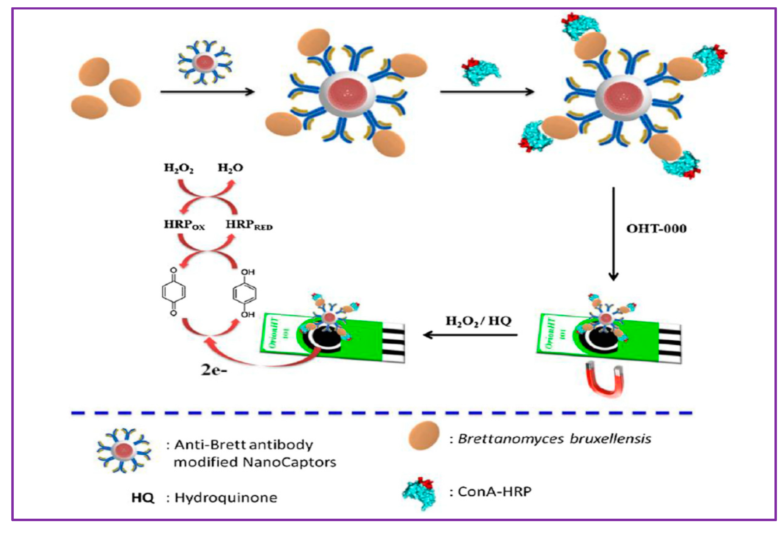
An impedimetric electrochemical aptasensor allowed rapid and sensitive detection of Escherichia coli [109]. The sensor system contained biotinylated polyclonal antibodies bound strongly to streptavidin-modified magnetic nanoparticles for selective separation of target bacteria from the sample in a coaxial capillary designed with high-gradient magnetic fields. For measuring impedance signals with the bacteria concentration, urease-catalyzed hydrolysis of urea into ammonium ions and carbonate ions in the capillary was used. A disposable amperometric sensor based on core/shell Fe3O4@SiO2 superparamagnetic nanoparticles conjugated with specific bioreceptors for selective detection and quantification of Brettanomyces bruxellensis (Brett) and total yeast content in wine has been developed (Figure 6) [110]. Maghemite nanoparticles (Fe2O3) have been identified to be an efficient and reliable material for removal of citrinin, a nephrotoxic mycotoxin, from food samples [111]. Table 2 presents various magnetic nanoparticles based optical sensors for detection of a wide range of analytes. Colorimetric, luminescence, surface plasmon resonance (SPR), and other surface-sensitive techniques are illustrated as representative transducers. Aptamers, peptides, antibodies, and MIPs are presented as recognition elements. The analytes ranged from proteins, hormones, peptides, miRNA, and bacteria to small molecules (Table 2).
Aptamer-functionalized magnetite (Fe3O4) conjugated upconversion nanoparticles were constructed to quantify trace enrofloxacin (a fluoroquinolone antibiotic) in fish samples; hybridization reaction-based luminescent intensity decreased when the analyte concentration was used as the signal transduction output [90] (Figure 4C). The “upconversion” terminology is related to the property of high-energy photon emission by a shorter wavelength excitation source of lower energy such as near-infrared photons. Other representative magnetic nanoparticle sensors for separation and/or detection include colorimetric tetracycline detection [117], chloramphenicol [61], lysozyme (an antimicrobial enzyme) [116], aflatoxins [118] (a review covering electrochemical, optical, and mass-sensitive biosensors), Listeria monocytogenes (a Gram-positive bacterium) [119], Staphylococcus aureus (a Gram-positive bacterium) [120], Salmonella enteritidis (a Gram-negative bacterium) [121], impedimetric hepatitis B virus DNA [122], and Vibrio cholerae DNA detection [123,124].
5. Environment
Environmental biosensors play significant roles in human safety and protecting the planet from devastations. Nanotechnology as a whole has contributed to the development of modern real-time environmental sensors (e.g., pollution monitoring, agriculture, renewable energy, water, and aviation) [47,125,126]. Herein, we illustrate literature reports on magnetic particle-based environmental biosensor system for organophosphorus pesticides, which are known to be toxic agricultural wastes despite their widespread use. Co-immobilization of carboxylic acid group-modified magnetic nanoparticles and acetylcholinesterase enzyme onto an electropolymerized surface of 4,7-di(furan-2-yl)benzo[c][1,2,5]-thiadiazole allowed sensitive micromolar detection of paraoxon and trichlorfon used as model organophosphates [127]. Amperometric current signals were used as the transduction method. Good reproducibility, long-term stability over 10 days, and applicability of the sensor for analysis in real tap water samples were demonstrated. Another acetylcholinesterase-magnetic particle electrochemical biosensor operated in flow injection analysis also delivered sensitive detection of organophosphate insecticides [128].
Summary/Prospects
The versatile nature of magnetic materials for applications with a range of transduction and biorecognition principles is detailed. Moreover, magnetic sensor designs for the detection of a broad range of analytes present in various complex real sample matrices of relevance to disease diagnosis, health, food, agriculture, energy, and environment are highlighted. Future directions in the field are aimed at synthesizing new multifunctional magnetic materials that offer simplicity and rapid detection platforms from the prior methods and quantitative insights into size and shape distributions as well as chemical and surface functionality aspects of magnetic materials. Moreover, efficient strategies for oriented and other modes of immobilization of bioprobes, their quantitative characterizations, activity, bioconjugation and catalytic efficiency, need of reproducibility, and long-term stability of sensor designs are being addressed. Emerging magnetic particle biosensor designs with multifunctional and multiplex features are discussed. Improvements on the scientific rigor of methodologies and approaches developed and standardization of protocols for reliable end applications are some of the currently emphasized benchmarks for successful magnetic particle sensor designs.
Author Contributions
S.K. and K.Y.G. contributed equally in the preparation of this review article.
Funding
This research received no external funding.
Conflicts of Interest
The authors declare no conflicts of interest.
References
- Hasanzadeh, M.; Shadjou, N.; de la Guardia, M. Iron and iron-oxide magnetic nanoparticles as signal-amplification elements in electrochemical biosensing. TrAC Trends Anal. Chem. 2015, 72, 1–9. [Google Scholar] [CrossRef]
- Kudr, J.; Haddad, Y.; Richtera, L.; Heger, Z.; Cernak, M.; Adam, V.; Zitka, O. Magnetic nanoparticles: From design and synthesis to real world applications. Nanomaterials 2017, 7, 243. [Google Scholar] [CrossRef] [PubMed]
- Jamshaid, T.; Neto, E.T.T.; Eissa, M.M.; Zine, N.; Kunita, M.H.; El-Salhi, A.E.; Elaissari, A. Magnetic particles: From preparation to lab-on-a-chip, biosensors, microsystems and microfluidics applications. TrAC Trends Anal. Chem. 2016, 79, 344–362. [Google Scholar] [CrossRef]
- Xianyu, Y.; Wang, Q.; Chen, Y. Magnetic particles-enabled biosensors for point-of-care testing. TrAC Trends Anal. Chem. 2018, 106, 213–224. [Google Scholar] [CrossRef]
- Giouroudi, I.; Kokkinis, G. Recent advances in magnetic microfluidic biosensors. Nanomaterials 2017, 7, 171. [Google Scholar] [CrossRef]
- Ravalli, A.; Marrazza, G. Gold and magnetic nanoparticles-based electrochemical biosensors for cancer biomarker determination. J. Nanosci. Nanotechnol. 2015, 15, 3307–3319. [Google Scholar] [CrossRef]
- Lan, L.; Yao, Y.; Ping, J.; Ying, Y. Recent advances in nanomaterial-based biosensors for antibiotics detection. Biosens. Bioelectron. 2017, 91, 504–514. [Google Scholar] [CrossRef]
- Kurbanoglu, S.; Ozkan, S.A.; Merkoçi, A. Nanomaterials-based enzyme electrochemical biosensors operating through inhibition for biosensing applications. Biosens. Bioelectron. 2017, 89, 886–898. [Google Scholar] [CrossRef]
- Yoo, S.M.; Lee, S.Y. Optical biosensors for the detection of pathogenic microorganisms. Trends Biotechnol. 2016, 34, 7–25. [Google Scholar] [CrossRef]
- Yu, E.Y.; Bishop, M.; Zheng, B.; Ferguson, R.M.; Khandhar, A.P.; Kemp, S.J.; Krishnan, K.M.; Goodwill, P.W.; Conolly, S.M. Magnetic particle imaging: A novel in vivo imaging platform for cancer detection. Nano Lett. 2017, 17, 1648–1654. [Google Scholar] [CrossRef]
- Salamon, J.; Hofmann, M.; Jung, C.; Kaul, M.G.; Werner, F.; Them, K.; Reimer, R.; Nielsen, P.; Vom Scheidt, A.; Adam, G.; et al. Magnetic particle/magnetic resonance imaging: In-vitro MPI-guided real time catheter tracking and 4D angioplasty using a road map and blood pool tracer approach. PLoS ONE 2016, 11, e0156899. [Google Scholar] [CrossRef] [PubMed]
- Arami, H.; Teeman, E.; Troksa, A.; Bradshaw, H.; Saatchi, K.; Tomitaka, A.; Gambhir, S.S.; Häfeli, U.O.; Liggitt, D.; Krishnan, K.M. Tomographic magnetic particle imaging of cancer targeted nanoparticles. Nanoscale 2017, 9, 18723–18730. [Google Scholar] [CrossRef] [PubMed]
- Shen, Z.; Wu, A.; Chen, X. Iron oxide nanoparticle based contrast agents for magnetic resonance imaging. Mol. Pharm. 2017, 14, 1352–1364. [Google Scholar] [CrossRef] [PubMed]
- Knopp, T.; Hofmann, M. Online reconstruction of 3D magnetic particle imaging data. Phys. Med. Biol. 2016, 61, N257–N267. [Google Scholar] [CrossRef]
- Arami, H.; Khandhar, A.P.; Tomitaka, A.; Yu, E.; Goodwill, P.W.; Conolly, S.M.; Krishnan, K.M. Invivo multimodal magnetic particle imaging (MPI) with tailored magneto/optical contrast agents. Biomaterials 2015, 52, 251–261. [Google Scholar] [CrossRef]
- Teeman, E.; Shasha, C.; Evans, J.E.; Krishnan, K.M. Intracellular dynamics of superparamagnetic iron oxide nanoparticles for magnetic particle imaging. Nanoscale 2019, 11, 7771–7780. [Google Scholar] [CrossRef]
- Möddel, M.; Knopp, T.; Werner, R.; Weller, D.; Salamon, J.M. Toward Employing the Full Potential of Magnetic Particle Imaging: Exploring Visualization Techniques and Clinical Use Cases for Real-Time 3D Vascular Imaging; SPIE-Intl Soc Optical Eng: Washington, DC, USA, 2019; p. 65. [Google Scholar]
- Vasilescu, C.; Todea, A.; Nan, A.; Circu, M.; Turcu, R.; Benea, I.C.; Peter, F. Enzymatic synthesis of short-chain flavor esters from natural sources using tailored magnetic biocatalysts. Food Chem. 2019, 296, 1–8. [Google Scholar] [CrossRef]
- Zeng, Y.; Zhu, Z.; Du, D.; Lin, Y. Nanomaterial-based electrochemical biosensors for food safety. J. Electroanal. Chem. 2016, 781, 147–154. [Google Scholar] [CrossRef]
- Rotariu, L.; Lagarde, F.; Jaffrezic-Renault, N.; Bala, C. Electrochemical biosensors for fast detection of food contaminants—Trends and perspective. TrAC Trends Anal. Chem. 2016, 79, 80–87. [Google Scholar] [CrossRef]
- Pon-On, W.; Tithito, T.; Maneeprakorn, W.; Phenrat, T.; Tang, I.M. Investigation of magnetic silica with thermoresponsive chitosan coating for drug controlled release and magnetic hyperthermia application. Mater. Sci. Eng. C 2019, 97, 23–30. [Google Scholar] [CrossRef]
- Zahn, D.; Weidner, A.; Saatchi, K.; Häfeli, U.O.; Dutz, S. Biodegradable magnetic microspheres for drug targeting, temperature controlled drug release, and hyperthermia. Curr. Dir. Biomed. Eng. 2019, 5, 161–164. [Google Scholar] [CrossRef]
- Chandra, S.; Noronha, G.; Dietrich, S.; Lang, H.; Bahadur, D. Dendrimer-magnetic nanoparticles as multiple stimuli responsive and enzymatic drug delivery vehicle. J. Magn. Magn. Mater. 2015, 380, 7–12. [Google Scholar] [CrossRef]
- Legge, C.J.; Colley, H.E.; Lawson, M.A.; Rawlings, A.E. Targeted magnetic nanoparticle hyperthermia for the treatment of oral cancer. J. Oral Pathol. Med. 2019. [Google Scholar] [CrossRef] [PubMed]
- Yao, X.; Niu, X.; Ma, K.; Huang, P.; Grothe, J.; Kaskel, S.; Zhu, Y. Graphene quantum dots-capped magnetic mesoporous Silica nanoparticles as a multifunctional platform for controlled drug delivery, magnetic hyperthermia, and photothermal therapy. Small 2017, 13, 1602225. [Google Scholar] [CrossRef]
- Alcantara, D.; Lopez, S.; García-Martin, M.L.; Pozo, D. Iron oxide nanoparticles as magnetic relaxation switching (MRSw) sensors: Current applications in nanomedicine. Nanomed. Nanotechnol. Biol. Med. 2016, 12, 1253–1262. [Google Scholar] [CrossRef]
- Lam, T.; Devadhasan, J.P.; Howse, R.; Kim, J. A chemically patterned microfluidic paper-based analytical device (C-μPAD) for point-of-care diagnostics. Sci. Rep. 2017, 7, 1188. [Google Scholar] [CrossRef]
- Dhavalikar, R.; Bohórquez, A.C.; Rinaldi, C. Image-guided thermal therapy using magnetic particle imaging and magnetic fluid hyperthermia. In Nanomaterials for Magnetic and Optical Hyperthermia Applications; Elsevier: Amsterdam, The Netherlands, 2019; pp. 265–286. [Google Scholar]
- Chałupniak, A.; Morales-Narváez, E.; Merkoçi, A. Micro and nanomotors in diagnostics. Adv. Drug Deliv. Rev. 2015, 95, 104–116. [Google Scholar] [CrossRef]
- Zhang, Y.; Nguyen, N.T. Magnetic digital microfluidics—A review. Lab Chip 2017, 17, 994–1008. [Google Scholar] [CrossRef]
- Kim, K.; Guo, J.; Liang, Z.; Fan, D. Artificial micro/nanomachines for bioapplications: Biochemical delivery and diagnostic sensing. Adv. Funct. Mater. 2018, 28. [Google Scholar] [CrossRef]
- Ferhan, A.R.; Jackman, J.A.; Park, J.H.; Cho, N.J. Nanoplasmonic sensors for detecting circulating cancer biomarkers. Adv. Drug Deliv. Rev. 2018, 125, 48–77. [Google Scholar] [CrossRef]
- Liu, B.; Zhang, Z. Catalytic conversion of biomass into chemicals and fuels over magnetic catalysts. ACS Catal. 2016, 6, 326–338. [Google Scholar] [CrossRef]
- Vaghari, H.; Jafarizadeh-Malmiri, H.; Mohammadlou, M.; Berenjian, A.; Anarjan, N.; Jafari, N.; Nasiri, S. Application of magnetic nanoparticles in smart enzyme immobilization. Biotechnol. Lett. 2016, 38, 223–233. [Google Scholar] [CrossRef] [PubMed]
- Meffre, A.; Mehdaoui, B.; Connord, V.; Carrey, J.; Fazzini, P.F.; Lachaize, S.; Respaud, M.; Chaudret, B. Complex nano-objects displaying both magnetic and catalytic properties: A proof of concept for magnetically induced heterogeneous catalysis. Nano Lett. 2015, 15, 3241–3248. [Google Scholar] [CrossRef] [PubMed]
- Ye, Z.; Li, C.; Skillen, N.; Xu, Y.; McCabe, H.; Kelly, J.; Robertson, P.; Bell, S.E.J. A one-pot method for building colloidal nanoparticles into bulk dry powders with nanoscale magnetic, plasmonic and catalytic functionalities. Appl. Mater. Today 2019, 15, 398–404. [Google Scholar] [CrossRef] [Green Version]
- Premaratne, G.; Nerimetla, R.; Matlock, R.; Sunday, L.; Hikkaduwa Koralege, R.S.; Ramsey, J.D.; Krishnan, S. Stability, scalability, and reusability of a volume efficient biocatalytic system constructed on magnetic nanoparticles. Catal. Sci. Technol. 2016, 6, 2361–2369. [Google Scholar] [CrossRef] [Green Version]
- Krishnan, S.; Walgama, C. Electrocatalytic features of a heme protein attached to polymer-functionalized magnetic nanoparticles. Anal. Chem. 2013, 85, 11420–11426. [Google Scholar] [CrossRef]
- Jiang, W.; Dong, L.; Li, H.; Jia, H.; Zhu, L.; Zhu, W.; Li, H. Magnetic supported ionic liquid catalysts with tunable pore volume for enhanced deep oxidative desulfurization. J. Mol. Liq. 2019, 274, 293–299. [Google Scholar] [CrossRef]
- Kluender, E.J.; Hedrick, J.L.; Brown, K.A.; Rao, R.; Meckes, B.; Du, J.S.; Moreau, L.M.; Maruyama, B.; Mirkin, C.A. Catalyst discovery through megalibraries of nanomaterials. Proc. Natl. Acad. Sci. USA 2019, 116, 40–45. [Google Scholar] [CrossRef] [Green Version]
- Wang, Y.; Chen, Q.; Gan, C.; Yan, B.; Han, Y.; Lin, J. A review on magnetophoretic immunoseparation. J. Nanosci. Nanotechnol. 2016, 16, 2152–2163. [Google Scholar] [CrossRef]
- Wu, J.; Wei, X.; Gan, J.; Huang, L.; Shen, T.; Lou, J.; Liu, B.; Zhang, J.X.J.; Qian, K. Multifunctional magnetic particles for combined circulating tumor cells isolation and cellular metabolism detection. Adv. Funct. Mater. 2016, 26, 4016–4025. [Google Scholar] [CrossRef]
- Arduini, F.; Micheli, L.; Moscone, D.; Palleschi, G.; Piermarini, S.; Ricci, F.; Volpe, G. Electrochemical biosensors based on nanomodified screen-printed electrodes: Recent applications in clinical analysis. TrAC Trends Anal. Chem. 2016, 79, 114–126. [Google Scholar] [CrossRef] [Green Version]
- Rufus, A.; Sreeju, N.; Philip, D. Size tunable biosynthesis and luminescence quenching of nanostructured hematite (α-Fe2O3) for catalytic degradation of organic pollutants. J. Phys. Chem. Solids 2019, 124, 221–234. [Google Scholar] [CrossRef]
- Zhou, Y.; Tang, L.; Zeng, G.; Zhang, C.; Zhang, Y.; Xie, X. Current progress in biosensors for heavy metal ions based on DNAzymes/DNA molecules functionalized nanostructures: A review. Sens. Actuators B Chem. 2016, 223, 280–294. [Google Scholar] [CrossRef]
- Reverté, L.; Prieto-Simón, B.; Campàs, M. New advances in electrochemical biosensors for the detection of toxins: Nanomaterials, magnetic beads and microfluidics systems. A review. Anal. Chim. Acta 2016, 908, 8–21. [Google Scholar] [CrossRef] [PubMed]
- Giraldo, J.P.; Wu, H.; Newkirk, G.M.; Kruss, S. Nanobiotechnology approaches for engineering smart plant sensors. Nat. Nanotechnol. 2019, 14, 541–553. [Google Scholar] [CrossRef]
- Farka, Z.; Juřík, T.; Kovář, D.; Trnková, L.; Skládal, P. Nanoparticle-based immunochemical biosensors and assays: Recent advances and challenges. Chem. Rev. 2017, 117, 9973–10042. [Google Scholar] [CrossRef]
- Lei, K.M.; Mak, P.I.; Law, M.K.; Martins, R.P. CMOS biosensors for: In vitro diagnosis-transducing mechanisms and applications. Lab Chip 2016, 16, 3664–3681. [Google Scholar] [CrossRef] [Green Version]
- Schrittwieser, S.; Pelaz, B.; Parak, W.J.; Lentijo-Mozo, S.; Soulantica, K.; Dieckhoff, J.; Ludwig, F.; Guenther, A.; Tschöpe, A.; Schotter, J. Homogeneous biosensing based on magnetic particle labels. Sensors 2016, 16, 828. [Google Scholar] [CrossRef] [Green Version]
- Schelhas, L.T.; Banholzer, M.J.; Mirkin, C.A.; Tolbert, S.H. Magnetic confinement and coupling in narrow-diameter Au-Ni nanowires. J. Magn. Magn. Mater. 2015, 379, 239–243. [Google Scholar] [CrossRef] [Green Version]
- Moraes Silva, S.; Tavallaie, R.; Sandiford, L.; Tilley, R.D.; Gooding, J.J. Gold coated magnetic nanoparticles: From preparation to surface modification for analytical and biomedical applications. Chem. Commun. 2016, 52, 7528–7540. [Google Scholar] [CrossRef] [Green Version]
- Ren, W.; Mohammed, S.I.; Wereley, S.; Irudayaraj, J. Magnetic focus lateral flow sensor for detection of cervical cancer biomarkers. Anal. Chem. 2019, 91, 2876–2884. [Google Scholar] [CrossRef] [PubMed]
- He, Y.; Wang, Y.; Yang, X.; Xie, S.; Yuan, R.; Chai, Y. Metal organic frameworks combining CoFe 2 O 4 magnetic nanoparticles as highly efficient SERS sensing platform for ultrasensitive detection of N-terminal pro-brain natriuretic peptide. ACS Appl. Mater. Interfaces 2016, 8, 7683–7690. [Google Scholar] [CrossRef] [PubMed]
- Pakapongpan, S.; Poo-arporn, R.P. Self-assembly of glucose oxidase on reduced graphene oxide-magnetic nanoparticles nanocomposite-based direct electrochemistry for reagentless glucose biosensor. Mater. Sci. Eng. C 2017, 76, 398–405. [Google Scholar] [CrossRef] [PubMed]
- Li, J.; Skeete, Z.; Shan, S.; Yan, S.; Kurzatkowska, K.; Zhao, W.; Ngo, Q.M.; Holubovska, P.; Luo, J.; Hepel, M.; et al. Surface enhanced raman scattering detection of cancer biomarkers with bifunctional nanocomposite probes. Anal. Chem. 2015, 87, 10698–10702. [Google Scholar] [CrossRef] [PubMed] [Green Version]
- Chung, S.; Moon, J.M.; Choi, J.; Hwang, H.; Shim, Y.B. Magnetic force assisted electrochemical sensor for the detection of thrombin with aptamer-antibody sandwich formation. Biosens. Bioelectron. 2018, 117, 480–486. [Google Scholar] [CrossRef]
- Wen, T.; Zhu, W.; Xue, C.; Wu, J.; Han, Q.; Wang, X.; Zhou, X.; Jiang, H. Novel electrochemical sensing platform based on magnetic field-induced self-assembly of Fe3O4@Polyaniline nanoparticles for clinical detection of creatinine. Biosens. Bioelectron. 2014, 56, 180–185. [Google Scholar] [CrossRef]
- He, C.; Xie, M.; Hong, F.; Chai, X.; Mi, H.; Zhou, X.; Fan, L.; Zhang, Q.; Ngai, T.; Liu, J. A highly sensitive glucose biosensor based on gold nanoparticles/bovine serum albumin/Fe3O4 biocomposite nanoparticles. Electrochim. Acta 2016, 222, 1709–1715. [Google Scholar] [CrossRef]
- Hu, Y.; Li, L.; Guo, L. The sandwich-type aptasensor based on gold nanoparticles/DNA/magnetic beads for detection of cancer biomarker protein AGR2. Sens. Actuators B Chem. 2015, 209, 846–852. [Google Scholar] [CrossRef]
- Yang, K.; Hu, Y.; Dong, N. A novel biosensor based on competitive SERS immunoassay and magnetic separation for accurate and sensitive detection of chloramphenicol. Biosens. Bioelectron. 2016, 80, 373–377. [Google Scholar] [CrossRef]
- Tavallaie, R.; McCarroll, J.; Le Grand, M.; Ariotti, N.; Schuhmann, W.; Bakker, E.; Tilley, R.D.; Hibbert, D.B.; Kavallaris, M.; Gooding, J.J. Nucleic acid hybridization on an electrically reconfigurable network of gold-coated magnetic nanoparticles enables microRNA detection in blood. Nat. Nanotechnol. 2018, 13, 1066–1071. [Google Scholar] [CrossRef]
- Singh, V.; Krishnan, S. An electrochemical mass sensor for diagnosing diabetes in human serum. Analyst 2014, 139, 724–728. [Google Scholar] [CrossRef] [PubMed]
- Singh, V.; Krishnan, S. Voltammetric immunosensor assembled on carbon-pyrenyl nanostructures for clinical diagnosis of type of diabetes. Anal. Chem. 2015, 87, 2648–2654. [Google Scholar] [CrossRef] [PubMed] [Green Version]
- Niroula, J.; Premaratne, G.; Ali Shojaee, S.; Lucca, D.A.; Krishnan, S. Combined covalent and noncovalent carboxylation of carbon nanotubes for sensitivity enhancement of clinical immunosensors. Chem. Commun. 2016, 52, 13039–13042. [Google Scholar] [CrossRef] [PubMed] [Green Version]
- Bayramoglu, G.; Ozalp, C.; Oztekin, M.; Guler, U.; Salih, B.; Arica, M.Y. Design of an aptamer-based magnetic adsorbent and biosensor systems for selective and sensitive separation and detection of thrombin. Talanta 2019, 191, 59–66. [Google Scholar] [CrossRef] [PubMed]
- Singh, V.; Rodenbaugh, C.; Krishnan, S. Magnetic optical microarray imager for diagnosing type of diabetes in clinical blood serum samples. ACS Sens. 2016, 1, 437–443. [Google Scholar] [CrossRef] [PubMed] [Green Version]
- Otieno, B.A.; Krause, C.E.; Latus, A.; Chikkaveeraiah, B.V.; Faria, R.C.; Rusling, J.F. On-line protein capture on magnetic beads for ultrasensitive microfluidic immunoassays of cancer biomarkers. Biosens. Bioelectron. 2014, 53, 268–274. [Google Scholar] [CrossRef] [PubMed] [Green Version]
- Walgama, C.; Al Mubarak, Z.H.; Zhang, B.; Akinwale, M.; Pathiranage, A.; Deng, J.; Berlin, K.D.; Benbrook, D.M.; Krishnan, S. Label-free real-time microarray imaging of cancer protein-protein interactions and their inhibition by small molecules. Anal. Chem. 2016, 88, 3130–3135. [Google Scholar] [CrossRef]
- Singh, V.; Nerimetla, R.; Yang, M.; Krishnan, S. Magnetite-quantum dot immunoarray for plasmon-coupled-fluorescence imaging of blood insulin and glycated hemoglobin. ACS Sens. 2017, 2, 909–915. [Google Scholar] [CrossRef]
- Sun, B.; Ni, X.; Cao, Y.; Cao, G. Electrochemical sensor based on magnetic molecularly imprinted nanoparticles modified magnetic electrode for determination of Hb. Biosens. Bioelectron. 2017, 91, 354–358. [Google Scholar] [CrossRef]
- Ben Messaoud, N.; Ait Lahcen, A.; Dridi, C.; Amine, A. Ultrasound assisted magnetic imprinted polymer combined sensor based on carbon black and gold nanoparticles for selective and sensitive electrochemical detection of Bisphenol A. Sens. Actuators B Chem. 2018, 276, 304–312. [Google Scholar] [CrossRef]
- Dehdashtian, S.; Gholivand, M.B.; Shamsipur, M.; Azadbakht, A.; Karimi, Z. Fabrication of a highly sensitive and selective electrochemical sensor based on chitosan-coated Fe3O4magnetic nanoparticle for determination of antibiotic ciprofloxacin and its application in biological samples. Can. J. Chem. 2016, 94, 803–811. [Google Scholar] [CrossRef]
- Jiang, H.; Jiang, D.; Shao, J.; Sun, X. Magnetic molecularly imprinted polymer nanoparticles based electrochemical sensor for the measurement of Gram-negative bacterial quorum signaling molecules (N-acyl-homoserine-lactones). Biosens. Bioelectron. 2016, 75, 411–419. [Google Scholar] [CrossRef] [PubMed]
- Long, F.; Zhang, Z.; Yang, Z.; Zeng, J.; Jiang, Y. Imprinted electrochemical sensor based on magnetic multi-walled carbon nanotube for sensitive determination of kanamycin. J. Electroanal. Chem. 2015, 755, 7–14. [Google Scholar] [CrossRef]
- Waifalkar, P.P.; Chougale, A.D.; Kollu, P.; Patil, P.S.; Patil, P.B. Magnetic nanoparticle decorated graphene based electrochemical nanobiosensor for H2O2 sensing using HRP. Colloids Surf. B Biointerfaces 2018, 167, 425–431. [Google Scholar] [CrossRef] [PubMed]
- Dehdashtian, S.; Gholivand, M.B.; Shamsipur, M.; Kariminia, S. Construction of a sensitive and selective sensor for morphine using chitosan coated Fe3O4 magnetic nanoparticle as a modifier. Mater. Sci. Eng. C 2016, 58, 53–59. [Google Scholar] [CrossRef] [PubMed]
- Tajyani, S.; Babaei, A. A new sensing platform based on magnetic Fe3O4@NiO core/shell nanoparticles modified carbon paste electrode for simultaneous voltammetric determination of Quercetin and Tryptophan. J. Electroanal. Chem. 2018, 808, 50–58. [Google Scholar] [CrossRef]
- Liang, H.; Xu, H.; Zhao, Y.; Zheng, J.; Zhao, H.; Li, G.; Li, C.-P. Ultrasensitive electrochemical sensor for prostate specific antigen detection with a phosphorene platform and magnetic covalent organic framework signal amplifier. Biosens. Bioelectron. 2019, 144, 111691. [Google Scholar] [CrossRef]
- Poo-arporn, Y.; Pakapongpan, S.; Chanlek, N.; Poo-arporn, R.P. The development of disposable electrochemical sensor based on Fe3O4-doped reduced graphene oxide modified magnetic screen-printed electrode for ractopamine determination in pork sample. Sens. Actuators B Chem. 2019, 284, 164–171. [Google Scholar] [CrossRef]
- Li, N.N.; Kang, T.F.; Zhang, J.J.; Lu, L.P.; Cheng, S.Y. Fe3O4@ZrO2 magnetic nanoparticles as a new electrode material for sensitive determination of organophosphorus agents. Anal. Methods 2015, 7, 5053–5059. [Google Scholar] [CrossRef]
- Arvand, M.; Hemmati, S. Magnetic nanoparticles embedded with graphene quantum dots and multiwalled carbon nanotubes as a sensing platform for electrochemical detection of progesterone. Sens. Actuators B Chem. 2017, 238, 346–356. [Google Scholar] [CrossRef]
- Sharafeldin, M.; Bishop, G.W.; Bhakta, S.; El-Sawy, A.; Suib, S.L.; Rusling, J.F. Fe3O4 nanoparticles on graphene oxide sheets for isolation and ultrasensitive amperometric detection of cancer biomarker proteins. Biosens. Bioelectron. 2017, 91, 359–366. [Google Scholar] [CrossRef] [PubMed] [Green Version]
- Tang, C.K.; Vaze, A.; Shen, M.; Rusling, J.F. High-throughput electrochemical microfluidic immunoarray for multiplexed detection of cancer biomarker proteins. ACS Sens. 2016, 1, 1036–1043. [Google Scholar] [CrossRef] [PubMed] [Green Version]
- Li, S.S.; Lin, C.W.; Wei, K.C.; Huang, C.Y.; Hsu, P.H.; Liu, H.L.; Lu, Y.J.; Lin, S.C.; Yang, H.W.; Ma, C.C.M. Non-invasive screening for early Alzheimer’s disease diagnosis by a sensitively immunomagnetic biosensor. Sci. Rep. 2016, 6, 25155. [Google Scholar] [CrossRef] [PubMed] [Green Version]
- Orlov, A.V.; Nikitin, M.P.; Bragina, V.A.; Znoyko, S.L.; Zaikina, M.N.; Ksenevich, T.I.; Gorshkov, B.G.; Nikitin, P.I. A new real-time method for investigation of affinity properties and binding kinetics of magnetic nanoparticles. J. Magn. Magn. Mater. 2015, 380, 231–235. [Google Scholar] [CrossRef]
- Xu, L.; Xie, S.; Du, J.; He, N. Porous magnetic pseudo-carbon paste electrode electrochemical biosensor for DNA detection. J. Nanosci. Nanotechnol. 2017, 17, 238–243. [Google Scholar] [CrossRef] [PubMed]
- Wignarajah, S.; Suaifan, G.A.R.Y.; Bizzarro, S.; Bikker, F.J.; Kaman, W.E.; Zourob, M. Colorimetric assay for the detection of typical biomarkers for periodontitis using a magnetic nanoparticle biosensor. Anal. Chem. 2015, 87, 12161–12168. [Google Scholar] [CrossRef]
- Martinkova, P.; Opatrilova, R.; Kruzliak, P.; Styriak, I.; Pohanka, M. Colorimetric glucose assay based on magnetic particles having pseudo-peroxidase activity and immobilized glucose oxidase. Mol. Biotechnol. 2016, 58, 373–380. [Google Scholar] [CrossRef]
- Liu, X.; Su, L.; Zhu, L.; Gao, X.; Wang, Y.; Bai, F.; Tang, Y.; Li, J. Hybrid material for enrofloxacin sensing based on aptamer-functionalized magnetic nanoparticle conjugated with upconversion nanoprobes. Sens. Actuators B Chem. 2016, 233, 394–401. [Google Scholar] [CrossRef]
- Borisova, B.; Sánchez, A.; Jiménez-Falcao, S.; Martín, M.; Salazar, P.; Parrado, C.; Pingarrón, J.M.; Villalonga, R. Reduced graphene oxide-carboxymethylcellulose layered with platinum nanoparticles/PAMAM dendrimer/magnetic nanoparticles hybrids. Application to the preparation of enzyme electrochemical biosensors. Sens. Actuators B Chem. 2016, 232, 84–90. [Google Scholar] [CrossRef]
- Baghayeri, M.; Veisi, H.; Ghanei-Motlagh, M. Amperometric glucose biosensor based on immobilization of glucose oxidase on a magnetic glassy carbon electrode modified with a novel magnetic nanocomposite. Sens. Actuators B Chem. 2017, 249, 321–330. [Google Scholar] [CrossRef]
- Fock, J.; Parmvi, M.; Strömberg, M.; Svedlindh, P.; Donolato, M.; Hansen, M.F. Comparison of optomagnetic and AC susceptibility readouts in a magnetic nanoparticle agglutination assay for detection of C-reactive protein. Biosens. Bioelectron. 2017, 88, 94–100. [Google Scholar] [CrossRef] [PubMed] [Green Version]
- Choi, G.; Kim, E.; Park, E.; Lee, J.H. A cost-effective chemiluminescent biosensor capable of early diagnosing cancer using a combination of magnetic beads and platinum nanoparticles. Talanta 2017, 162, 38–45. [Google Scholar] [CrossRef] [PubMed]
- Premaratne, G.; Niroula, J.; Patel, M.K.; Zhong, W.; Suib, S.L.; Kaan Kalkan, A.; Krishnan, S. Electrochemical and surface-plasmon correlation of a serum-autoantibody immunoassay with binding insights: Graphenyl surface versus mercapto-monolayer surface. Anal. Chem. 2018, 90, 12456–12463. [Google Scholar] [CrossRef] [PubMed]
- Premaratne, G.; Dharmaratne, A.C.; Al Mubarak, Z.H.; Mohammadparast, F.; Andiappan, M.; Krishnan, S. Multiplexed surface plasmon imaging of serum biomolecules: Fe3O4@Au Core/shell nanoparticles with plasmonic simulation insights. Sens. Actuators B Chem. 2019, 299, 126956. [Google Scholar] [CrossRef]
- Sanaeifar, N.; Rabiee, M.; Abdolrahim, M.; Tahriri, M.; Vashaee, D.; Tayebi, L. A novel electrochemical biosensor based on Fe3O4 nanoparticles-polyvinyl alcohol composite for sensitive detection of glucose. Anal. Biochem. 2017, 519, 19–26. [Google Scholar] [CrossRef] [Green Version]
- Zhang, C.; Si, S.; Yang, Z. A highly selective photoelectrochemical biosensor for uric acid based on core-shell Fe3O4@C nanoparticle and molecularly imprinted TiO2. Biosens. Bioelectron. 2015, 65, 115–120. [Google Scholar] [CrossRef]
- Rouhi, M.; Mansour Lakouraj, M.; Baghayeri, M.; Hasantabar, V. Novel conductive magnetic nanocomposite based on poly (indole-co-thiophene) as a hemoglobin diagnostic biosensor: Synthesis, characterization and physical properties. Int. J. Polym. Mater. Polym. Biomater. 2017, 66, 12–19. [Google Scholar] [CrossRef]
- Zhang, W.; Li, X.; Zou, R.; Wu, H.; Shi, H.; Yu, S.; Liu, Y. Multifunctional glucose biosensors from Fe3O4 nanoparticles modified chitosan/graphene nanocomposites. Sci. Rep. 2015, 5, 11129. [Google Scholar] [CrossRef] [Green Version]
- Yuan, Y.H.; Wu, Y.D.; Chi, B.Z.; Wen, S.H.; Liang, R.P.; Qiu, J.D. Simultaneously electrochemical detection of microRNAs based on multifunctional magnetic nanoparticles probe coupling with hybridization chain reaction. Biosens. Bioelectron. 2017, 97, 325–331. [Google Scholar] [CrossRef]
- Pal, M.K.; Rashid, M.; Bisht, M. Multiplexed magnetic nanoparticle-antibody conjugates (MNPs-ABS) based prognostic detection of ovarian cancer biomarkers, CA-125, β-2M and ApoA1 using fluorescence spectroscopy with comparison of surface plasmon resonance (SPR) analysis. Biosens. Bioelectron. 2015, 73, 146–152. [Google Scholar] [CrossRef]
- Ahmed, A.; Hassan, I.; Mosa, I.M.; Elsanadidy, E.; Sharafeldin, M.; Rusling, J.F.; Ren, S. An ultra-shapeable, smart sensing platform based on a multimodal ferrofluid-infused surface. Adv. Mater. 2019, 31, 1807201. [Google Scholar] [CrossRef] [PubMed]
- Nerimetla, R.; Premaratne, G.; Liu, H.; Krishnan, S. Improved electrocatalytic metabolite production and drug biosensing by human liver microsomes immobilized on amine-functionalized magnetic nanoparticles. Electrochim. Acta 2018, 280, 101–107. [Google Scholar] [CrossRef]
- Bakshi, S.F.; Guz, N.; Zakharchenko, A.; Deng, H.; Tumanov, A.V.; Woodworth, C.D.; Minko, S.; Kolpashchikov, D.M.; Katz, E. Magnetic field-activated sensing of mRNA in living cells. J. Am. Chem. Soc. 2017, 139, 12117–12120. [Google Scholar] [CrossRef] [PubMed] [Green Version]
- Bakshi, S.; Zakharchenko, A.; Minko, S.; Kolpashchikov, D.; Katz, E. Towards nanomaterials for cancer theranostics: A system of DNA-modified magnetic nanoparticles for detection and suppression of RNA marker in cancer cells. Magnetochemistry 2019, 5, 24. [Google Scholar] [CrossRef] [Green Version]
- Pang, Y.; Wang, C.; Wang, J.; Sun, Z.; Xiao, R.; Wang, S. Fe3O4@Ag magnetic nanoparticles for microRNA capture and duplex-specific nuclease signal amplification based SERS detection in cancer cells. Biosens. Bioelectron. 2016, 79, 574–580. [Google Scholar] [CrossRef]
- Bülbül, G.; Hayat, A.; Andreescu, S. Portable nanoparticle-based sensors for food safety assessment. Sensors 2015, 15, 30736–30758. [Google Scholar] [CrossRef]
- Wang, L.; Huang, F.; Cai, G.; Yao, L.; Zhang, H.; Lin, J. An electrochemical aptasensor using coaxial capillary with magnetic nanoparticle, urease catalysis and PCB electrode for rapid and sensitive detection of escherichia coli O157:H7. Nanotheranostics 2017, 1, 403. [Google Scholar] [CrossRef]
- Villalonga, M.L.; Borisova, B.; Arenas, C.B.; Villalonga, A.; Arévalo-Villena, M.; Sánchez, A.; Pingarrón, J.M.; Briones-Pérez, A.; Villalonga, R. Disposable electrochemical biosensors for Brettanomyces bruxellensis and total yeast content in wine based on core-shell magnetic nanoparticles. Sens. Actuators B Chem. 2019, 279, 15–21. [Google Scholar] [CrossRef]
- Magro, M.; Moritz, D.E.; Bonaiuto, E.; Baratella, D.; Terzo, M.; Jakubec, P.; Malina, O.; Čépe, K.; De Aragao, G.M.F.; Zboril, R.; et al. Citrinin mycotoxin recognition and removal by naked magnetic nanoparticles. Food Chem. 2016, 203, 505–512. [Google Scholar] [CrossRef]
- Chen, H.; Qi, F.; Zhou, H.; Jia, S.; Gao, Y.; Koh, K.; Yin, Y. Fe3O4@Au nanoparticles as a means of signal enhancement in surface plasmon resonance spectroscopy for thrombin detection. Sens. Actuators B Chem. 2015, 212, 505–511. [Google Scholar] [CrossRef]
- Chen, Q.; Huang, F.; Cai, G.; Wang, M.; Lin, J. An optical biosensor using immunomagnetic separation, urease catalysis and pH indication for rapid and sensitive detection of Listeria monocytogenes. Sens. Actuators B Chem. 2018, 258, 447–453. [Google Scholar] [CrossRef]
- Suaifan, G.A.R.Y.; Alhogail, S.; Zourob, M. Paper-based magnetic nanoparticle-peptide probe for rapid and quantitative colorimetric detection of Escherichia coli O157:H7. Biosens. Bioelectron. 2017, 92, 702–708. [Google Scholar] [CrossRef] [PubMed]
- Peterson, R.D.; Chen, W.; Cunningham, B.T.; Andrade, J.E. Enhanced sandwich immunoassay using antibody-functionalized magnetic iron-oxide nanoparticles for extraction and detection of soluble transferrin receptor on a photonic crystal biosensor. Biosens. Bioelectron. 2015, 74, 815–822. [Google Scholar] [CrossRef] [PubMed] [Green Version]
- Duan, H.; Wang, X.; Wang, Y.; Sun, Y.; Li, J.; Luo, C. An ultrasensitive lysozyme chemiluminescence biosensor based on surface molecular imprinting using ionic liquid modified magnetic graphene oxide/β-cyclodextrin as supporting material. Anal. Chim. Acta 2016, 918, 89–96. [Google Scholar] [CrossRef] [PubMed]
- Wang, Y.; Sun, Y.; Dai, H.; Ni, P.; Jiang, S.; Lu, W.; Li, Z.; Li, Z. A colorimetric biosensor using Fe3O4 nanoparticles for highly sensitive and selective detection of tetracyclines. Sens. Actuators B Chem. 2016, 236, 621–626. [Google Scholar] [CrossRef]
- Wang, X.; Niessner, R.; Tang, D.; Knopp, D. Nanoparticle-based immunosensors and immunoassays for aflatoxins. Anal. Chim. Acta 2016, 912, 10–23. [Google Scholar] [CrossRef] [PubMed]
- Wang, D.; Chen, Q.; Huo, H.; Bai, S.; Cai, G.; Lai, W.; Lin, J. Efficient separation and quantitative detection of Listeria monocytogenes based on screen-printed interdigitated electrode, urease and magnetic nanoparticles. Food Control 2017, 73, 555–561. [Google Scholar] [CrossRef]
- Suaifan, G.A.R.Y.; Alhogail, S.; Zourob, M. Rapid and low-cost biosensor for the detection of Staphylococcus aureus. Biosens. Bioelectron. 2017, 90, 230–237. [Google Scholar] [CrossRef]
- Liu, X.; Hu, Y.; Zheng, S.; Liu, Y.; He, Z.; Luo, F. Surface plasmon resonance immunosensor for fast, highly sensitive, and in situ detection of the magnetic nanoparticles-enriched Salmonella enteritidis. Sens. Actuators B Chem. 2016, 230, 191–198. [Google Scholar] [CrossRef]
- Mashhadizadeh, M.H.; Talemi, R.P. Synergistic effect of magnetite and gold nanoparticles onto the response of a label-free impedimetric hepatitis B virus DNA biosensor. Mater. Sci. Eng. C 2016, 59, 773–781. [Google Scholar] [CrossRef]
- Low, K.F.; Rijiravanich, P.; Singh, K.K.B.; Surareungchai, W.; Yean, C.Y. An electrochemical genosensing assay based on magnetic beads and gold nanoparticle-loaded latex microspheres for vibrio cholerae detection. J. Biomed. Nanotechnol. 2015, 11, 702–710. [Google Scholar] [CrossRef] [PubMed]
- Narmani, A.; Kamali, M.; Amini, B.; Kooshki, H.; Amini, A.; Hasani, L. Highly sensitive and accurate detection of Vibrio cholera O1 OmpW gene by fluorescence DNA biosensor based on gold and magnetic nanoparticles. Process Biochem. 2018, 65, 46–54. [Google Scholar] [CrossRef]
- Antonacci, A.; Arduini, F.; Moscone, D.; Palleschi, G.; Scognamiglio, V. Nanostructured (Bio)sensors for smart agriculture. TrAC Trends Anal. Chem. 2018, 98, 95–103. [Google Scholar] [CrossRef]
- Abegaz, B.W.; Datta, T.; Mahajan, S.M. Sensor technologies for the energy-water nexus—A review. Appl. Energy 2018, 210, 451–466. [Google Scholar] [CrossRef]
- Dzudzevic Cancar, H.; Soylemez, S.; Akpinar, Y.; Kesik, M.; Göker, S.; Gunbas, G.; Volkan, M.; Toppare, L. A novel Acetylcholinesterase biosensor: Core-shell magnetic nanoparticles incorporating a conjugated polymer for the detection of organophosphorus pesticides. ACS Appl. Mater. Interfaces 2016, 8, 8058–8067. [Google Scholar] [CrossRef]
- Dominguez, R.B.; Alonso, G.A.; Muñoz, R.; Hayat, A.; Marty, J.L. Design of a novel magnetic particles based electrochemical biosensor for organophosphate insecticide detection in flow injection analysis. Sens. Actuators B Chem. 2015, 208, 491–496. [Google Scholar] [CrossRef]
Figure 1.
(I) (A) Sensing system composed of an electrochemical sensor, a pair of electromagnets, and a sample chamber. (B–D) The process of magnetic force-assisted electrochemical sandwich assays (MESA) is illustrated: (B) the sample loading step to mix the sample solution and the magnetic nanoparticle (MNP) bioconjugates with antibody and toluidine blue O (MNP@Ab-TBO); (C) the reaction step to form sandwich complexes on the electrode surface; and (D) the removal step to remove unbound MNP@Ab-TBO from the electrode surface. Reproduced with permission from Reference [57]. Copyright 2018 Elsevier. (II) Schematic representation of the preparation of the molecularly imprinted electrochemical sensor (MIES) for detection of creatinine. Reproduced with permission from Reference [58]. Copyright 2014 Elsevier. (III) Schematic illustration of the preparation of a GOx/AuNPs/BSA/Fe3O4/Pt electrode. Reproduced with permission from Reference [59]. Copyright 2016 Elsevier. Abbreviations: TBA - 2,2´:5´,5″-terthiophene-3´-p-benzoic acid; MGCE - magnetic glassy carbon electrode; Fe3O4@PANI - magnetite@polyaniline; BSA - bovine serum albumin; AuNPs - gold nanoparticles; GOx - glucose oxidase.
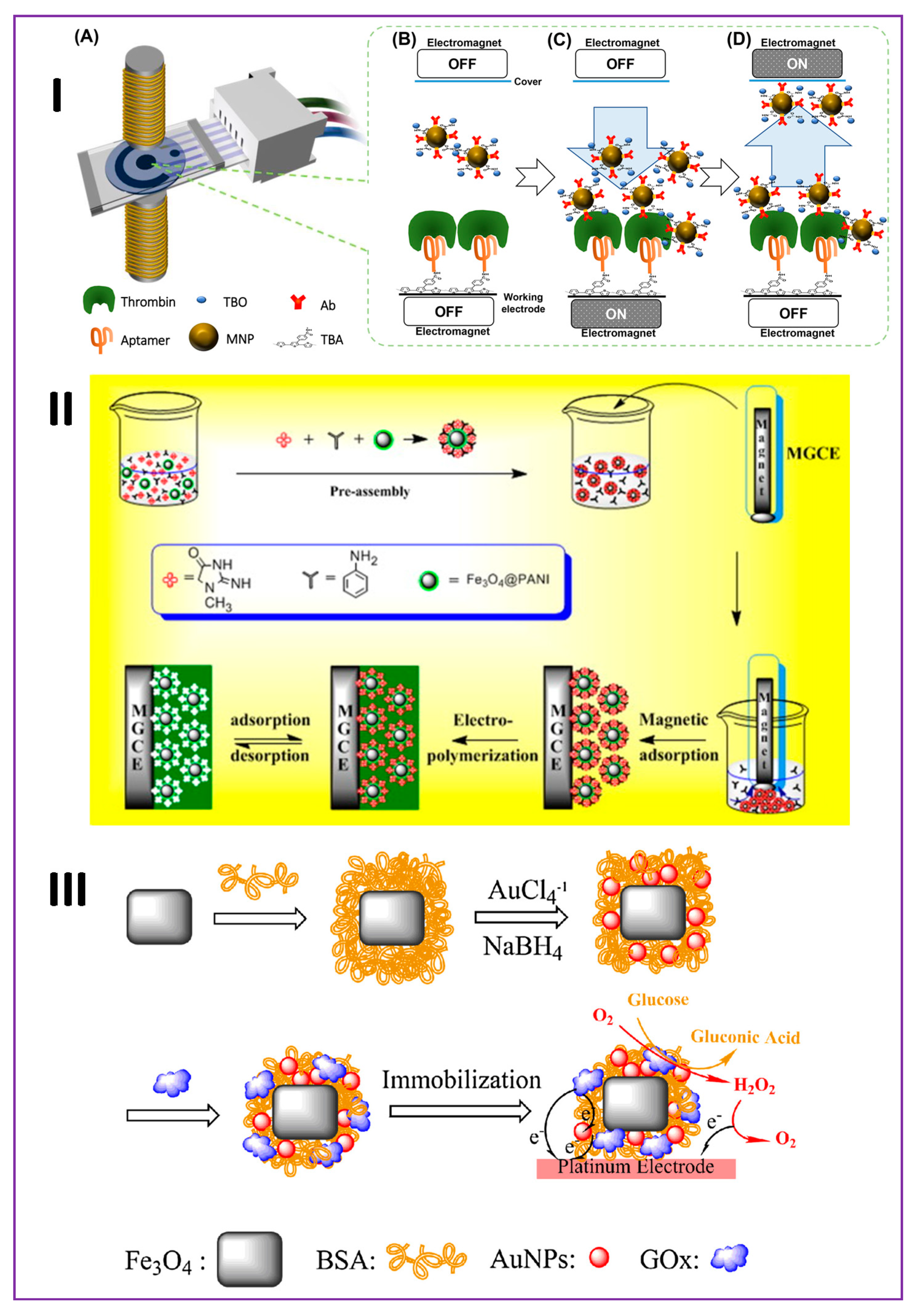
Figure 2.
(I) Schematic representation of the surface-enhanced Raman spectroscopy (SERS)-based sandwich immunosensor for the detection of N-terminal pro-brain natriuretic peptides (NT-proBNPs). Reproduced with permission from Reference [54]. (II) Schematic representation of anterior gradient homolog 2 (AGR2) protein detection procedure. Reproduced with permission from Reference [60]. Copyright 2015 Elsevier. (III) (A) Preparation of MNPs modified with chloramphenicol antibody. (B) Preparation of SERS tags. (C) SERS-based magnetic immunosensor for chloramphenicol. Reproduced with permission from Reference [61]. Copyright 2016 Elsevier. Abbreviations: IRMOF- amino-functionalized metal–organic framework; AuTPs - gold tetrapods; AuNPs - gold nanoparticles; TB - toluidine blue; GNPs - gold nanoparticles; 4,4′-DP - 4,4′-dipyridyl; EDC - 1-Ethyl-3-(3-dimethylaminopropyl)carbodiimide; NHS - N-Hydroxysuccinimide.
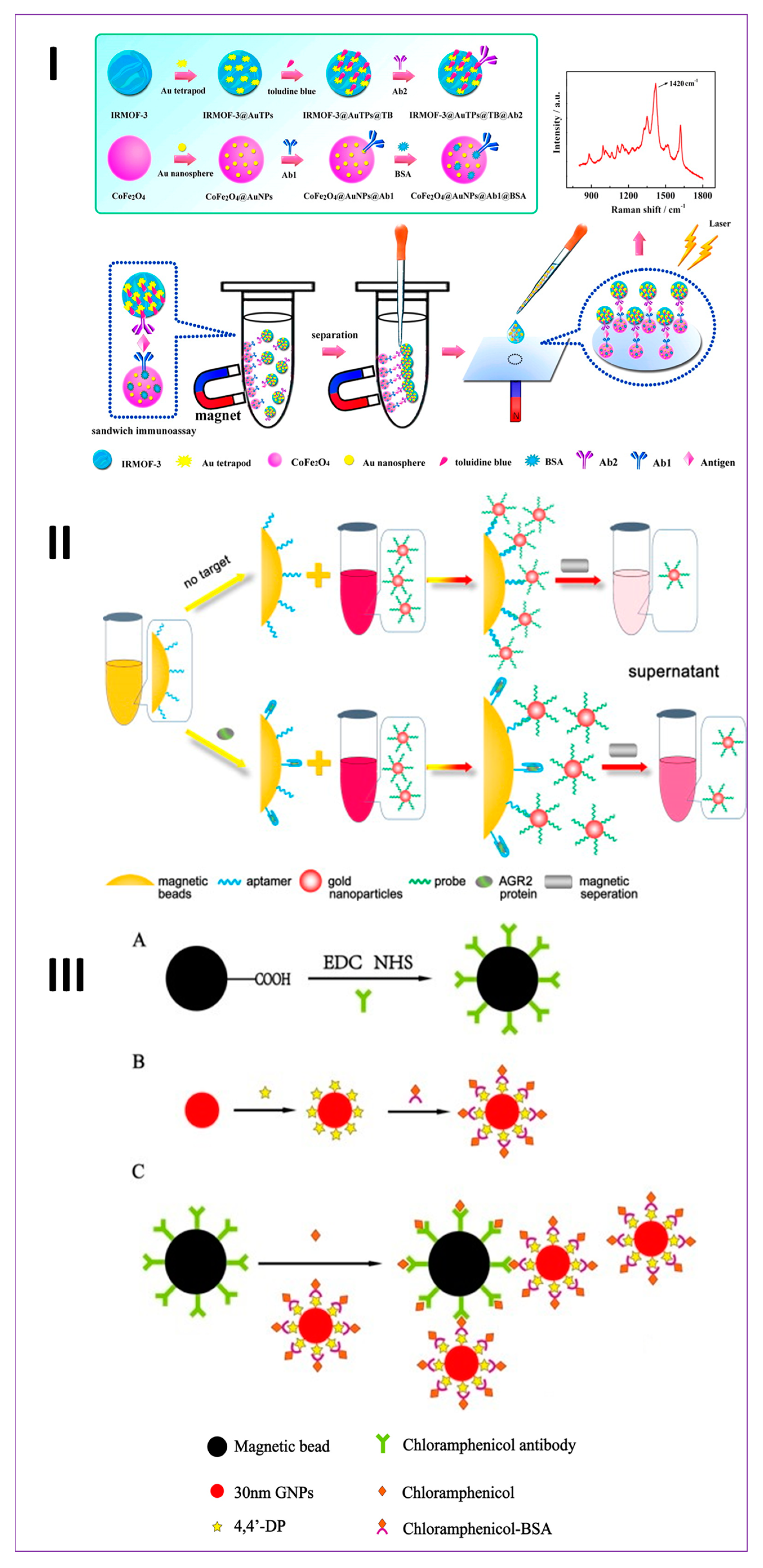
Figure 3.
(A) An electrochemical mass sensor for clinically relevant detection of insulin in human serum conjugated to magnetic nanoparticles and captured onto antibody immobilized gold-coated quartz resonators was reported in this study: (a) Schematic of the electrochemical quartz crystal microbalance (eQCM) sensor and (b) sensor responses (QCM and Faradaic impedance) showing the significant reduction of non-specific signals for control serum due to the magnetic nanoparticle capturing of insulin and detection upon binding onto the surface antibody sensor, enabling picomolar detection limits. This is not possible by doing the assay from a solution form of the serum insulin sample due to the inability of ultra-low picomolar signals to overcome the large background signal response of the un-spiked serum solution control. Reproduced with permission from Reference [63]. Copyright 2014 Royal Society of Chemistry. (B) This report presented the first serum insulin voltammetric immunosensor for diagnosis of type-1 and type-2 diabetic disorders based on fasting serum insulin levels. The sensor was composed of multiwalled carbon nanotube-pyrenebutyric acid frameworks on edge plane pyrolytic graphite electrodes to which anti-insulin antibody was covalently attached. Magnetically captured insulin from 50% serum in buffer was detected upon binding onto the surface insulin-antibody sensor. Reproduced with permission from Reference [64]. Copyright 2015 American Chemical Society. (C) Schematic of a combined covalent and noncovalent carboxylation of carbon nanotubes for sensitivity enhancement of clinical immunosensors (demonstrated here with serum insulin). Reproduced with permission from Reference [65]. Copyright 2016 Royal Society of Chemistry. (D) Schematic of a magnetic optical microarray imager for diagnosing type of diabetes in clinical fasting blood serum samples based on picomolar insulin concentrations. Reproduced with permission from Reference [67]. Copyright 2016 American Chemical Society. (E) Schematic of a magnetite-quantum dot immunoarray for plasmon-coupled fluorescence imaging of blood insulin and glycated hemoglobin. Reproduced with permission from Reference [70]. Copyright 2017 American Chemical Society.
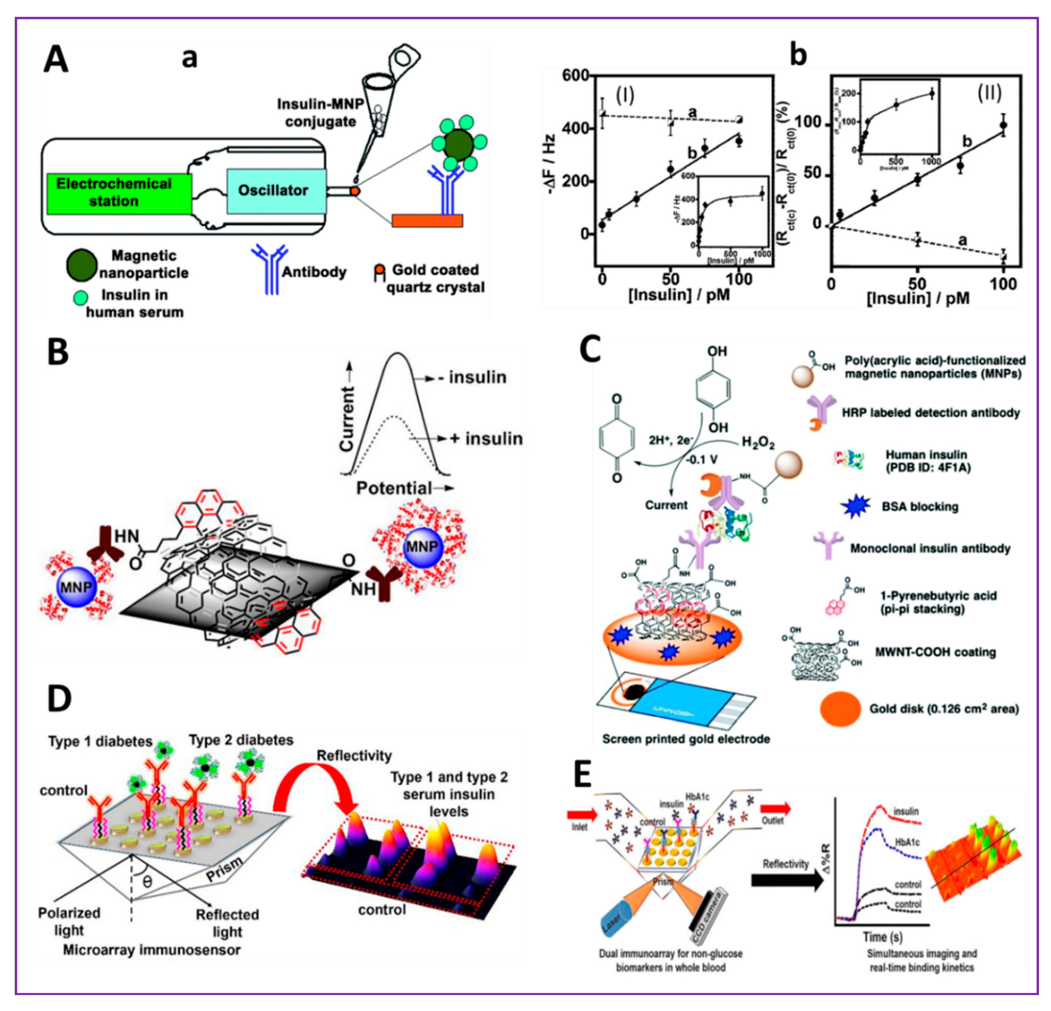
Figure 4.
(A) (a) Schematic depicting the effect of magnetic focus lateral flow biosensor. Without the magnet, the magnetic probe-labeled targets move along with the sample flow on the lateral flow strip, resulting in a low capture efficiency (left). With magnet, the probe-labeled targets are focused at the signal generation zone due to the magnetic focus thus increasing the capture efficiency of labeled targets (right). (b) Comparison of the detection results with and without magnet. Reproduced with permission from Reference [53]. Copyright 2019 American Chemical Society. (B) Sandwich immunoassay for t-PSA: on the left—sensograms demonstrating all assay steps in absence of t-PSA (lower blue curve) and in the presence of 1 µg/ml t-PSA (upper red curve); on the right—scheme of sandwich assay: 1—capture antibody, 2—antigen, 3—biotinylated tracer antibody, 4—magnetic nanoparticle coated by streptavidin. Reproduced with permission from Reference [86]. Copyright 2016 Elsevier. (C) Schematic diagram of upconversion nanoparticles (UCNPs) luminescence-based aptasensor for the selective detection of ENR. Reproduced with permission from Reference [90]. Copyright 2016 Elsevier. Abbreviations: TMB - Tetramethyl benzidine; HRP - Horseradish peroxidase; UCNPs - Upconversion nanoparticles; ENR – Enrofloxacin.
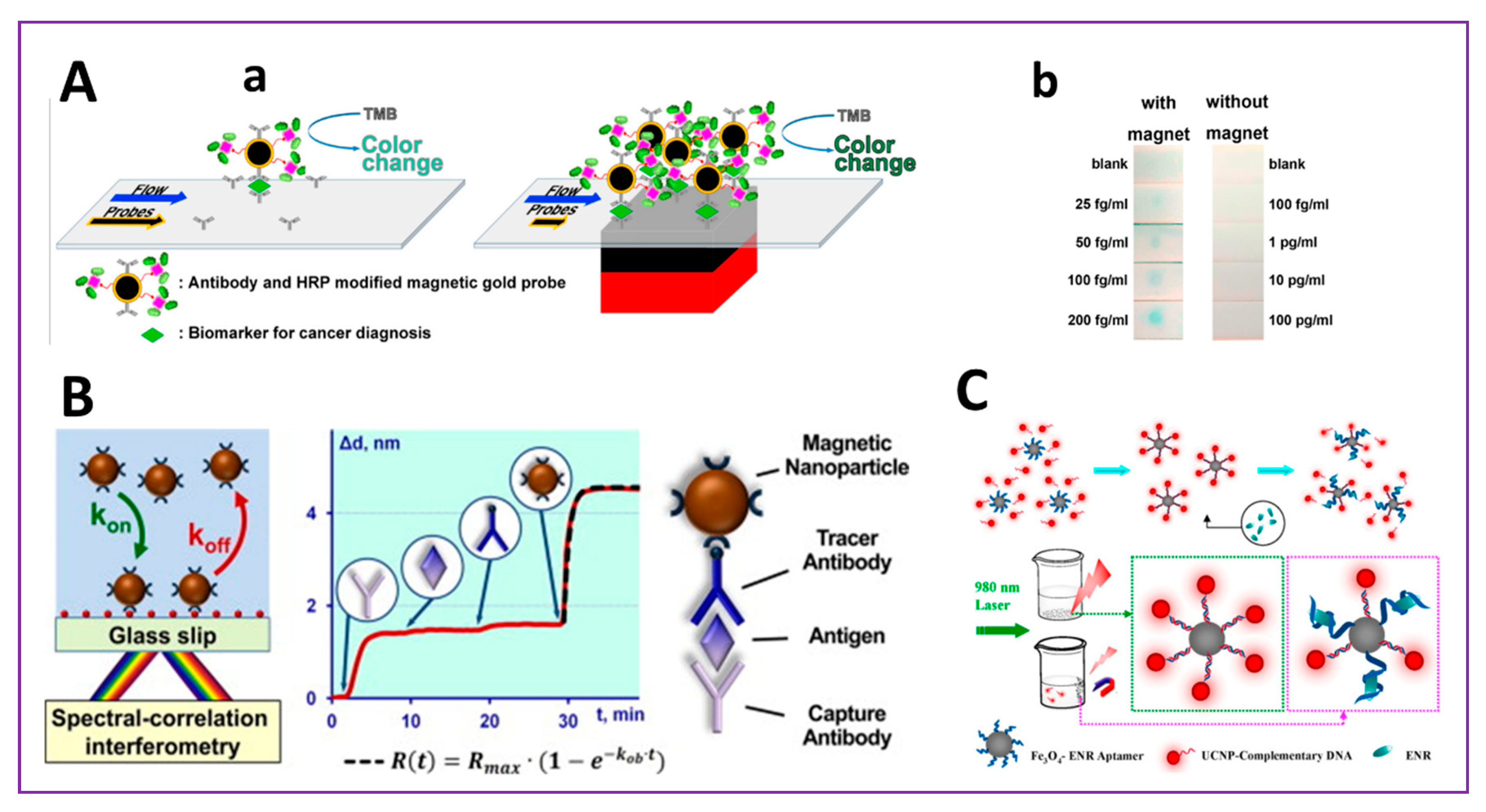
Figure 5.
(A) (a) Electrochemical and surface plasmon correlation of serum autoantibody immunosensors with binding insights. In this report, a comparative account of carboxylated graphenyl and mercapto-monolayer surfaces was made for the detection of serum glutamic acid decarboxylase-65 autoantibody (GADA), a biomarker of type 1 diabetes and (b) schematic representation. Reproduced with permission from Reference [95]. Copyright 2018 American Chemical Society. (B) Schematic representation of a multiplexed surface plasmon imaging of serum biomolecules. In this report, Fe3O4@Au core/shell nanoparticles carrying analyte-specific second biorecognition element (the first one was immobilized on the sensor surface) were used for signal amplification purpose and presented with plasmonic simulation insights. Reproduced with permission from Reference [96]. Copyright 2019 Elsevier. (C) Schematic of a biocatalytic system constructed by electrostatically immobilizing drug-metabolizing human liver microsomes (HLMs, negatively charged due to the phospholipids) onto positively charged amine-functionalized magnetic nanoparticles (100 nm hydrodynamic diameter). This report presented electrochemical biosensing of cytochrome P450 (CYP)-specific drug candidates and electrocatalyzing drug conversion into metabolites with electron mediation in the biofilm by cytochrome P450-reductase (CPR). Reproduced with permission from Reference [104]. Copyright 2018 Elsevier.
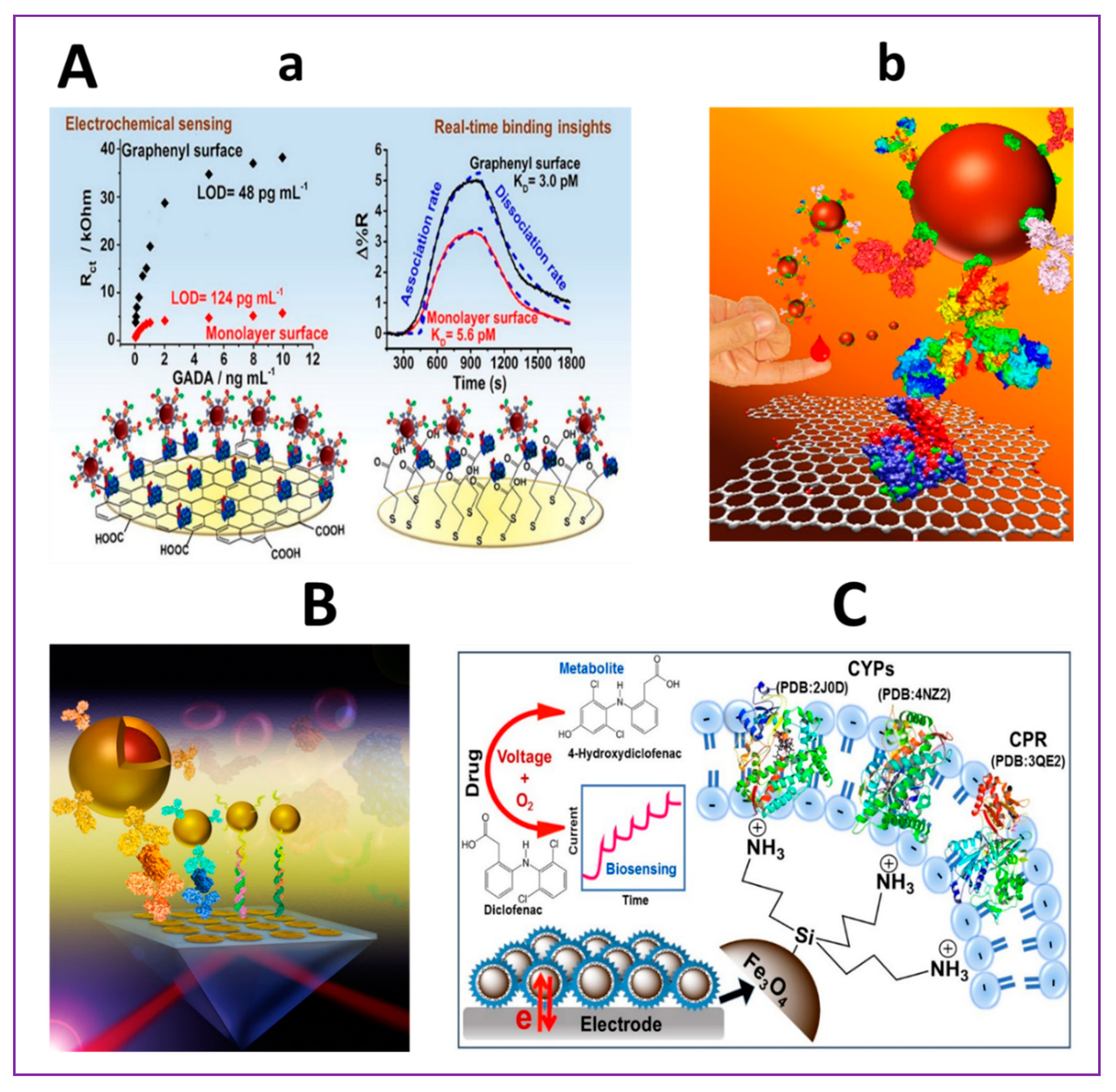
Figure 6.
Schematic display of the steps involved in the preparation and performance of the biosensor for Brett (Ab-B sensor). Reproduced with permission from Reference [110]. Copyright 2019 Elsevier. Abbreviations: ConA - Concanavalin A.
Figure 6.
Schematic display of the steps involved in the preparation and performance of the biosensor for Brett (Ab-B sensor). Reproduced with permission from Reference [110]. Copyright 2019 Elsevier. Abbreviations: ConA - Concanavalin A.

Table 1.
Magnetic nanoparticle (MNP)-based electrochemical sensors for the detection of various analytes.
Table 1.
Magnetic nanoparticle (MNP)-based electrochemical sensors for the detection of various analytes.
| S. No. | Application | Transduction Method | Recognition Element | Analyte | Interface | Real Sample | Range | LOD | Reference |
|---|---|---|---|---|---|---|---|---|---|
| 1 | Medical | Voltammetry | Molecularly Imprinted Polymers (MIPs) | Hemoglobin | GCE1/Fe3O4@SiO2/MMIP | Blood | 0.005–0.1 mg mL−1 | 0.001 mg mL−1 | [71] |
| 2 | Environmental | Differential Pulse Voltammetry (DPV) | MIP | Bisphenol A | SPCE2/AuNPs/CBNPs5/Fe3O4/MMIP3 | Mineral water | 0.07–10 μM | 8.8 nM | [72] |
| 3 | Medical | DPV | Chemical | Ciprofloxacin | CPE/Fe3O4/CMNPs | Serum, Urine | 0.05–75 µM L−1 | 0.01 µM L−1 | [73] |
| 4 | Food Clinical | DPV | MIP | N-Acyl-homoserine-lactones | MGCE5/Fe3O4@SiO2-MIP | 2.5 × 10⁻⁹–1.0 × 10⁻⁷ mol L−1 | 8 × 10⁻¹⁰ mol L−1 | [74] | |
| 5 | Food | DPV | MIP | Kanamycin | CE4/MWCNTs/Fe3O4/PMMA | Milk, Chicken, Pig | 1 × 10⁻¹⁰–1.0 × 10⁻⁶ mol L−1 | 2.3 × 10⁻¹¹ mol L−1 | [75] |
| 6 | Food | Cyclic Voltammetry (CV) | Enzyme | Peroxide | Pt/MRGO6/chit/HRP | Orange juice | 20–1000 μM 48.08 μA μM−1· cm−2 | 2 μM | [76] |
| 7 | Security | DPV | Morphine | CPE/CHT7/Fe3O4 | Serum, Urine | 10–2000 nM | 3 nM | [77] | |
| 8 | Food | DPV | Quercetin and Tryptophan | CPE/Fe3O4@NiO core/shell nanoparticles | Human breast milk, cow milk, and honey | 0.08–60 μM 0.1–120 μM | 2.18 nM 14.23 nM | [78] | |
| 9 | Medical | DPV | Antibody (Ab) | Prostate specific antigen (PSA) | Ab2/MB8/Au@Fe3O4@COF/GCE | Serum | 0.0001–10 ng mL−1 | 30 fg mL−1 | [79] |
| 10 | Medical | Amperometry | Enzyme | Glucose | GOx14/AuNPs/BSA/Fe3O4/PtE | - | 0.25–7.0 mM | 3.54 μM | [59] |
| 11 | Medical | DPV | Ractopamine | MSPE/RGO/Fe3O4 | Pork meat | 0.05–100 μM | 13 nM | [80] | |
| 12 | Medical | Amperometry | Aptamer-Antibody | Thrombin | pTBA/Apt/thrombin/MNP@Ab-TBO9/SPCE | Serum | 1–500 nM | 0.49 | [57] |
| 13 | Security | Square Wave Voltammetry (SWV) | Organo-phosphates | Fe3O4@ZrO2/MGCE | - | 7.60 × 10⁻⁸–9.12 × 10⁻⁵ M | 1.52 × 10⁻⁸ M | [81] | |
| 14 | Medical | DPV | MIP | Creatinine | MGCE/Fe3O4@PANI10 NPs/MIP | Plasma, Urine | 0.02–1 µM L−1 | 0.35 nM L−1 | [58] |
| 15 | Medical | DPV | Progesterone | Fe3O4@GQD11/f–MWCNTs12/GCE | Serum | 0.01–3.0 μM | 2.18 nM | [82] | |
| 16 | Medical | Amperometry | PSA, Prostate specific membrane antigen (PSMA) Cancer biomarker | Fe3O4@GO/Ab/PSMA | 61 fg mL−1–3.9 pg mL−1 9.8 fg mL−1–10 pg mL−1 | 15 fg mL−1 4.8 fg mL−1 | [83] | ||
| 17 | Medical | DPV | PSA, PSMA, IL6, Platelet factor 4 (PF4) | MNP/HRP13-Ab | 0.05–2 pg mL−1 | [84] |
GCE1: Glassy carbon electrode; SPCE2: screen-printed carbon electrode; MMIPs3: magnetic molecularly imprinted polymers; CE4: carbon electrode; CBNPs4: carbon black nanoparticles; MGCE5: magnetic glassy carbon electrode; MRGO6: magnetic reduced graphene oxide; CHT7: chitosan; MB8: methylene blue; TBO9: toluidine blue O; PANI10: polyaniline; GQD11: graphene quantum dots; MWCNTs12: multiwalled carbon nanotubes; HRP13: horseradish peroxidase; GOx: glucose oxidase.
Table 2.
Magnetic nanoparticle (MNP)-based optical sensors for the detection of various analytes.
| S.No. | Transduction Method | Recognition Element | Analyte | Assay | Range | LOD | Reference |
|---|---|---|---|---|---|---|---|
| 1 | Ultraviolet-Visible (UV-Vis) | Aptamer | Cancer biomarker AGR21 | Au NPs/DNA/MBs | 10–1280 pM | 6.6 pM | [60] |
| 2 | Surface Plasmon Resonance (SPR) | Aptamer | Thrombin | Fe3O4@Au NPs/Apt | 0.1–100 nM | 0.1 nM | [112] |
| 3 | Colorimetric | Ab | Listeria | MNB-MAb-Listeria-PAb-AuNP-urease | 1.1 × 10²–1.1 × 10⁶ CFU mL−1 | 102 CFU mL−1 | [113] |
| 4 | Colorimetric | Peptide | E coli. | MNP2/Peptide/AuNPs/SAM | 30–300 CFU mL−1 | 12 CFU mL−1 | [114] |
| 5 | Photonic crystal | Ab | soluble transferrin receptor | fAb-IONs3 | 0.01–0.2 µg mL−1 | - | [115] |
| 6 | Surface Enhanced Raman Spectroscopy (SERS) | Ab | chloramphenicol | AuNPs/MNPs/Ab | 0–10 ng mL−1 | 1 pg mL−1 | [61] |
| 7 | Chemiluminescence | MIP | Lysozyme | ILs3-Fe3O4@DA/GO6/β-CD4 | 1.0 × 10⁻⁹–8.0 × 10⁻⁸ mg mL−1 | 3.0 × 10⁻¹⁰ mg mL−1 | [116] |
| 8 | Chemiluminescence | Enzyme-Ab | Thyroid stimulant hormone | Pt NPs/HRP-Ab/magnetic beads | 0.013–12 mU L−1 | 0.005 mU L−1 | [94] |
| 9 | SERS | MiRNA let-7b (cancer cells) | Fe3O4@Ag NPs | 0–1000 pM | 0.3 fM | [107] | |
| 10 | SERS | Ab | Ovarian cancer Multiplexed (CA-125, β2-M and ApoA1) | MNPs/Abs | 0.26 U mL−1, 0.55 ng mL−1, and 7.7 ng mL−1 | [102] | |
| 11 | SERS | Ab | N-Terminal pro-brain natriuretic peptide (heart failure) | CoFe2O4@AuNPs/MOFs | 1 fg mL−1–1 ng mL−1 | 0.75 fg mL−1 | [54] |
| 12 | SERS | Carcinoembryonic antigen | MBA-labeled NiFe@Au NPs | 0–1 ng mL−1 | 0.1 pM | [56] | |
| 13 | SERS | Fe3O4@Au Core/shell nanoparticles | [96] |
AGR21: anterior gradient homolog 2; MNP2: magnetic nanoparticles; IONs3: iron oxide nanoparticles; Ils3: ionic liquids; β-CD4: β-cyclodextrins5; GO6: graphene oxide.
© 2019 by the authors. Licensee MDPI, Basel, Switzerland. This article is an open access article distributed under the terms and conditions of the Creative Commons Attribution (CC BY) license (http://creativecommons.org/licenses/by/4.0/).
Share and Cite
MDPI and ACS Style
Krishnan, S.; Goud, K.Y. Magnetic Particle Bioconjugates: A Versatile Sensor Approach. Magnetochemistry 2019, 5, 64. https://doi.org/10.3390/magnetochemistry5040064
AMA Style
Krishnan S, Goud KY. Magnetic Particle Bioconjugates: A Versatile Sensor Approach. Magnetochemistry. 2019; 5(4):64. https://doi.org/10.3390/magnetochemistry5040064
Chicago/Turabian StyleKrishnan, Sadagopan, and K. Yugender Goud. 2019. "Magnetic Particle Bioconjugates: A Versatile Sensor Approach" Magnetochemistry 5, no. 4: 64. https://doi.org/10.3390/magnetochemistry5040064
Note that from the first issue of 2016, this journal uses article numbers instead of page numbers. See further details here.