Review

Magnetic Particles-Based Analytical Platforms for Food Safety Monitoring

1
Department of Chemistry and Biomolecular Science, Clarkson University, Potsdam, New York, NY 13699, USA
2
Interdisciplinary Research Centre in Biomedical Materials (IRCBM), COMSATS University Islamabad (CUI), Lahore Campus, Lahore 54000, Pakistan
*
Authors to whom correspondence should be addressed.
Magnetochemistry 2019, 5(4), 63; https://doi.org/10.3390/magnetochemistry5040063
Submission received: 29 September 2019 / Revised: 13 November 2019 / Accepted: 14 November 2019 / Published: 18 November 2019
(This article belongs to the Special Issue Magnetic Nanoparticles)

Abstract

:
Magnetic nanoparticles (MNPs) have attracted growing interest as versatile materials for the development of analytical detection and separation platforms for food safety monitoring. This review discusses recent advances in the synthesis, functionalization and applications of MNPs in bioanalysis. A special emphasis is given to the use of MNPs as an immobilization support for biomolecules and as a target capture and pre-concentration to increase selectivity and sensitivity of analytical platforms for the monitoring of food contaminants. General principles and examples of MNP-based platforms for separation, amplification and detection of analytes of interest in food, including organic and inorganic constituents are discussed.

1. Introduction

Nanotechnology provides a broad range of opportunities for designing nanomaterials with unique physicochemical characteristics offering new and unique capabilities for designing analytical platforms for detection, separation and tracking of food constituents. Magnetic nanoparticles (MNPs) with sizes ranging from 10–100 nm consisting of magnetic elements such as iron, manganese, chromium, gadolinium, cobalt, nickel, and their alloys have found a broad range of applications in sensors, diagnostics, biomedical devices and in the food sector [1]. The reduction of material dimensions form macro to nanoscale changes their optical, electrical and magnetic characteristics. MNPs of sizes typically below 30 nm exhibit superparamagnetic properties with zero coercivity and hysteresis. The application of an external magnetic field causes magnetization of MNPs. When MNPs are functionalized with specific biorecognition molecules, for example, antibodies (Ab), application of a magnetic field enables facile separation and pre-concentration of target analytes from complex matrices such as food. Therefore, the MNPs’ location and motion can be controlled by an externally applied magnetic field and visualized by using magnetic resonance imaging (MRI). The toxicity and biocompatibility of MNPs is dependent on the nature, particle size, coating and the core material of the magnetically responsive component (Fe, Mg, Mn, Ni, Co).
The most commonly employed MNPs are iron oxides such as magnetite (Fe3O4) and its oxidized form, maghemite (γ-Fe2O3). These particles have been explored for biomedical and food applications owing to their biocompatibility, non-immunogenic and non-toxic characteristics and the ability to prepare them in small particle sizes [2]. Ferrous oxide NPs are the only United States Fodo and Drug Administration (US FDA) approved MNPs that retain zero magnetism after removal of an external magnetic field [1]. MNPs based on other materials, such as cobalt and nickel, despite a high magnetism have received little interest due to their toxicity and tendency to rapidly oxidize at the surface [3]. The magnetic behavior of individual NPs arise from their narrow, finite-size and surface effects, which all affect their properties [4]. MNPs can be used in bare form, surface coated or functionalized with molecular recognition sites for specific binding to enable applications in bio-separation, biosensing, information storage, catalysis and diagnostic imaging [1]. The physicochemical characteristics of NPs depend on the size, surface area, effective sedimentation rate and dispersity [1,5].
In the food sector, MNPs can be used as colorants and sources of bioavailable iron. MNPs modified with immunorecognition reagents have been used to improve detection of food borne pathogens [6]. MNPs are commonly used in magnetic solid-phase extraction for the recovery of analytes in the preparation of biological, environmental and food samples [7]. The use of MNPs can simplify the extraction process and increase sensitivity of measurements by providing selective isolation and enrichment of analytes using an external magnetic field. In this article, we discuss the synthesis, structure, properties and applications of MNPs in the development of analytical assays and sensors for food monitoring and highlight the advantages and limitations of MNPs for sample pre-treatment and pre-concentration in the food sector. Several examples of magneto-switchable devices with controlled sensing and actuation properties are provided.

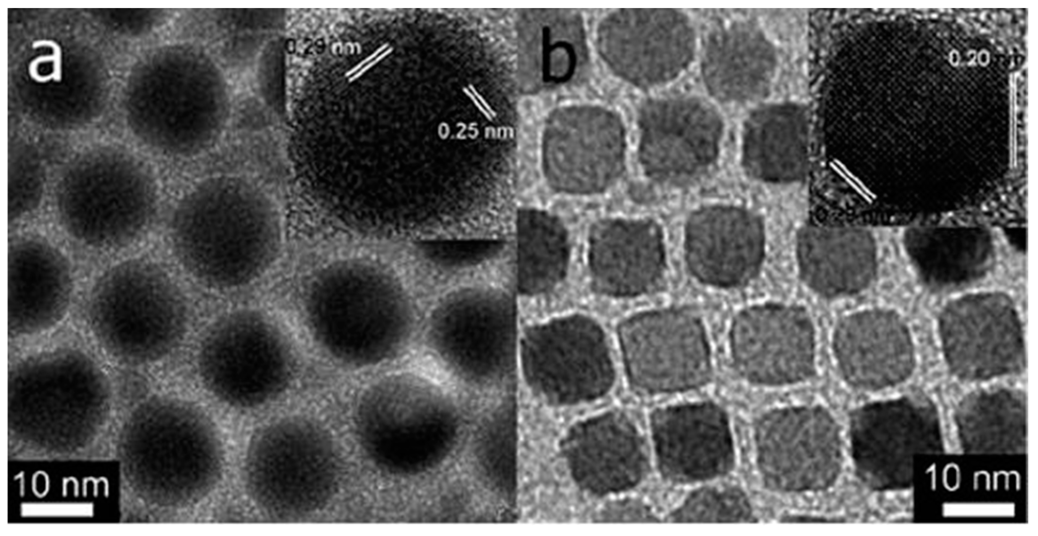
2. Synthesis of MNPs

Various forms and sizes of MNPs are available and can be purchased from several companies or they can be prepared using established procedures [8,9,10]. Methods to synthesize MNPs (iron oxide, metal, metal alloys, magnetic composite materials) include biomineralization, physical and chemical methods [11,12,13]. Biomineralization involves the use of living organisms to prepare magnetic particles. MNPs fabricated by biomineralization are prepared as natural magnetosomes (nanosized magnetic iron oxide crystals coated with protein) involving magnetotactic bacteria. Magnetosomes of 20 to 45 nm have been synthesized in laboratory under simulated environment of bacterial anaerobic habitat. There is significant research focusing on the development of chemical methods which emulate the biomineralization process to synthesize MNPs [14,15,16].
Physical methods for MNPs synthesis can be either “top down” or “bottom up” procedures. Top down methods employ the size reduction of coarse macroscopic magnetic materials to the nanometer range by milling. The main limitation of this method is the difficulty to control the particle size and shape [15]. Bottom up techniques rely on condensation of NPs from their liquid or gaseous phase. An example if the laser evaporation technique employed for the synthesis of MNPs in which coarse particles of metal oxides are temperature treated to evaporate the solvent, followed by condensation and nucleation resulting in synthesis of 20–50 nm particles [15,17,18].
Wet chemical methods for the preparation of MNPs can be distinguished as (a) high-temperature thermal decomposition and/or reduction, (b) co-precipitation and (c) template synthesis in the interior of micelles [1]. Other methods are template-directed and can involve micro emulsion, thermal decomposition, solvo-thermal, solid state, spray pyrolysis, self-assembly, lithography, sono-chemical, microwave assisted, carbon arc techniques or glycothermal synthesis, among others [19,20]. The most common chemical method employed for MNPs synthesis involve co-precipitation and nucleation of particles from ions in solution. During the co-precipitation process, a super saturation stage followed by a burst nucleation occurs with formation of nuclei which gradually grow in size and cause the monodisperse particles to diffuse from solution into solute. The co-precipitation principle to achieve monodisperse particles is represented by the La Mer-Dinegar model [18] of homogeneous precipitation shown in Figure 1. In general, the co-precipitation method requires low temperatures and metal salts and the properties of MNPs can be tailored by controlling the reaction pH, temperature, stirring speed and metal ion concentration.
MNPs synthesis can also be achieved by thermal decomposition of the chemical structure of organometallic compounds at elevated temperatures by cleavage of chemical bonds. This method is mostly employed for organometallic compounds (acetyl acetonates) in organic solvents (benzyl ether, carbonyls and Ethylene diamine) with surface active agent such as oleic acid, oleyl amine, polyvinyl pyrrolidone (PVP), cetyltrimethyl ammonium bromide (CTAB) and hexadecyl amine. Morphology and size of MNPs can be controlled by varying the precursor composition (Figure 2) [21]. The thermal decomposition method is used to synthesize MNPs with good crystallinity, controlled morphology and size distribution (4–45 nm) [22,23]. For example thermal decomposition of FeCup3 (Cup: N-nitroso phenyl hydroxyl amine) at elevated temperatures of 250 °C–300 °C allowed fabrication of maghemite nanocrystals with a size range of 3–9 nm. The method can also be used to synthesize MNPs of transition metals (Co, Ni and Fe) [24]. To obtain the 3D MNPs, hot solutions of metal precursor and surfactant is mixed with a reducing agent. The morphology of the MNPs shown in Figure 2 was controlled by controlling the decomposition time. A shorter duration of (2–4 h) resulted in spherical particles while longer decomposition time (10–12 h) resulted in cubic MNPs [21,24].
Excellent control of particle size and shape can be achieved by hydrothermal synthesis. This procedure involves the synthesis of MNPs at high vapor pressure and high boiling point. During this reaction, phase separation occurs at the solid-liquid interface resulting in monodisperse Fe3O4 and MFe2O4 nanocrystals [25]. This synthesis method has been used to fabricate a variety of metal, rare earth, fluorescent, polymeric and magnetic metallic nanocrystals at different reaction conditions [26,27,28]. High MNPs yield with short reaction times was achieved bv microwave radiation. Wang et al. [6] reported the synthesis of a highly crystalline cubical spinel MIIFe2O4 (M = Co, Mn, Ni) structure by exposing the precursors to microwave radiation for 10 min. The fabrication of magnetite (Fe3O4) and hematite (α-Fe2O3) particle by microwave radiation involved the use of FeCl3, polyethylene glycol and N2H4.H2O precursors. The final condition of Fe3O4 was controlled by controlling the quantity of N2H4.H2O [6]. Another method to prepare MNPs involves a multistep template assisted fabrication which requires the use of a base template tailed by subsequent deposition of a magnetic material. MNPs nuclei grow at the hole and defects of the template resulting in MNPs of a morphology tailored to the template. The physical properties (size, morphology) of MNPs can be controlled by the selection of a base template of desired properties. This technique allows fabricating MNPs of complex structure (nanotubes, nanorods, nanocubes, hexagonal and octahedrons) with controlled size and morphology [29].

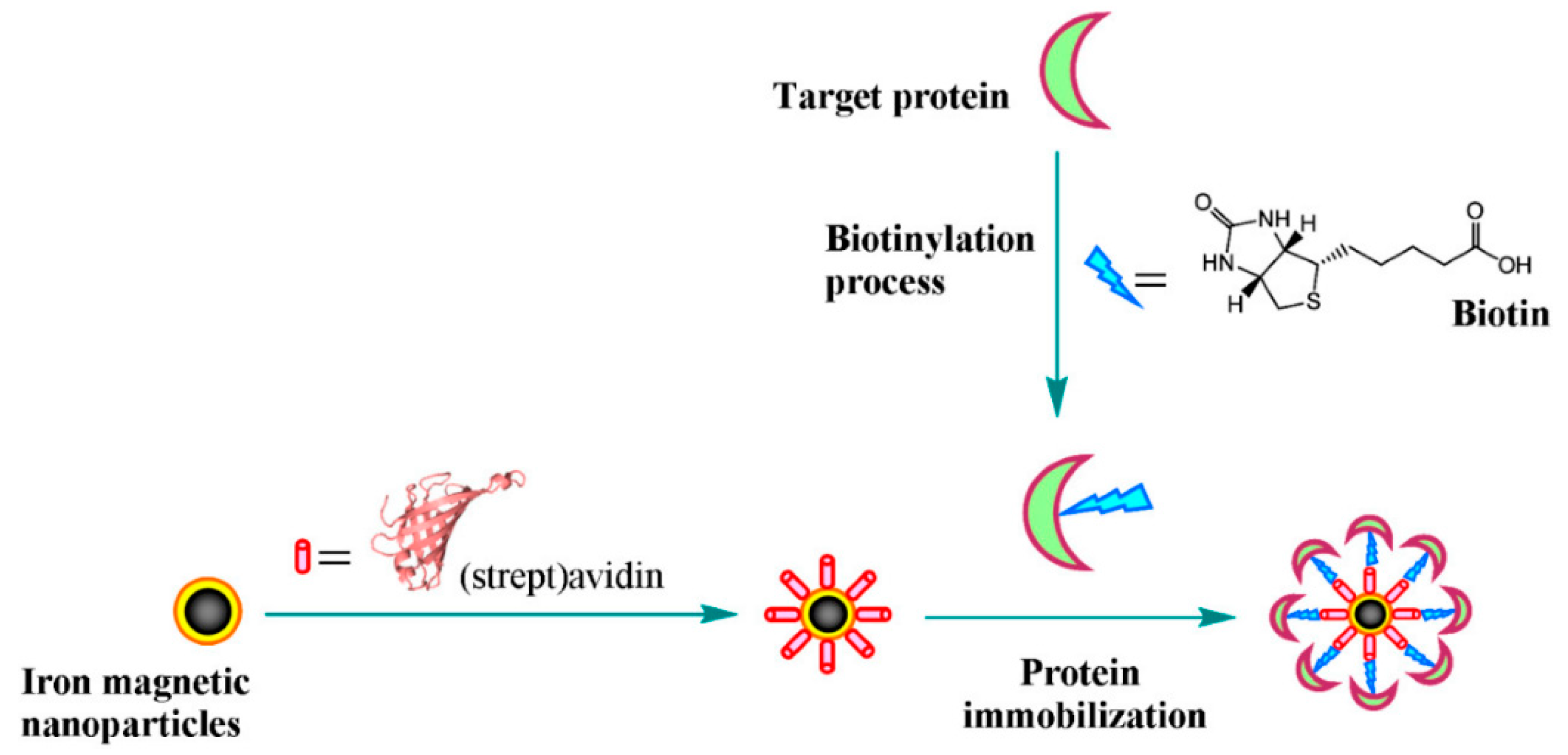
3. Surface Modification of MNPs

To enhance solubility, stability and biocompatibility and achieve target specificity, MNPs are generally modified with surface ligands. MNPs which are hydrophobic in nature can be functionalized by ligand exchange [25,27] or encapsulated within a phospholipid bilayer to make them hydrophilic [6,28]. Other methods involve encapsulation within materials that have affinity for the iron oxide core such as gold [30,31], SiO2 [31,32] or carbon [33] which enhances their surface properties while maintaining the magnetic functions for applications [19].
Modification of super paramagnetic NPs (Fe2O3, 13.5 nm in diameter) with lactoferrin or ceruloplasmin protein enabled stabilization of MNPs at room temperature and prevented non-specific adsorption. To achieve target specificity through the attachment of antibodies commercial tosylated polystyrene MNPs (Fe2O3) were coated with a gold layer. Other methods involve coating with polymers, silica [34] or organic layers [17]. MNPs modified by charged layers facilitated adsorption of biomolecules by electrostatic interactions. Matsunaga et al. [35] devised a technique for electrostatic-based DNA extraction in which negatively charged DNA is extracted by electrostatic interaction of positively charged NH2-MNPs. MNPs coated with a dense amino layer using an aminosilane reagent, 3-[2-2-aminoethy(amino)-ethylamino-propyetrimethoxysilane] in toluene solution enabled fabrication of NH2-functionalizaed MNPs. [35]. Polyamidoamine dendrimer remodified [36] and aminosilane-coated MNPs [37] were also used in adsorption-based DNA extraction techniques. For such applications, the MNPs surface was chemically modified with carboxyl and amino groups that were further used to covalently immobilize biomolecules. In addition to surface stabilizers, the coatings can include fluorescent labels [38] such that the particles can be manipulated through the use of an external magnetic field and simultaneously visualizing their position through fluorescence methods. Surface modification can also be achieved through physical means via encapsulation and sorption [39]. Various natural (gelatin, dextran, starch chitosan) and synthetic polymers (PVA, PEG, PMMA, PLA, PANI etc.) have been used to functionalize the surface of MNPs to prevent the agglomeration and enhance the selectivity for specific target. [39].

4. Applications of MNPs in Bioanalysis

4.1. Enzyme Immobilization Support

Owing to their biocomptability and magnetic properties, MNPs have various applications in the food industry as enzyme immbolization support and separator for protein purification. Modification of the MNPs with bioactive molecules tipically involves surface modification with hydroxyl -OH, carboxyl –COOH and amino –NH2 functional groups [40]. Most food applications invloves catalytic enzymes such as, that is, carbohydrase, protease, lipase, lysozymes and oxidoreductase. Free enzymes are unstable to changes in pH, temperature and ionic solutions and are difficult to recycle if they are used in solution. Their stability and reuseability can be improved by immobilization [41] on various supports throug organic and inorganic linkers [42,43,44,45]. MNPsprovide a large surface area with a high enzyme loading capability. Enzyme immobilized on MNPs offers lower diffusion coefficient in solution [46]. Immobilized enzyme @ MNPs can be collected with ease and at low cost from enzyme complex mixture with the aid of external magnetic field [47,48].
Enymes immobilization on MNPs can be carried out by physical adsorption and covalent bonding. Covalent bonding provides strong enzyme adherence to MNPs surface but limits the enzyme activity, as this immobilization involves strong covalent interaction between the surface of MNPs and the functional groups of the enzymes [49]. The physical adsorption method is based on simple enzyme adhesion through weak ionic, H-bonding and van der Waals forces between the surface-modified MNPs and enzymes. Physically-immobilized enzymes on MNPs are more succeptable to changes in temperature, pH and ionic strength [49]. Immobilization though (strept)avidin–biotin affinity bonds can also be used for protein immoblization (Figure 3). The high specificity and binding affinity (Kd ≈ 10−15 M) of (strept)avidin and biotin makes this approach appealing for protein immobilization applications [50].
In order to enable protein binding on MNPs, the particles are first functionalized with amino or carboxyl functional groups. An example is the use of organic (lauric acid C12H24O2) and inorganic (silicone dioxide SiO2) materials. Covalent cross-linking involves coupling agents (e.g., glutaraldehyde) which provide the aldehyde group for covalent interaction between MNPs and enzymes. Enzymes immobilized via covalent bonding are more stable than those immobilized by physical adsorption [51,52]. However, covalent bonding can cause conformational changes and possible alteration of the enzyme active site reducing the activity of the immobilized enzyme. It was reported that when the concentration of the coupling agent was <0.4% the effect on the recovered enzyme activity is minimal. However greater concentrations (>4%) were found to reduce the enzyme activity due to confrmational changes induced by the interaction with the coupling agent for example, (glutraldehyde) [53,54]. Huang et al. [55] reported a simple method for the immobilization of lipase enzyme via direct bounding to MNPs by using 1-ethyl-3-(3-dimethylaminopropyl) carbodiimide hydrochloride (EDC), which enhanced its specific activity. It was reported that the specific activity of immobilized lipase was enhanced up to 1.41 times than the free form [55]. In a similar way, Liu et al. [56] immobilized lipase on lauric acid stabilized-MNPs by using EDC as a coupling agent; the specific activity of the immobilized lipase was 1.8 times higher than that of the free enzyme. A number of studies concluded that enzymes activity can be enhanced by immobilization on functionalized MNPs [56].

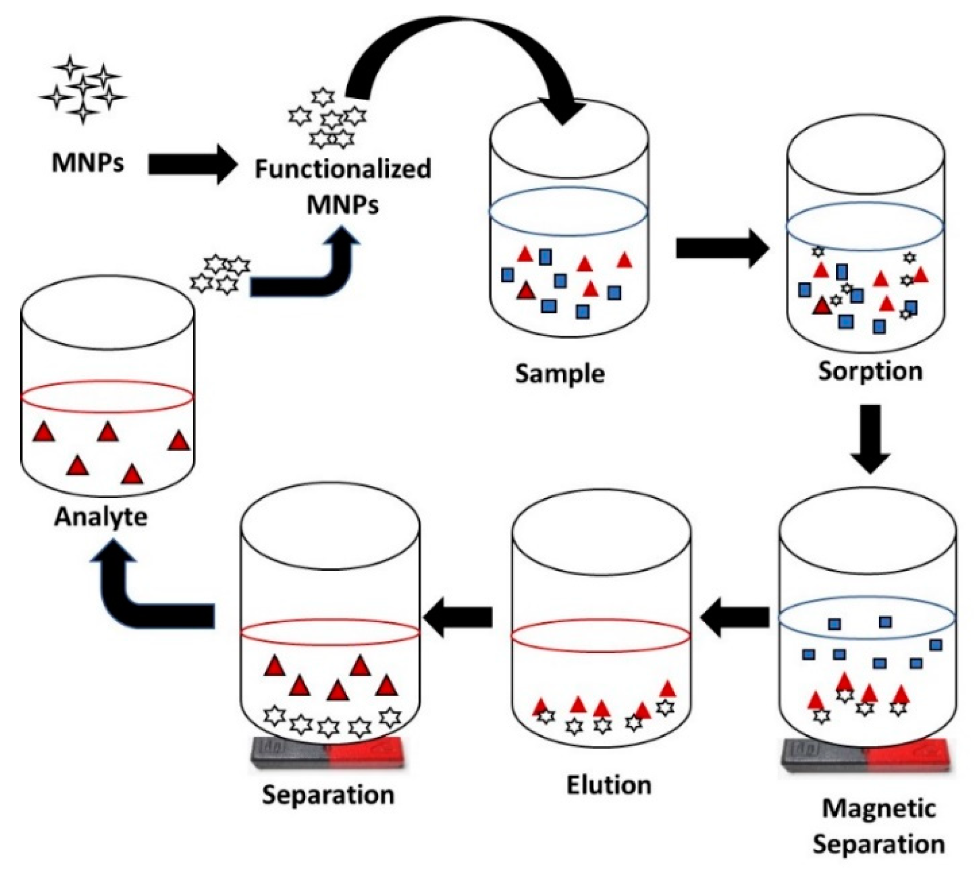
4.2. MNPs as Pre-Concentration and Capture Probe

Sample preparation is a critical step in almost all analytical procedures and directly influences the accuracy of measurements. Inappropriate sample preparation can cause contamination, loss of analyte, erroneous compositional data and so forth, particularly in environmental, biological or food samples in which the analytes are present in trace or ultra-trace levels or when samples are complex. Therefore, isolation or pre-concentration of analyte before analysis is a necessary step for achieving accurate qualitative and quantitative analysis. Isolation of an analyte from the primary matrix is meant to reduce the effect of interfering species but the conventional separation methods are high cost, time consuming and involve laborious procedures.
Separation of analytes from food samples can be achieved by solid phase extraction (SPE) that provides rapid enrichment of the analyte using an economical and simple procedure. MNPs represent an alternative approach and can be used to concentrate and then magnetically separate a large amount of analytes by virtue of their large surface area and magnetic properties [57]. Integration of MNPs in conventional SPE was the basis for the development of an advanced, automated and time saving extraction technique [58]. In this combined use, called magnetic-dispersive solid phase extraction (M-DSPE), the MNPs served as a sorbent in SPE. Contrary to conventional SPE where the sorbent is always packed into cartridges, the M-DSPE utilize MNPs that are directly placed into the sample which after extraction are isolated by applying an external magnetic field. Such magnetic isolation of MNPs after analyte adsorption eliminates post centrifugation or filtration processes. The analyte-sorbent interaction mainly depends on the surface chemistry of the MNPs and the tailoring of their properties to selectively interact with the analyte of interest. In general, the interaction between the sorbent and the analyte are polarity-based, ionic, van der Waals, hydrophobicity based, dipole-dipole, pi-pi and dipole induced dipole type interactions. Due to irreversibility, chemical bonding is usually avoided in MSPE. After the extraction step, the analyte is eluted from the surface of the magnetic sorbent by using a suitable solvent and thus the sorbent is regenerated and can be reused (Figure 4). The quantity of extract depends on the strength of interaction between sorbent and analyte and the elution strength of the solvent.
In such applications, the magnetic core of the sorbent is mainly composed of nickel, cobalt, iron and their oxides. Pure iron oxide, for example, magnetite and maghemite are most commonly used but MNPs based on these oxides tend to be highly reactive and prone to aggregation. Therefore, surface modification with stabilizing groups is usually required. The major challenges associated with the use of MNPs are: the acquisition of high magnetic gradient to control transport properties of the MNPs present inside the matrix, poor dispersibility, strong aggregation tendency, inadequate stability in aqueous/acidic media and low selectivity for a specific target and so forth. Some of these challenges have been addressed by tuning the MNPs surface with appropriate functional groups in order to make them stable and selective for the analyte of interest, enabling them to be used as capture probes. As an example of application in the food sector, a magnetic nanocomposite composed of iron oxide NPs and polythionine was used as a sorbent in MSPE to capture and enrich the cobalt II ions [59]. The procedure enabled extraction and concentration of Co ions by 50 folds, with a limit of detection (LOD) of 0.3 ng L−1. Similarly, a Schiff base/magnetite/silica nanocomposite was used as magnetic sorbent to pre-concentrate trace metals including Pb, Cu, Cd in biological and environmental samples [60]. Table 1 summarizes MNPs-based procedures for sample pretreatment.
Apart from being used as sorbent in MSPE, modified MNPs are also being used directly as capture probes. A nanoprobe based affinity mass spectrometry NBAMS technique was developed by Lin et al. to capture and analyze small molecule targets. MNPs served dual function, at first as capture probe to extract and enrich the analyte from a complex matrix and secondly as a solid laser desorption to detect the analyte from nanoprobe. The technique enabled separation and preconcentration of mefenamic acid, salicylamide, flufenamic acid, ketoprofen, prednisolone, mannose and sulindac. Chen et al. designed an immunoassay to capture proteins from a matrix using antibody conjugated iron oxide NPs. Analyte capturing and detection efficiency of the antibody-modified MNPs were compared with superparamagnetic microbeads, demonstrating higher performance of MNPs for such applications [61]. Alumina coated iron oxide NPs were also used to capture viruses. Iron oxide NPs modified with nitrilotriacetic acid (NTA), acting as a chelating agent for transition metals such as Zr(IV), Gd(III) and Ni(II) and so forth, formation of MNP-NTA-metal ion complexes were efficiently utilized to capture protein molecules [62].
Protein separation and purification is highly demanded in food, biosciences and biomedical applications. Precipitation, co-precipitation, chromatography, filtration, ultrafiltration, centrifugation and dialysis are the most commonly used methods for protein bioprocessing. However, these methods suffer limitations such as time consuming, the need for sample pre-treatment and skilled operators and so forth, which could be overcome through magnetic separation. MNPs-based magnetic separation is a powerful technique for such applications requiring protein isolation and purification. Figure 5 presents the different stages involved in the magnetic separation of proteins using MNPs modified with ligands having high affinity for targeted proteins. Ligand-functionalized MNPs are designed to specifically recognize and capture the target protein followed by magnetic separation. Separation using an indirect approach is also possible, in which ligands are first added into the crude sample, to form make complexes with corresponding proteins. Meanwhile, MNPs functionalized with suitable functional groups recognize the ligand-protein complex and will be separated by applying an external magnetic field. Subsequently, separated proteins are eluted from the surface of MNPs after several rounds of magnetic separation and washing.
The selection and design of the ligand and its binding to MNPs are the most important steps in order to obtain a high yield of purified protein. A good ligand must have high physical and chemical stability, low cost, high specificity for target and high bonding capacity. Several ligands are being used in magnetic affinity separation of proteins such as enzymes, antibodies, DNA and aptamers [79]. Such ligands offer high specificity but are expensive and some have low stability and bonding capacity. As an alternative, pseudo affinity ligands [80] and ion exchange groups [81] having well defined chemical structure have been designed to capture proteins. There are basically three classes of such ligands—amino acids, metal chelates and triazine dyes [82]. These ligands possess relatively low specificity but have higher physical and chemical stability, are easy to elute, have low cost and high bonding capacity. In general, pseudo affinity ligands are preferable over ion exchange groups due to their higher bonding capacity and greater specificity towards targeted proteins. Triazine dyes such as Cibacron Blue F3GA and some metal ions have been widely used as pseudo affinity ligands due to their high selectivity for large number of proteins [83].
Elution methods and buffer conditions used for the separation of proteins from ligand-immobilized MNPs are equally important to obtain high yields. The basic principle of elution is to change the pH or salt concentration of buffer that changes the surface charge of protein, which causes weakening of the interaction between the ligand and the protein. In this way, MNPs are regenerated and ready to be used for the next cycle of protein purification. Two buffers 0.05 mol/L glycine NaOH (pH 11) and 0.05 mol/L citrate (pH 3) have been reported to desorb IgG protein from modified MNPs with elution rates 35% and 64%, respectively [84]. The protein recoveries were satisfactory; however, extreme pH values significantly decreased the bioactivity of protein. Likewise, an increase in salt concentration reduces the electrostatic interaction between protein and ligands. Therefore, buffers containing higher salt concentration are good eluting reagents that can completely desorb proteins. Proteins that could not easily desorb from the surface of ligand-functionalized MNPs due to strong interaction between ligand and protein require specific elution such as affinity elution to displace the adsorbed proteins. A summary of different MNP-based protein purification procedures is provided in Table 2.

5. Food Analysis Using MNPs

Food analysis, and the need to develop analytical methods for the analysis of food samples, has gained significant attention during the past decades. The presence of chemical contaminants in food poses a major concern affecting the food safety and security for public health. The main application of MNPs in food analysis is for sample pre-concentration and analyte extraction, typically for inorganic and organic contaminants. Other uses have been reported in the development of emerging electroanalytical methods in which MNPs are used as electrode modifier. In most cases, MNPs are stabilized using silane chemistries, which can be further functionalized with specific ligands selected to capture their corresponding analyte. For enhancing specificity and detection capabilities of food contaminants, magnetic extraction methods are coupled with instrumental techniques such as spectrophotometry or chromatography, for example, High Performance Liquid Chromatography ( HPLC), Polymerase chain reaction (PCR), immunoassays, Gas Chromatography (GC)/ Liquid Chromatography- mass spectrometry (LC-MS) and so forth, Enzyme immunoassays and aptamer-based assays are commonly used in food analysis to detect different immunogenic (antibody-antigen based) and biochemical reactions. MNPs have been integrated with these techniques to increase the efficiency. Contaminants such as mycotoxins, pesticide residue and food allergens and so forth, have been analyzed by using MNPs integrated immunoassays. Immunomagnetic separation (IMS) using antibody functionalized magnetic particles has been used successfully in a number of studies for affinity capture of targets and subsequent enzyme-linked immunosorbent assay (ELISA) or real-time polymerase chain reaction (PCR) detection. Speroni et al. reported a magnetic particle-based ELISA assay for detection and quantification of a peanut allergen. PAMAM-sodium, carboxylate modified magnetic micro-particles were used as a solid support to immobilize antibodies. Afterwards, these antibody coated magnetic microparticles were suspended in a sample solution to capture and detect the Ara h3/4 peanut allergen in food matrices. The limit of detection (LOD) obtained using this method was 0.2 mg/Kg with good precision and reliability [93]. PCR is generally used for the pathogens detection. The reliability of PCR depends on the purity and amount of target DNA in the sample. In the presence of various inhibitors in the food matrix, rapid enrichment of target DNA is mandatory to yield the full strength of PCR [94]. MNPs are the best pre-concentrator in this regard. Yang et al., used submicron sized superparamagnetic anion exchangers to pre-concentrate bacteria for multiplexed PCR-based detection. By integrating MNPs in PCR, the LOD was lowered from 105 CFU/mL to 102 CFU/mL using Agrobacterium tumefaciens and Escherichia coli as model bacteria [95]. Integration of superparamagnetic anion exchangers has also been studied in freshly produced samples and a slight change in LOD (i.e., 103 CFU/mL) was observed. Furthermore, integration of MNPs in chromatographic detection techniques such as HPLC, LC-MS and optical methods have been reported for detection and extraction of contaminants for example, mycotoxins and veterinary medicines and so forth. Fe2O3 NPs based magnetic molecularly imprinted polymers (MIPs) were used for the extraction of β-lactam antibiotics from milk. Subsequently, the antibiotic was detected by LC-MS with LOD and recovery valued of 1.6–2.8 ng/mL and 71.6–90.7%, respectively [96].
An area of continuous development is the biosensors field which holds promise for fast and easy detection of different compounds. MNPs have been integrated in biosensors to increase sensitivity. Varshney et al., reported a MNPs-antibody based impedimetric biosensor for the detection of E. coli demonstrating a 35% increase in sensitivity due to incorporation of MNPs. The developed biosensor was based on interdigitated array microelectrode (IDAM) coupled with magnetic nanoparticle–antibody conjugates (MNAC). MNACs were prepared by immobilizing biotin-labeled polyclonal goat anti-E. coli antibodies onto streptavidin-coated magnetic nanoparticles, which were used to separate and concentrate E. coli O157:H7. The sensor had a LOD of 7.4 × 10⁴ CFU/mL in pure culture and 8.0 × 10⁵ CFU/mL in ground beef samples, requiring a total assay time of 35 min [97]. The same biosensing design can be applied for the detection of other pathogens by changing the type of immobilized antibodies [98]. Another MNP based biosensor has been developed for the detection of an herbicide 2,4-dichlorophenoxyacetic acid (2,4-D). The biosensor is based on immobilized alkaline phosphatase (ALP)-MNPs and the reaction of ALP with ascorbic acid 2-phosphate (AA2P). Fe3O4 nanoparticles and ALP were incorporated into a sol gel/chitosan biosensor membrane which led to an enhancement of the biosensor response. Using the inhibition property of the ALP, the biosensor was applied to the determination of the 2,4-D. The use of MNPs gives a two fold increase in sensitivity (LOD 0.3–0.4 µg/L with 95–100% recoveries) but the method suffered from interferences from heavy metals such as Hg+ Cu+ Pb+ and Ag+ and so forth. [99]. Similarly, MNPs have been incorporated in glucose biosensors for the detection of glucose in food samples. Kaushik et al. developed a Fe2O3 NPs-chitosan composite based glucose biosensor. MNPs were dispersed in chitosan (CH) solution to fabricate a nanocomposite film on an indium–tin oxide (ITO) glass plate. Glucose oxidase (GOx) was immobilized onto this CH–MNPs nanocomposite film via physical adsorption. The incorporation of MNPs increased the stability of the oxidase enzyme and subsequently the shelf life of the biosensor to up to 8 weeks under refrigeration conditions. This biosensor demonstrated a good linear range between 0.1–4 mg/mL, a sensing time of 5 s and a LOD of 9.3 × 10⁻² mA/(mg mL cm2) [100].

5.1. Analysis of Inorganic Species in Food Using MNPs

Metals such as lead, cadmium, mercury, silver and gold and so forth, have been analyzed in food samples using bare and functionalized MNPs. Functionalization normally increases the efficiency and selectivity of MNPs for different analytes. Mirabi and coworkers used sodium dodecyl sulfate (SDS) to functionalize MNPs with diphenylcarbazone enabling extraction of Cd through MDSPE with a LOD of 1 ng L−1 [101]. Huang achieved a lower LOD by using γ-mercaptopropyl trimethoxysilane (MPTMS) functionalized MNPs where the -SH group interacted more efficiently with the metal ions [63]. Pirouz et al. functionalized calcium ferrite NPs with (3-Aminopropyl)triethoxysilane (APTES) and phthalic anhydride PA, reporting higher selectivity and stability for these modified particles [102]. To improve performance, ionic liquids (ILs) have been used as an MNPs modifier for enhancing selectivity for extraction of food contaminants such as metal ions (Cd, Pb, Cu), synthetic dyes and pesticides [103,104] and so forth. ILs can be used in two different ways to enhance extraction of metal ions: modification of MNPs with ILs and using the ILs as ferro-fluid carriers.
Mahdinia et al. [105] reported the use of tricaprylmethyl ammoniumchloride thiosalicylate (Aliquat®336, [A336] [TS]) modified Fe3O4 MNPs for the isolation of cadmium from fruits (apple, orange, banana) and water samples by DMSPE with a pre-concentration factor of 50. The thiol groups present in Aliquat®336 increase the selectivity of the functionalized MNPs for Cd. After optimization of all the important parameters of DMSPE, a quantification range of 2.5–260 ng mL−1 and a LOD of 0.5 ng mL−1 was achieved using atomic absorption spectroscopy as detection method. Soylak et al. developed a method based on the complexation of Cd2+ with pyrrolidine dithiocarbamate used as chelate, which was then extracted by adding fine droplets of an ionic liquid (1-butyl-3-methylimidazolium hexafluorophosphate) with subsequent acid elution of Cd2+, quantified using flame atomic absorption spectroscopy, with a LOD of 0.32 ng mL−1 and a preconcentration factor of 80. The method was applied to determine the amount of Cd2+ in spinach leaves and other fruits and vegetables [106]. Comparing these two methods for Cd analysis, the one reported by Soylak and Yilmaz (2015) showed good sensitivity and reliability based on the LOD and enrichment factors reported by the authors. A similar methodology was adopted for Cu analysis, in which Cu was complexed with diethyldithiocarbamate, followed by extraction of the chelate with ILs mixed with MNPs. Cu ions were then eluted from the surface of MNPs by acid digestion. Several ILs were tested for the extraction of chelates and a maximum recovery (90%) was found with 1-hexyl-3-methylimidazolium tetrafluoroborate [Hmim][BF4] [107]. Table 3 summarizes representative work for the analysis of metals in food using functionalized MNPs.

5.2. Analysis of Organic Species in Food Using MNPs

MNPs have also been used as a solid support in the extraction and analysis of organic food contaminants, including drugs such as sulphonamides (sulfisoxazole, sulfadoxine, sulfamethizole, sulfamethoxazole and sulfamerazine) in milk and honey samples, β-agonists in meat, different hormones for example, estrogens (estrone, estradiol and diethylstilbestrol) and cytoquinine. Other applications include the extraction of various bioactive compounds such as chlorogenic acid, phenolic acid and gallic acid and so forth. Selective analysis of gallic acid was achieved by Hao et al. who designed a magnetic molecularly imprinted polymer (MIPs) consisting of magnetic carbon nanotubes (CNTs) acting as a carrier and branched polyethylineimine as a functional monomer [110]. Extraction and detection of chlorogenic acid by using MNPs -MIPs was also reported, with a LOD and a linear range of 0.01 μg mL−1 and 0.05–100 μg mL−1 respectively. For the preparation of the MIP polymer, PEI was used as a functional monomer and NH2 functionalized MNPs as carriers while chlorogenic acid was used as template. Application of this methodology was demonstrated for the analysis of chlorogenic acid in apples, peaches and grape juice [111].
MNPs have also been used to extract synthetic food dyes such as sudan IV, allura red and safranin T. SiO2-MNPs coupled with [Hmim][PF6] as ionic liquid were used for the fluorimetric analysis of safranin T in tomatoes and tomato sauce. Later, the ionic liquid and safranin T were recovered from the surface of magnetic NPs using ultrasound and an organic solvent. The optimized method was characterized by a linear range between 5–300 ng mL−1 with a LOD of 0.48 ng mL−1 [112]. Piao and Chen reported the extraction of Sudan dyes from chili powder by using magnetic MIPs. These imprinted polymers were composed of silica modified Fe3O4 NPs, methacrylic acid as a functional monomer and ethylene glycol dimethacrylate as cross-linking agent. The LOD reported by using this method was 6.2 ng g−1 with a linear range between 25–5000 ng g−1 [113]. Moreover, polystyrene covered MNPs were also tested for the extraction and analysis of Sudan food dyes from wine and grape juice and vinegar. In other applications, phathalates and bisphenol-A which are present in packaging of food and beverages were analyzed using functionalized MNPs. CNT functionalized magnetite NPs were used to extract phthalic esters from juices, carbonated beverages and mineral water. The excellent adsorption capability of these magnetic CNTs towards hydrophobic compounds was utilized to capture the organic target. The LOD and linear range obtained by using these MNPs were 0.013 ng ML−1 and 0.2–50 ng mL−1 respectively [114].

6. Conclusions

Magnetic NPs have found a large number of applications in the development of analytical platforms for food analysis because of their unique physical, magnetic and chemical properties. MNPs are characterized by a large surface area, rich functionalities and magnetic properties enabling their easy separation upon the application of an external magnetic field. This review paper summarized recent efforts dedicated to the synthesis and surface functionalization of MNPs and their application as capture probes and target amplifiers in a variety of analytical assays for the detection of organic and inorganic species. It was shown that the surface of MNPs can be modulated with other materials and linkers to perform a variety of functions. The functionalization of their surface was shown to enhance and improve the analytical features of the MNPs. Moreover, controlled modification is of vital importance to retain the original features of the particles, while imparting other characteristics. This review also summarized applications of MNPs such as support for bimolecular immobilization, protein purification and capture probes. The integration of MNPs in an analytical platform for food safety is widely explored to improve the food technology and this area is expected to continue. However, the potential risk of MNPs to human health and their impact on the environment should also be considered in future research.

Author Contributions

R.K. worked on the Section 3, Section 4 and Section 5 of the review paper. A.R. worked on the Section 1 and Section 2 of the review paper. A.H. wrote abstract and conclusion part of the review paper, along with contribution in the design of review paper and final correction. S.A. planned the theme and idea of the review paper and performed the final correction.

Funding

This work was carried under NSF grant # 1561491 to S.A. R.K. and A.R. are financially supported by the Higher Education Commission (HEC) of Pakistan and the US-Pakistan Knowledge Corridor Program.

Acknowledgments

This work was supported by NSF grant # 1561491 to S.A. R.K. and A.R. gratefully acknowledge the Higher Education Commission (HEC) of Pakistan and the US-Pakistan Knowledge Corridor Program for support of their PhD studies at Clarkson University.

Conflicts of Interest

The authors declare no conflict of interest

References

  1. Beveridge, J.S.; Stephens, J.R.; Williams, M.E. The Use of Magnetic Nanoparticles in Analytical Chemistry. Annu. Rev. Anal. Chem. 2011, 4, 251–273. [Google Scholar] [CrossRef] [PubMed]
  2. Muldoon, L.L.; Sàndor, M.; Pinkston, K.E.; Neuwelt, E.A. Imaging, distribution, and toxicity of superparamagnetic iron oxide magnetic resonance nanoparticles in the rat brain and intracerebral tumor. Neurosurgery 2005, 57, 785–796. [Google Scholar] [CrossRef] [PubMed]
  3. Puntes, V.F.; Krishnan, K.M.; Alivisatos, A.P. Colloidal nanocrystal shape and size control: The case of cobalt. Science 2001, 291, 2115–2117. [Google Scholar] [CrossRef] [PubMed]
  4. Akbarzadeh, A.; Samiei, M.; Davaran, S. Magnetic nanoparticles: Preparation, physical properties, and applications in biomedicine. Nanoscale Res. Lett. 2012, 7, 144. [Google Scholar] [CrossRef] [PubMed]
  5. Denizot, B.; Tanguy, G.; Hindre, F.; Rump, E.; Lejeune, J.; Jallet, P. The preparation of magnetite nanoparicles for biomedical. J. Colloid Interface Sci. 1999, 209, 10.1006. [Google Scholar]
  6. Wang, W.-W.; Zhu, Y.-J.; Ruan, M.-L. Microwave-assisted synthesis and magnetic property of magnetite and hematite nanoparticles. J. Nanopart. Res. 2007, 9, 419–426. [Google Scholar] [CrossRef]
  7. Wierucka, M.; Biziuk, M. Application of magnetic nanoparticles for magnetic solid-phase extraction in preparing biological, environmental and food samples. Trac-Trends Anal. Chem. 2014, 59, 50–58. [Google Scholar] [CrossRef]
  8. Latham, A.H.; Williams, M.E. Controlling transport and chemical functionality of magnetic nanoparticles. Acc. Chem. Res. 2008, 41, 411–420. [Google Scholar] [CrossRef]
  9. Burda, C.; Chen, X.B.; Narayanan, R.; El-Sayed, M.A. Chemistry and properties of nanocrystals of different shapes. Chem. Rev. 2005, 105, 1025–1102. [Google Scholar] [CrossRef]
  10. Qiao, R.R.; Yang, C.H.; Gao, M.Y. Superparamagnetic iron oxide nanoparticles: From preparations to in vivo MRI applications. J. Mater. Chem. 2009, 19, 6274–6293. [Google Scholar] [CrossRef]
  11. Gupta, A.K.; Gupta, M. Synthesis and surface engineering of iron oxide nanoparticles for biomedical applications. Biomaterials 2005, 26, 3995–4021. [Google Scholar] [CrossRef] [PubMed]
  12. Laurent, S.; Forge, D.; Port, M.; Roch, A.; Robic, C.; Elst, L.V.; Muller, R.N. Magnetic iron oxide nanoparticles: Synthesis, stabilization, vectorization, physicochemical characterizations, and biological applications. Chem. Rev. 2008, 108, 2064–2110. [Google Scholar] [CrossRef] [PubMed]
  13. Gao, J.H.; Gu, H.W.; Xu, B. Multifunctional Magnetic Nanoparticles: Design, Synthesis, and Biomedical Applications. Acc. Chem. Res. 2009, 42, 1097–1107. [Google Scholar] [CrossRef] [PubMed]
  14. Baumgartner, J.; Antonietta Carillo, M.; Eckes, K.M.; Werner, P.; Faivre, D. Biomimetic magnetite formation: From biocombinatorial approaches to mineralization effects. Langmuir 2014, 30, 2129–2136. [Google Scholar] [CrossRef] [PubMed]
  15. Biehl, P.; von der Lühe, M.; Dutz, S.; Schacher, F. Synthesis, characterization, and applications of magnetic nanoparticles featuring polyzwitterionic coatings. Polymers 2018, 10, 91. [Google Scholar] [CrossRef]
  16. Timko, M.; Molcan, M.; Hashim, A.; Skumiel, A.; Muller, M.; Gojzewski, H.; Jozefczak, A.; Kovac, J.; Rajnak, M.; Makowski, M. Hyperthermic effect in suspension of magnetosomes prepared by various methods. IEEE Trans. Magn. 2012, 49, 250–254. [Google Scholar] [CrossRef]
  17. Claudio, L.; Mitchell, B. Nanoparticles from Mechanical Attrition. Synthesis, Functionalization and Surface Treatment of Nanoparticles, Chapter 1; American Scientific Publishers: Valencia, CA, USA, 2002. [Google Scholar]
  18. LaMer, V.K.; Dinegar, R.H. Theory, production and mechanism of formation of monodispersed hydrosols. J. Am. Chem. Soc. 1950, 72, 4847–4854. [Google Scholar] [CrossRef]
  19. Khan, K.; Rehman, S.; Rahman, H.U.; Khan, Q. Synthesis and Application of Magnetic Nanoparticles; One Central Press (OCP): Atlantic Business Centre: Altrincham, UK, 2014; pp. 135–169. [Google Scholar]
  20. Kudr, J.; Haddad, Y.; Richtera, L.; Heger, Z.; Cernak, M.; Adam, V.; Zitka, O. Magnetic nanoparticles: From design and synthesis to real world applications. Nanomaterials 2017, 7, 243. [Google Scholar] [CrossRef]
  21. Salazar-Alvarez, G.; Qin, J.; Sepelak, V.; Bergmann, I.; Vasilakaki, M.; Trohidou, K.; Ardisson, J.; Macedo, W.; Mikhaylova, M.; Muhammed, M. Cubic versus spherical magnetic nanoparticles: The role of surface anisotropy. J. Am. Chem. Soc. 2008, 130, 13234–13239. [Google Scholar] [CrossRef]
  22. Jana, N.R.; Chen, Y.; Peng, X. Size-and shape-controlled magnetic (Cr, Mn, Fe, Co, Ni) oxide nanocrystals via a simple and general approach. Chem. Mater. 2004, 16, 3931–3935. [Google Scholar] [CrossRef]
  23. Rockenberger, J.; Scher, E.C.; Alivisatos, A.P. A new nonhydrolytic single-precursor approach to surfactant-capped nanocrystals of transition metal oxides. J. Am. Chem. Soc. 1999, 121, 11595–11596. [Google Scholar] [CrossRef]
  24. Zhang, H.; Ding, J.; Chow, G.; Ran, M.; Yi, J. Engineering magnetic properties of Ni nanoparticles by non-magnetic cores. Chem. Mater. 2009, 21, 5222–5228. [Google Scholar] [CrossRef]
  25. Sun, S.; Zeng, H.; Robinson, D.B.; Raoux, S.; Rice, P.M.; Wang, S.X.; Li, G. Monodisperse MFe2O4 (M = Fe, Co, Mn) nanoparticles. J. Am. Chem. Soc. 2004, 126, 273–279. [Google Scholar] [CrossRef] [PubMed]
  26. Adschiri, T.; Hakuta, Y.; Sue, K.; Arai, K. Hydrothermal synthesis of metal oxide nanoparticles at supercritical conditions. J. Nanopart. Res. 2001, 3, 227–235. [Google Scholar] [CrossRef]
  27. Yu, J.; Yu, X. Hydrothermal synthesis and photocatalytic activity of zinc oxide hollow spheres. Environ. Sci. Technol. 2008, 42, 4902–4907. [Google Scholar] [CrossRef]
  28. Yang, T.; Li, Y.; Zhu, M.; Li, Y.; Huang, J.; Jin, H.; Hu, Y. Room-temperature ferromagnetic Mn-doped ZnO nanocrystal synthesized by hydrothermal method under high magnetic field. Mater. Sci. Eng. B 2010, 170, 129–132. [Google Scholar] [CrossRef]
  29. Sander, D.; Oka, H.; Corbetta, M.; Stepanyuk, V.; Kirschner, J. New insights into nano-magnetism by spin-polarized scanning tunneling microscopy. J. Electron Spectrosc. Relat. Phenom. 2013, 189, 206–215. [Google Scholar] [CrossRef]
  30. Wu, W.; He, Q.; Chen, H.; Tang, J.; Nie, L. Sonochemical synthesis, structure and magnetic properties of air-stable Fe3O4/Au nanoparticles. Nanotechnology 2007, 18, 145609. [Google Scholar] [CrossRef]
  31. Ban, Z.; Barnakov, Y.A.; Li, F.; Golub, V.O.; O’Connor, C.J. The synthesis of core–shell iron@ gold nanoparticles and their characterization. J. Mater. Chem. 2005, 15, 4660–4662. [Google Scholar] [CrossRef]
  32. Hernández-Hernández, A.A.; Álvarez-Romero, G.A.; Contreras-López, E.; Aguilar-Arteaga, K.; Castañeda-Ovando, A. Food analysis by microextraction methods based on the use of magnetic nanoparticles as supports: Recent advances. Food Anal. Methods 2017, 10, 2974–2993. [Google Scholar] [CrossRef]
  33. Lu, H.; Yi, G.; Zhao, S.; Chen, D.; Guo, L.-H.; Cheng, J. Synthesis and characterization of multi-functional nanoparticles possessing magnetic, up-conversion fluorescence and bio-affinity properties. J. Mater. Chem. 2004, 14, 1336–1341. [Google Scholar] [CrossRef]
  34. Yang, H.H.; Zhang, S.Q.; Chen, X.L.; Zhuang, Z.X.; Xu, J.G.; Wang, X.R. Magnetite-containing spherical silica nanoparticles for biocatalysis and bioseparations. Anal. Chem. 2005, 77, 354. [Google Scholar] [CrossRef] [Green Version]
  35. Nakagawa, T.; Hashimoto, R.; Maruyama, K.; Tanaka, T.; Takeyama, H.; Matsunaga, T. Capture and release of DNA using aminosilane-modified bacterial magnetic particles for automated detection system of single nucleotide polymorphisms. Biotechnol. Bioeng. 2006, 94, 862–868. [Google Scholar] [CrossRef] [PubMed]
  36. Yoza, B.; Arakaki, A.; Maruyama, K.; Takeyama, H.; Matsunaga, T. Fully automated DNA extraction from blood using magnetic particles modified with a hyperbranched polyamidoamine dendrimer. J. Biosci. Bioeng. 2003, 95, 21–26. [Google Scholar] [CrossRef]
  37. Nakagawa, T.; Tanaka, T.; Niwa, D.; Osaka, T.; Takeyama, H.; Matsunaga, T. Fabrication of amino silane-coated microchip for DNA extraction from whole blood. J. Biotechnol. 2005, 116, 105–111. [Google Scholar] [CrossRef] [PubMed]
  38. Anker, J.N.; Kopelman, R. Magnetically modulated optical nanoprobes. Appl. Phys. Lett. 2003, 82, 1102–1104. [Google Scholar] [CrossRef] [Green Version]
  39. Baker, I. Magnetic nanoparticle synthesis. In Nanobiomaterials; Elsevier: Amsterdam, The Netherlands, 2018; pp. 197–229. [Google Scholar]
  40. Berry, C.C.; Curtis, A.S. Functionalisation of magnetic nanoparticles for applications in biomedicine. J. Phys. D Appl. Phys. 2003, 36, R198. [Google Scholar]
  41. Mateo, C.; Palomo, J.M.; Fernandez-Lorente, G.; Guisan, J.M.; Fernandez-Lafuente, R. Improvement of enzyme activity, stability and selectivity via immobilization techniques. Enzym. Microb. Technol. 2007, 40, 1451–1463. [Google Scholar] [CrossRef]
  42. Bolivar, J.M.; Wilson, L.; Ferrarotti, S.A.; Fernandez-Lafuente, R.; Guisan, J.M.; Mateo, C. Stabilization of a formate dehydrogenase by covalent immobilization on highly activated glyoxyl-agarose supports. Biomacromolecules 2006, 7, 669–673. [Google Scholar] [CrossRef]
  43. Li, Y.; Xu, X.; Deng, C.; Yang, P.; Zhang, X. Immobilization of trypsin on superparamagnetic nanoparticles for rapid and effective proteolysis. J. Proteome Res. 2007, 6, 3849–3855. [Google Scholar] [CrossRef]
  44. Oh, C.; Lee, J.-H.; Lee, Y.-G.; Lee, Y.-H.; Kim, J.-W.; Kang, H.-H.; Oh, S.-G. New approach to the immobilization of glucose oxidase on non-porous silica microspheres functionalized by (3-aminopropyl) trimethoxysilane (APTMS). Colloids Surf. B Biointerfaces 2006, 53, 225–232. [Google Scholar] [CrossRef] [PubMed]
  45. Palomo, J.M.; Muñoz, G.; Fernández-Lorente, G.; Mateo, C.; Fuentes, M.; Guisan, J.M.; Fernández-Lafuente, R. Modulation of Mucor miehei lipase properties via directed immobilization on different hetero-functional epoxy resins: Hydrolytic resolution of (R, S)-2-butyroyl-2-phenylacetic acid. J. Mol. Catal. B Enzym. 2003, 21, 201–210. [Google Scholar] [CrossRef]
  46. Kim, J.; Grate, J.W.; Wang, P. Nanostructures for enzyme stabilization. Chem. Eng. Sci. 2006, 61, 1017–1026. [Google Scholar] [CrossRef]
  47. Liu, X.; Lei, L.; Li, Y.; Zhu, H.; Cui, Y.; Hu, H. Preparation of carriers based on magnetic nanoparticles grafted polymer and immobilization for lipase. Biochem. Eng. J. 2011, 56, 142–149. [Google Scholar] [CrossRef]
  48. Xie, T.; Wang, A.; Huang, L.; Li, H.; Chen, Z.; Wang, Q.; Yin, X. Recent advance in the support and technology used in enzyme immobilization. Afr. J. Biotechnol. 2009, 8, 4724–4733. [Google Scholar]
  49. Zhu, H.; Pan, J.; Hu, B.; Yu, H.-L.; Xu, J.-H. Immobilization of glycolate oxidase from Medicago falcata on magnetic nanoparticles for application in biosynthesis of glyoxylic acid. J. Mol. Catal. B Enzym. 2009, 61, 174–179. [Google Scholar] [CrossRef]
  50. Xu, J.; Sun, J.; Wang, Y.; Sheng, J.; Wang, F.; Sun, M. Application of iron magnetic nanoparticles in protein immobilization. Molecules 2014, 19, 11465–11486. [Google Scholar] [CrossRef]
  51. López-Gallego, F.; Betancor, L.; Mateo, C.; Hidalgo, A.; Alonso-Morales, N.; Dellamora-Ortiz, G.; Guisán, J.M.; Fernández-Lafuente, R. Enzyme stabilization by glutaraldehyde crosslinking of adsorbed proteins on aminated supports. J. Biotechnol. 2005, 119, 70–75. [Google Scholar] [CrossRef]
  52. Mateo, C.; Palomo, J.M.; Fuentes, M.; Betancor, L.; Grazu, V.; López-Gallego, F.; Pessela, B.C.; Hidalgo, A.; Fernández-Lorente, G.; Fernández-Lafuente, R. Glyoxyl agarose: A fully inert and hydrophilic support for immobilization and high stabilization of proteins. Enzym. Microb. Technol. 2006, 39, 274–280. [Google Scholar] [CrossRef]
  53. Chang, M.-Y.; Juang, R.-S. Use of chitosan–clay composite as immobilization support for improved activity and stability of β-glucosidase. Biochem. Eng. J. 2007, 35, 93–98. [Google Scholar] [CrossRef]
  54. Pan, C.; Hu, B.; Li, W.; Sun, Y.; Ye, H.; Zeng, X. Novel and efficient method for immobilization and stabilization of β-d-galactosidase by covalent attachment onto magnetic Fe3O4–chitosan nanoparticles. J. Mol. Catal. B Enzym. 2009, 61, 208–215. [Google Scholar] [CrossRef]
  55. Huang, S.H.; Liao, M.H.; Chen, D.H. Direct binding and characterization of lipase onto magnetic nanoparticles. Biotechnol. Prog. 2003, 19, 1095–1100. [Google Scholar] [CrossRef] [PubMed]
  56. Liu, W.; Bai, S.; Sun, Y. Preparation of magnetic nanoparticles and its application to enzyme immobilization. Chin. J. Process Eng. 2004, 4, 362–366. [Google Scholar]
  57. Lu, A.H.; Salabas, E.e.L.; Schüth, F. Magnetic nanoparticles: Synthesis, protection, functionalization, and application. Angew. Chem. Int. Ed. 2007, 46, 1222–1244. [Google Scholar] [CrossRef] [PubMed]
  58. Hemmati, M.; Rajabi, M.; Asghari, A. Magnetic nanoparticle based solid-phase extraction of heavy metal ions: A review on recent advances. Microchim. Acta 2018, 185, 160. [Google Scholar] [CrossRef] [PubMed]
  59. Shegefti, S.; Mehdinia, A.; Shemirani, F. Preconcentration of cobalt (II) using polythionine-coated Fe3O4 nanocomposite prior its determination by AAS. Microchim. Acta 2016, 183, 1963–1970. [Google Scholar] [CrossRef]
  60. Bagheri, H.; Afkhami, A.; Saber-Tehrani, M.; Khoshsafar, H. Preparation and characterization of magnetic nanocomposite of Schiff base/silica/magnetite as a preconcentration phase for the trace determination of heavy metal ions in water, food and biological samples using atomic absorption spectrometry. Talanta 2012, 97, 87–95. [Google Scholar] [CrossRef]
  61. Chou, P.-H.; Chen, S.-H.; Liao, H.-K.; Lin, P.-C.; Her, G.-R.; Lai, A.C.-Y.; Chen, J.-H.; Lin, C.-C.; Chen, Y.-J. Nanoprobe-based affinity mass spectrometry for selected protein profiling in human plasma. Anal. Chem. 2005, 77, 5990–5997. [Google Scholar] [CrossRef]
  62. Li, Y.-C.; Lin, Y.-S.; Tsai, P.-J.; Chen, C.-T.; Chen, W.-Y.; Chen, Y.-C. Nitrilotriacetic acid-coated magnetic nanoparticles as affinity probes for enrichment of histidine-tagged proteins and phosphorylated peptides. Anal. Chem. 2007, 79, 7519–7525. [Google Scholar] [CrossRef]
  63. Huang, C.; Hu, B. Silica-coated magnetic nanoparticles modified with γ-mercaptopropyltrimethoxysilane for fast and selective solid phase extraction of trace amounts of Cd, Cu, Hg, and Pb in environmental and biological samples prior to their determination by inductively coupled plasma mass spectrometry. Spectrochim. Acta Part B At. Spectrosc. 2008, 63, 437–444. [Google Scholar]
  64. Shishehbore, M.R.; Afkhami, A.; Bagheri, H. Salicylic acid functionalized silica-coated magnetite nanoparticles for solid phase extraction and preconcentration of some heavy metal ions from various real samples. Chem. Cent. J. 2011, 5, 41. [Google Scholar] [CrossRef] [PubMed] [Green Version]
  65. Huang, C.; Hu, B. Speciation of inorganic tellurium from seawater by ICP-MS following magnetic SPE separation and preconcentration. J. Sep. Sci. 2008, 31, 760–767. [Google Scholar] [CrossRef] [PubMed]
  66. Huo, S.-H.; Yan, X.-P. Facile magnetization of metal–organic framework MIL-101 for magnetic solid-phase extraction of polycyclic aromatic hydrocarbons in environmental water samples. Analyst 2012, 137, 3445–3451. [Google Scholar] [CrossRef] [PubMed]
  67. Bianchi, F.; Chiesi, V.; Casoli, F.; Luches, P.; Nasi, L.; Careri, M.; Mangia, A. Magnetic solid-phase extraction based on diphenyl functionalization of Fe3O4 magnetic nanoparticles for the determination of polycyclic aromatic hydrocarbons in urine samples. J. Chromatogr. A 2012, 1231, 8–15. [Google Scholar] [CrossRef] [PubMed]
  68. Huang, C.; Xie, W.; Li, X.; Zhang, J. Speciation of inorganic arsenic in environmental waters using magnetic solid phase extraction and preconcentration followed by ICP-MS. Microchim. Acta 2011, 173, 165–172. [Google Scholar] [CrossRef]
  69. Zhang, N.; Peng, H.; Wang, S.; Hu, B. Fast and selective magnetic solid phase extraction of trace Cd, Mn and Pb in environmental and biological samples and their determination by ICP-MS. Microchim. Acta 2011, 175, 121. [Google Scholar] [CrossRef]
  70. Suleiman, J.S.; Hu, B.; Peng, H.; Huang, C. Separation/preconcentration of trace amounts of Cr, Cu and Pb in environmental samples by magnetic solid-phase extraction with Bismuthiol-II-immobilized magnetic nanoparticles and their determination by ICP-OES. Talanta 2009, 77, 1579–1583. [Google Scholar] [CrossRef]
  71. Zhang, N.; Peng, H.; Hu, B. Light-induced pH change and its application to solid phase extraction of trace heavy metals by high-magnetization Fe3O4@ SiO2@ TiO2 nanoparticles followed by inductively coupled plasma mass spectrometry detection. Talanta 2012, 94, 278–283. [Google Scholar] [CrossRef]
  72. Wang, Y.; Tian, T.; Wang, L.; Hu, X. Solid-phase preconcentration of cadmium (II) using amino-functionalized magnetic-core silica-shell nanoparticles, and its determination by hydride generation atomic fluorescence spectrometry. Microchim. Acta 2013, 180, 235–242. [Google Scholar] [CrossRef]
  73. Ozmen, E.Y.; Sezgin, M.; Yilmaz, A.; Yilmaz, M. Synthesis of β-cyclodextrin and starch based polymers for sorption of azo dyes from aqueous solutions. Bioresour. Technol. 2008, 99, 526–531. [Google Scholar] [CrossRef]
  74. Ji, Y.; Liu, X.; Guan, M.; Zhao, C.; Huang, H.; Zhang, H.; Wang, C. Preparation of functionalized magnetic nanoparticulate sorbents for rapid extraction of biphenolic pollutants from environmental samples. J. Sep. Sci. 2009, 32, 2139–2145. [Google Scholar] [CrossRef] [PubMed]
  75. Wang, Y.; Sun, Y.; Wang, Y.; Jiang, C.; Yu, X.; Gao, Y.; Zhang, H.; Song, D. Determination of Sudan dyes in environmental water by magnetic mesoporous microsphere-based solid phase extraction ultra fast liquid chromatography. Anal. Methods 2013, 5, 1399–1406. [Google Scholar] [CrossRef]
  76. Zhai, Y.; He, Q.; Han, Q. Solid-phase extraction of trace metal ions with magnetic nanoparticles modified with 2, 6-diaminopyridine. Microchim. Acta 2012, 178, 405–412. [Google Scholar] [CrossRef]
  77. Zhai, Y.; He, Q.; Yang, X.; Han, Q. Solid phase extraction and preconcentration of trace mercury (II) from aqueous solution using magnetic nanoparticles doped with 1, 5-diphenylcarbazide. Microchim. Acta 2010, 169, 353–360. [Google Scholar] [CrossRef]
  78. Tian, H.; Li, J.; Shen, Q.; Wang, H.; Hao, Z.; Zou, L.; Hu, Q. Using shell-tunable mesoporous Fe3O4@ HMS and magnetic separation to remove DDT from aqueous media. J. Hazard. Mater. 2009, 171, 459–464. [Google Scholar] [CrossRef] [PubMed]
  79. Fang, X.; Zhang, W.-W. Affinity separation and enrichment methods in proteomic analysis. J. Proteom. 2008, 71, 284–303. [Google Scholar] [CrossRef] [PubMed]
  80. Sahu, S.K.; Chakrabarty, A.; Bhattacharya, D.; Ghosh, S.K.; Pramanik, P. Single step surface modification of highly stable magnetic nanoparticles for purification of His-tag proteins. J. Nanopart. Res. 2011, 13, 2475–2484. [Google Scholar] [CrossRef]
  81. Zhang, G.; Cao, Q.; Li, N.; Li, K.; Liu, F. Tris (hydroxymethyl) aminomethane-modified magnetic microspheres for rapid affinity purification of lysozyme. Talanta 2011, 83, 1515–1520. [Google Scholar] [CrossRef]
  82. Vijayalakshmi, M. Pseudo-biospecific affinity ligand chromatography. In Molecular Interactions in Bioseparations; Springer: Berlin, Germany, 1993; pp. 257–275. [Google Scholar]
  83. Hilbrig, F.; Freitag, R. Protein purification by affinity precipitation. J. Chromatogr. B 2003, 790, 79–90. [Google Scholar] [CrossRef]
  84. Batalha, I.L.; Hussain, A.; Roque, A. Gum Arabic coated magnetic nanoparticles with affinity ligands specific for antibodies. J. Mol. Recognit. 2010, 23, 462–471. [Google Scholar] [CrossRef]
  85. Huang, S.-H.; Liao, M.-H.; Chen, D.-H. Fast and efficient recovery of lipase by polyacrylic acid-coated magnetic nano-adsorbent with high activity retention. Sep. Purif. Technol. 2006, 51, 113–117. [Google Scholar] [CrossRef]
  86. Meyer, A.; Hansen, D.B.; Gomes, C.S.; Hobley, T.J.; Thomas, O.R.; Franzreb, M. Demonstration of a strategy for product purification by high-gradient magnetic fishing: Recovery of superoxide dismutase from unconditioned whey. Biotechnol. Prog. 2005, 21, 244–254. [Google Scholar] [CrossRef] [PubMed]
  87. Ma, Z.-Y.; Liu, X.-Q.; Guan, Y.-P.; Liu, H.-Z. Synthesis of magnetic silica nanospheres with metal ligands and application in affinity separation of proteins. Colloids Surf. A Physicochem. Eng. Asp. 2006, 275, 87–91. [Google Scholar] [CrossRef]
  88. Chen, L.; Guo, C.; Guan, Y.; Liu, H. Isolation of lactoferrin from acid whey by magnetic affinity separation. Sep. Purif. Technol. 2007, 56, 168–174. [Google Scholar] [CrossRef]
  89. Chen, D.-H.; Huang, S.-H. Fast separation of bromelain by polyacrylic acid-bound iron oxide magnetic nanoparticles. Process Biochem. 2004, 39, 2207–2211. [Google Scholar] [CrossRef]
  90. Başar, N.; Uzun, L.; Güner, A.; Denizli, A. Lysozyme purification with dye-affinity beads under magnetic field. Int. J. Biol. Macromol. 2007, 41, 234–242. [Google Scholar] [CrossRef] [PubMed]
  91. Sun, J.; Su, Y.; Rao, S.; Yang, Y. Separation of lysozyme using superparamagnetic carboxymethyl chitosan nanoparticles. J. Chromatogr. B 2011, 879, 2194–2200. [Google Scholar] [CrossRef]
  92. Liao, M.-H.; Chen, D.-H. Fast and efficient adsorption/desorption of protein by a novel magnetic nano-adsorbent. Biotechnol. Lett. 2002, 24, 1913–1917. [Google Scholar] [CrossRef]
  93. Speroni, F.; Elviri, L.; Careri, M.; Mangia, A. Magnetic particles functionalized with PAMAM-dendrimers and antibodies: A new system for an ELISA method able to detect Ara h3/4 peanut allergen in foods. Anal. Bioanal. Chem. 2010, 397, 3035–3042. [Google Scholar] [CrossRef]
  94. Yang, H.; Qu, L.; Wimbrow, A.N.; Jiang, X.; Sun, Y. Rapid detection of Listeria monocytogenes by nanoparticle-based immunomagnetic separation and real-time PCR. Int. J. Food Microbiol. 2007, 118, 132–138. [Google Scholar] [CrossRef]
  95. Yang, K.; Jenkins, D.M.; Su, W.W. Rapid concentration of bacteria using submicron magnetic anion exchangers for improving PCR-based multiplex pathogen detection. J. Microbiol. Methods 2011, 86, 69–77. [Google Scholar] [CrossRef] [PubMed]
  96. Zhang, X.; Chen, L.; Xu, Y.; Wang, H.; Zeng, Q.; Zhao, Q.; Ren, N.; Ding, L. Determination of β-lactam antibiotics in milk based on magnetic molecularly imprinted polymer extraction coupled with liquid chromatography–tandem mass spectrometry. J. Chromatogr. B 2010, 878, 3421–3426. [Google Scholar] [CrossRef] [PubMed]
  97. Varshney, M.; Li, Y. Interdigitated array microelectrode based impedance biosensor coupled with magnetic nanoparticle–antibody conjugates for detection of Escherichia coli O157: H7 in food samples. Biosens. Bioelectron. 2007, 22, 2408–2414. [Google Scholar] [CrossRef] [PubMed]
  98. Varshney, M.; Li, Y. Interdigitated array microelectrodes based impedance biosensors for detection of bacterial cells. Biosens. Bioelectron. 2009, 24, 2951–2960. [Google Scholar] [CrossRef]
  99. Loh, K.-S.; Lee, Y.; Musa, A.; Salmah, A.; Zamri, I. Use of Fe3O4 nanoparticles for enhancement of biosensor response to the herbicide 2, 4-dichlorophenoxyacetic acid. Sensors 2008, 8, 5775–5791. [Google Scholar] [CrossRef]
  100. Kaushik, A.; Khan, R.; Solanki, P.R.; Pandey, P.; Alam, J.; Ahmad, S.; Malhotra, B. Iron oxide nanoparticles–chitosan composite based glucose biosensor. Biosens. Bioelectron. 2008, 24, 676–683. [Google Scholar] [CrossRef]
  101. Mirabi, A.; Dalirandeh, Z.; Rad, A.S. Preparation of modified magnetic nanoparticles as a sorbent for the preconcentration and determination of cadmium ions in food and environmental water samples prior to flame atomic absorption spectrometry. J. Magn. Magn. Mater. 2015, 381, 138–144. [Google Scholar] [CrossRef]
  102. Pirouz, M.J.; Beyki, M.H.; Shemirani, F. Anhydride functionalised calcium ferrite nanoparticles: A new selective magnetic material for enrichment of lead ions from water and food samples. Food Chem. 2015, 170, 131–137. [Google Scholar] [CrossRef]
  103. Yang, M.; Wu, X.; Jia, Y.; Xi, X.; Yang, X.; Lu, R.; Zhang, S.; Gao, H.; Zhou, W. Use of magnetic effervescent tablet-assisted ionic liquid dispersive liquid–liquid microextraction to extract fungicides from environmental waters with the aid of experimental design methodology. Anal. Chim. Acta 2016, 906, 118–127. [Google Scholar] [CrossRef]
  104. Chen, J.; Zhu, X. Ionic liquid coated magnetic core/shell Fe3O4@ SiO2 nanoparticles for the separation/analysis of linuron in food samples. Spectrochim. Acta Part A Mol. Biomol. Spectrosc. 2015, 137, 456–462. [Google Scholar] [CrossRef]
  105. Mehdinia, A.; Shegefti, S.; Shemirani, F. A novel nanomagnetic task specific ionic liquid as a selective sorbent for the trace determination of cadmium in water and fruit samples. Talanta 2015, 144, 1266–1272. [Google Scholar] [CrossRef] [PubMed]
  106. Soylak, M.; Yilmaz, E. Determination of cadmium in fruit and vegetables by ionic liquid magnetic microextraction and flame atomic absorption spectrometry. Anal. Lett. 2015, 48, 464–476. [Google Scholar] [CrossRef]
  107. Farahani, M.D.; Shemirani, F.; Ramandi, N.F.; Gharehbaghi, M. Ionic liquid as a ferrofluid carrier for dispersive solid phase extraction of copper from food samples. Food Anal. Methods 2015, 8, 1979–1989. [Google Scholar] [CrossRef]
  108. Najafi, E.; Aboufazeli, F.; Zhad, H.R.L.Z.; Sadeghi, O.; Amani, V. A novel magnetic ion imprinted nano-polymer for selective separation and determination of low levels of mercury (II) ions in fish samples. Food Chem. 2013, 141, 4040–4045. [Google Scholar] [CrossRef] [PubMed]
  109. Mashhadizadeh, M.H.; Amoli-Diva, M.; Shapouri, M.R.; Afruzi, H. Solid phase extraction of trace amounts of silver, cadmium, copper, mercury, and lead in various food samples based on ethylene glycol bis-mercaptoacetate modified 3-(trimethoxysilyl)-1-propanethiol coated Fe3O4 nanoparticles. Food Chem. 2014, 151, 300–305. [Google Scholar] [CrossRef]
  110. Hao, Y.; Gao, R.; Liu, D.; Tang, Y.; Guo, Z. Selective extraction of gallic acid in pomegranate rind using surface imprinting polymers over magnetic carbon nanotubes. Anal. Bioanal. Chem. 2015, 407, 7681–7690. [Google Scholar] [CrossRef] [PubMed]
  111. Hao, Y.; Gao, R.; Liu, D.; He, G.; Tang, Y.; Guo, Z. Selective extraction and determination of chlorogenic acid in fruit juices using hydrophilic magnetic imprinted nanoparticles. Food Chem. 2016, 200, 215–222. [Google Scholar] [CrossRef]
  112. Zhang, L.; Wu, H.; Liu, Z.; Gao, N.; Du, L.; Fu, Y. Ionic liquid-magnetic nanoparticle microextraction of safranin T in food samples. Food Anal. Methods 2015, 8, 541–548. [Google Scholar] [CrossRef]
  113. Piao, C.; Chen, L. Separation of Sudan dyes from chilli powder by magnetic molecularly imprinted polymer. J. Chromatogr. A 2012, 1268, 185–190. [Google Scholar] [CrossRef]
  114. Luo, Y.-B.; Yu, Q.-W.; Yuan, B.-F.; Feng, Y.-Q. Fast microextraction of phthalate acid esters from beverage, environmental water and perfume samples by magnetic multi-walled carbon nanotubes. Talanta 2012, 90, 123–131. [Google Scholar] [CrossRef]
Figure 1. Mechanism of La Mer’s nucleation condition. Reprinted with permission from Reference [18] Copyrights 1950, American Chemical Society.
Magnetochemistry 05 00063 g001
Figure 2. Ttransmission electron microscopy (HR TEM) images of monodisperse (a) nanosphere, (b) nanocubes by thermal decomposition method. Reprinted with permission from reference [21] Copyrights 2008, American Chemical Society.
Magnetochemistry 05 00063 g002
Figure 3. The (strept) avidin–biotin method for protein immobilization onto iron magnetic nanoparticles. Reproduced with permission from Reference [50], MDPI, [2014].
Magnetochemistry 05 00063 g003
Figure 4. Schematic illustration of magnetic solid phase extraction.
Magnetochemistry 05 00063 g004
Figure 5. Schematic illustration of magnetic affinity separation of proteins.
Magnetochemistry 05 00063 g005
Table 1. Application of silica modified magnetic nanoparticles (MNPs) as capture probe for pre-concentration of analytes.
Magnetic NanomaterialAnalytesDetection TechniqueAmount of Sorbent
(mg)
Sample Volume
(mL)
LOD
(ng L−1)
Enrichment FactorAdsorption Capacity
(mg g−1)
Precision
(RSD, %)
Recovery (%)Ref.
Fe3O4-SiO2-γ-MPTMSCd
Cu
Hg
Pb
ICP-MS502500.024
0.092
0.107
0.056
50045.2
56.8
83.8
70.4
6.7
9.6
8.3
3.7
94[63]
Fe3O4-SiO2-SACr(III)
Cu(II)
Ni(II)
Cd(II)
FAAS1104150
220
270
110
20039.9
39.8
27.8
17.3
4.0
3.1
2.2
3.3
-[64]
Fe3O4-SiO2-γ-MPTMSTe(IV)ICP-MS501600.07932010.17.088–109[65]
Fe3O4-SiO2-LPb(II)
Cd(II)
Cu(II)
FAAS130350140
190
120
--1.4
1.6
1.8
97.8–102.9[60]
Fe3O4-SiO2-MIL-101PAHsHPLC-PDA0.6+1.0202.8–27.2101-180-3.1-8.781.3–105[66]
Fe3O4-SiO2-diphenylPAHsGC-MS250.8----88–97[67]
Fe3O4-SiO2–AAPTSAs(V)ICP-MS501500.21300113.16.8104.1[68]
Fe3O4-SiO2-IDACd(II)
Mn(II)
Pb(II)
ICP-MS401000.16
0.26
0.26
20045.1
30.5
73.1
4.8
4.6
7.4
95–106.6[69]
Fe3O4-SiO2-Bismuthiol-IICr
Cu
Pb
ICP-OES10010043
58
85
96
95
87
8.6
5.3
9.4
3.5
4.6
3.7
90–104[70]
Fe3O4-SiO2-TiO2Cd(II)
Cr(III)
Mn(II)
Cu(II)
ICP-MS40504.0
2.6
1.6
2.3
10059.3
27.8
15.4
33.2
3.6
4.5
4.0
4.1
100–109[71]
Aminated-CoFe2O4-SiO2Cd(II)HG-AFS20503.15505.04.998.0–100.4[72]
Fe3O4-SiO2-ZinconPbGFAAS201001020021.57.8–9.284–104[73]
Fe3O4-SiO2-β-CDBPA
DES
HPLC10025020.0
23.0
100-390-<780–105[74]
Fe3O4-SiO2Sudan dyesUFLC40482–120500-1.93–8.1187.10–111.4[75]
Fe3O4-SiO2-DAPDCu(II)
Zn(II)
FAAS10100140
220
12545
32
2.3
3.6
97–104[76]
Fe3O4-SiO2-DPCHg(II)AAS100200160100-2.297.5[77]
Fe3O4-HMSDDTGC-MS1035-----[78]
DCP 1,5-diphenylcarbazide, HMS hexagonal mesoporous silica, HPLC-PAD High performanace liquid chromatography-photodiode array detection, ICP-OES Inductively coupled plasma optical emission spectroscopy, HG-AFS Hydride genreation atomic fluorescence spectroscopy.
Table 2. Protein purification using functionalized MNPs.
ProteinMagnetic CarrierLigandElution MethodBinding Capacity (mg/g)Reference
LipaseFe3O4-PAA–COOHPhosphate buffer (pH 9)605[85]
AntibodyFe3O4-gum Arabic-Artificial ProteinArtificial Protein A/artificial Protein LCitrate buffer (pH 3) and Glycine NaOH buffer (pH 11)133
65
[84]
Superoxide dismutaseFe3O4-IDA-Cu+2IDA-Cu+2Potassium phosphate containing NH4Cln/a[86]
Bovine haemoglobinFe3O4-SiO2-GPS–IDA-Zn+2IDA-Zn+2n/a207.2[87]
LactoferrinFe3O4-PGMA-EA-heparinHeparinNaCl Solution164[88]
His-Tagged ProteinFe3O4-PMIDA-Ni+2PMIDA-Ni+2Sodium phosphate, imidazole and NaCln/a[80]
BromelainFe3O4-PAA–COOHPhosphate buffer containing NaCl476[89]
LysozymeMagnetic PHEMA beads-Cibacron Blue-F3GACibacron Blue F3GATris/HCl buffer containing NaCl342[90]
Fe3O4-PEG-CMCTs–COOHPBS + NaCl256.6[91]
Fe3O4-PAA–COOHPBS + NaSCN224[92]
Fe3O4-SiO2-GPS-TrisTris 108.6[81]
PAA polyacrylic acid, IDA iminodiacetic acid, PHEMA poly(2-hydroxyethyl methacrylate, PGMA polyglycidyl methacrylate, PMIDA N-phosphonomethyl iminodiacetic acid, CM-CTS carboxymethyl chitosan, PEG polyethylene glycol, GPS 3-glycidoxypropyltrimethoxysilane.
Table 3. MNPs-based analysis of metals in food samples.
AnalyteSamplesMagnetic Functionalized MaterialDetection TechniqueEluentLOD (ng mL−1)Recovery %Reference
HgFishFe3O4-IIPICP-OESEDTA0.0398.4–102.4[108]
PbWater, milk, canned tuna fish, parsley and canned tomato pasteCaFe2O4-SiO2-NH2-PAFAASHNO30.7891.3–100[102]
AgRice, tuna fish and tea leavesFe3O4-SiO2-SHICP-OES
CV-AAS
Thiourea0.0796.2[109]
Cu0.0999.8
Cd0.0698.4
Pb0.0895.4
Hg0.0197.1
CdGreen tea. Lettuce, ginseng, rice, spice and carrotFe3O4-SDS-carbazoneFAASHCL3.71NR[101]
CdMilk powderFe3O4-SiO2-SHICP-MSHCL2 × 10−597[63]
Cu9 × 10−596
Hg1.1× 10−4104
Pb6 × 10−597
PbTuna fish, rice and shrimpFe3O4-SiO2-3-(4-methoxybenzylideneamino)-2-Thioxothiazolodin-4-0neFAASMeOH-HNO30.1497.8–102.9[60]
Cd0.1998.7–101.4
Cu0.1298.6–102.6

