Article

Magnetic and Electrochemical Properties of Lantern-Type Dinuclear Ru(II,III) Complexes with Axial Chloride Ions or Water Molecules

1
Department of Chemistry, Graduate School of Natural Science and Technology, Shimane University, 1060 Nishikawatsu, Matsue 690-8504, Japan
2
Department of Chemistry, Faculty of Science, Okayama University of Science, 1-1 Ridaicho, Kita-Ku, Okayama 700-0005, Japan
3
Department of Science, Faculty of Science, Yamagata University, 1-4-12 Kojirakawa, Yamagata 990-8560, Japan
4
Department of Biological Sciences, Faculty of Science, Kanagawa University, Hiratsuka, Kanagawa 259-1293, Japan
5
Department of Applied Chemistry for Environment, School of Science and Technology, Kwansei Gakuin University, 2-1 Gakuen, Sanda 669-1337, Japan
6
Research Institute of Natural Sciences, Okayama University of Science, 1-1 Ridai-cho, Kita-ku, Okayama 700-0005, Japan
*
Authors to whom correspondence should be addressed.
Magnetochemistry 2019, 5(1), 18; https://doi.org/10.3390/magnetochemistry5010018
Submission received: 31 December 2018 / Revised: 22 February 2019 / Accepted: 25 February 2019 / Published: 6 March 2019
(This article belongs to the Special Issue Coordination Compounds for Coordination Molecule-Based Devices)

Abstract

:
By using [Ru2(O2CC3H7)4Cl]n (1) as a starting material, nBu4N[Ru2(O2CC3H7)4Cl2] (nBu4N+ = tetra(n-butyl)ammonium cation) (2) and [Ru2(O2CC3H7)4(H2O)2]BF4 (3) were prepared. The lantern-type dinuclear structures with axial chloride ions or water molecules were confirmed for 2 and 3 by X-ray crystal structure analyses. The crystal structures of 2 and 3 were compared with that of 1. In the crystal of 2, there were three crystallographically different dinuclear units; the Ru–Ru distances of each unit were 2.3094(3), 2.3046(4), and 2.3034(4) Å, respectively, which were longer than those of 1 (2.281(4) Å) and 3 (2.2584 (7) Å). Temperature dependent magnetic susceptibility measurements were performed for 1 and 2 as well as 3. The effective magnetic moments (µeff) at 300 K were 3.97 (for 1), 4.00 (for 2), and 3.97 µB (for 3), respectively. The decreases in the µeff value were confirmed for all of the complexes due to the large zero-field splitting (D): D = 68 cm−1 for 1, 78 cm−1 for 2, and 60 cm−1 for 3. Cyclic voltammograms measured in CH2Cl2 with a electrolyte of nBu4N(BF4) showed the Ru25+/Ru24+ process at −0.2–−0.4 V (vs. SCE) and the Ru26+/Ru25+ one at 1.3–1.4 V (vs. SCE), of which potentials were confirmed by the DFT calculation for nBu4N[Ru2(O2CC3H7)4Cl2].

Graphical Abstract

1. Introduction

There has been much interest devoted to lantern-type dinuclear complexes with a direct metal–metal (M–M) interaction giving a wide range of remarkable physical–chemical properties based on the direct M–M interaction [1,2,3,4,5]. In the case of the ruthenium dinuclear complexes, the crystal structure was first reported in 1969 by Cotton and co-workers for [Ru2(O2CC3H7)4Cl]n (1), where the mixed-valent diruthenium(II,III) dinuclear (Ru25+) units are linked by axial chloride ions to give a zig-zag chain structure with a Ru–Clax–Ru bond angle of 125.4°, as shown in Scheme 1 [6].
Temperature-dependent magnetic susceptibility was later reported for [Ru2(O2CC3H7)4Cl]n (1) by Telser et al., showing a large zero-field splitting (D = 76.8 cm−1) with g = 2.10 with the support of EPR (electron paramagnetic resonance) spectral data [7]. In the study, they did not mention the interaction through the axial Cl ions between the paramagnetic dinuclear units (S = 3/2), although we have lately described the importance of the axial bond through interaction to understand the magnetic behaviors of the polymer complexes of Ru25+ units linked by axial ligands [5]. An electrochemical study has been also performed for [Ru2(O2CC3H7)4Cl]n (1); the complicated electrochemical behaviors found in CV (cyclic voltammogram) with two redox waves for Ru25+/Ru24+ at E1/2 = 0.00 V (vs. SCE) and −0.34 V (vs. SCE) in dichloromethane containing 0.1 M tetrabutylammonium perchlorate (nBu4N(ClO4)) were interpreted in terms of equilibrium [Ru2(O2CC3H7)4]+ + nCl ↔ [Ru2(O2CC3H7)4Cln](n−1)− [8]. That is, the Ru25+/Ru24+ redox waves were observed at E1/2 = 0.00 V for [Ru2(O2CC3H7)]+ and −0.34 V for {Ru2(O2CC3H7)4Cln}(n−l), respectively. The axial coordination of chloride ions is considered to magnetically and electrochemically affect the properties of the Ru25+ complexes. However, systematic investigation has not been conducted by changing the number of axial chloride ions. In this study, the dinuclear complexes nBu4N[Ru2(O2CC3H7)4Cl2] (2) and [Ru2(O2CC3H7)4(H2O)2]BF4 (3) were prepared, characterized, and compared with [Ru2(O2CC3H7)4Cl]n (1) for their crystal structures, magnetic properties, and electrochemical properties. Furthermore, DFT calculations were also performed to estimate the redox potentials for the Ru25+ complexes.

2. Results and Discussion

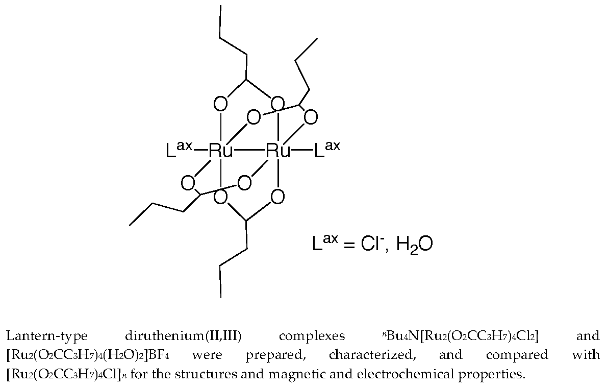
2.1. Synthesis and Characterizations

The addition of an excess amount of nBu4NCl to the dinuclear units of [Ru2(O2CC3H7)4Cl]n (1) with stirring at room temperature for 24 h in dichloromethane solution gave a dichloridodiruthenium(II,III) complex nBu4N[Ru2(O2CC3H7)4Cl2] (2) with a yield of 85% (based on Ru25+ unit of 1). On the other hand, the axial chloride ligand of 1 could be removed by the reaction with AgBF4 in THF at room temperature for 24 h with stirring to give a diaquadiruthenium(II,III) complex [Ru2(O2CC3H7)4(H2O)2]BF4 (3) with a yield of 69%. Their chemical formula were confirmed by elemental analyses. The IR spectra of 2 and 3 showed the COO vibrations as a set of two distinctive bands (νasym (COO) 1465 cm−1 and νsym (COO) 1426 cm−1 for 2; νasym (COO) 1455 cm−1 and νsym (COO) 1428 cm−1 for 3) in a similar energy region to those of 1asym (COO) 1462 cm−1 and νsym (COO) 1425 cm−1). These facts suggest that the dinuclear skeleton is preserved in the above-mentioned reactions to 2 and 3. The stretching vibrations of BF4 appear as a broad band around 1090 cm−1 in 3, which indicate no coordination of the ion to the Ru25+ core [9].

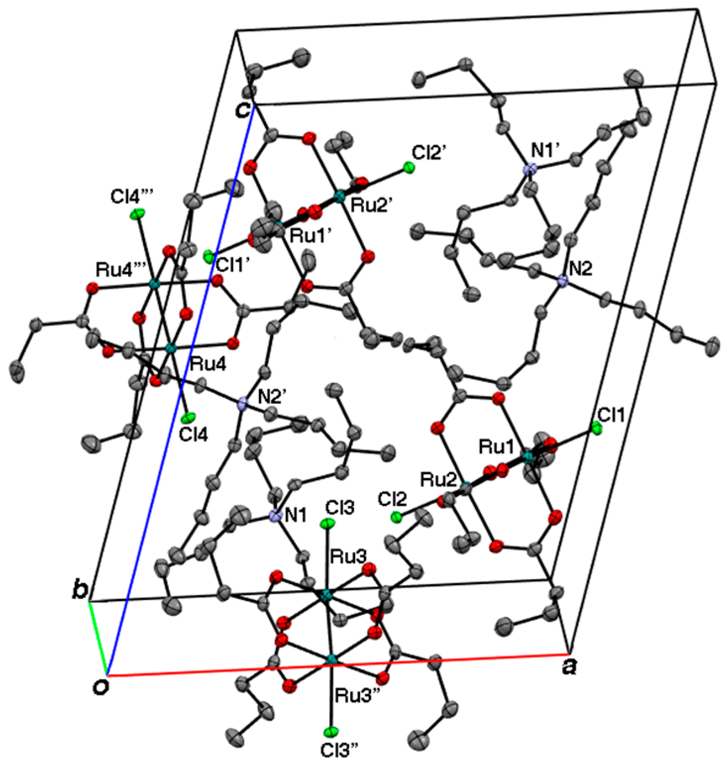
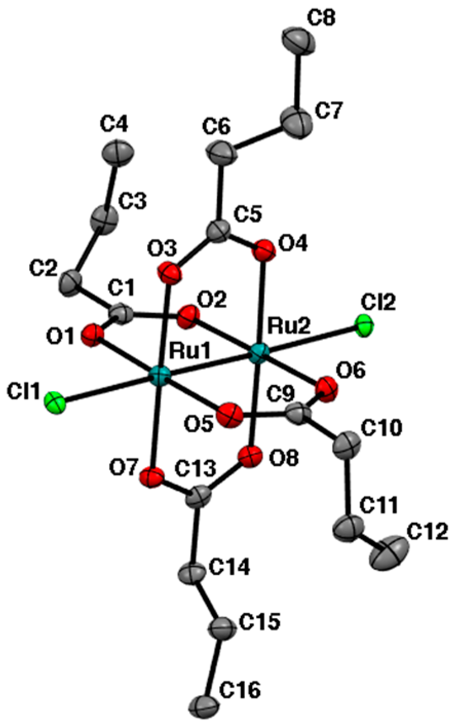
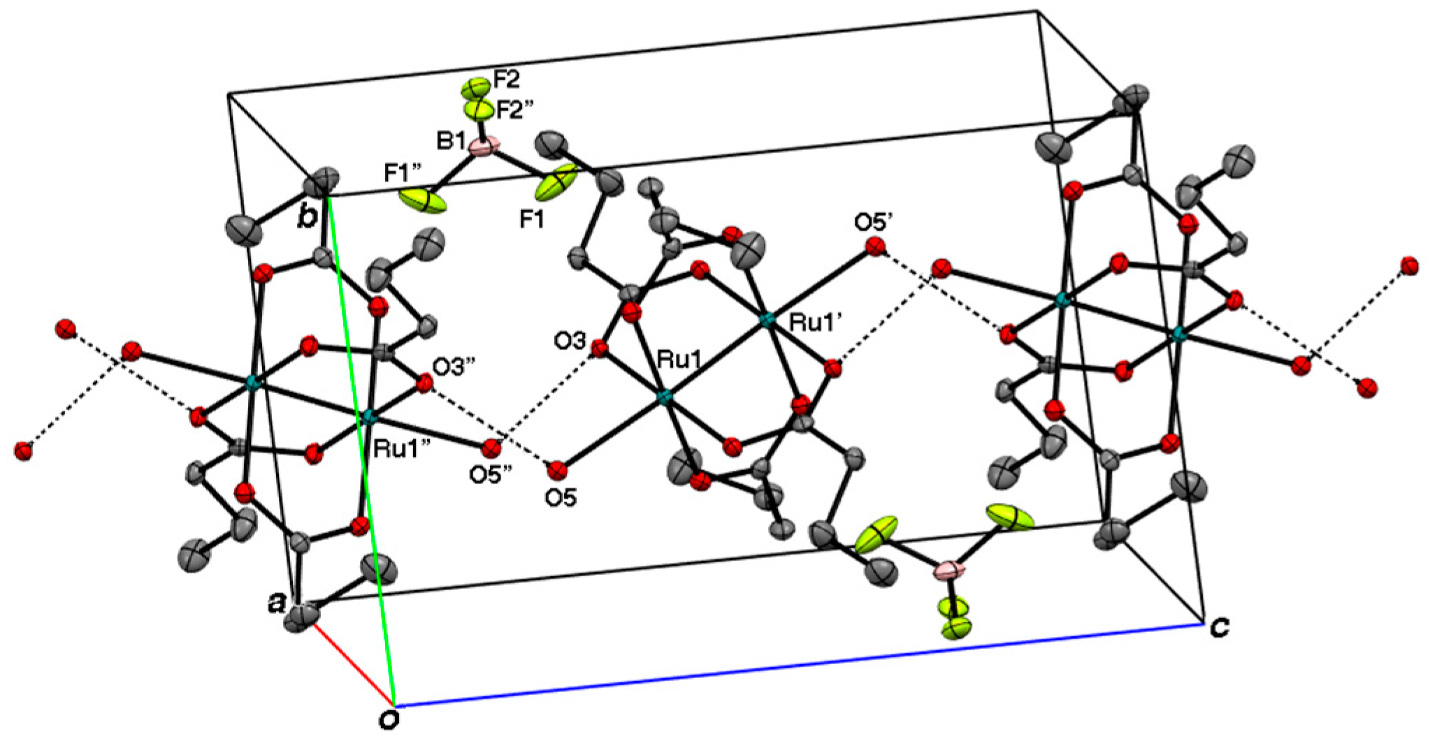
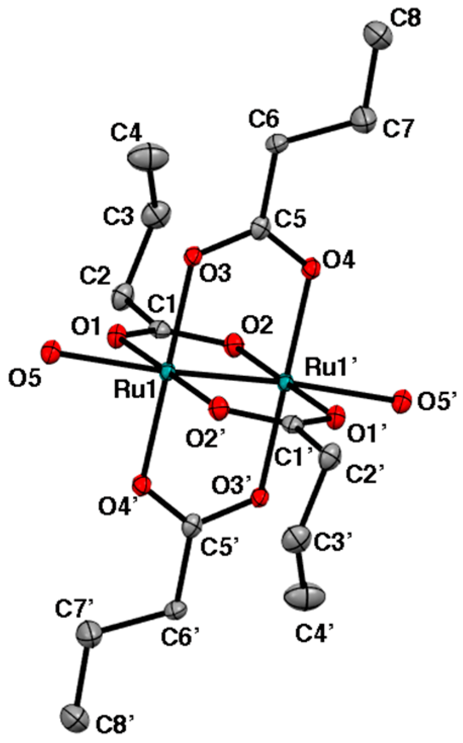
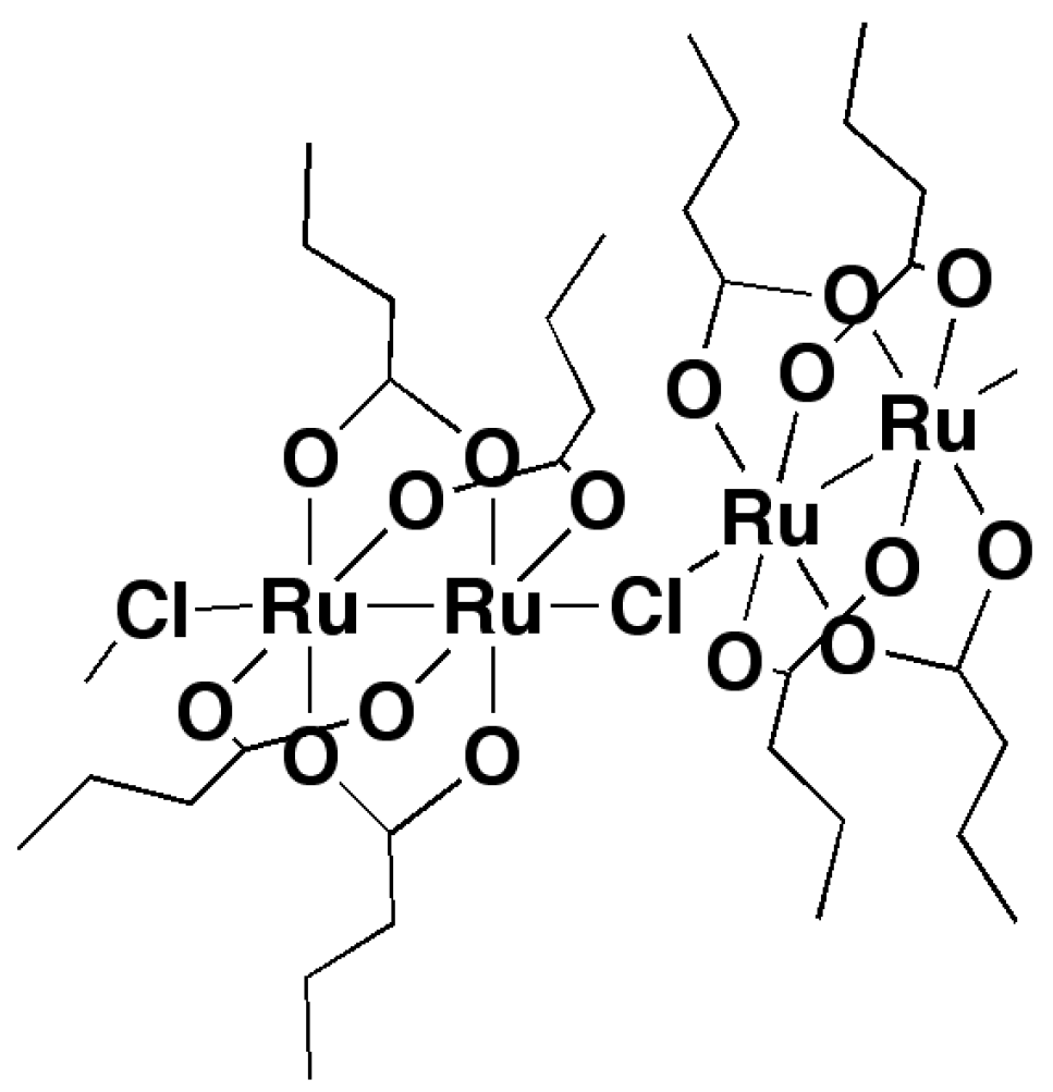
2.2. Crystal Structures

Crystals of 2 and 3 suitable for X-ray crystal structure analyses were obtained by recrystallizations from dichloromethane–diethyl ether and dichloromethane–benzene mixed solvents, respectively. The crystal packing diagram of nBu4N[Ru2(O2CC3H7)4Cl2] (2) is shown in Figure 1. In this crystal, there are crystallographically different dinuclear (Cl–Ru–Ru–Cl) anionic units designated as (Cl1–Ru1–Ru2–Cl2), (Cl3–Ru3–Ru3″–Cl3″), and (Cl4–Ru4–Ru4‴–Cl4‴), respectively, while nBu4N+ counter cations exist among the (Cl–Ru–-Ru–-Cl) anionic units without any important short contacts with the (Cl–Ru–Ru–Cl) units in the crystal. The crystallographical inversion centers are located at the centers of the (Cl3–Ru3–Ru3″–Cl3″) and (Cl4–Ru4–Ru4‴–Cl4‴) dinuclear units. The ORTEP drawing for one of the anionic dinuclear units, (Cl1–Ru1–Ru2–Cl2), is depicted in Figure 2. Including the other dinuclear units, (Cl3–Ru3–Ru3″–Cl3″) and (Cl4–Ru4–Ru4‴–Cl4‴), of which structures are shown in Figures S1 and S2, respectively, the lantern-type dinuclear structures with axial chloride ligands are basically the same as those reported for nBu4N[Ru2(O2CCH3)4Cl2] (4) [10]. The Ru–Ru and Ru–Clax bond distances were 2.3094(3) (for Ru1–Ru2), 2.3046(4) (for Ru3–Ru3″), 2.3034(4) Å (Ru4–Ru4‴), 2.5344(6) (for Ru1–Cl1), 2.5524(6) (for Ru2–Cl2), 2.5335(6) (for Ru3–Cl3), and 2.5127(6) Å (for Ru4–Cl4), respectively. The Ru–Ru and Ru–Cl bond distances were similar to those for 4 (Ru–Ru = 2.3019(4) and 2.3006(5) Å; Ru–Clax = 2.5316(6) and 2.5181(7) Å). The Ru–Ru bond distances of 2 seemed to be large when compared with that of 1 (2.281(4)Å), while the Ru–Clax distances of 2 were rather small when compared with that of 1 (2.587(5) Å). The ORTEP drawing of the cationic unit of [Ru2(O2CC3H7)4(H2O)2]BF4 (3) is depicted in Figure 3. The crystallographic inversion center exists at the center of the lantern-type dinuclear Ru25+ core. The Ru1–Ru1′ and Ru1–O5 bond distances were 2.2584(7) and 2.267(3) Å, respectively, which were comparable to the corresponding distances for the previously reported lantern-type Ru25+ complexes with axial water molecules, [Ru2(O2CCH3)4(H2O)2]BF4 (Ru–Ru = 2.248(1) Å and Ru–Oax = 2.34(1) and 2.27(1) Å) [11], [Ru2(O2CCH3)4(H2O)2]PF6•3H2O (Ru–Ru = 2.2648(9) Å and Ru–Oax = 2.279(4) Å) [12], [Ru2{O2CC(CH3)3}4(H2O)2]BF4 (Ru–Ru =2.259(1) Å and Ru–Oax = 2.280(5) and 2.323(5) Å), and [Ru2{O2CC(CH3)3}4(H2O)2]BF4•CH2Cl2 (Ru–Ru = 2.256(1) Å and Ru–Oax = 2.330(5) and 2.248 (4) Å) [13]. As depicted in the crystal packing diagram (Figure 4), the BF4 counter anions are located among the dinuclear Ru25+ units without any important contacts between them. Alternatively, there are hydrogen bonds between an axially coordinating water molecule and a bridging butanoate oxygen within the neighboring dinuclear unit as shown by dashed line (O5---O3″ = 2.800 Å) in Figure 4 where the hydrogen bonds connect to Ru25+ to give a zig-zag chain. This is in contrast to the hydrogen bonds between the axially coordinated water molecules and BF4 anions that were found for [Ru2{O2CC(CH3)3}4(H2O)2]BF4 and [Ru2{O2CC(CH3)3}4(H2O)2]BF4•CH2Cl2 (O(water)---F (BF4) = 2.742~2.960 Å), leading to two-dimensional sheets [13].

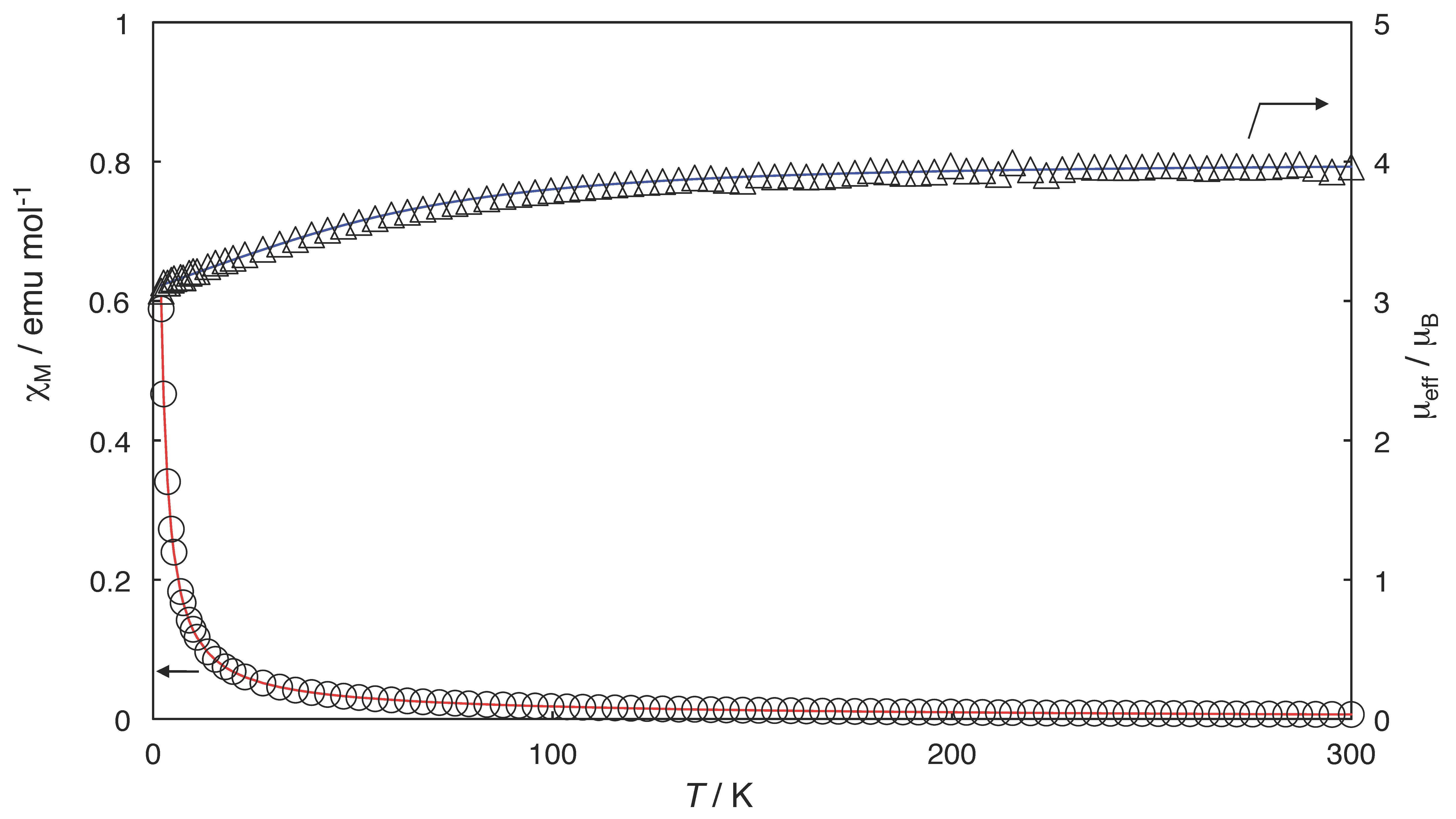
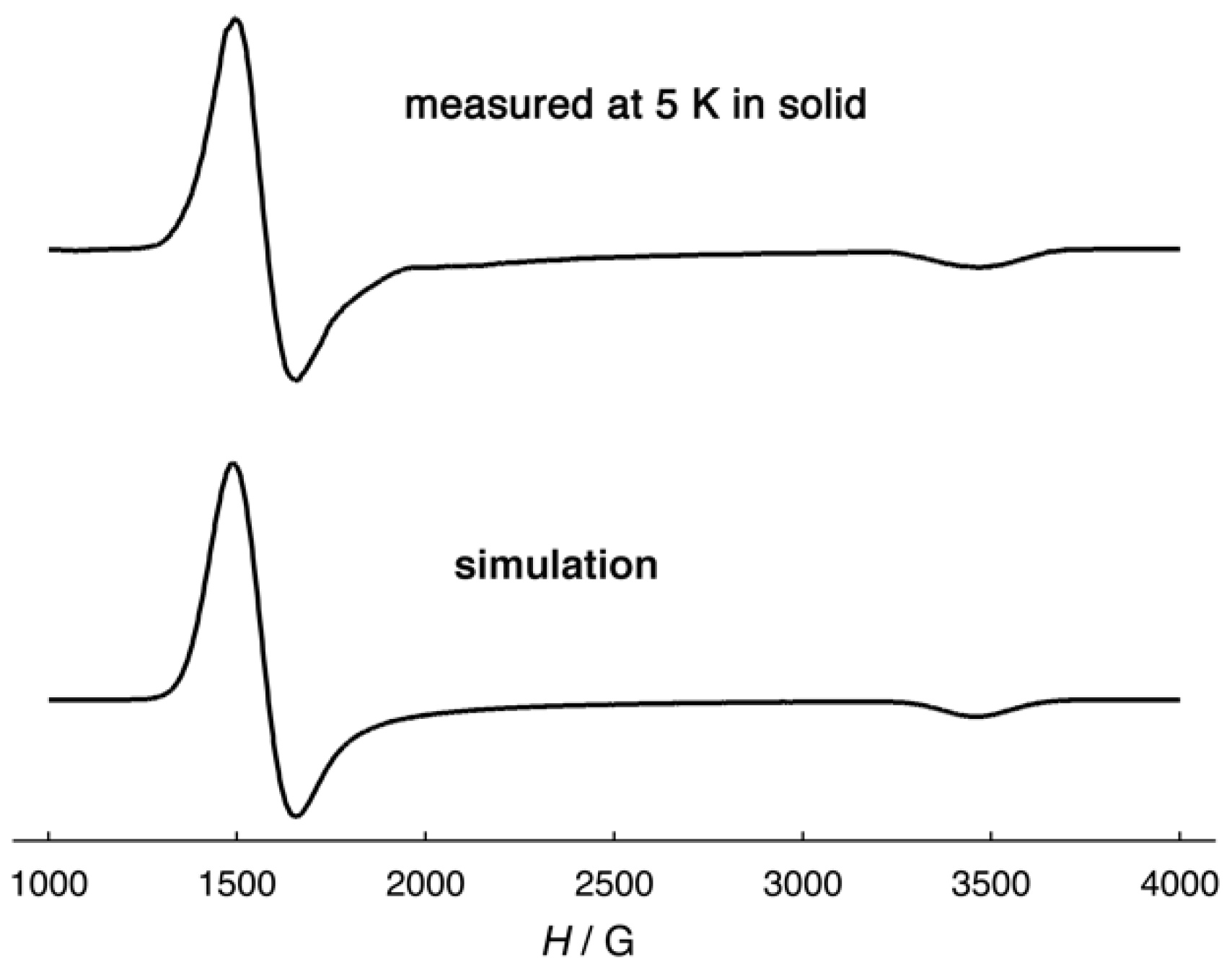
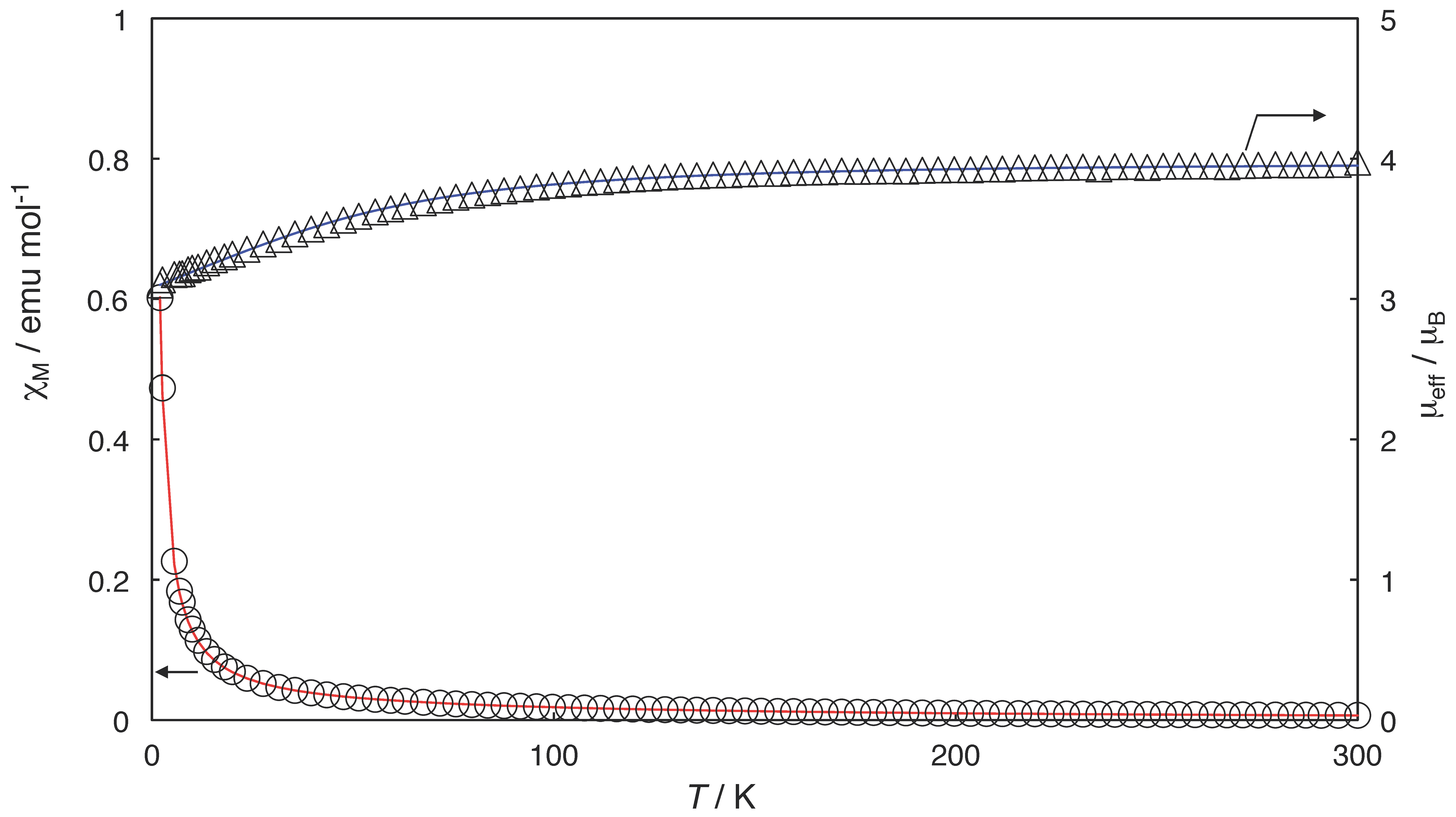
2.3. Magnetic Properties

The temperature dependences of magnetic susceptibilities and effective magnetic moments of 1, 2, and 3 are shown in Figure 5, Figure 6 and Figure 7, respectively. The magnetic moments at 300 K were 3.97 (for 1), 4.00 (for 2), and 3.97 µB (for 3), respectively, indicating the existence of three unpaired electrons within the dinuclear Ru25+ cores like the other lantern-type tetrakis(carboxylato)diruthenium(II,III) with a formal electron configuration of σ2π4δ2(δ*π*)3 (i.e., S = 3/2 ground state) [1]. All of the complexes showed decreases in the moments by lowering the temperature due to the strong zero-field splitting (D). The magnetic behaviors were simulated using the Equations (1)–(3) [3,5,7,14]:
χ = (χ// + 2χ)/3,
where χ is the magnetic susceptibility and χ// and χ are magnetic susceptibility terms defined as follows:
χ// = (Ng2µB2/kT){1 + 9exp(−2D/kT)}/4{1 + exp(−2D/kT)},
χ = (Ng2µB2/kT)[4 + (3kT/D){1 − exp(−2D/kT)}]/4{1 + exp(−2D/kT)}.
The simulation results provided the following parameter values: g = 2.06, D = 68 cm−1 for 1, g = 2.08, D = 78 cm−1 for 2, and g = 2.05, D = 60 cm−1 for 3. Large D values are common for the lantern-type Ru25+ complexes [1,2,3,4,5] and are in the range of D = 50–100 cm−1. Although the magnetic interactions between dinuclear units can be estimated using zJ, which means the exchange energy multiplied by the number of interacting neighboring units and is defined by χ’ = χ/{1 − (2zJ/Ng2µB2)χ}, when a molecular field approximation is applied [3,5,7,14], the temperature-dependent profiles of magnetic susceptibilities and moments of 13 could all be reproduced well without the zJ term. That is, the magnetic interactions were negligible for the complexes (zJ = 0 cm−1). This is reasonable for 2 and 3 because the X-ray crystal structural data showed that the Ru25+ units were distant from each other without any axial linkers mediating the interaction. As for 1, the antiferromagnetic interaction could be possible through an axial chloride linker ligand. The negligible interaction may be due to the zig-zag chain structure with a smaller Ru–Clax-Ru bond angle (125.4°). According to an empirical linear relationship between zJ and the structural parameter Ru–X/Ru–X–Ru, which was proposed for lantern-type Ru25+ complexes with axial halide (X) linkers by Delgado-Martinez et al., smaller Ru–X–Ru bond angles decrease the antiferromagnetic interaction [15]. Although the Ru–X/Ru–X–Ru value of 1 (2.587/125.4 = 0.0206) seems to be for weak antiferromagnetic interaction (zJ = −2 ~ −4 cm−1), we expect that the negligible interaction comes from the small Ru–Clax-Ru bond angle (125.4°). A similar explanation has been presented in terms of MO overlap by Cukiernik et al. for zJ = 0 cm−1 of [Ru2(O2CC3C7)4Cl]n (1) [16]. The magnetic simulation has been reported for 1 as g// = 2.02, g = 2.14 (gav = 2.10), and D = 76.8 cm−1 by Telser et al. as well as g// = 2.14, g = 2.25 (gav = 2.21), and D = 69 cm−1 by Cukiernik et al. Although the g values shown by Cukiernik et al. were rather large, our results for the g, D, and zJ values obtained in this study for complex 1 were not against the previously reported results.
The EPR spectra measured at 5 K in solid for 13 are given in Figure 8, Figures S3 and S4. The signal intensities were strong enough for 2 and 3 to analyze the spectra. Despite the weak signal intensities for 1, the g values were barely estimated. The estimated g values were g// = 2.040 and g = 4.390 for 1; g// = 1.980 and g = 4.385 for 2; and g// = 1.975 and g = 4.335 for 3. For the S =3/2 system with D >> gβH, the estimated effective g values (ge = /βH) are g//e ≈ g// and ge ≈ 2g [7,17]. Thus obtained g values (g// = 2.040 and g = 2.195 for 1; g// = 1.980 and g = 2.1925 for 2; g// = 1.975 and g = 2.168 for 3) are typical of the lantern-type Ru25+ complexes [3,7,8,10,17]. The axial signal pattern was observed in 1:1 toluene/CH2Cl2 at 3.4 K for 1 (g// = 1.9465 and g = 4.400) [7].

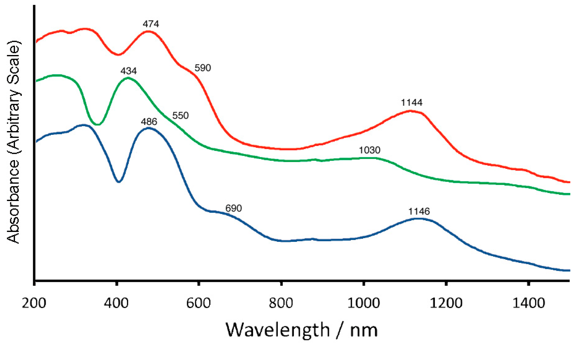
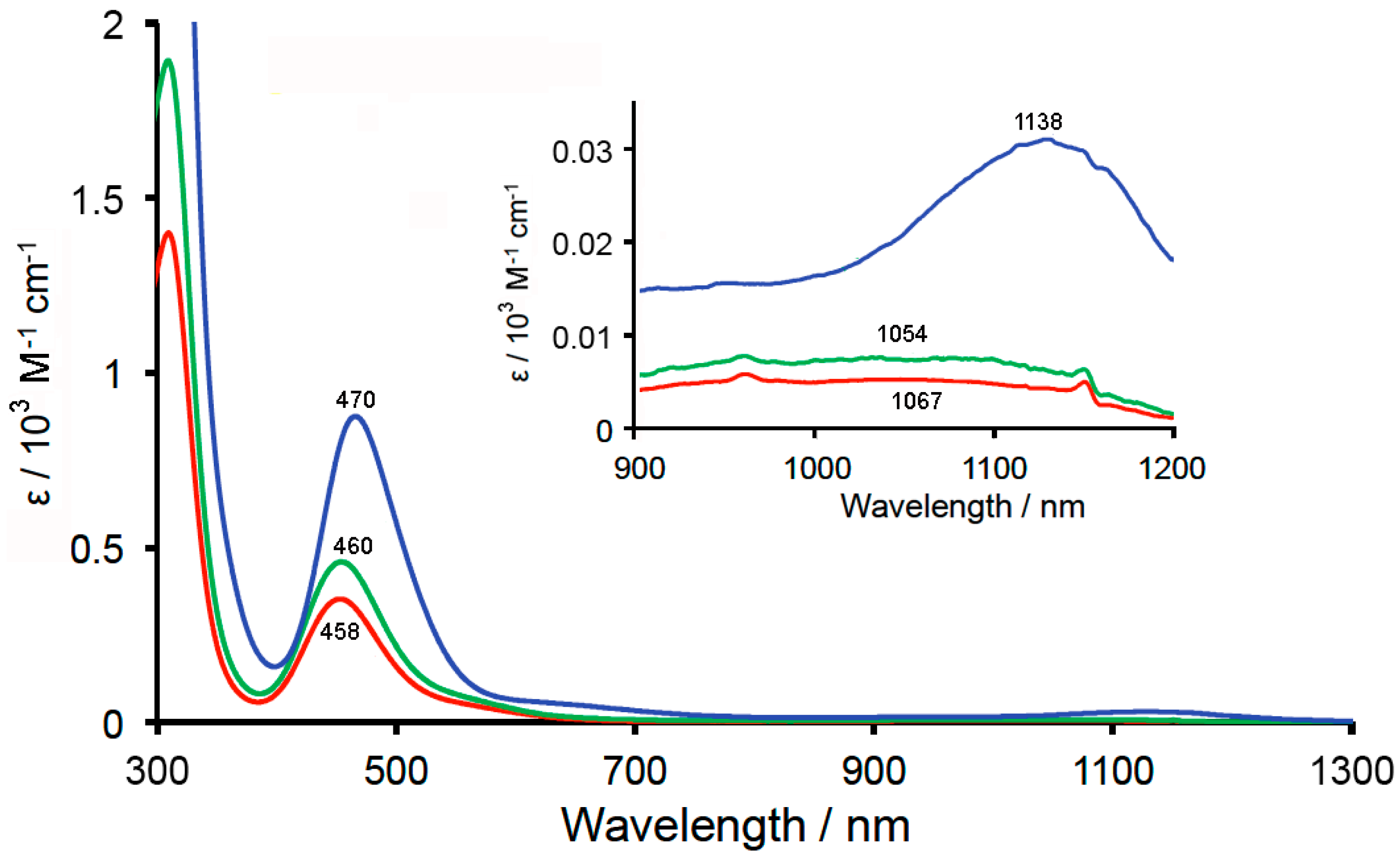
2.4. Reflectance and Absorption Spectra

The diffuse reflectance spectra for the powder samples of 13 are given in Figure 9. All of the complexes showed a distinctive band at 430–490 nm with a discernible shoulder band at 550–690 nm and a broad band at 1030–1150 nm. These spectral features seem to be typical of lantern-type Ru25+ dinuclear complexes [1]. In fact, [Ru2{O2CC(CH3)3}4]BF4 has been reported as having corresponding bands; a band at 427 nm with a shoulder band at 545 nm and a band at 990 nm in the diffuse reflectance spectrum [18], which were assigned as π (Ru–O, Ru2) → π* (Ru2), δ*/π* (Ru2) → δ* (Ru–O), and δ(Ru2) → δ* (Ru2), respectively, according to their assignment in the literature [19]. Absorption spectra (measured in CH2Cl2) are shown in Figure 10. Absorption peaks are found in the near-ultraviolet (450–470 nm) and near-infrared region (1000–1150 nm) for all complexes. The similarity in the spectral features between the reflectance and absorption spectra indicates that the Ru25+ dinuclear skeletons were maintained in the solution.

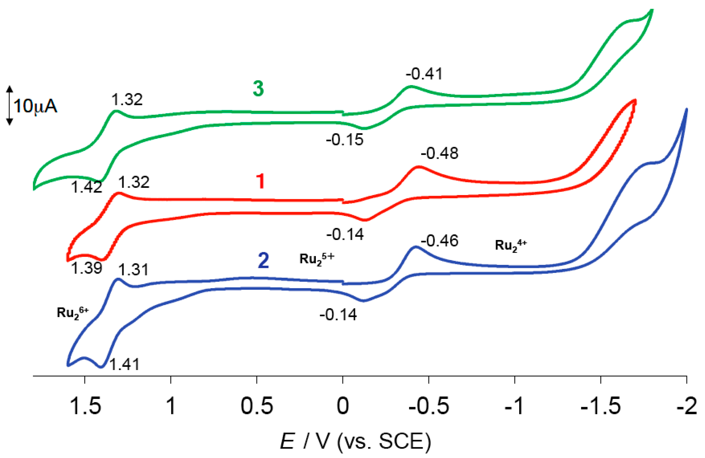
2.5. Cyclic Voltammogram (CV)

Cyclic voltammograms (CVs) were obtained in the dichloromethane solutions containing nBu4N(BF4) (Figure 11). All complexes showed the Ru25+ → Ru24+ process at −0.2–−0.4 V and the Ru25+ → Ru24+ one at 1.3–1.4 V, respectively. Cotton et al. reported that [Ru2(O2CC3H7)4Cl]n exhibited a two-step Ru25+ → Ru24+ reduction process (E1/2 = 0.00 and −0.34 V (vs. SCE) in CH2Cl2 containingnBu4N(ClO4)) although a one-step reduction was observed at E1/2 = −0.34 V when nBu4NCl was used as the electrolyte, which was due to the existence of equilibrium shown by [Ru2(O2CC3H7)4]+ + n(Cl) ↔ [Ru2(O2CC3H7)4Cln](n−1)− [8]. It seems reasonable that the bis-adduct species [Ru2(O2CC3H7)4Cl2] is predominant in the CH2Cl2 solution containing nBu4NCl, and the observed redox couple at E1/2 = −0.34 V can be attributed to that of the Ru25+ → Ru24+ process of the bis-adduct species. We confirmed that nBu4N[Ru2(O2CCH3)4Cl2] (4) exhibited a redox couple attributed to the Ru25+ → Ru24+ process at E1/2 = −0.34 V in a CH2Cl2 solution with an electrolyte nBu4N(ClO4); in addition, the redox wave observed at E1/2 = −0.32 V for [Ru2(O2CCH3)4Cl]n dissolved in a CH2Cl2 solution with an electrolyte nBu4NCl [10]. That is, the axial coordination of Cl to the Ru25+ unit was kept in the measured CH2Cl2 solution containing the nBu4N(ClO4) electrolyte. Hence, the axial chloride ligations of 2 could also be considered as kept in the measured CH2Cl2 solution, although the reversibility of the redox couple (Epc = −0.46 V and Epa = −0.14 V) was not good when compared with that of 4 (Epc = −0.40 V and Epa = −0.28 V) [10]. We performed DFT calculations to estimate the redox potentials (Ecalc1/2) for Ru25+ → Ru24+ as well as the Ru26+ → Ru25+ processes for [Ru2(O2CC3H7)4Cl2] by using our previous treatment for [Ru2(O2CCH3)4Cl2] [10]. The calculated values were Ecalc1/2 (for Ru25+ → Ru24+) = −0.42 V and Ecalc1/2 (for Ru26+ → Ru25+) = 1.25 V. The results support the assignment of the Ru25+ → Ru24+ process at –0.2– –0.4 V and the Ru26+ → Ru25+ one at 1.3–1.4 V for 2. We further performed calculations on [Ru2(O2CC3H7)4Cl] and [Ru2(O2CC3H7)4(H2O)2]+ as the model compounds of 1 and 3, respectively. The calculated Ecalc1/2 (for Ru25+ → Ru24+) and Ecalc1/2 (for Ru26+ → Ru25+) values were −0.05 and 2.09 V for [Ru2(O2CC3H7)4Cl], and 0.34 and 2.77 V for [Ru2(O2CC3H7)4(H2O)2]+. At present, it is difficult to explain the reason why complexes 13 showed redox waves at similar potentials of −0.2– −0.4 V and 1.3–1.4 V. Many factors such as the coordination of the Cl ion and BF4 ion of the electrolyte nBu4N(BF4) as well as the oligomerization of Ru25+ dinuclear units should be taken further into consideration.

3. Materials and Methods

3.1. General Aspects

All reagents and solvents were used as received. The complex [Ru2(O2CC3H7)4Cl]n (1) was prepared according to a published procedure [8].
Elemental analyses for carbon, hydrogen, and nitrogen were performed using a Yanako CHN Corder MT-6. Infrared spectra (KBr pellets) were measured with a JASCO FT/IR-4600. Absorption spectra and diffuse reflectance spectra were obtained using JASCO V-670 and Shimadzu UV-3100 spectrometers, respectively. The temperature dependent magnetic susceptibilities were measured over the temperature range of 2–300 K at the constant field of 0.5 T with a Quantum Design MPMS XL-5. The measured data were corrected for diamagnetic contributions [20]. EPR spectra were measured at 5 K in solid by a BRUKER ELEXSYS E500 equipped with OXFORD ESR900 and OXFORD ITC503 attachments. The EPR simulation was conducted using the “Hyperfine Spectrum” program with spin Hamiltonian, Hs = βBgS [21]. Cyclic voltammograms (CVs) were measured in dichloromethane containing nBu4N(BF4) on a BAS ALS-DY2325 electrochemical analyzer. A glassy carbon disk (1.5 mm radius), platinum wire, and saturated calomel electrodes were used as the working, counter, and reference electrodes, respectively. All of the potential values are described versus SCE.

3.2. Syntheses of Complexes

3.2.1. Synthesis of nBu4N[Ru2(O2CC3H7)4Cl2] (2)

A suspension of [Ru2(O2CC3H7)4Cl]n (50 mg, 0.085 mmol (based on Ru2 dinuclear unit)) was stirred with nBu4NCl (29.5 mg, 0.11 mmol) in dichloromethane (20 mL) for 24 h at room temperature. The resulting solution was concentrated to a small portion and stood to give a brown precipitation, which was collected by suction and dried under vacuum overnight. The yield was 62.9 mg (85% based on [Ru2(O2CC3H7)4Cl] unit). Anal. Found: C, 44.93; H, 7.38; N, 2.00%. Cacld. for C32H64Cl2NO8Ru2, C, 44.49, H, 7.47; N, 1.62%. IR (KBr disk, cm−1): 2964 s, 2936 m, 2874 m, 1465 s, 1426 vs, 1313 m, 1261 w, 1200 vw, 1172 vw, 1102 vw, 889 w, 798 w, 729 w, 677 w, 628 w, and 461 m.

3.2.2. Synthesis of [Ru2(O2CC3H7)4(H2O)2]BF4 (3)

A suspension of [Ru2(O2CC3H7)4Cl]n (90.9 mg, 0.15 mmol (based on Ru2 dinuclear unit)) was stirred with AgBF4 (31.1 mg, 0.16 mmol) in THF (20 mL) for 24 h at room temperature, and the reaction vessel was covered with aluminum foil to shield against the light. The precipitate of AgCl was removed by filtration through celite. The filtrate solution was concentrated to a small portion by evaporating under reduced pressure and stood overnight to give a brown microcrsytalline solid, which was separated by filtration, washed with hexane, and dried under vacuum overnight. The yield was 71.8 mg (69% based on [Ru2(O2CC3H7)4Cl] unit). Anal. Found: C, 28.33; H, 4.41%. Cacld. for C16H32BF4O10Ru2, C, 28.54, H, 4.79%. IR (KBr disk, cm−1): 2965 s, 2932 m, 2878 m, 1455 vs, 1428 vs, 1320 m, 1266 w, 1213 w, 1090 vs, 801 w, 739 m, 673 m, 525 vw, and 465 m.

3.3. Crystal Structure Determination

Single crystals of 2 and 3 suitable for X-ray crystal structure analysis were obtained by the recrystallization from dichloromethane–diethyl ether and dichloromethane–benzene mixed solvents, respectively. X-ray crystallographic data (Table 1) were collected for a single crystal at 90 K on a Bruker CCD X-ray diffractometer (SMART APEX) using graphite-monochromated Mo Kα radiation (λ = 0.71073 Å) for 2 and a RIGAKU Saturn 724 CCD system equipped with a Mo rotating-anode X-ray generator with monochromated Mo Kα radiation (λ = 0.71075 Å) for 3. Diffraction data of 2 and 3 were processed using APEX2 (Bruker) and CrystalClear-SM (RIGAKU), respectively. The structures of 2 and 3 were solved by intrinsic phasing methods (SHELEX) and direct methods (SIR-2011), respectively and refined using the full-matrix least-squares technique (F2) with SHELXL-2014 as part of the SAINT (Bruker) (Billerica, MS, USA) and CrystalStructure 4.2.5 (RIGAKU) (Tokyo, Japan) software, respectively. Non-hydrogen atoms were refined with anisotropic displacement parameters, and all hydrogen atoms were refined with a riding model. Selected bond distances and angles for 2 and 3 are given in Tables S1 and S2, respectively.
CCDC-1887475 and 1887753 contain the supplementary crystallographic data for nBu4N[Ru2(O2CC3H7)4Cl2] (2) and [Ru2(O2CC3H7)4(H2O)2]BF4 (3), respectively. These data can be obtained free of charge from the Cambridge Crystallographic Data Centre [22].

3.4. Computational Details

The unrestricted density functional theory (uDFT) calculations applied in this study were performed with the long-range and dispersion correlated hybrid DFT functional method, ωB97XD, on the Gaussian 09 program [23]. The Los Alamos effective core potential LANL08(f) and Pople’s 6-311 + G* basis sets were applied for the Ru and other atoms, respectively. All molecular geometries were fully optimized and checked by the vibrational frequency analyses. The solvent effect of CH2Cl2 was considered by the polarizable continuum model (PCM). The redox potentials were estimated by using the standard method with the Born–Harbor cycle and Gibbs free energy changes, which was defined by Noodleman [24]. In order to estimate the redox potentials (Ecalc1/2) for the Ru25+ → Ru24+ and Ru26+ → Ru25+ processes, the atomic coordinates of optimized geometries for Ru24+, Ru25+, and Ru26+ species are needed for [Ru2(O2CC3H7)4Cl] (model compound of 1), [Ru2(O2CC3H7)4Cl2] (model compound of 2), and [Ru2(O2CC3H7)4(H2O)2]+ (model compound of 3), respectively. All of the coordinates used for the estimations are given in Tables S3–S11. We subtracted 4.68 V (IUPAC value) [25] from the calculated absolute potentials of the Ru2 complexes to make a direct comparison to the experimental CV data referenced to the SCE.

4. Conclusions

By using [Ru2(O2CC3H7)4Cl]n as a starting material, nBu4N[Ru2(O2CC3H7)4Cl2] and [Ru2(O2CC3H7)4(H2O)2]BF4 were prepared. Their lantern-type dinuclear structures with axial ligands of Cl or H2O were confirmed by X-ray crystal structure analyses. Temperature dependent magnetic susceptibility measurements were performed to show that all of the complexes ([Ru2(O2CC3H7)4Cl]n, nBu4N[Ru2(O2CC3H7)4Cl2], and [Ru2(O2CC3H7)4(H2O)2]BF4) had an S = 3/2 ground state, with a large zero-field splitting (D = 60–80 cm−1). No important magnetic interaction was observed between the dinuclear units for the complexes. Cyclic voltammograms (measured in CH2Cl2 with an electrolyte of nBu4N(BF4)) showed the Ru25+/Ru24+ process at −0.2–−0.4 V (vs. SCE) and the Ru26+/Ru25+ one at 1.3–1.4 V (vs. SCE), where the potentials were confirmed by the DFT calculation for [Ru2(O2CC3H7)4Cl2].

Supplementary Materials

The following are available at https://www.mdpi.com/2312-7481/5/1/18/s1. Selected bond distances and angles of 2 (Table S1); selected bond distances and angles of 3 (Table S2); atomic coordinates of optimized geometries of Ru24+, Ru25+, and Ru26+ species for [Ru2(O2CC3H7)4Cl], [Ru2(O2CC3H7)4Cl2], and [Ru2(O2CC3H7)4(H2O)2]+ (Tables S3–S11); structure of an anionic dinuclear unit designated as (Cl3–Ru3–Ru3″–Cl3″) (Figure S1); structure of an anionic dinuclear unit designated as (Cl4–Ru4–Ru4‴–Cl4‴) (Figure S2); EPR spectra of 1 (Figure S3); and EPR spectra of 3 (Figure S4).

Author Contributions

M.H. conceived and designed the experiment, analyzed the data, and wrote the paper; H.Y. and N.Y. performed the experiments. M.M. (Minoru Mitsumi) and H.A. helped with the SQUID measurements and x-ray crystal structure determination. H.S. analyzed the magnetic susceptibility and EPR data. M.K. and I.I. measured the EPR spectra. R.M. determined the x-ray crystal structure of 2. M.M. (Masahiro Mikuriya) measured the diffuse reflectance spectra. Y.K. measured SQUID, determined the X-ray crystal structure of 3, performed DFT calculations, and wrote the part of the DFT calculation results in the paper.

Funding

The present work was partially supported by Grants-in-Aid for Scientific Research Nos. 15K17897, 16K05722, and 17K05820 from the Ministry of Education, Culture, Sports, Science and Technology (MEXT), Japan.

Acknowledgments

Y. Yano acknowledges the Research Fellowships of the Japan Society for the Promotion of Science for Young Scientists. The authors are grateful to Michiko Egawa (Shimane University) for her measurements of the elemental analyses.

Conflicts of Interest

The authors declare no conflict of interest.

References

  1. Cotton, F.A.; Murillo, C.A.; Walton, R.A. Multiple Bonds between Metal Atoms, 3rd ed.; Springer: New York, NY, USA, 2005. [Google Scholar]
  2. Liddle, S.T. Molecular Metal-Metal Bonds, Compounds, Synthesis, Properties; Wiley-VCH: Weinheim, Germany, 2015. [Google Scholar]
  3. Aquino, M.A.S. Diruthenium and diosmium tetracarboxylates: Synthesis, physical properties and applications. Coord. Chem. Rev. 1998, 170, 141–202. [Google Scholar] [CrossRef]
  4. Aquino, M.A.S. Recent developments in the synthesis and properties of diruthnium tetracarboxylates. Coord. Chem. Rev. 2004, 248, 1025–1045. [Google Scholar] [CrossRef]
  5. Mikuriya, M.; Yoshioka, D.; Handa, M. Magnetic interactions in one-, two-, and three-dimensional assemblies of dinuclear ruthenium carboxylates. Coord. Chem. Rev. 2006, 250, 2194–2211. [Google Scholar] [CrossRef]
  6. Bennett, M.J.; Caulton, K.G.; Cotton, F.A. The structure of tetra-n-butyratodiruthenium chloride, a compound with a strong metal-metal bond. Inorg. Chem. 1969, 8, 1–6. [Google Scholar] [CrossRef]
  7. Telser, J.; Drago, R.S. Reinvesigation of the Electronic and Magnetic Properties of Ruthenium Butyrate Chloride. Inorg. Chem. 1984, 23, 3114–3120, Erratum in 1985, 24, 4765, doi:10.1021/ic00220a601. [Google Scholar] [CrossRef]
  8. Cotton, F.A.; Pedersen, E. Magnetic and electrochemical properties of transition metal complexes with multiple metal-to-metal bonds. II. [Ru2(C3H7COO)4]n+ with n = 0 and 1. Inorg. Chem. 1975, 14, 388–391. [Google Scholar] [CrossRef]
  9. Nakamoto, K. Infrared and Raman Spectra, 4th ed.; John Wiley & Sons: New York, NY, USA, 1986. [Google Scholar]
  10. Hiraoka, Y.; Ikeue, T.; Sakiyama, H.; Guégan, F.; Luneau, D.; Gillon, B.; Hiromitsu, I.; Yoshioka, D.; Mikuriya, M.; Kataoka, Y.; et al. An unprecedented up-field shift in the 13C NMR spectrum of the carboxyl carbons of the lantern-type dinuclear complex TBA[Ru2(O2CCH3)4Cl2] (TBA+ = tetra(n-butyl)ammonium cation. Dalton Trans. 2015, 44, 13439–13443. [Google Scholar] [CrossRef] [PubMed]
  11. Bino, A.; Cotton, F.A.; Felthouse, T.R. Structural studies of some multiply bonded diruthenium tetracarboxylate compounds. Inorg. Chem. 1979, 18, 2599–2604. [Google Scholar] [CrossRef]
  12. Drysdale, K.D.; Beck, E.J.; Cameron, T.S.; Robertson, K.N.; Aquino, M.A.S. Crystal structures and physico-chemical properties of a series of [Ru2(O2CCH3)4L2](PF6) adducts (L = H2O, DMF, DMSO). Inorg. Chim. Acta 1997, 256, 243–252. [Google Scholar] [CrossRef]
  13. Sayama, Y.; Handa, M.; Nukada, R.; Hiromitsu, I.; Kasuga, K. Mixed-Valent Ruthenium Pivalate and Its Nitroxide Adduct. In Coordination Chemistry at the Turn of The Chemistry; Ondrejovic, G., Sirota, A., Eds.; Slovak Technical Universty Press: Bratislava, Slovakia, 1999; pp. 447–452. [Google Scholar]
  14. O’Connor, C.J. Magnetochemistry—Advances in Theory and Experimentaion. Prog. Inorg. Chem. 1982, 29, 203–283. [Google Scholar] [CrossRef]
  15. Delgado-Martinez, P.; González-Prieto, R.; Gómez-García, C.J.; Jiménez-Aparicio, R.; Priego, J.L.; Torres, M.R. Structural, magnetic and electrical properties of one-dimensional tetraamidatodiruthnium compounds. Dalton Trans. 2014, 43, 3227–3237. [Google Scholar] [CrossRef] [PubMed]
  16. Cukiernik, F.D.; Luneau, D.; Machon, J.-C.; Maldivi, P. Mixed-Valent Diruthenium Long-Chain Carboxylates. 2. Magnetic Properties. Inorg. Chem. 1998, 37, 3698–3704. [Google Scholar] [CrossRef] [PubMed]
  17. Handa, M.; Sayama, Y.; Mikuriya, M.; Nukada, R.; Hiromitsu, I.; Kasuga, K. Structure and Magnetic Properties of a Nitroxide Diruthenium(II,III) Complex, [Ru2(O2CCMe3)4(tempo)2][Ru2(O2CCMe3)4(H2O)2] (BF4)2 (tempo = 2,2,6,6-Tetramethylpiperidine-1-oxyl). Bull. Chem. Soc. Jpn. 1995, 68, 1647–1653. [Google Scholar] [CrossRef]
  18. Yoshioka, D.; Mikuirya, M.; Handa, M. Synthsis and Characterization of Polynuclear Chain and Tetranuclear Complexes of Mixed-Valent Ruthenium(II,III) Pivalate with N,N’-Didentate Ligands. Bull. Chem. Soc. Jpn. 2004, 77, 2205–2211. [Google Scholar] [CrossRef]
  19. Miskowski, V.M.; Gray, H.B. Electronic spectra of Ru2(carboxylate)4+ complexes. Higher energy electronic excited states. Inorg. Chem. 1988, 27, 2501–2506. [Google Scholar] [CrossRef]
  20. Kahn, O. Molecular Magnetism; VCH: Cambridge, UK, 1993; Chapter 1. [Google Scholar]
  21. Hagen, W.R. Biomolecular EPR Spectroscopy; CRC: Boca Raton, FL, USA, 2009. [Google Scholar]
  22. Access Structures. Available online: www.ccdc.cam.ac.uk/data_request/cif (accessed on 5 March 2019).
  23. Frisch, M.J.; Trucks, G.W.; Schlegel, H.B.; Scuseria, G.E.; Robb, M.A.; Cheeseman, J.R.; Scalmani, G.; Barone, V.; Mennucci, B.; Petersson, G.A.; et al. Gaussian 09, Revision C.01; Gaussian, Inc.: Wallingford, CT, USA, 2009. [Google Scholar]
  24. Li, J.; Fisher, C.L.; Chen, J.L.; Bashford, D.; Noodleman, L. Calculation of Redox Potentials and pKa Values of Hydrated Transition Metal Cations by a Combined Density Functional and Continuum Dielectric Theory. Inorg. Chem. 1996, 35, 4694–4702. [Google Scholar] [CrossRef]
  25. Trasatti, S. The Absolute Electrode Potential: An Explanatory Note. Pure Appl. Chem. 1986, 58, 955–966. [Google Scholar] [CrossRef]
Scheme 1. Chain structure of [Ru2(O2CC3H7)4Cl]n.
Magnetochemistry 05 00018 sch001
Figure 1. Packing diagram of 2. Primes (′), double primes (″), and triple primes (‴) refer to the equivalent positions (1 − x, 1 − y, 1 − z), (−x, −1 − y, −z), and (−x, 2 − y, 1 − z), respectively.
Magnetochemistry 05 00018 g001
Figure 2. Structure of an anionic dinuclear unit designated as (Cl1–Ru1–Ru2–Cl2). Hydrogen atoms were omitted for clarity. The thermal ellipsoids are shown at the 50% probability level.
Magnetochemistry 05 00018 g002
Figure 3. Structure of the cationic dinuclear unit of [Ru2(O2CC3H7)4(H2O)2]BF4 (3). Hydrogen atoms were omitted for clarity. Primes (′) refer to the equivalent positions (−x, −y, −z). The thermal ellipsoids are shown at the 50% probability level.
Magnetochemistry 05 00018 g003
Figure 4. Packing diagram of 3. Primes (′) and double primes (″) refer to the equivalent positions (−x, −y, −z) and (−x, y, 1/2 − z), respectively.
Magnetochemistry 05 00018 g004
Figure 5. Temperature dependences of magnetic susceptibility χM (circles) and moment µeff (triangles) for [Ru2(O2CC3C7)4Cl]n (1). The red and blue solid lines were calculated and drawn with the parameter values described in the text.
Magnetochemistry 05 00018 g005
Figure 6. Temperature dependences of magnetic susceptibility χM (circles) and moment µeff (triangles) for nBu4N[Ru2(O2CC3C7)4Cl2] (2). The red and blue solid lines were calculated and drawn with the parameter values described in the text.
Magnetochemistry 05 00018 g006
Figure 7. Temperature dependences of magnetic susceptibility χM (circles) and moment µeff (triangles) for [Ru2(O2CC3C7)4(H2O)2]BF4 (3). The red and blue solid lines were calculated and drawn with the parameter values described in the text.
Magnetochemistry 05 00018 g007
Figure 8. EPR spectra of 2. The simulated spectrum was drawn with the parameter values of gz = 1.980, gx = gy = 4.385, Wz = 120 G, and Wx = Wy = 85 G.
Magnetochemistry 05 00018 g008
Figure 9. Diffuse reflectance spectra of 1 (), 2 (), and 3 ().
Magnetochemistry 05 00018 g009
Figure 10. Absorption spectra (measured in CH2Cl2) of 1 (), 2 (), and 3 ().
Magnetochemistry 05 00018 g010
Figure 11. Cyclic voltammograms of 13 at 1.0 × 10⁻³ M in CH2Cl2 containing 0.1 M TBA(BF)4 (glassy carbon working electrode; scan rate = 50 mV/s).
Magnetochemistry 05 00018 g011
Table 1. Crystallographic data of nBu4N[Ru2(O2CC3H7)4Cl2] (2) and [Ru2(O2CC3H7)4(H2O)2]BF4 (3) a.
23
Empirical formulaC32H64Cl2NO8Ru2C16H32BF4O10Ru2
Formula mass863.88673.37
Temperature90 K 90 K
Crystal systemTriclinic Monoclinic
Space group P 1 ¯ P 2/c
a16.0297(11) Å12.600(4) Å
b16.0936(11) Å8.808(3) Å
c18.6813(13) Å13.968(4) Å
α68.4640(10)°90°
β68.5090(10)°106.581(4)°
γ64.3420(10)°90°
Unit-cell volume, V3915.3(5) Å31485.7(8) Å3
Formula per unit cell, Z42
Density, Dcalcd1.466 g cm−31.505 g cm−3
Crystal size0.330 × 0.300 × 0.200 mm30.300 × 0.160 × 0.060 mm3
Absorption coefficient, µ0.953 mm−11.080 mm−1
θ range for data collection1.641–28.500°2.768–24.496°
Reflections collected/unique25828/182448821/2430
R indices [I > 2σ(I)] bR1 = 0.0348, wR2 = 0.0867R1 = 0.0265, wR2 = 0.0783
Goodness-of-fit on F21.0721.164
a Standard deviations in parentheses; b R1 = ∑||Fo| − |Fc||/∑|Fo|; wR2 = [∑w(Fo2Fc2)2/∑(Fo2)2]1/2.

