Magnetic nanoparticles and nanocomposites continue to garner considerable interest due to their versatility in biomedical applications, ranging from diagnostics and therapy to catalysis and sensing. Typically, they consist of magnetic core material and coating, providing stability or functionality. Their magnetic properties allow them to be guided in specific tissue using external magnetic fields, enabling targeted drug or gene delivery. Furthermore, magnetic nanoparticles can serve as contrast agents for magnetic resonance imaging, providing clear visualization of specific tissues. This combination of multiple functions into a single platform makes them ideal candidates for theranostics.
This Special Issue of Magnetochemistry, entitled “Magnetic Nanospecies: Synthesis, Properties, Physical and Biomedical Applications,” features a total of seven contributions covering synthesis, characterization, and vital biological applications of magnetic nanocomposites for therapy and diagnostics (theranostics). It consists of five research articles and two review manuscripts, addressing recent progress in the synthesis, characterization, and biomedical utilization of magnetic nanocomposites.
The articles in this Special Issue cover the following specific areas:
- 1.
- Preparation, Characterization, Coating, and Modification of Magnetic Nanoparticles
Magnetic nanoparticles have great potential in the field of drug delivery. However, challenges such as low solubility and colloidal stability, the formation of reactive oxygen species, and potential toxicity remain major obstacles for their in vivo application. The review article “Serum Albumin for Magnetic Nanoparticles Coating” by Chubarov, A. [1], from the Institute of Chemical Biology and Fundamental Medicine (Russia), focuses on the use of serum albumin as a coating agent for magnetic nanoparticles. The review highlights the role of albumin in organisms, cell–receptor interaction, and its features that enable it to be used as a coating material for magnetic nanoparticles. Key benefits include reduced toxicity, targeted delivery to cancer tissue, and enhanced relaxivity r2, which improves the efficacy of T2-weighted magnetic resonance imaging (Figure 1). The article also presents various strategies for noncovalent and covalent albumin modification for diagnostic probe or drug attachment. As one of the major plasma proteins, albumin interacts with each type of nanoparticle, forming a protein “corona” on the surface. Such a coating affects not only the physical properties but also cell–receptor interaction, biodistribution, and pharmacokinetics. The combination of chemically modified albumin for the coating of magnetic nanoparticles is a possible method for the fabrication of multifunctional platforms capable of simultaneous targeted drug delivery and imaging for cancer treatment.
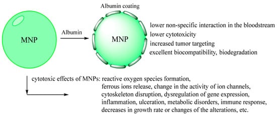
Figure 1.
Features of albumin protein coating on MNPs properties [1]. (Reproduced from Magnetochemistry. 2022, 8, 13; open access article distributed under the terms and conditions of the Creative Commons Attribution (CC BY 4.0)).
The contribution “Magnetism and EPR Spectroscopy of Nanocrystalline and Amorphous TiO2: Fe upon Al Doping” by Yermakov, A., Uimin, M., Borodin, K., Minin, A., Boukhvalov, D., Starichenko, D., Volegov, A., Eremina, R., Yatsyk, I., Zakharova, G. and Gaviko, V. [2] from the M.N. Mikheev Institute of Metal Physics (Russia), the Ural Federal University (Russia), the Nanjing Forestry University (China), the Federal Research Center “Kazan Scientific Center of RAS” (Russia), and the Institute of Solid State Chemistry (Russia), explores the effects of Electron Paramagnetic Resonance (EPR) spectroscopy of TiO2:Fe nanoparticles doped with Al in amorphous and crystalline (3–20 nm) states. Their characterization shows that Al prefers to be localized near Fe ions and influences the crystallinity of nanoparticles. However, aluminum doping does not significantly alter the magnitude of the magnetic moment of the Fe ions.
- 2.
- Biosensors Based on Magnetic Nanoparticles
Magnetite nanoparticles are extensively used for magnetic solid-phase extraction of nucleic acids, enabling subsequent detection or analysis. Almost any laboratory working with nucleic acids relies on these nanoparticles due to their ability to provide a high level of purity of nucleic acids and excellent yield. However, most commercially available magnetic nanoparticles for magnetic separation are designed primarily for DNA extraction rather than RNA, tend to be costly, and are not sequence-specific.
The article “The Effect of pH and Buffer on Oligonucleotide Affinity for Iron Oxide Nanoparticles” by Bobrikova, E., Chubarov, A. and Dmitrienko, E. [3], from the Institute of Chemical Biology and Fundamental Medicine (Russia), investigates a straightforward method for DNA capture using non-coated magnetite nanoparticles. The procedure is simple and consists of the incubation of freshly synthesized nanoparticles with oligonucleotides in acetic buffer pH 3–4, achieving a capacity of 48 nmol of oligonucleotides per mg of particle. According to the authors, this method is suitable for nucleic acid capture, enabling further analysis or therapeutic applications.
The study “Magnetic Nylon 6 Nanocomposites for the Microextraction of Nucleic Acids from Biological Samples” by Bulgakova, A., Chubarov, A. and Dmitrienko, E. [4], from the Institute of Chemical Biology and Fundamental Medicine (Russia), describes the synthesis of oligonucleotide-modified magnetic nanoparticles for magnetic extraction of specific DNA or RNA from the pull (Figure 2). For this purpose, capture-probe oligonucleotides were covalently immobilized onto two types of magnetic nanocomposites (with silica and nylon coating). The developed system was successfully applied to isolate target RNA from mixtures containing large amounts of non-target RNA, achieving an isolation efficiency of 60% with high specificity.
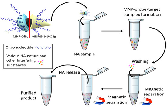
Figure 2.
Nucleic acid (NA) magnetic solid-phase extraction [4]. (Reproduced from Magnetochemistry. 2022, 8, 85; open access article distributed under the terms and conditions of the Creative Commons Attribution (CC BY 4.0)).
- 3.
- Magnetic Nanoparticles for Therapy and Diagnostics (Theranostics) and Other Applications
Magnetic nanoparticles offer a versatile tool for biomedical applications, as discussed in the review article, “Magnetic Nanocomposites and Imprinted Polymers for Biomedical Applications of Nucleic Acids” by Popova, V., Dmitrienko, E. and Chubarov, A. [5], from the Institute of Chemical Biology and Fundamental Medicine (Russia). Magnetic nanocomposites are designed for nucleic acid-based applications, such as diagnostics, drug and gene delivery, therapy, biosensors, biocatalysis, and magnetic separation (Figure 3). The review highlights the use of organic molecules and polymers, inorganic substances, such as noble metals, silica, calcium carbonate, carbon, metal–organic frameworks, biomolecules, and bioinspired materials for surface modification of magnetic nanoparticles. Coating nanoparticles with biomolecules such as polysaccharides, peptides, or nucleic acids imparts “bioinspired” properties, enabling specific interactions with cells, tissues, and the body. Such bioinspired magnetic nanocomposites have attracted significant attention in recent years, particularly in therapeutic applications and biosensor development. In addition to general composite types, such as core–shell, doping, and multilayer, a unique magnetic molecularly imprinted polymer is also presented.
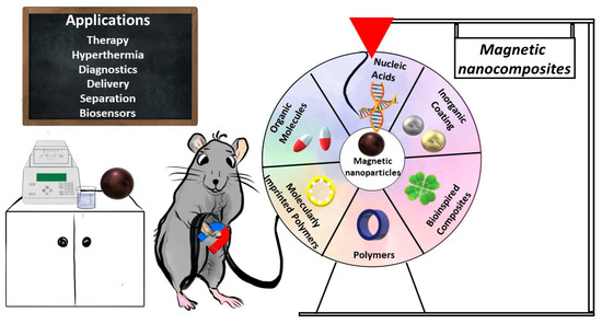
Figure 3.
The combination of nucleic acids and magnetic nanocomposites applications [5]. (Reproduced from Magnetochemistry. 2023, 9, 12; open access article distributed under the terms and conditions of the Creative Commons Attribution (CC BY 4.0)).
The contribution, “Fluorescent Single-Core and Multi-Core Nanoprobes as Cell Trackers and Magnetic Nanoheaters” by Acevedo, P.G., Gómez, M.A.G., Prieto, Á.A., Alves, L.C., Gudiña, R.S., Piñeiro, Y. and Rivas, J. [6], from the Universidade de Santiago de Compostela (Spain), presents different single-core and multi-core iron oxide magnetic nanoparticles functionalized with organic (polydopamine, polyethylene glycol, oleic acid, citrate, and chitosan) and inorganic (silica) coating shells. The single-core nanoparticles were labeled with fluorescent dyes and evaluated in magnetic transfection and cell tracking via confocal microscopy. The multi-core materials were synthesized for magnetic hyperthermia applications.
The article, “High Drug Capacity Doxorubicin-Loaded Iron Oxide Nanocomposites for Cancer Therapy” by Kovrigina, E., Chubarov, A. and Dmitrienko, E. [7], from the Institute of Chemical Biology and Fundamental Medicine (Russia), reports the synthesis of magnetite nanoparticles coated with oleic acid, polyethylene glycol, and Tween. These magnetic nanoparticles were evaluated for stability in aqueous solution with different pH, reactive oxygen species production, and cytotoxicity. The most promising oleic-acid-coated nanoparticles were loaded with anticancer drug doxorubicin (DOX), achieving an extremely high capacity of up to 1.76 mg of DOX per mg of nanoparticles. The DOX-loaded magnetic nanoparticles exhibited pH-responsive drug release with high efficiency at acidic pH, and effectively inhibited the growth of A549 cancer cells (IC50 1.13 mM per DOX concentration).
This Special Issue offers valuable insights into the synthesis, properties, and biomedical applications of magnetic nanoparticles. I sincerely thank all the authors for their significant and outstanding contributions. Additionally, I would like to express my great respect and gratitude to the support team of the Magnetochemistry Journal, whose assistance has played a crucial role in preparing this Special Issue.
Conflicts of Interest
The authors declare no conflict of interest.
References
- Chubarov, A.S. Serum Albumin for Magnetic Nanoparticles Coating. Magnetochemistry 2022, 8, 13. [Google Scholar] [CrossRef]
- Yermakov, A.; Uimin, M.; Borodin, K.; Minin, A.; Boukhvalov, D.; Starichenko, D.; Volegov, A.; Eremina, R.; Yatsyk, I.; Zakharova, G.; et al. Magnetism and EPR Spectroscopy of Nanocrystalline and Amorphous TiO2: Fe upon Al Doping. Magnetochemistry 2023, 9, 26. [Google Scholar] [CrossRef]
- Bobrikova, E.; Chubarov, A.; Dmitrienko, E. The Effect of PH and Buffer on Oligonucleotide Affinity for Iron Oxide Nanoparticles. Magnetochemistry 2021, 7, 128. [Google Scholar] [CrossRef]
- Bulgakova, A.; Chubarov, A.; Dmitrienko, E. Magnetic Nylon 6 Nanocomposites for the Microextraction of Nucleic Acids from Biological Samples. Magnetochemistry 2022, 8, 85. [Google Scholar] [CrossRef]
- Popova, V.; Dmitrienko, E.; Chubarov, A. Magnetic Nanocomposites and Imprinted Polymers for Biomedical Applications of Nucleic Acids. Magnetochemistry 2023, 9, 12. [Google Scholar] [CrossRef]
- García Acevedo, P.; González Gómez, M.A.; Arnosa Prieto, Á.; De Castro Alves, L.; Seco Gudiña, R.; Piñeiro, Y.; Rivas, J. Fluorescent Single-Core and Multi-Core Nanoprobes as Cell Trackers and Magnetic Nanoheaters. Magnetochemistry 2022, 8, 83. [Google Scholar] [CrossRef]
- Kovrigina, E.; Chubarov, A.; Dmitrienko, E. High Drug Capacity Doxorubicin-Loaded Iron Oxide Nanocomposites for Cancer Therapy. Magnetochemistry 2022, 8, 54. [Google Scholar] [CrossRef]
Disclaimer/Publisher’s Note: The statements, opinions and data contained in all publications are solely those of the individual author(s) and contributor(s) and not of MDPI and/or the editor(s). MDPI and/or the editor(s) disclaim responsibility for any injury to people or property resulting from any ideas, methods, instructions or products referred to in the content. |
© 2025 by the author. Licensee MDPI, Basel, Switzerland. This article is an open access article distributed under the terms and conditions of the Creative Commons Attribution (CC BY) license (https://creativecommons.org/licenses/by/4.0/).