Review

Magnetic-Responsive Material-Mediated Magnetic Stimulation for Tissue Engineering

1
Jiangsu Institute of Metrology (Jiangsu Energy Measurement Data Center), Nanjing 210023, China
2
School of Pharmacy, Taizhou University, Taizhou 225300, China
3
Zhejiang Institute of Quality Sciences, Hangzhou 310018, China
4
School of Bioscience and Medical Engineering, Southeast University, Nanjing 210009, China
*
Author to whom correspondence should be addressed.
These authors contributed equally to this work.
Magnetochemistry 2025, 11(10), 82; https://doi.org/10.3390/magnetochemistry11100082
Submission received: 11 August 2025 / Revised: 17 September 2025 / Accepted: 19 September 2025 / Published: 23 September 2025
(This article belongs to the Section Applications of Magnetism and Magnetic Materials)

Abstract

Tissue repair is a significant challenge in biomedical research. Traditional treatments face limitations such as donor shortage, high costs, and immune rejection. Recently, magnetic-responsive materials, particularly magnetic nanoparticles have been introduced into tissue engineering due to their ability to respond to external magnetic fields, generating electrical, thermal, and mechanical effects. These effects enable precise regulation of cellular behavior and promote tissue regeneration. Compared to traditional physical stimulation, magnetic-responsive material-mediated stimulation offers advantages such as non-invasiveness, deep tissue penetration, and high spatiotemporal precision. This review summarizes the classification, fabrication, magnetic effects and applications of magnetic-responsive materials, focusing on their mechanisms and therapeutic effects in neural and bone tissue engineering, and discusses future directions.

1. Introduction

The repair of tissue damage and loss, such as nerve injury and bone defects, are key focuses in biomedical research. Traditional treatment methods, including autografts and allografts, face challenges such as limited donor availability, high costs, and immune rejection [1,2]. To promote tissue repair and regeneration, biomaterial-based tissue engineering has emerged as a promising strategy [3,4,5]. Scaffolds, as essential tools in tissue engineering, mimic the extracellular matrix (ECM) to promote cell migration, adhesion, proliferation, and differentiation [6,7,8]. An effective strategy to enhance scaffold performance is the incorporation of functional materials [9]. Since external physical signals (e.g., electrical, magnetic, thermal, and mechanical) can regulate cellular functions, there has been growing interest in using exogenous stimulation to promote tissue regeneration [10]. However, traditional physical stimulation methods are limited by their invasiveness and low precision [11]. Stimuli-responsive materials combined with external physical signals enable precise and wireless physical stimulation, demonstrating significant potential in tissue engineering [12,13].
Among various external stimulation methods, magnetic stimulation mediated by magnetic-responsive materials has garnered significant attention [14]. Compared to optical, electrical, and ultrasonic stimulation, magnetic stimulation stands out due to its high spatiotemporal resolution, non-invasiveness, deep tissue penetration, and excellent biosafety [15,16]. Researchers have confirmed that magnetic-responsive materials can generate various physical effects under magnetic fields, converting magnetic energy into signals that cells can perceive, such as electricity, force, and heat [17]. These physical effects have been demonstrated to promote cell proliferation, angiogenesis, wound healing, and bone regeneration [18,19,20]. Magnetic-responsive nanomaterials are typically based on magnetic nanoparticles (MNPs), which have been widely used in biomedical fields [21,22]. Notably, some magnetic nanoparticles have received approval from the U.S. Food and Drug Administration (FDA) for clinical use due to their high chemical stability, strong magnetization, and excellent biocompatibility, providing opportunities for the clinical translation of magnetic-responsive material-mediated magnetic stimulation [23]. However, comprehensive reviews on the use of magnetic-responsive material-mediated magnetic stimulation in tissue engineering remain scarce.
This review focuses on the role of magnetic-responsive materials combined with magnetic fields in tissue engineering (Figure 1). First, we introduce magnetic-responsive materials and fabrication techniques, with an emphasis on recent advances in magnetic nanomaterials, polymer scaffolds, and hydrogels. Additionally, we discuss the physical effects of magnetic-responsive materials under magnetic fields. Next, we explore the specific applications of magnetic-responsive material-mediated magnetic stimulation in tissue engineering, particularly in neural and bone tissue engineering. Finally, we discuss potential future directions for this field.

2. Magnetic-Responsive Materials

2.1. Magnetic Nanoparticles

MNPs are a significant magnetic-responsive nanomaterial with particle sizes ranging between 1 and 100 nm [24,25]. MNPs commonly consist of iron, cobalt, nickel and their corresponding oxides [25,26]. Among these, iron oxide nanoparticles (IONPs) are widely recognized in biomedicine including magnetite (Fe3O4), hematite (α-Fe2O3), maghemite (γ-Fe2O3), et al. [27] Fe3O4 and γ-Fe2O3 nanoparticles typically exhibit high magnetic magnetization and are referred to as superparamagnetic iron oxide nanoparticles (SPIONs), characterized by their excellent biocompatibility, fine biodegradability, and stability [28]. Those SPIONs have found a wide range of biomedical applications such as magnetic resonance imaging (MRI), drugs delivery, hyperthermia and biosensing [24,29,30,31].
The magnetic properties of IONPs form the foundation for their medical applications. To enhance the magnetic properties of IONPs, several synthesis methods have been developed to control their size, composition, dispersity and morphology [32,33]. Common synthesis methods for IONPs are chemical methods, including co-precipitation, thermal decomposition, sol–gel, microemulsion and et al. [24,27]. The co-precipitation method is the most used and effective technique, which synthesizes IONPs from aqueous salt solutions [34,35,36]. IONPs can be produced at high yield, while they usually have broad size distributions and low crystallinity. The thermal decomposition method allows to prepare IONPs with high crystallinity and uniform size under high-temperature conditions [37,38]. However, the use of toxic organic-soluble solvents during synthesis could limit their applicability in biomedical applications. The microemulsion method involves mixing oil, water, and surfactants to form microemulsions, where nanoscale droplets serve as reaction templates to control particle size and morphology [39,40]. However, this method relies on surfactants and organic solvents, and their residues can negatively impact the performance and biomedical applications of the resulting IONPs. The sol–gel method, based on the condensation and hydrolysis of metal alkoxides, enables the synthesis of IONPs with high purity and narrow size distribution [41,42]. However, this method often results in contamination by byproducts. Shape, size, and crystallinity are the primary factors determining the characteristics of IONPs, which also influence their interactions with biological systems [32,43]. The shape of IONPs (e.g., spherical, cubic, rod-like, disk-like) determines their bio-clearance and biodistribution, as well as their biocompatibility [28]. Compared with regular spherical IONPs, elongated IONPs exhibit reduced cell uptake and a longer blood circulation time [27,28]. The size of IONPs also has a major impact on their biodistribution [33]. IONPs with diameters ranging from 10 to 100 nm exhibit optimal pharmacokinetic properties for in vivo applications [28]. It should be noted that the magnetic property of IONP s is dependent on their shape, size, and crystallinity [43]. Typically, the size of IONPs is a critical factor governing superparamagnetic behavior, as the saturation magnetization significantly increases with larger particle dimensions [29]. The anisotropy of IONPs also significantly influences their magnetic response [38]. Additionally, superior crystallinity generally results in higher saturation magnetization [29]. Therefore, for biomedical applications, systematic research on synthesis methods is necessary to tailor IONPs with customized properties.
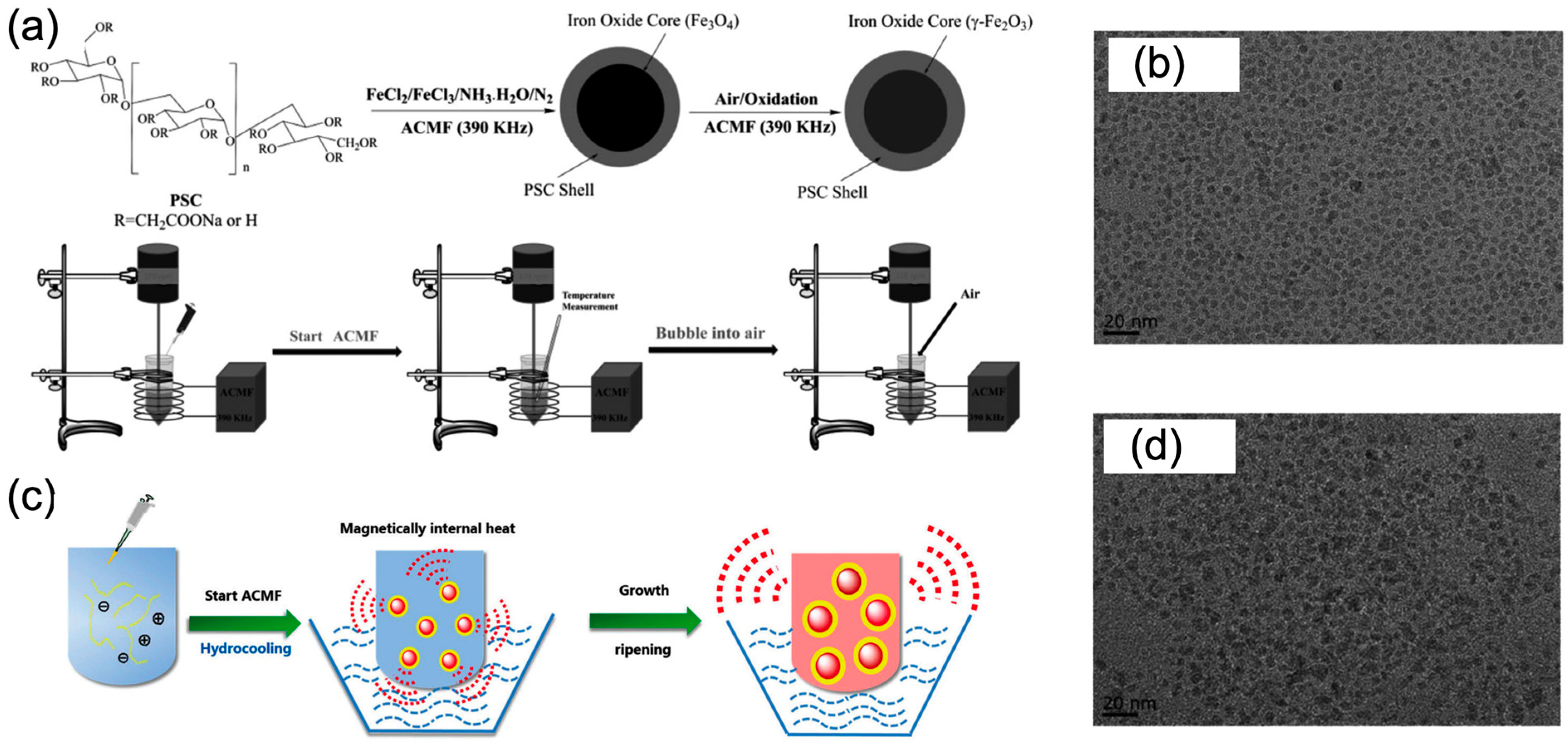
Moreover, the surface modifications profoundly influence the stability and biocompatibility of IONPs. Common coating materials include hydrophilic polymers such as polysaccharides, polyethylene glycol, and polyvinyl alcohol, which can decrease the macro-aggregation and control degradation of IONPs [43,44]. To achieve target specificity in vivo, antibodies can also be used for surface functionalization [45]. In biomedical applications, the type and thickness of the coating material can significantly affect the bio-interactions between IONPs and biological systems [44,46,47]. It should be mentioned that several polysaccharide-coated IONPs produced from co-precipitation method have been approved for the treatment of iron deficiency anemia and as MRI contrast agents by the FDA [47,48]. In this sense, co-precipitation is probably the most suitable methodology for the preparation of IONPs for biomedical applications. To optimize the co-precipitation method, Chen et al. designed a novel synthesis method of IONPs involved endogenous heating process [49]. An alternating-current magnetic field was used to control the spontaneous heat production of IONPs coated with polydextrose-sorbitol carboxymethyl ether (PSC), bringing them more uniform size, better crystallinity and stronger magnetism (Figure 2a,b). Based on this magnetically internal heating co-precipitation, they further introduced a long water-cooling annealing process to drive the equilibrium of colloid formation toward the desired product (Figure 2c,d) [50]. This method produces iron oxide with improved crystallinity and magnetic properties, making it a promising approach for iron oxide synthesis.
The clinical translation of magnetic-responsive materials relies on their biocompatibility, which encompasses both the inherent material properties and their bio-interactions with biological systems. IONPs are generally considered to exhibit good biocompatibility, with only mild cytotoxicity observed when iron concentrations reach 100 μg/mL [30]. However, the toxicity of IONPs requires careful and systematic investigation, as it is closely related to concentration, shape, size, surface properties and structure [32,43]. It is well-established that elevated iron levels can lead to an increase in reactive oxygen species through the Fenton reaction, which may regulate cellular processes such as death, proliferation, motility, and phagocytic capacity [30]. For instance, polyethyleneimine-coated IONPs can induce M1-like activation in macrophages, and neutrally charged PSC-coated IONPs reduce the release of free-iron before being uptake by macrophages in the reticuloendothelial system, thereby mitigating toxicity [30,48]. It is noteworthy that polysaccharide-coated IONPs demonstrate significant potential for clinical translation, with several formulations already approved by FDA for specific medical applications. For example, dextran-coated Fermoxtran-10 and Ferumoxide have been approved as MRI contrast agents, while carboxymethyl-dextran-coated Ferumoxytol has been approved for both MRI contrast enhancement and the treatment of iron deficiency anemia [30]. For tissue engineering, future research should investigate the effects of magnetic nanoparticles’ magnetic properties, surface coatings, and functional groups on biodistribution, biocompatibility and biological functionality.

2.2. Magnetic-Responsive Polymer Scaffolds

Polymer scaffolds are fundamental components of tissue engineering, featuring a three-dimensional porous structure [51,52]. Scaffolds function as an artificial ECM for cell growth that promotes cell proliferation, differentiation, and attachment [6,53]. Commonly, scaffolds can be made from either natural or synthetic polymers owing to their biodegradability, biocompatibility and mechanical strength [54,55,56]. Natural polymers scaffolds made of proteins, polypeptides, or polysaccharides typically exhibit excellent biocompatibility and bioactivity [57,58]. Synthetic polymers (e.g., polylactic Acid, polycaprolactone, polyglycolic acid) are more easily functionalized than natural polymers in terms of degradation rate, mechanical strength and porosity [55]. For tissue engineering, scaffold efficiency depends on several critical factors, including material type, scaffold shape, porosity, mechanical properties, and the integration of bioactive molecules [58]. For instance, in bone tissue engineering, scaffolds require high mechanical strength and stiffness [59,60], whereas in soft tissue engineering, such as vascular applications, flexibility is essential [61,62,63].
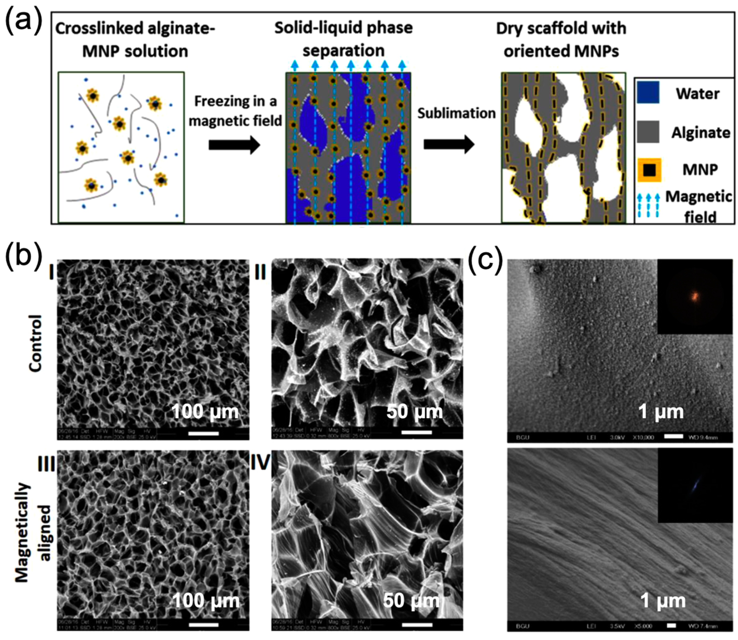
Recently, magnetic-responsive nanomaterials have been incorporated into polymeric scaffolds, enhancing bioactivity and biocompatibility [64,65,66]. MNPs combined with polymers can increase mechanical strength and impart magnetic responsiveness [66]. The simplest method to obtain magnetic scaffolds is to immerse the scaffold in a solution containing MNPs, thereby introducing MNPs into the three-dimensional structure [67]. To improve the dispersion of MNPs within the scaffold, MNPs can also be mixed directly with the polymer solution to fabricate magnetic scaffolds [68]. However, MNPs may aggregate locally within the polymer matrix, negatively impacting their magnetic effects. To uniformly nucleate Fe3O4 nanoparticles within scaffold, Zhao et al. employed an in situ biomimetic strategy, achieving nanoscale homogeneous dispersion of MNPs [69]. Moreover, MNPs can also be aligned and oriented within scaffolds using magnetic fields. This alignment can replicate the anisotropic structure of tissues and improve their interaction with cells and tissues (Figure 3) [70,71].
Specifically, common methods for preparing magnetic scaffolds include solvent casting, freeze drying, electrospinning, 3D printing, and other methods [66,72]. In solvent casting, a polymer solution is poured into a mold and the scaffold is formed as the solvent evaporates [73]. During casting, salt particles can be mixed with polymer solution and removed after evaporating the solvent to control the porous structure. This method is simple but struggles to fabricate scaffolds with complex structures. Freeze-drying involves freezing a polymer slurry and then freeze-drying it to create scaffolds with low stiffness and small pores [74,75]. However, freeze-drying is limited by its time-consuming preparation process and the use of toxic reagents. Electrospinning serves as a robust technique for creating fibrous polymer scaffolds. In a high-voltage electric field, the polymer solution is propelled by electrostatic force, overcoming surface tension to be ejected from the needle and forming nanofibers as the solvent evaporates [76]. Despite advancements in scaffold fabrication using above methods, they usually struggle to control pore size, porosity, and geometry effectively. Three-dimensional printing technology enables the fabrication of scaffolds with complex geometries using layered materials, while also allowing control over the size and connectivity of internal pores [61,76]. However, 3D-printed scaffolds have certain limitations that must be addressed, including limited material options, expensive devices, and technical complexity. In the future, it is essential to develop simple and cost-effective methods for producing magnetic scaffolds to advance their clinical translation in medical applications.

2.3. Magnetic-Responsive Hydrogels

Hydrogels are cross-linked polymer networks containing a large amount of water, characterized by their hydrophilicity, flexibility, biocompatibility, and degradability [77]. Hydrogels can also serve as scaffolds in tissue engineering, and their flexibility makes them particularly suitable for soft tissues [78,79]. Currently, various hydrogel materials, including collagen, alginate, chitosan, and hyaluronic acid, have been approved by the FDA and the European Medicines Agency as medical devices for the clinical treatment of diseases such as tumors, heart failure, and osteoarthritis [80]. Hydrogels typically undergo slow degradation in vivo over periods ranging from several weeks to months, making them highly suitable for customized applications in tissue engineering. By combining hydrogel materials with magnetic-responsive nanomaterials, magnetic-responsive hydrogels can be fabricated, retaining the remote controllability and rapid magnetic responsiveness of the magnetic field [81,82]. Since the hydrogel network can encapsulate nanomaterials internally, the diffusion of nanoparticles within the body is significantly reduced, making them suitable for long-term disease treatment [83].
Common methods for fabricating magnetic hydrogels include the blending method, in situ precipitation method and the grafting-onto method [84,85]. The blending method, the most widely used approach, involves mixing an MNP suspension with a hydrogel precursor solution, followed by gelation under specific conditions [86]. This method is the most widely used method owing to its simplicity, while the added MNPs tend to aggregate inside hydrogels. In situ precipitation method. The in situ precipitation method uses a preformed hydrogel matrix as a chemical reactor to synthesize IONPs within the hydrogel via co-precipitation [87]. This method facilitates the uniform dispersion of MNPs; however, the alkaline reaction conditions limit the types of gels that can be used. In the grafting-onto method, functionalized MNPs act as crosslinkers, forming magnetic hydrogels through covalent coupling with polymers [88]. Although this method is complex and costly, it ensures the uniform distribution and stability of MNPs within the hydrogel. To mimic the anisotropic structures of tissues (e.g., nerves, muscles, cartilage), researchers have attempted to construct biomimetic structures (such as aligned and hierarchical patterns) in magnetic hydrogels to promote tissue reconstruction [84]. Typically, by applying a static magnetic field during gelation, MNPs can form chain-like or columnar structures within the hydrogel [89]. While using rotating magnetic field, more complex two-dimensional structure could be formed [90,91]. The assembly of MNPs improved the microstructural alignment and mechanical properties of the hydrogel [92], while can also enable anisotropic responses under magnetic stimulation [93].
Microhydrogels (1–1000 μm) are promising candidates for tissue engineering due to their injectability, high surface area, and modularity [77]. In most cases, magnetic microhydrogels are prepared by first generating pre-crosslinked droplets, which are then solidified using various crosslinking strategies [94]. Common methods for microhydrogel fabrication include bulk emulsification, fluid electrospraying, mechanical fragmentation, microfluidics, and 3D printing [94,95,96]. Among these, microfluidics stands out as a promising technology due to its versatility and precise control over microgel properties [97]. In our recent work, we proposed a microfluidic method capable of synthesizing magnetic hydrogel microspheres with diverse structures from a single device [98]. This approach uses interfacial tension-controlled microfluidic laminar flow as a soft template to directly regulate microhydrogel structures, offering simplicity and flexibility. The method is compatible with various hydrogel materials and operates solely in aqueous solutions, making it highly suitable for biomedical applications.
In the future, it is essential to continue developing advanced hydrogel fabrication techniques and design appropriate hydrogel matrices and magnetic structures tailored to specific medical applications. Additionally, careful investigation into the biocompatibility and biodegradability of hydrogel materials is necessary.

3. Magnetic-Responsive Effects

3.1. Magneto-Electric Effects

Magneto-electric nanomaterials can generate strong localized electric fields under external magnetic field or be magnetized under electric fields [99,100]. Single-phase magneto-electric materials (multiferroic materials) possess a non-centrosymmetric crystal structure with both magnetic and electric ordering [100]. However, their low room-temperature magneto-electric coefficient limits their applications in biomedicine. Magneto-electric composites, which combine magnetostrictive and piezoelectric properties, exhibit excellent magneto-electric conversion capabilities [20]. These materials are typically fabricated as core–shell structures, where magnetostrictive nanoparticles (CoFe2O4, Fe3O4) are embedded within a piezoelectric matrix (BaTiO3) [17]. The strain-induced coupling between the magnetostrictive and piezoelectric phases, facilitated by lattice matching, enhances the efficiency of magneto-electric conversion [101,102]. These nanoparticles can exhibit a magneto-electric coefficient on the order of 10 V/cm/Oe [20,103]. Another method for producing electrical signals involves the electromagnetic induction of a conductor, which generates an electric current in response to a fluctuating magnetic field [104,105]. In tissue engineering, various conductive biomaterials have been extensively studied, including carbon-based nanomaterials [106], conductive polymers [107], and metallic nanomaterials [104].
Physiological electric fields are closely related to the functions of human tissues, regulating nerves, muscles, and other tissues through cell membrane potentials and action potentials [108,109]. Traditional electrical stimulation relies on external devices and wires, which not only cause significant trauma but also suffer from current attenuation within tissues, reducing stimulation precision and depth [105]. When using magneto-electric nanomaterials, electrical stimulation can be conducted precisely without external power sources or invasive wiring [108,110,111]. And magneto-electric stimulation can activate voltage-gated ion channels, integrins, and intracellular signaling pathways, ultimately modulating biological processes such as cell migration, proliferation, differentiation, and tissue repair [109,112]. For example, electrical stimulation can activate voltage-gated ion channels to modulate neuronal activity [17,105]. The expression of proteins associated with neural regeneration, such as GAP-43, α1-tubulin, trkB and BDNF in neurons, can be activated, and electrical signals can also promote Schwann cell migration and myelination [113]. It can also activate the MEK/ERK pathway to drive osteoblast differentiation, muscle cell proliferation, and neurite outgrowth [114]. Additionally, by inhibiting GSK-3, electrical stimulation regulates the Wnt/β-catenin/GSK-3 pathway, promoting osteocyte differentiation and growth [115]. Although magneto-electric material-mediated electrical stimulation is non-invasive or minimally invasive, the safety of the materials and the long-term stability of the stimulation are critical considerations. Future development of magneto-electric stimulation materials should focus on achieving high electromagnetic conversion efficiency, excellent biocompatibility, and customizable biodegradability.

3.2. Magneto-Mechanical Effects

Under a magnetic field, MNPs experience magnetic forces that align their magnetic moments with the direction of the applied field, inducing movement [19,116]. Different magnetic field modes can drive various mechanical motions. For example, a static magnetic field (SMF) induces directional movement [117], a rotating magnetic field (RMF) causes rotation around a point and vibration [118,119], and an alternating magnetic field (AMF) leads to vibration and spinning [120,121]. These motions generate different mechanical forces (compression, tension, and torque) that can act on cells or tissues. The shape of MNPs also determines their motion and the resulting mechanical forces under magnetic fields. Any magnetic material can generate compression or tension under a magnetic field, while anisotropic magnetic materials are more likely to produce torque under RMF or AMF [122,123]. Therefore, different magneto-mechanical stimulation modes can be customized by tailoring the shape of MNPs and the applied magnetic field patterns.
Mechanical forces are ubiquitous in the cellular microenvironment and play a crucial role in numerous physiological processes [124,125]. Magnetic stimulation mediated by magnetic-responsive materials enables precise manipulation of cell surface proteins through magneto-mechanical forces, thereby activating adhesion, proliferation, and differentiation signaling pathways essential for tissue regeneration [19,126,127]. For example, magneto-mechanical stimulations can activate mechanosensitive ion channels (Piezo1, Piezo2, N-type Ca2+ channel et al.) on neurons to directly regulate neural activity [128,129,130,131]. In bone regeneration, magneto-mechanical stimulation promotes skeletal repair by regulating cell distribution, activating mechano-transduction membrane receptors, and modulating mechanosensitive signaling pathways [127]. In vitro, magneto-mechanical forces can drive cells to form specific structures, enabling the reconstruction of three-dimensional tissues, such as sheets, tubes, and spherical clusters [132,133]. However, magneto-mechanical stimulation faces challenges such as weak force effects from individual MNPs and limited operating distances of magnetic field devices. Therefore, future efforts should focus on designing MNPs with higher magnetic-responsive ability, regulating their collective behavior, and developing advanced magnetic field devices.

3.3. Magneto-Thermal Effects

Magnetic nanomaterials also exhibit magneto-thermal effects [134]. When exposed to a high-frequency AMF, MNPs can absorb a significant amount of electromagnetic energy and convert it into heat through mechanisms such as magnetic hysteresis or relaxation [82]. For SPIONs, heat generation is associated with Néel relaxation and Brownian relaxation. In Néel relaxation, the internal magnetic moments of SPIONs undergo thermal rotation as they overcome magnetic anisotropy. In Brownian relaxation, the physical rotation of SPIONs in a solution causes frictional interactions between the nanoparticles themselves and the surrounding fluid, resulting in heat production. Smaller SPIONs are primarily influenced by Néel relaxation, while larger nanoparticles predominantly experience Brownian relaxation [83].
Current research indicates that thermal stimulation can influence tissue regeneration processes. Mild localized heating (2–4 °C above body temperature) activates heat shock factor 1 (HSF1), leading to the upregulation of heat shock proteins (HSPs). This process stimulates local microcirculation, enhances blood flow, and promotes cellular activities such as fibroblast proliferation, angiogenesis, wound healing, and bone regeneration [16,17]. One of the hallmarks of tissue repair is the generation of reactive oxygen species (ROS), which can regulate cell proliferation. Tommasini et al. found that MNP-mediated magneto-thermal stimulation can achieve biphasic modulation of ROS levels, demonstrating potential for tissue regeneration applications [135]. The magneto-thermal effect of magnetic nanomaterials can also activate thermosensitive ion channels, enabling neural modulation. TRPV1, one of the most extensively studied thermosensitive ion channels, is a Ca2+-permeable cation channel with an activation temperature of 43 °C [84]. Thus, rapid heating of nanoparticles to 43 °C under an alternating magnetic field can activate this channel, facilitating neural stimulation. However, it is important to note that excessive thermal stimulation can disrupt the structure of proteins and peptides inside cells, leading to cellular damage [18].

4. Tissue Engineering

Owing to their excellent biocompatibility, tunable mechanical properties, and remote responsiveness to magnetic fields, magnetic-responsive materials have enabled the widespread application of magnetic stimulation in tissue engineering (Table 1).

4.1. Neural Tissue Engineering

Physical stimulation can positively regulate the nervous system and has shown progress in improving degenerative neurological diseases and promoting neural regeneration [105]. Magnetic-responsive materials, under magnetic fields, can generate various forms of physical stimulation and offer advantages such as wireless operation, precision, and deep tissue penetration [17]. Therefore, magnetic stimulation mediated by magnetic-responsive materials holds significant research potential for treating neurological disorders and nerve injuries.
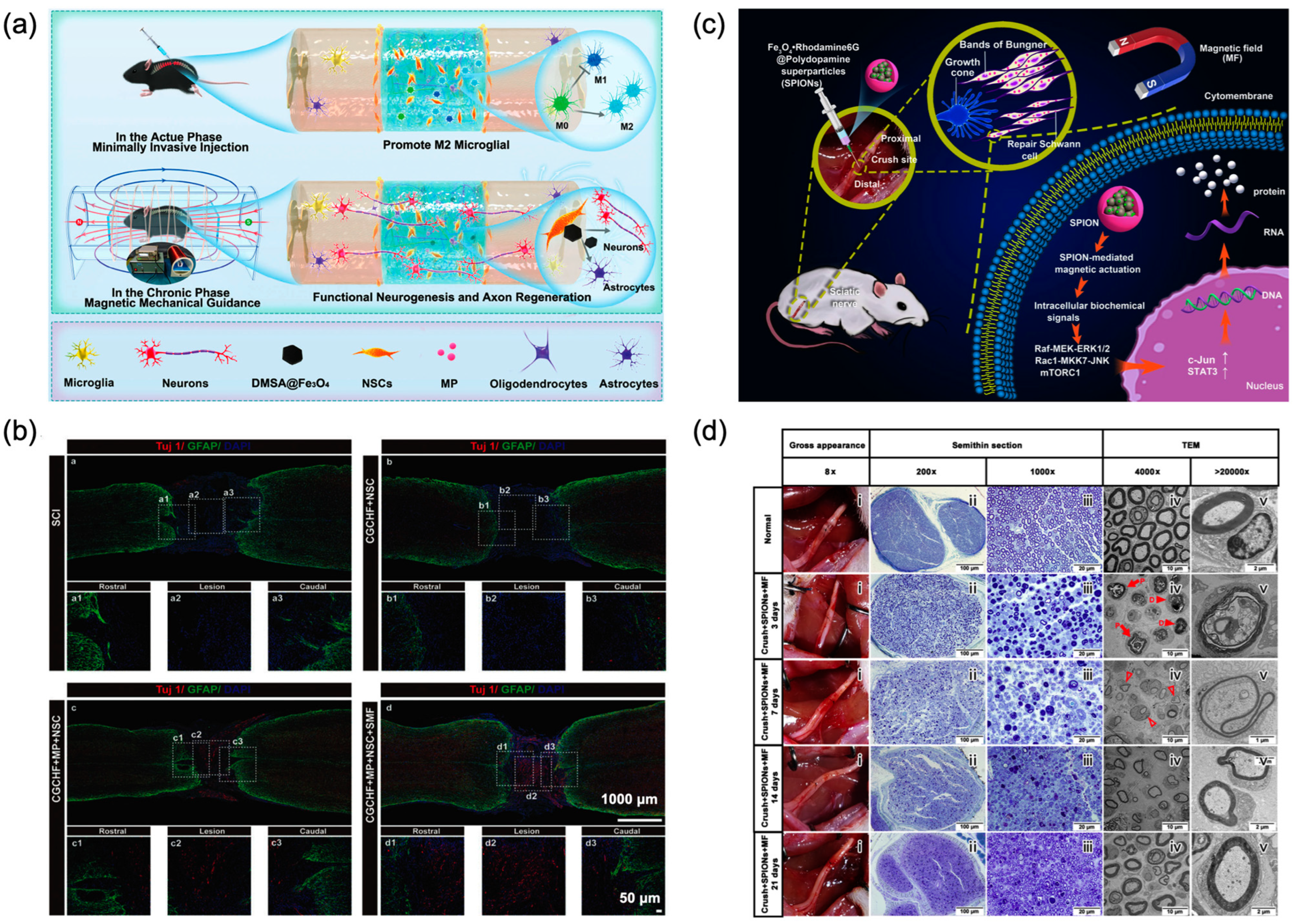
When magneto-electric nanomaterials convert external magnetic fields into localized electric fields near cell membranes, they can induce membrane depolarization and activate voltage-gated ion channels, enabling precise neural modulation [154]. As shown in Figure 4, Zhang et al. combined Fe3O4@BaTiO3 magneto-electric nanoparticles with hyaluronic acid/collagen hydrogels [136]. Under an external pulsed magnetic field, this system promoted the repair of injured spinal cords through wireless electrical stimulation. In addition to magnetic-piezoelectric materials, conductive materials can also generate electrical signals via electromagnetic induction. For example, Liu et al. developed a reduced graphene oxide membrane (rGO-M) that mediates wireless electrical stimulation under a rotating magnetic field (RMF), inducing neuron-like differentiation of mesenchymal stem cells (MSCs) [137]. In vitro experiments demonstrated that RMF stimulation promoted the expression of neuron-specific genes and proteins in MSCs cultured on rGO-M. Further in vivo experiments in rats confirmed that rGO-M-mediated magnetic stimulation could drive exogenous MSCs to differentiate into neuron-like cells, highlighting its potential for neural disease applications. In vivo, Wang et al. reported that reduced graphene oxide combined with a pulsed magnetic field could generate microcurrents, promoting the regeneration of injured nerves [138]. Their findings showed that magneto-electric stimulation effectively enhanced functional recovery in rats with injured sciatic nerves, increasing the expression of S100β, NF200, and GAP43. Jiao et al. developed a triple-network hydrogel based on sodium alginate/calcium, polyacrylamide, and polypyrrole, encapsulating nerve growth factor (NGF). Polypyrrole, a biocompatible conductive polymer, endowed the hydrogel with excellent conductivity (0.32 S/m) [113]. By slowly releasing NGF and combining it with transcranial magnetic stimulation, the system promoted nerve regeneration and functional recovery in rats with sciatic nerve defects, achieving results comparable to autograft transplantation.
Magnetic-responsive nanomaterials can carry drug molecules and achieve spatiotemporally controlled drug release via magnetic fields, integrating magnetic stimulation with chemical molecular effects [17]. For example, nitric oxide (NO) is involved in various biological processes, including immune responses, neurotransmission, and cardiovascular homeostasis [155]. However, a major challenge for tissue engineering is the spatiotemporal control of NO release. Chan et al. developed a magneto-electric stimulation system combined with controlled NO release for neuronal repair [139]. Molybdenum carbide octahedra conjugated with NO donors exhibited AMF-induced electrical currents and controlled NO release. Electrical stimulation promoted the differentiation of neural stem cells, while the NO donors released NO to enhance neural stem cell growth. In vivo results demonstrated that magnetic stimulation reduced glial scarring and immune responses while promoting angiogenesis and neural regeneration.
Another mechanism of the neuron modulation is based on the magneto-mechanical conversion [156]. Magnetic force stimulation leverages the sensitivity of ion channels to mechanical forces, modulating intracellular calcium levels and thereby influencing downstream processes such as cell signaling and synaptic plasticity. In our previous study, we developed a magnetic microhydrogel capable of generating pN-scale magnetic forces under a 10 Hz magnetic field, activating calcium influx in neurons [130]. Furthermore, compared to dispersed SPIONs, the magnetic microhydrogel demonstrated higher efficiency in neural activation. Magneto-mechanical stimulation has made significant progress in regulating cell growth and neural regeneration. As shown in Figure 5a,b, Zhang et al. found that SPION-mediated magneto-mechanical stimulation under a SMF promoted the differentiation of neural stem cells into functional neurons by activating the PI3K/AKT/mTOR pathway [140]. Fattah et al. developed a method for targeted mechanical stimulation of organoids using embedded MNPs [157]. Under a magnetic field, MNPs can precisely apply mechanical stimulation to human neural tube organoids, leading to the remodeling of the cytoskeleton. In neural regeneration, Liu et al. demonstrated that magnetic force stimulation can also act on Schwann cells to promote nerve regeneration and functional recovery (Figure 5c,d) [141]. In a rat sciatic nerve injury model, SPION-mediated magnetic force stimulation induced and maintained the repair-supportive phenotype of Schwann cells, facilitating nerve regeneration and repair.
Neurons are highly sensitive to heat, and excessive heat often inhibits neural function. However, mild magneto-thermal stimulation can activate thermosensitive ion channels, such as TRPV1, enabling the modulation of neural activity [18]. Rosenfeld et al. proved that magneto-thermal stimulation can induce Ca2+ influx by activating TRPV1 in dorsal root ganglion neurons [142]. This stimulation accelerated axon growth and enhanced Schwann cell migration, demonstrating the potential of magneto-thermal modulation to promote nerve regeneration.
Notably, magnetic stimulation of nerves can also regulate other tissues through neural circuits. In our previous work, we developed a novel magnetic vagus nerve stimulation system using an injectable hydrogel loaded with SPIONs, enabling precise and minimally invasive neuromodulation [158]. This system significantly improved cardiac function and reduced infarct size in myocardial infarction rats by restoring autonomic balance and suppressing inflammation, demonstrating its potential for clinical applications. Li et al. utilized SPION-mediated magnetic stimulation to target the gigantocellular reticular nucleus (GRN) for treating spinal cord contusion in mice [159]. The results demonstrated that magnetic stimulation combined with treadmill training significantly improved motor function, enhanced GRN fiber recovery, and promoted the reconstruction of the cortico-reticulo-spinal circuit.
The above results demonstrate the potential of magnetic-responsive materials in mediating magnetic nerve stimulation and their promising applications in tissue engineering. While various magnetic stimulation modalities have demonstrated efficacy in neural repair, there is still a lack of comparative studies across different stimulation paradigms. Therefore, systematic research on distinct magnetic effects is necessary to select optimal stimulation modes or to develop multi-physical coupling strategies that enhance the efficiency of neural repair.

4.2. Bone Tissue Engineering

In bone tissue engineering, magnetic scaffolds with highly porous and interconnected 3D structures can serve as guiding frameworks [64,160]. Under magnetic fields, these scaffolds demonstrate enhanced benefits during bone repair, promoting bone tissue formation, repair, and regeneration [161].
Under a magnetic field, magneto-electric materials can generate electrical signals that activate intracellular Ca2+ and Ca2+-dependent signaling pathways, as well as cell membrane receptor-associated signaling pathways [162]. This activation can enhance the osteogenic differentiation of MSCs, osteoblast precursors, and osteoclasts. Qi et al. developed an iron oxide-doped poly-l-lactic acid scaffold for magneto-electric stimulation, which exhibited a magneto-electric coupling coefficient of 10 mV/cm·Oe [143]. Under a magnetic field (100 Oe, 1400 Hz, 40 min/day), the scaffold promoted the proliferation and differentiation of mouse bone marrow mesenchymal stem cells (BMSCs). And the expression of osteogenesis-related genes, including Col-I, OCN, and Runx2.were and upregulated, demonstrating the potential of magneto-electric stimulation for bone repair applications. In a recent study, Zhao et al. developed a magnetic-responsive flexible membrane composed of CoFe2O4@BaTiO3/poly(vinylidene fluoride-trifluoroethylene) [P(VDF-TrFE)] for magneto-electric microenvironment modulation [163]. Under a direct current magnetic field of 2300–2400 Oe, BMSCs exhibited a successful metabolic shift from glycolysis to oxidative phosphorylation, indicating a strong tendency toward osteogenic differentiation. This study elucidates the intrinsic mechanism of magneto-electric microenvironment regulation in osteogenesis from the perspective of cellular energy metabolism, providing a new direction for bone repair therapies. Mushtaq et al. found that magneto-electric nanoparticles in 3D scaffolds induced stronger electrical stimulation and promoted greater proliferation of human-derived MG63 osteoblasts compared to 2D membranes (134% vs. 43%) [144]. This result highlights the beneficial effects of magneto-electric stimulation on regulating cellular functions and underscores the importance of designing scaffolds with 3D characteristics.
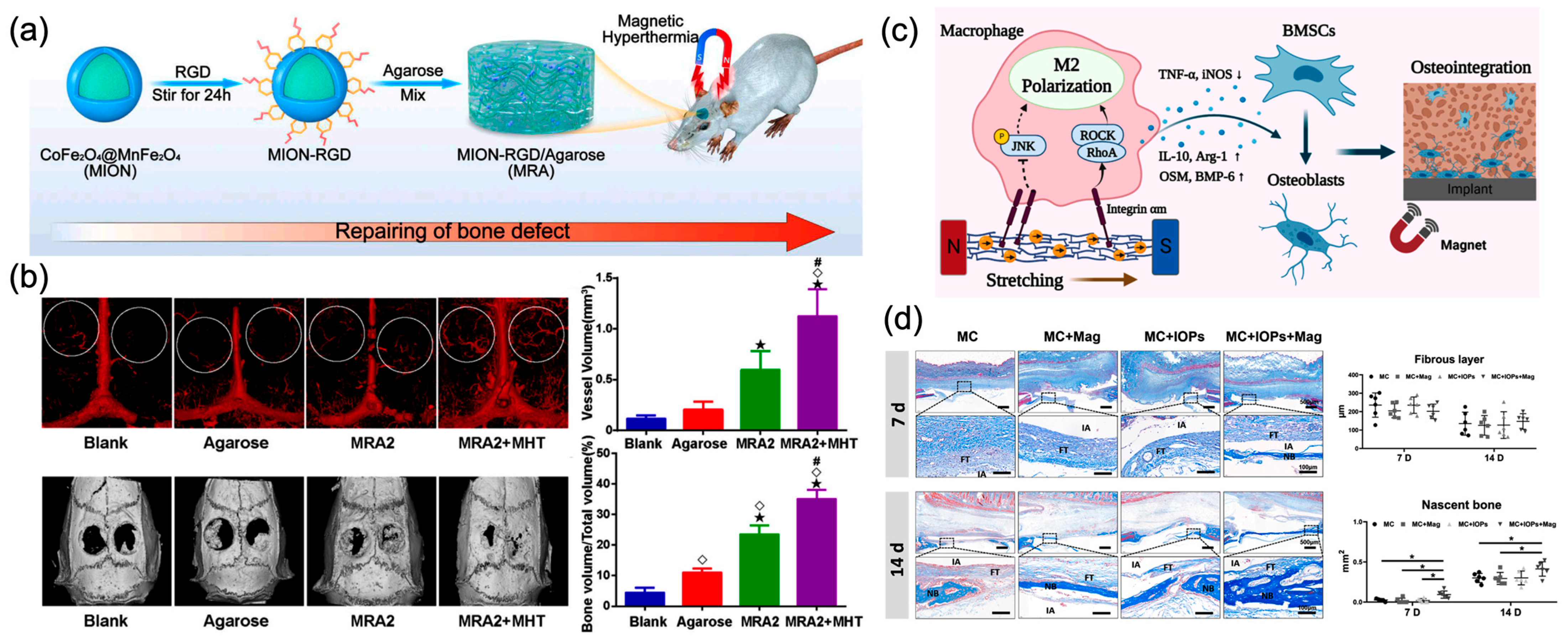
During bone repair, heat stress acts as a significant stimulus that enhances osteogenesis and mineralization [164]. Thermal stimulation can promote angiogenesis at bone defect sites, enhance the migration, proliferation, and differentiation of BMSCs and osteoblasts, and facilitate mineralization during new bone formation [18,165]. As shown in Figure 6a, Wang et al. synthesized a magnetic-responsive scaffold using CoFe2O4@MnFe2O4 nanoparticles for bone regeneration [145]. Under an alternating magnetic field, the scaffold generated mild magneto-thermal stimulation. This thermal stimulation promoted the expression of HSP90, leading to the activation of the phosphatidylinositol 3-kinase (PI3K)/Akt pathway, which enhanced osteogenesis. Additionally, it upregulated the expression of hypoxia-inducible factor (HIF)−1α, which is beneficial for angiogenesis, thereby accelerating the formation of new blood vessels. In rat model of cranial defect, this magneto-thermal stimulation promoted osteogenesis and biomineralization (Figure 6b).
Mechanical stress also has a significant impact on the bone microenvironment, as it can activate mechanical sensors within cells that trigger intracellular signaling pathways [146,166,167]. These mechanical sensors include ion channels, integrins, connexins, actin filaments, focal adhesion kinase, and ECM components, ultimately promoting the proliferation and formation of osteoclasts and osteoblasts [146,168]. With the assistance of magnetic nanomaterials, bone tissue can be mechanically stimulated remotely under magnetic fields. In particular, superparamagnetic iron oxide nanoparticles enable this process through the application of low-intensity magnetic fields. Recently, Xu et al. found that magnetic scaffolds doped with SPIONs could promote the proliferation and adhesion of BMSCs under a mild magnetic field (25–30 mT) [147]. Estévez et al. found that applying magnetic stimulation when SPIONs come into contact with the membrane of human bone marrow mesenchymal stem cells (hBM-MSCs) to initiate endocytosis can induce mechano-transduction effects [146]. SPIONs functionalized with RGD can target integrin receptors on the cell membrane. Under a magnetic field (1 Hz, 250 mT), the expression of osteogenic genes Runx2 and ALP, as well as ALP activity in hBM-MSCs, significantly increased, indicating enhanced osteogenic differentiation. Osteoimmuno-modulation is also a promising strategy for enhancing bone integration. As shown in Figure 6c, Shao et al. developed a magnetic-responsive mineralized collagen coating that can modulate macrophage behavior through magneto-mechanical effects [148]. The magneto-mechanical stimulation triggers integrin-associated RhoA/ROCK signaling cascades and inhibits the phosphorylation of JNK in the MAPK pathway, thereby promoting M2 macrophage polarization. In vitro experiments demonstrated that magnetic stimulation enhanced the osteogenic differentiation of bone marrow-derived MSCs, and the osteogenic effects in vivo were further validated using a rat calvarial defect model (Figure 6d).

4.3. Other Tissue Engineering

Besides nerves and bones, magnetic-responsive material-mediated magnetic stimulation can also enhance the regeneration of cartilage, cardiovascular tissues, and wounds [85,108]. Articular cartilage exhibits a depth-dependent gradient structure, making the adaptation to natural tissue a key challenge in cartilage tissue engineering [169]. Brady et al. developed an advanced three-layer nanocomposite magnetic hydrogel. Under a magnetic field, the MNP-doped hydrogel exhibited elastic, depth-dependent strain gradients, offering a promising tool for cartilage tissue engineering [170]. Additionally, magnetic hydrogels can mediate magneto-thermal [149] and magneto-mechanical stimulation [150,151] to promote chondrogenic differentiation of stem cells. In a recent study, MNP-mediated magnetic force stimulation was shown to activate mechanosensitive pathways, enhancing endochondral ossification [171]. Magnetic stimulation can also be used for control of angiogenesis [172]. For example, Pires et al. fabricated a magnetic gelatin scaffold that, under a 0.08 T magnetic field, enhanced the expression levels of genes and proteins related to angiogenesis in MSCs [152]. Besides angiogenesis, inhibiting bacterial growth and biofilm formation is crucial for promoting wound healing in skin wound tissue engineering [173]. Magneto-thermal responsive bilayer microneedles can control the heating of Fe3O4 under an alternating magnetic field, providing antibacterial and anti-biofilm effects in deep tissues and improving the quality of wound repair [153]. The above results demonstrate the potential of magnetic-responsive materials in mediating magnetic stimulation for tissue engineering, and it is anticipated that magnetic stimulation will play a role in a wider range of tissue engineering applications in the future.

5. Conclusions and Prospects

In this review, we have introduced the magnetic-responsive material-mediated stimulation for tissue engineering. Magnetic-responsive materials can wirelessly convert magnetic energy into mechanical, thermal, or electrical signals, which enable precise control over cellular behaviors such as proliferation, differentiation, and migration, making them highly promising for tissue regeneration. We introduced synthetic strategies of magnetic-responsive materials and their magneto-effects. The latest progress and applications of magnetic-responsive materials mediated magnetic stimulation in tissue engineering have also been summarized in detail. Finally, the challenges and development opportunities of magnetic-responsive material-mediated magnetic stimulation in tissue engineering applications were also discussed.
Although numerous remarkable achievements have been made in magnetic stimulation for tissue engineering, several challenges remain that require further interdisciplinary efforts. The following introduces the challenges that need to be addressed and the key future research directions, providing insights into the advancement of tissue engineering.
(1) The safety of magnetic-responsive materials is of paramount importance, necessitating rigorous evaluation of their biocompatibility both in vitro and in vivo. Developing magnetic-responsive materials based on clinically approved substances and utilizing green, user-friendly fabrication technologies may serve as promising directions to accelerate future clinical translation. Additionally, tissue regeneration often requires long treatment periods, placing high demands on the in vivo stability of these materials. Considering the specific structures of tissues, ideal magnetic stimulation materials should possess biomimetic and bioactive properties to achieve better tissue compatibility and enhanced tissue regeneration capabilities. Four-dimensional printing is emerging as a promising technology that introduces the fourth dimension of time, enabling the fabrication of magnetic-responsive shape-memory polymers [174,175]. This innovation holds potential to establish 4D printing as a preferred method in tissue engineering applications.
(2) In tissue repair, current material-mediated magnetic stimulation typically employs a single mode of stimulation, which often fails to achieve perfect tissue regeneration and functional recovery. Developing materials capable of responding to multiple stimulation modes (e.g., ultrasound, light, and magnetic fields) or coupling multiple stimulation modes through different magnetic field parameters may offer a promising approach to improving therapeutic outcomes. Additionally, since stem cells are essential tools in tissue engineering, we believe that combining stem cell therapy with magnetic stimulation could bring breakthroughs in tissue engineering.
(3) For magnetic stimulation, the frequency and intensity of the magnetic field are crucial. Therefore, future research should focus on investigating the physical effects of magnetic-responsive materials under different magnetic field parameters and evaluating their tissue repair efficacy. Additionally, the mechanisms by which these magnetic effects regulate cellular behavior remain to be fully elucidated. A key priority is the development of measurement techniques for magnetically induced physical effects, as this will help establish a functional relationship between magnetic field stimulation parameters and their biological outcomes.
(4) Material-mediated magnetic stimulation often focuses on disease treatment but lacks feedback on therapeutic efficacy. Integrating treatment and diagnostics through magnetic stimulation could enable closed-loop regulation of tissue regeneration. Achieving this goal in the future will require a comprehensive combination of device development, material fabrication, and artificial intelligence.
(5) Last but not least, current research is primarily conducted on small animals, with a lack of studies on tissue regeneration in large animal models. Future efforts should focus on developing advanced magnetic field generation devices to meet the needs of in vivo magnetic stimulation in large animals, thereby facilitating clinical translation.
Overall, magnetic stimulation mediated by magnetic-responsive materials is a vital tool in tissue engineering, with broad potential for future development.

Author Contributions

Writing—original draft preparation, J.G., L.G. and L.X.; writing—review and editing, L.X.; visualization, L.G.; supervision, D.Y. and X.X.; project administration, Z.X.; funding acquisition, D.Y. and J.G. All authors have read and agreed to the published version of the manuscript.

Funding

This research was funded by the Science and Technology Program of Zhejiang Provincial Market Supervision Administration, grant number ZC2023006; the Science Program of Jiangsu Province Administration for Market Regulation, grant number KJ2024010; and the Fundamental Research Funds of Taizhou University, grant number TZXYQD2024A040.

Institutional Review Board Statement

Not applicable.

Informed Consent Statement

Not applicable.

Conflicts of Interest

The authors declare no conflicts of interest.

Abbreviations

The following abbreviations are used in this manuscript:
AMFAlternating magnetic field
BMSCsBone marrow mesenchymal stem cells
ECMExtracellular matrix
FDAU.S. Food and Drug Administration
GRNGigantocellular reticular nucleus
hBM-MSCshuman bone marrow mesenchymal stem cells
HSFHeat shock factor
HSPHeat shock proteins
MNPsMagnetic nanoparticles
MSCsMesenchymal stem cells
NGFNerve growth factor
NONitric oxide
PSCPolydextrose-sorbitol carboxymethyl ether
rGO-Mreduced graphene oxide membrane
RMFRotating magnetic field
SMFStatic magnetic field
SPIONsSuperparamagnetic iron oxide nanoparticles

References

  1. Bello, S.A.; Cruz-Lebron, J.; Rodriguez-Rivera, O.A.; Nicolau, E. Bioactive scaffolds as a promising alternative for enhancing critical-size bone defect regeneration in the craniomaxillofacial region. ACS Appl. Bio Mater. 2023, 6, 4465–4503. [Google Scholar] [CrossRef] [PubMed]
  2. Safa, B.; Buncke, G. Autograft substitutes: Conduits and processed nerve allografts. Hand Clin. 2016, 32, 127–140. [Google Scholar] [CrossRef]
  3. Tupe, A.; Patole, V.; Ingavle, G.; Kavitkar, G.; Mishra Tiwari, R.; Kapare, H.; Baheti, R.; Jadhav, P. Recent advances in biomaterial-based scaffolds for guided bone tissue engineering: Challenges and future directions. Polym. Adv. Technol. 2024, 35, e6619. [Google Scholar] [CrossRef]
  4. Wu, H.; Feng, E.; Yin, H.; Zhang, Y.; Chen, G.; Zhu, B.; Yue, X.; Zhang, H.; Liu, Q.; Xiong, L. Biomaterials for neuroengineering: Applications and challenges. Regen. Biomater. 2025, 12, rbae137. [Google Scholar] [CrossRef]
  5. Yao, X.; Xue, T.; Chen, B.; Zhou, X.; Ji, Y.; Gao, Z.; Liu, B.; Yang, J.; Shen, Y.; Sun, H.; et al. Advances in biomaterial-based tissue engineering for peripheral nerve injury repair. Bioact. Mater. 2025, 46, 150–172. [Google Scholar] [CrossRef]
  6. Koushik, T.M.; Miller, C.M.; Antunes, E. Bone tissue engineering scaffolds: Function of multi-material hierarchically structured scaffolds. Adv. Healthc. Mater. 2023, 12, 2202766. [Google Scholar] [CrossRef]
  7. Krishani, M.; Shin, W.Y.; Suhaimi, H.; Sambudi, N.S. Development of scaffolds from bio-based natural materials for tissue regeneration applications: A review. Gels 2023, 9, 100. [Google Scholar] [CrossRef]
  8. Zielinska, A.; Karczewski, J.; Eder, P.; Kolanowski, T.; Szalata, M.; Wielgus, K.; Szalata, M.; Kim, D.; Shin, S.R.; Slomski, R.; et al. Scaffolds for drug delivery and tissue engineering: The role of genetics. J. Control. Release 2023, 359, 207–223. [Google Scholar] [CrossRef]
  9. Sultana, N.; Cole, A.; Strachan, F. Biocomposite scaffolds for tissue engineering: Materials, fabrication techniques and future directions. Materials 2024, 17, 5577. [Google Scholar] [CrossRef]
  10. Luo, T.; Tan, B.; Liao, J.; Shi, K.; Ning, L. A review on external physical stimuli with biomaterials for bone repair. Chem. Eng. J. 2024, 496, 153749. [Google Scholar] [CrossRef]
  11. Liu, Y.; Li, B.; Shi, D.; Xiao, R.; Kang, H.; Li, F.; Ling, D. Stimuli-responsive nanomaterials for wireless and precise neuromodulation. Small Methods 2025, e01275. [Google Scholar] [CrossRef]
  12. Ansari, M.A.A.; Dash, M.; Camci-Unal, G.; Jain, P.K.; Nukavarapu, S.; Ramakrishna, S.; Falcone, N.; Dokmeci, M.R.; Najafabadi, A.H.; Khademhosseini, A.; et al. Engineered stimuli-responsive smart grafts for bone regeneration. Curr. Opin. Biomed. Eng. 2023, 28, 100493. [Google Scholar] [CrossRef]
  13. Bai, Y.; Wang, Z.; He, X.; Zhu, Y.; Xu, X.; Yang, H.; Mei, G.; Chen, S.; Ma, B.; Zhu, R. Application of bioactive materials for osteogenic function in bone tissue engineering. Small Methods 2024, 8, 2301283. [Google Scholar] [CrossRef]
  14. Ding, H.; Hao, L.; Mao, H. Magneto-responsive biocomposites in wound healing: From characteristics to functions. J. Mater. Chem. B 2024, 12, 7463–7479. [Google Scholar] [CrossRef]
  15. Ge, C.; Masalehdan, T.; Baghini, M.S.; Toro, V.D.; Signorelli, L.; Thomson, H.; Gregurec, D.; Heidari, H. Microfabrication technologies for nanoinvasive and high-Resolution magnetic neuromodulation. Adv. Sci. 2024, 11, 2404254. [Google Scholar] [CrossRef] [PubMed]
  16. Ribeiro, T.P.; Flores, M.; Madureira, S.; Zanotto, F.; Monteiro, F.J.; Laranjeira, M.S. Magnetic bone tissue engineering: Reviewing the effects of magnetic stimulation on bone regeneration and angiogenesis. Pharmaceutics 2023, 15, 1045. [Google Scholar] [CrossRef] [PubMed]
  17. Lu, X.; Li, G.; Jiao, W.; Li, K.; Zhang, T.; Liu, X.; Fan, H. Magnetic nanomaterials-mediated neuromodulation. WIREs Nanomed. Nanobiotechnol. 2023, 15, e1890. [Google Scholar] [CrossRef] [PubMed]
  18. Li, T.; Zhang, L.; Qu, X.; Lei, B. Advanced thermoactive nanomaterials for thermomedical tissue regeneration: Opportunities and challenges. Small Methods 2025, 9, 2400510. [Google Scholar] [CrossRef]
  19. Yao, J.; Yao, C.; Zhang, A.; Xu, X.; Wu, A.; Yang, F. Magnetomechanical force: An emerging paradigm for therapeutic applications. J. Mater. Chem. B 2022, 10, 7136–7147. [Google Scholar] [CrossRef]
  20. Martins, P.; Brito-Pereira, R.; Ribeiro, S.; Lanceros-Mendez, S.; Ribeiro, C. Magneto-electrics for biomedical applications: 130 years later, bridging materials, energy, and life. Nano Energy 2024, 126, 109569. [Google Scholar] [CrossRef]
  21. Materón, E.M.; Miyazaki, C.M.; Carr, O.; Joshi, N.; Picciani, P.H.; Dalmaschio, C.J.; Davis, F.; Shimizu, F.M. Magnetic nanoparticles in biomedical applications: A review. Appl. Surf. Sci. Adv. 2021, 6, 100163. [Google Scholar] [CrossRef]
  22. Stueber, D.D.; Villanova, J.; Aponte, I.; Xiao, Z.; Colvin, V.L. Magnetic nanoparticles in biology and medicine: Past, present, and future trends. Pharmaceutics 2021, 13, 943. [Google Scholar] [CrossRef]
  23. Wang, S.; Cheng, K.; Chen, K.; Xu, C.; Ma, P.; Dang, G.; Yang, Y.; Lei, Q.; Huang, H.; Yu, Y. Nanoparticle-based medicines in clinical cancer therapy. Nano Today 2022, 45, 101512. [Google Scholar] [CrossRef]
  24. Cardoso, V.F.; Francesko, A.; Ribeiro, C.; Bañobre-López, M.; Martins, P.; Lanceros-Mendez, S. Advances in magnetic nanoparticles for biomedical applications. Adv. Healthc. Mater. 2018, 7, 1700845. [Google Scholar] [CrossRef] [PubMed]
  25. Mishra, S.; Yadav, M.D. Magnetic Nanoparticles: A comprehensive review from synthesis to biomedical frontiers. Langmuir 2024, 40, 17239–17269. [Google Scholar] [CrossRef] [PubMed]
  26. Stiufiuc, G.F.; Stiufiuc, R.I. Magnetic nanoparticles: Synthesis, characterization, and their use in biomedical field. Appl. Sci. 2024, 14, 1623. [Google Scholar] [CrossRef]
  27. Meng, Y.Q.; Shi, Y.N.; Zhu, Y.P.; Liu, Y.Q.; Gu, L.W.; Liu, D.D.; Ma, A.; Xia, F.; Guo, Q.Y.; Xu, C.C.; et al. Recent trends in preparation and biomedical applications of iron oxide nanoparticles. J. Nanobiotechnol. 2024, 22, 24. [Google Scholar] [CrossRef]
  28. Vangijzegem, T.; Lecomte, V.; Ternad, I.; Van Leuven, L.; Muller, R.N.; Stanicki, D.; Laurent, S. Superparamagnetic iron oxide nanoparticles (SPION): From fundamentals to state-of-the-art innovative applications for cancer therapy. Pharmaceutics 2023, 15, 236. [Google Scholar] [CrossRef]
  29. Kandasamy, G.; Maity, D. Recent advances in superparamagnetic iron oxide nanoparticles (SPIONs) for in vitro and in vivo cancer nanotheranostics. Int. J. Pharm. 2015, 496, 191–218. [Google Scholar] [CrossRef]
  30. Dadfar, S.M.; Roemhild, K.; Drude, N.I.; von Stillfried, S.; Knüchel, R.; Kiessling, F.; Lammers, T. Iron oxide nanoparticles: Diagnostic, therapeutic and theranostic applications. Adv. Drug Deliv. Rev. 2019, 138, 302–325. [Google Scholar] [CrossRef]
  31. McNamara, K.; Tofail, S.A.M. Nanoparticles in biomedical applications. Adv. Phys. X 2017, 2, 54–88. [Google Scholar] [CrossRef]
  32. Ling, D.; Lee, N.; Hyeon, T. Chemical synthesis and assembly of uniformly sized iron oxide nanoparticles for medical applications. Acc. Chem. Res. 2015, 48, 1276–1285. [Google Scholar] [CrossRef]
  33. Xie, W.; Guo, Z.; Gao, F.; Gao, Q.; Wang, D.; Liaw, B.-S.; Cai, Q.; Sun, X.; Wang, X.; Zhao, L. Shape-, size- and structure-controlled synthesis and biocompatibility of iron oxide nanoparticles for magnetic theranostics. Theranostics 2018, 8, 3284–3307. [Google Scholar] [CrossRef] [PubMed]
  34. Majidi, S.; Zeinali Sehrig, F.; Farkhani, S.M.; Soleymani Goloujeh, M.; Akbarzadeh, A. Current methods for synthesis of magnetic nanoparticles. Artif. Cells Nanomed. Biotechnol. 2016, 44, 722–734. [Google Scholar] [CrossRef] [PubMed]
  35. Rajput, S.; Pittman, C.U.; Mohan, D. Magnetic magnetite (Fe3O4) nanoparticle synthesis and applications for lead (Pb2+) and chromium (Cr6+) removal from water. J. Colloid Interface Sci. 2016, 468, 334–346. [Google Scholar] [CrossRef]
  36. Trifoi, A.R.; Matei, E.; Râpă, M.; Berbecaru, A.-C.; Panaitescu, C.; Banu, I.; Doukeh, R. Coprecipitation nanoarchitectonics for the synthesis of magnetite: A review of mechanism and characterization. React. Kinet. Mech. Catal. 2023, 136, 2835–2874. [Google Scholar] [CrossRef]
  37. Hufschmid, R.; Arami, H.; Ferguson, R.M.; Gonzales, M.; Teeman, E.; Brush, L.N.; Browning, N.D.; Krishnan, K.M. Synthesis of phase-pure and monodisperse iron oxide nanoparticles by thermal decomposition. Nanoscale 2015, 7, 11142–11154. [Google Scholar] [CrossRef]
  38. Rezaei, B.; Yari, P.; Sanders, S.M.; Wang, H.; Chugh, V.K.; Liang, S.; Mostufa, S.; Xu, K.; Wang, J.-P.; Gómez-Pastora, J.; et al. Magnetic nanoparticles: A review on synthesis, characterization, functionalization, and biomedical applications. Small 2024, 20, 2304848. [Google Scholar] [CrossRef]
  39. Foroughi, F.; Hassanzadeh-Tabrizi, S.A.; Bigham, A. In situ microemulsion synthesis of hydroxyapatite-MgFe2O4 nanocomposite as a magnetic drug delivery system. Mater. Sci. Eng. C Mater. Biol. Appl. 2016, 68, 774–779. [Google Scholar] [CrossRef]
  40. Salvador, M.; Gutiérrez, G.; Noriega, S.; Moyano, A.; Blanco-López, M.C.; Matos, M. Microemulsion synthesis of superparamagnetic nanoparticles for bioapplications. Int. J. Mol. Sci. 2021, 22, 427. [Google Scholar] [CrossRef]
  41. Fatmawati, T.; Shiddiq, M.; Armynah, B.; Tahir, D. Synthesis methods of Fe3O4 nanoparticles for biomedical applications. Chem. Eng. Technol. 2023, 46, 2356–2366. [Google Scholar] [CrossRef]
  42. Tavakoli, A.; Sohrabi, M.; Kargari, A. A review of methods for synthesis of nanostructured metals with emphasis on iron compounds. Chem. Pap. 2007, 61, 151–170. [Google Scholar] [CrossRef]
  43. Wu, W.; Jiang, C.Z.; Roy, V.A.L. Designed synthesis and surface engineering strategies of magnetic iron oxide nanoparticles for biomedical applications. Nanoscale 2016, 8, 19421–19474. [Google Scholar] [CrossRef] [PubMed]
  44. Muthukumaran, T.; Philip, J. A review on synthesis, capping and applications of superparamagnetic magnetic nanoparticles. Adv. Colloid Interface Sci. 2024, 334, 103314. [Google Scholar] [CrossRef] [PubMed]
  45. Gao, J.; Gu, H.; Xu, B. Multifunctional magnetic nanoparticles: Design, synthesis, and biomedical applications. Acc. Chem. Res. 2009, 42, 1097–1107. [Google Scholar] [CrossRef]
  46. Dik, G.; Ulu, A.; Ates, B. Synthesis and biomedical applications of polymer-functionalized magnetic nanoparticles. Nanofabrication 2023, 8, 1–33. [Google Scholar] [CrossRef]
  47. Anderson, S.D.; Gwenin, V.V.; Gwenin, C.D. Magnetic functionalized nanoparticles for biomedical, drug delivery and imaging applications. Nanoscale Res. Lett. 2019, 14, 188. [Google Scholar] [CrossRef] [PubMed]
  48. Long, M.; Li, Y.; He, H.; Gu, N. The story of ferumoxytol: Synthesis production, current clinical applications, and therapeutic potential. Adv. Healthc. Mater. 2024, 13, 2302773. [Google Scholar] [CrossRef]
  49. Chen, B.; Li, Y.; Zhang, X.; Liu, F.; Liu, Y.; Ji, M.; Xiong, F.; Gu, N. An efficient synthesis of ferumoxytol induced by alternating-current magnetic field. Mater. Lett. 2016, 170, 93–96. [Google Scholar] [CrossRef]
  50. Chen, B.; Sun, J.; Fan, F.; Zhang, X.; Qin, Z.; Wang, P.; Li, Y.; Zhang, X.; Liu, F.; Liu, Y.; et al. Ferumoxytol of ultrahigh magnetization produced by hydrocooling and magnetically internal heating co-precipitation. Nanoscale 2018, 10, 7369–7376. [Google Scholar] [CrossRef]
  51. Wang, C.; Huang, W.; Zhou, Y.; He, L.; He, Z.; Chen, Z.; He, X.; Tian, S.; Liao, J.; Lu, B.; et al. 3D printing of bone tissue engineering scaffolds. Bioact. Mater. 2020, 5, 82–91. [Google Scholar] [CrossRef]
  52. Chocholata, P.; Kulda, V.; Babuska, V. Fabrication of scaffolds for bone-tissue regeneration. Materials 2019, 12, 568. [Google Scholar] [CrossRef] [PubMed]
  53. Bruzauskaite, I.; Bironaite, D.; Bagdonas, E.; Bernotiene, E. Scaffolds and cells for tissue regeneration: Different scaffold pore sizes-different cell effects. Cytotechnology 2016, 68, 355–369. [Google Scholar] [CrossRef] [PubMed]
  54. Reddy, M.S.B.; Ponnamma, D.; Choudhary, R.; Sadasivuni, K.K. A comparative review of natural and synthetic biopolymer composite scaffolds. Polymers 2021, 13, 1105. [Google Scholar] [CrossRef] [PubMed]
  55. Chaudhari, A.A.; Vig, K.; Baganizi, D.R.; Sahu, R.; Dixit, S.; Dennis, V.; Singh, S.R.; Pillai, S.R. Future prospects for scaffolding methods and biomaterials in skin tissue engineering: A review. Int. J. Mol. Sci. 2016, 17, 1974. [Google Scholar] [CrossRef]
  56. Guo, L.; Liang, Z.; Yang, L.; Du, W.; Yu, T.; Tang, H.; Li, C.; Qiu, H. The role of natural polymers in bone tissue engineering. J. Control. Release 2021, 338, 571–582. [Google Scholar] [CrossRef]
  57. Mondschein, R.J.; Kanitkar, A.; Williams, C.B.; Verbridge, S.S.; Long, T.E. Polymer structure-property requirements for stereolithographic 3D printing of soft tissue engineering scaffolds. Biomaterials 2017, 140, 170–188. [Google Scholar] [CrossRef]
  58. Sathiya, K.; Ganesamoorthi, S.; Mohan, S.; Shanmugavadivu, A.; Selvamurugan, N. Natural polymers-based surface engineering of bone scaffolds—A review. Int. J. Biol. Macromol. 2024, 282, 136840. [Google Scholar] [CrossRef]
  59. Liu, D.; Liu, J.; Zhao, P.; Peng, Z.; Geng, Z.; Zhang, J.; Zhang, Z.; Shen, R.; Li, X.; Wang, X.; et al. 3D bioprinted tissue-engineered bone with enhanced mechanical strength and bioactivities: Accelerating bone defect repair through sequential immunomodulatory properties. Adv. Healthc. Mater. 2024, 13, 2401919. [Google Scholar] [CrossRef]
  60. El-Bahrawy, N.R.; Elgharbawy, H.; Elmekawy, A.; Salem, M.; Morsy, R. Development of porous hydroxyapatite/PVA/gelatin/alginate hybrid flexible scaffolds with improved mechanical properties for bone tissue engineering. Mater. Chem. Phys. 2024, 319, 129332. [Google Scholar] [CrossRef]
  61. Lee, W.; Xu, C.; Fu, H.; Ploch, M.; D’Souza, S.; Lustig, S.; Long, X.; Hong, Y.; Dai, G. 3D bioprinting highly elastic PEG-PCL-DA hydrogel for soft tissue fabrication and biomechanical stimulation. Adv. Funct. Mater. 2024, 34, 2313942. [Google Scholar] [CrossRef]
  62. Xie, R.; Cao, Y.; Sun, R.; Wang, R.; Morgan, A.; Kim, J.; Callens, S.J.P.; Xie, K.; Zou, J.; Lin, J.; et al. Magnetically driven formation of 3D freestanding soft bioscaffolds. Sci. Adv. 2024, 10, 1549. [Google Scholar] [CrossRef] [PubMed]
  63. Wang, Z.; Huang, C.; Shi, Z.; Liu, H.; Han, X.; Chen, Z.; Li, S.; Wang, Z.; Huang, J. A soft receiving platform for coaxial bioprinting cell-laden microtubes with uniform wall thickness. Appl. Mater. Today 2025, 42, 102599. [Google Scholar] [CrossRef]
  64. Li, Z.; Xue, L.; Wang, P.; Ren, X.; Zhang, Y.; Wang, C.; Sun, J. Biological scaffolds assembled with magnetic nanoparticles for bone tissue engineering: A review. Materials 2023, 16, 1429. [Google Scholar] [CrossRef] [PubMed]
  65. Mortimer, C.J.; Wright, C.J. The fabrication of iron oxide nanoparticle-nanofiber composites by electrospinning and their applications in tissue engineering. Biotechnol. J. 2017, 12, 1600693. [Google Scholar] [CrossRef]
  66. Ebrahimzadeh, M.H.; Nakhaei, M.; Gharib, A.; Mirbagheri, M.S.; Moradi, A.; Jirofti, N. Investigation of background, novelty and recent advance of iron (II,III) oxide- loaded on 3D polymer based scaffolds as regenerative implant for bone tissue engineering: A review. Int. J. Biol. Macromol. 2024, 259, 128959. [Google Scholar] [CrossRef]
  67. Li, Z.; Zhang, Y.; Ye, Q.; Wang, L.; Chen, H.; Xu, C.; Wang, P.; Sun, J. Endowing improved osteogenic activities with collagen membrane by incorporating biocompatible iron oxide nanoparticles. Front. Bioeng. Biotechnol. 2023, 11, 1259904. [Google Scholar] [CrossRef]
  68. Yang, W.; Zhong, Y.; Feng, P.; Gao, C.; Peng, S.; Zhao, Z.; Shuai, C. Disperse magnetic sources constructed with functionalized Fe3O4 nanoparticles in poly-l-lactic acid scaffolds. Polym. Test. 2019, 76, 33–42. [Google Scholar] [CrossRef]
  69. Zhao, Y.; Fan, T.; Chen, J.; Su, J.; Zhi, X.; Pan, P.; Zou, L.; Zhang, Q. Magnetic bioinspired micro/nanostructured composite scaffold for bone regeneration. Colloids Surf. B Biointerfaces 2019, 174, 70–79. [Google Scholar] [CrossRef]
  70. Margolis, G.; Polyak, B.; Cohen, S. Magnetic induction of multiscale anisotropy in macroporous alginate scaffolds. Nano Lett. 2018, 18, 7314–7322. [Google Scholar] [CrossRef]
  71. Taghizadeh, S.; Tayebi, L.; Akbarzadeh, M.; Lohrasbi, P.; Savardashtaki, A. Magnetic hydrogel applications in articular cartilage tissue engineering. J. Biomed. Mater. Res. Part A 2024, 112, 260–275. [Google Scholar] [CrossRef]
  72. Paltanea, G.; Manescu, V.; Antoniac, I.; Antoniac, A.; Nemoianu, I.V.; Robu, A.; Dura, H. A review of biomimetic and biodegradable magnetic scaffolds for bone tissue engineering and oncology. Int. J. Mol. Sci. 2023, 24, 4312. [Google Scholar] [CrossRef] [PubMed]
  73. Pamula, E.; Bacakova, L.; Filova, E.; Buczynska, J.; Dobrzynski, P.; Noskova, L.; Grausova, L. The influence of pore size on colonization of poly(L-lactide-glycolide) scaffolds with human osteoblast-like MG 63 cells in vitro. J. Mater. Sci. Mater. Med. 2008, 19, 425–435. [Google Scholar] [CrossRef]
  74. Ge, J.; Zhai, M.; Zhang, Y.; Bian, J.; Wu, J. Biocompatible Fe3O4 subchitosan scaffolds with high magnetism. Int. J. Biol. Macromol. 2019, 128, 406–413. [Google Scholar] [CrossRef] [PubMed]
  75. Guedes, D.G.; Guedes, G.G.; da Silva, J.d.O.; da Silva, A.L.; Luna, C.B.B.; Damasceno, B.P.G.d.L.; Costa, A.C.F.d.M. Development of scaffolds with chitosan magnetically activated with cobalt nanoferrite: A study on physical-chemical, mechanical, cytotoxic and antimicrobial Behavior. Pharmaceuticals 2024, 17, 1332. [Google Scholar] [CrossRef]
  76. Safavi, A.S.; Karbasi, S. A new path in bone tissue engineering: Polymer-based 3D-printed magnetic scaffolds (a comprehensive review of in vitro and in vivo studies). J. Biomater. Sci. Polym. Ed. 2025, 36, 1321–1341. [Google Scholar] [CrossRef]
  77. Daly, A.C.; Riley, L.; Segura, T.; Burdick, J.A. Hydrogel microparticles for biomedical applications. Nat. Rev. Mater. 2020, 5, 20–43. [Google Scholar] [CrossRef]
  78. Farrukh, A.; Nayab, S. Shape memory hydrogels for biomedical applications. Gels 2024, 10, 270. [Google Scholar] [CrossRef]
  79. Zhang, Y.S.; Khademhosseini, A. Advances in engineering hydrogels. Science 2017, 356, eaaf3627. [Google Scholar] [CrossRef]
  80. Mandal, A.; Clegg, J.R.; Anselmo, A.C.; Mitragotri, S. Hydrogels in the clinic. Bioeng. Transl. Med. 2020, 5, e10158. [Google Scholar] [CrossRef]
  81. Shi, W.; Huang, J.; Fang, R.; Liu, M. Imparting functionality to the hydrogel by magnetic-field-induced nano-assembly and macro-response. ACS Appl. Mater. Interfaces 2020, 12, 5177–5194. [Google Scholar] [CrossRef] [PubMed]
  82. Fragal, E.H.; Fragal, V.H.; Silva, E.P.; Paulino, A.T.; da Silva Filho, E.C.; Mauricio, M.R.; Silva, R.; Rubira, A.F.; Muniz, E.C. Magnetic-responsive polysaccharide hydrogels as smart biomaterials: Synthesis, properties, and biomedical applications. Carbohydr. Polym. 2022, 292, 119665. [Google Scholar] [CrossRef] [PubMed]
  83. Seo, B.-B.; Kwon, Y.; Kim, J.; Hong, K.H.; Kim, S.-E.; Song, H.-R.; Kim, Y.-M.; Song, S.-C. Injectable polymeric nanoparticle hydrogel system for long-term anti-inflammatory effect to treat osteoarthritis. Bioact. Mater. 2022, 7, 14–25. [Google Scholar] [CrossRef]
  84. Li, Z.; Li, Y.; Chen, C.; Cheng, Y. Magnetic-responsive hydrogels: From strategic design to biomedical applications. J. Control. Release 2021, 335, 541–556. [Google Scholar] [CrossRef] [PubMed]
  85. Liu, Z.; Liu, J.; Cui, X.; Wang, X.; Zhang, L.; Tang, P. Recent advances on magnetic sensitive hydrogels in tissue engineering. Front. Chem. 2020, 8, 124. [Google Scholar] [CrossRef]
  86. Ahmad, H.; Sultana, M.S.; Alam, M.A.; Rahman, M.M.; Tauer, K.; Gafur, M.A.; Sharafat, M.K. Evaluating a simple blending approach to prepare magnetic and stimuli-responsive composite hydrogel particles for application in biomedical field. Express Polym. Lett. 2016, 10, 664–678. [Google Scholar] [CrossRef]
  87. Li, B.; Jia, D.; Zhou, Y.; Hu, Q.; Cai, W. In situ hybridization to chitosan/magnetite nanocomposite induced by the magnetic field. J. Magn. Magn. Mater. 2006, 306, 223–227. [Google Scholar] [CrossRef]
  88. Hu, X.; Nian, G.; Liang, X.; Wu, L.; Yin, T.; Lu, H.; Qu, S.; Yang, W. Adhesive tough magnetic hydrogels with high Fe3O4 content. ACS Appl. Mater. Interfaces 2019, 11, 10292–10300. [Google Scholar] [CrossRef]
  89. Goudu, S.R.; Yasa, I.C.; Hu, X.; Ceylan, H.; Hu, W.; Sitti, M. Biodegradable untethered magnetic hydrogel milli-grippers. Adv. Funct. Mater. 2020, 30, 2004975. [Google Scholar] [CrossRef]
  90. Fan, F.; Sun, J.; Chen, B.; Li, Y.; Hu, K.; Wang, P.; Ma, M.; Gu, N. Rotating magnetic field-controlled fabrication of magnetic hydrogel with spatially disk-like microstructures. Sci. China Mater. 2018, 61, 1112–1122. [Google Scholar] [CrossRef]
  91. Dai, C.F.; Khoruzhenko, O.; Zhang, C.; Zhu, Q.L.; Jiao, D.; Du, M.; Breu, J.; Zhao, P.; Zheng, Q.; Wu, Z.L. Magneto-orientation of magnetic double stacks for patterned anisotropic hydrogels with multiple responses and modulable motions. Angew. Chem. Int. Ed. 2022, 61, e202207272. [Google Scholar] [CrossRef]
  92. Gila-Vilchez, C.; Manas-Torres, M.C.; Contreras-Montoya, R.; Alaminos, M.; Duran, J.D.G.; Alvarez de Cienfuegos, L.; Lopez-Lopez, M.T. Anisotropic magnetic hydrogels: Design, structure and mechanical properties. Philos. Trans. R. Soc. A Math. Phys. Eng. Sci. 2019, 377, 20180217. [Google Scholar] [CrossRef] [PubMed]
  93. Hu, K.; Sun, J.; Guo, Z.; Wang, P.; Chen, Q.; Ma, M.; Gu, N. A novel magnetic hydrogel with aligned magnetic colloidal assemblies showing controllable enhancement of magneto-thermal effect in the presence of alternating magnetic field. Adv. Mater. 2015, 27, 2507–2514. [Google Scholar] [CrossRef]
  94. Xue, L.; Sun, J. Magnetic hydrogels with ordered structure for biomedical applications. Front. Chem. 2022, 10, 1040492. [Google Scholar] [CrossRef] [PubMed]
  95. Xue, X.; Hu, Y.; Deng, Y.; Su, J. Recent advances in design of functional biocompatible hydrogels for bone tissue engineering. Adv. Funct. Mater. 2021, 31, 2009432. [Google Scholar] [CrossRef]
  96. Echeverria, C.; Fernandes, S.N.; Godinho, M.H.; Borges, J.P.; Soares, P.I.P. Functional stimuli-responsive gels: Hydrogels and microgels. Gels 2018, 4, 54. [Google Scholar] [CrossRef]
  97. Zhao, Z.Y.; Wang, Z.; Li, G.; Cai, Z.W.; Wu, J.Z.; Wang, L.; Deng, L.F.; Cai, M.; Cui, W.G. Injectable microfluidic hydrogel microspheres for cell and drug delivery. Adv. Funct. Mater. 2021, 31, 2103339. [Google Scholar] [CrossRef]
  98. Xue, L.; Wang, L.; Ding, X.; Sun, D.; Gu, N.; Sun, J. Soft-template regulation of magnetic microsphere topology from a microfluidic device. Chem. Eng. Sci. 2025, 305, 121106. [Google Scholar] [CrossRef]
  99. Nguyen, T.; Gao, J.; Wang, P.; Nagesetti, A.; Andrews, P.; Masood, S.; Vriesman, Z.; Liang, P.; Khizroev, S.; Jin, X. In vivo wireless brain stimulation via non-invasive and targeted delivery of magneto-electric nanoparticles. Neurotherapeutics 2021, 18, 2091–2106. [Google Scholar] [CrossRef]
  100. Smith, I.T.; Zhang, E.; Yildirim, Y.A.; Campos, M.A.; Abdel-Mottaleb, M.; Yildirim, B.; Ramezani, Z.; Andre, V.L.; Scott-Vandeusen, A.; Liang, P. Nanomedicine and nanobiotechnology applications of magneto-electric nanoparticles. WIREs Nanomed. Nanobiotechnol. 2023, 15, e1849. [Google Scholar] [CrossRef]
  101. Yoo, K.; Jeon, B.-G.; Chun, S.H.; Patil, D.R.; Lim, Y.-j.; Noh, S.-h.; Gil, J.; Cheon, J.; Kim, K.H. Quantitative measurements of size-dependent magneto-electric coupling in Fe3O4 nanoparticles. Nano Lett. 2016, 16, 7408–7413. [Google Scholar] [CrossRef] [PubMed]
  102. Song, H.Y.S.; Listyawan, M.A.; Ryu, J. Core-shell magnetoelectric nanoparticles: Materials, synthesis, magnetoelectricity, and applications. Actuators 2022, 11, 380. [Google Scholar] [CrossRef]
  103. Wang, P.; Zhang, E.; Toledo, D.; Smith, I.T.; Navarrete, B.; Furman, N.; Hernandez, A.F.; Telusma, M.; McDaniel, D.; Liang, P.; et al. Colossal magneto-electric effect in core-shell magneto-electric nanoparticles. Nano Lett. 2020, 20, 5765–5772. [Google Scholar] [CrossRef]
  104. Dong, R.; Ma, P.X.; Guo, B. Conductive biomaterials for muscle tissue engineering. Biomaterials 2020, 229, 119584. [Google Scholar] [CrossRef]
  105. Li, J.; Wu, C.; Zeng, M.; Zhang, Y.; Wei, D.; Sun, J.; Fan, H. Functional material-mediated wireless physical stimulation for neuro-modulation and regeneration. J. Mater. Chem. B 2023, 11, 9056–9083. [Google Scholar] [CrossRef]
  106. Cha, C.; Shin, S.R.; Annabi, N.; Dokmeci, M.R.; Khademhosseini, A. Carbon-based nanomaterials: Multifunctional materials for biomedical engineering. ACS Nano 2013, 7, 2891–2897. [Google Scholar] [CrossRef]
  107. Nezakati, T.; Seifalian, A.; Tan, A.; Seifalian, A.M. Conductive polymers: Opportunities and challenges in biomedical applications. Chem. Rev. 2018, 118, 6766–6843. [Google Scholar] [CrossRef] [PubMed]
  108. Park, J.; Akbaba, G.E.; Sharma, N.; Das, R.; Vinikoor, T.; Liu, Y.; Le, D.Q.; Angadi, K.; Nguyen, T.D. Electrically active biomaterials for stimulation and regeneration in tissue engineering. J. Biomed. Mater. Res. Part A 2025, 113, e37871. [Google Scholar] [CrossRef]
  109. Luo, R.; Liang, Y.; Yang, J.; Feng, H.; Chen, Y.; Jiang, X.; Zhang, Z.; Liu, J.; Bai, Y.; Xue, J.; et al. Reshaping the endogenous electric field to boost wound repair via electrogenerative dressing. Adv. Mater. 2023, 35, e2208395. [Google Scholar] [CrossRef]
  110. Zhang, X.; Wang, T.; Zhang, Z.; Liu, H.; Li, L.; Wang, A.; Ouyang, J.; Xie, T.; Zhang, L.; Xue, J.; et al. Electrical stimulation system based on electroactive biomaterials for bone tissue engineering. Mater. Today 2023, 68, 177–203. [Google Scholar] [CrossRef]
  111. Sun, J.; Xie, W.; Wu, Y.; Li, Z.; Li, Y. Accelerated bone healing via electrical stimulation. Adv. Sci. 2024, 12, 2404190. [Google Scholar] [CrossRef]
  112. Mortensen, P.; Gao, H.; Smith, G.; Simitev, R.D. Action potential propagation and block in a model of atrial tissue with myocyte-fibroblast coupling. Math. Med. Biol. 2021, 38, 106–131. [Google Scholar] [CrossRef]
  113. Jiao, J.; Wang, F.; Huang, J.-J.; Huang, J.-J.; Li, Z.-A.; Kong, Y.; Zhang, Z.-J. Microfluidic hollow fiber with improved stiffness repairs peripheral nerve injury through non-invasive electromagnetic induction and controlled release of NGF. Chem. Eng. J. 2021, 426, 131826. [Google Scholar] [CrossRef]
  114. Liu, Q.; Telezhkin, V.; Jiang, W.; Gu, Y.; Wang, Y.; Hong, W.; Tian, W.; Yarova, P.; Zhang, G.; Lee, S.M.; et al. Electric field stimulation boosts neuronal differentiation of neural stem cells for spinal cord injury treatment via PI3K/Akt/GSK-3β/β-catenin activation. Cell Biosci. 2023, 13, 4. [Google Scholar] [CrossRef] [PubMed]
  115. Liu, Z.; Liu, Y.; Yushan, M.; Yusufu, A. Enhanced nerve regeneration by bionic conductive nerve scaffold under electrical stimulation. Front. Neurosci. 2022, 16, 810676. [Google Scholar] [CrossRef] [PubMed]
  116. Nikitin, A.A.; Ivanova, A.V.; Semkina, A.S.; Lazareva, P.A.; Abakumov, M.A. Magneto-mechanical approach in biomedicine: Benefits, challenges, and future perspectives. Int. J. Mol. Sci. 2022, 23, 11134. [Google Scholar] [CrossRef]
  117. Li, W.; Tian, W.; Wu, Y.; Guo, S. A novel magnetic manipulation promotes directional growth of periodontal ligament stem cells. Tissue Eng. Part A 2023, 29, 620–632. [Google Scholar] [CrossRef]
  118. Wu, S.; Zhou, Y.; Wei, J.; Da, Z.; Chen, W.; Shu, X.; Luo, T.; Duan, Y.; Yang, R.; Ding, C.; et al. Alginate/GelMA microparticles via oil-free interface shearing for untethered magnetic microbots. Biomater. Sci. 2024, 12, 5562–5572. [Google Scholar] [CrossRef]
  119. Han, Y.; Fan, F.; Wang, P.; Liu, D.; Hu, F.; Zhu, P.; Sun, J. A Moire interference pattern formation of magnetic nanoparticles by rotational magnetic field controlled interfacial self-assembly. Appl. Phys. A Mater. Sci. Process. 2022, 128, 457. [Google Scholar] [CrossRef]
  120. Carrey, J.; Connord, V.; Respaud, M. Ultrasound generation and high-frequency motion of magnetic nanoparticles in an alternating magnetic field: Toward intracellular ultrasound therapy? Appl. Phys. Lett. 2013, 102, 232404. [Google Scholar] [CrossRef]
  121. Suzuki, S.; Satoh, A. The behavior and heat generation effect of a magnetic rod-like particle suspension in an alternating and a rotating magnetic field. Mol. Phys. 2023, 121, e2151523. [Google Scholar] [CrossRef]
  122. An, J.; Hong, H.; Won, M.; Rha, H.; Ding, Q.; Kang, N.; Kang, H.; Kim, J.S. Mechanical stimuli-driven cancer therapeutics. Chem. Soc. Rev. 2023, 52, 30–46. [Google Scholar] [CrossRef] [PubMed]
  123. Wang, X.; Cai, J.; Sun, L.; Zhang, S.; Gong, D.; Li, X.; Yue, S.; Feng, L.; Zhang, D. Facile fabrication of magnetic microrobots based on spirulina templates for targeted delivery and synergistic chemo-photothermal therapy. ACS Appl. Mater. Interfaces 2019, 11, 4745–4756. [Google Scholar] [CrossRef]
  124. Humphrey, J.D.; Dufresne, E.R.; Schwartz, M.A. Mechanotransduction and extracellular matrix homeostasis. Nat. Rev. Mol. Cell Biol. 2014, 15, 802–812. [Google Scholar] [CrossRef] [PubMed]
  125. Hamill, O.P.; Martinac, B. Molecular basis of mechanotransduction in living cells. Physiol. Rev. 2001, 81, 685–740. [Google Scholar] [CrossRef]
  126. Sensenig, R.; Sapir, Y.; MacDonald, C.; Cohen, S.; Polyak, B. Magnetic nanoparticle-based approaches to locally target therapy and enhance tissue regeneration in vivo. Nanomedicine 2012, 7, 1425–1442. [Google Scholar] [CrossRef]
  127. Li, Y.; Ye, D.; Li, M.; Ma, M.; Gu, N. Adaptive materials based on iron oxide nanoparticles for bone regeneration. Chemphyschem 2018, 19, 1965–1979. [Google Scholar] [CrossRef]
  128. Collier, C.; Muzzio, N.; Thevi Guntnur, R.; Gomez, A.; Redondo, C.; Zurbano, R.; Schuller, I.K.; Monton, C.; Morales, R.; Romero, G. Wireless force-inducing neuronal stimulation mediated by high magnetic moment microdiscs. Adv. Healthc. Mater. 2022, 11, 2101826. [Google Scholar] [CrossRef]
  129. Lee, J.U.; Shin, W.; Lim, Y.; Kim, J.; Kim, W.R.; Kim, H.; Lee, J.H.; Cheon, J. Non-contact long-range magnetic stimulation of mechanosensitive ion channels in freely moving animals. Nat. Mater. 2021, 20, 1029–1036. [Google Scholar] [CrossRef]
  130. Xue, L.; Ye, Q.; Wu, L.; Li, D.; Bao, S.; Lu, Q.; Liu, S.; Sung, D.; Sheng, Z.; Zhang, Z.; et al. Magneto-mechanical effect of magnetic microhydrogel for improvement of magnetic neuro-stimulation. Nano Res. 2023, 16, 7393–7404. [Google Scholar] [CrossRef]
  131. Tay, A.; Sohrabi, A.; Poole, K.; Seidlits, S.; Di Carlo, D. A 3D magnetic hyaluronic acid hydrogel for magnetomechanical neuromodulation of primary dorsal root ganglion neurons. Adv. Mater. 2018, 30, 1800927. [Google Scholar] [CrossRef]
  132. Ma, C.; Izumiya, M.; Nobuoka, H.; Ueno, R.; Mimura, M.; Ueda, K.; Ishida, H.; Tomotsune, D.; Johkura, K.; Yue, F.; et al. Three-dimensional modeling with osteoblast-like cells under external magnetic field conditions using magnetic nano-ferrite particles for the development of cell-derived artificial bone. Nanomaterials 2024, 14, 251. [Google Scholar] [CrossRef]
  133. Ito, A.; Takizawa, Y.; Honda, H.; Hata, K.; Kagami, H.; Ueda, M.; Kobayashi, T. Tissue engineering using magnetite nanoparticles and magnetic force: Heterotypic layers of cocultured hepatocytes and endothelial cells. Tissue Eng. 2004, 10, 833–840. [Google Scholar] [CrossRef] [PubMed]
  134. Dutz, S.; Hergt, R. Magnetic particle hyperthermia-a promising tumour therapy? Nanotechnology 2014, 25, 45. [Google Scholar] [CrossRef] [PubMed]
  135. Tommasini, G.; Sol-Fernández, S.D.; Flavián-Lázaro, A.C.; Lewinska, A.; Wnuk, M.; Tortiglione, C.; Moros, M. Remote magneto–thermal modulation of reactive oxygen species balance enhances tissue regeneration in vivo. Adv. Funct. Mater. 2024, 34, 2405282. [Google Scholar] [CrossRef]
  136. Zhang, Y.; Chen, S.; Xiao, Z.; Liu, X.; Wu, C.; Wu, K.; Liu, A.; Wei, D.; Sun, J.; Zhou, L.; et al. Magneto-electric nanoparticles incorporated biomimetic matrix for wireless electrical stimulation and nerve regeneration. Adv. Healthc. Mater. 2021, 10, 2100695. [Google Scholar] [CrossRef]
  137. Feng, Z.; Liu, Q.; Wang, W.; Zhang, S.; Dong, M.; Hu, S.; Yin, A.; Meng, L.; Wang, A.; Yu, X.; et al. Reduced graphene oxide-mediated magneto-electric effect drives neural differentiation of mesenchymal stem cells. Sci. China Mater. 2023, 66, 2504–2512. [Google Scholar] [CrossRef]
  138. Wang, Z.; Li, S.; Wu, Z.; Kang, Y.; Xie, S.; Cai, Z.; Shan, X.; Li, Q. Pulsed electromagnetic field-assisted reduced graphene oxide composite 3D printed nerve scaffold promotes sciatic nerve regeneration in rats. Biofabrication 2024, 16, 035013. [Google Scholar] [CrossRef]
  139. Chan, Y.-C.; Lin, Y.-H.; Liu, H.-C.; Hsu, R.-S.; Chiang, M.-R.; Wang, L.-W.; Chou, T.-C.; Lu, T.-T.; Lee, I.C.; Chu, L.-A.; et al. In situ magneto-electric generation of nitric oxide and electric stimulus for nerve therapy by wireless chargeable molybdenum carbide octahedrons. Nano Today 2023, 51, 101935. [Google Scholar] [CrossRef]
  140. Zhang, W.; Liu, M.; Ren, J.; Han, S.; Zhou, X.; Zhang, D.; Guo, X.; Feng, H.; Ye, L.; Feng, S.; et al. Magnetic nanoparticles and methylprednisolone based physico-chemical bifunctional neural stem cells delivery system for spinal cord injury repair. Adv. Sci. 2024, 11, 2308993. [Google Scholar] [CrossRef]
  141. Liu, T.; Wang, Y.; Lu, L.; Liu, Y. SPIONs mediated magnetic actuation promotes nerve regeneration by inducing and maintaining repair-supportive phenotypes in Schwann cells. J. Nanobiotechnol. 2022, 20, 159. [Google Scholar] [CrossRef]
  142. Rosenfeld, D.; Field, H.; Kim, Y.J.; Pang, K.K.L.; Nagao, K.; Koehler, F.; Anikeeva, P. Magneto-thermal modulation of calcium-dependent nerve growth. Adv. Funct. Mater. 2022, 32, 0224558. [Google Scholar] [CrossRef]
  143. Qi, F.; Gao, X.; Shuai, Y.; Peng, S.; Deng, Y.; Yang, S.; Yang, Y.; Shuai, C. Magnetic-driven wireless electrical stimulation in a scaffold. Compos. Part B Eng. 2022, 237, 109864. [Google Scholar] [CrossRef]
  144. Mushtaq, F.; Torlakcik, H.; Vallmajo-Martin, Q.; Siringil, E.C.; Zhang, J.; Röhrig, C.; Shen, Y.; Yu, Y.; Chen, X.-Z.; Müller, R.; et al. Magneto-electric 3D scaffolds for enhanced bone cell proliferation. Appl. Mater. Today 2019, 16, 290–300. [Google Scholar] [CrossRef]
  145. Wang, L.; Hu, P.; Jiang, H.; Zhao, J.; Tang, J.; Jiang, D.; Wang, J.; Shi, J.; Jia, W. Mild hyperthermia-mediated osteogenesis and angiogenesis play a critical role in magneto-thermal composite-induced bone regeneration. Nano Today 2022, 43, 101401. [Google Scholar] [CrossRef]
  146. Estévez, M.; Cicuéndez, M.; Colilla, M.; Vallet-Regí, M.; González, B.; Izquierdo-Barba, I. Magnetic colloidal nanoformulations to remotely trigger mechanotransduction for osteogenic differentiation. J. Colloid Interface Sci. 2024, 664, 454–468. [Google Scholar] [CrossRef]
  147. Xu, Z.; Zhang, Y.; Zheng, L.; Cai, H.; Yang, X.; Xue, Y.; Wan, Q.; Chen, J.; Li, Y. Magnetic scaffold constructing by micro-injection for bone tissue engineering under static magnetic field. J. Mater. Res. Technol. 2024, 29, 3554–3565. [Google Scholar] [CrossRef]
  148. Shao, J.; Li, J.; Weng, L.; Cheng, K.; Weng, W.; Sun, Q.; Wu, M.; Lin, J. Remote activation of M2 macrophage polarization via magneto-mechanical stimulation to promote osteointegration. ACS Biomater. Sci. Eng. 2023, 9, 2483–2494. [Google Scholar] [CrossRef]
  149. Kryuchkova, A.; Savin, A.; Kiseleva, A.; Dukhinova, M.; Krivoshapkina, E.; Krivoshapkin, P. Magnetothermal spider silk-based scaffolds for cartilage regeneration. Int. J. Biol. Macromol. 2023, 253, 127246. [Google Scholar] [CrossRef]
  150. Najafi, P.; Tamjid, E.; Abdolmaleki, P.; Behmanesh, M. Thermomagneto-responsive injectable hydrogel for chondrogenic differentiation of mesenchymal stem cells. Biomater. Adv. 2025, 168, 214115. [Google Scholar] [CrossRef]
  151. Son, B.; Kim, H.D.; Kim, M.; Kim, J.A.; Lee, J.; Shin, H.; Hwang, N.S.; Park, T.H. Physical stimuli-induced chondrogenic differentiation of mesenchymal stem cells using magnetic nanoparticles. Adv. Healthc. Mater. 2015, 4, 1339–1347. [Google Scholar] [CrossRef] [PubMed]
  152. Pires, F.; Silva, J.C.; Ferreira, F.C.; Portugal, C.A.M. Heparinized acellular hydrogels for magnetically induced wound healing applications. ACS Appl. Mater. Interfaces 2024, 16, 9908–9924. [Google Scholar] [CrossRef] [PubMed]
  153. He, D.; Liu, X.; Jia, J.; Peng, B.; Xu, N.; Zhang, Q.; Wang, S.; Li, L.; Liu, M.; Huang, Y.; et al. Magnetic field-directed deep thermal therapy via double-layered microneedle patch for promoting tissue regeneration in infected diabetic skin wounds. Adv. Funct. Mater. 2024, 34, 2306357. [Google Scholar] [CrossRef]
  154. Zhang, E.; Abdel-Mottaleb, M.; Liang, P.; Navarrete, B.; Yildirim, Y.A.; Campos, M.A.; Smith, I.; Wang, P.; Yildirim, B.; Yang, L. Magnetic-field-synchronized wireless modulation of neural activity by magneto-electric nanoparticles. Brain Stimul. 2022, 15, 1451–1462. [Google Scholar] [CrossRef]
  155. Calabrese, E.J.; Agathokleous, E.; Dhawan, G.; Kapoor, R.; Dhawan, V.; Manes, P.K.; Calabrese, V. Nitric oxide and hormesis. Nitric Oxide Biol. Chem. 2023, 133, 1–17. [Google Scholar] [CrossRef]
  156. Del Sol-Fernandez, S.; Martinez-Vicente, P.; Gomollon-Zueco, P.; Castro-Hinojosa, C.; Gutierrez, L.; Fratila, R.M.; Moros, M. Magnetogenetics: Remote activation of cellular functions triggered by magnetic switches. Nanoscale 2022, 14, 2091–2118. [Google Scholar] [CrossRef]
  157. Abdel Fattah, A.R.; Kolaitis, N.; Van Daele, K.; Daza, B.; Rustandi, A.G.; Ranga, A. Targeted mechanical stimulation via magnetic nanoparticles guides in vitro tissue development. Nat. Commun. 2023, 14, 5281. [Google Scholar] [CrossRef]
  158. Bao, S.; Lu, Y.; Zhang, J.; Xue, L.; Zhang, Y.; Wang, P.; Zhang, F.; Gu, N.; Sun, J. Rapid improvement of heart repair in rats after myocardial infarction by precise magnetic stimulation on the vagus nerve with an injectable magnetic hydrogel. Nanoscale 2023, 15, 3532–3541. [Google Scholar] [CrossRef]
  159. Li, J.; Zhou, T.; Wang, P.; Yin, R.; Zhang, S.; Cao, Y.; Zong, L.; Xiao, M.; Zhang, Y.; Liu, W.; et al. Magnetic stimulation of gigantocellular reticular nucleus with iron oxide nanoparticles combined treadmill training enhanced locomotor recovery by reorganizing cortico-reticulo-spinal circuit. Int. J. Nanomed. 2024, 19, 7473–7492. [Google Scholar] [CrossRef]
  160. Yi, H.; Rehman, F.U.; Zhao, C.; Liu, B.; He, N. Recent advances in nano scaffolds for bone repair. Bone Res. 2016, 4, 16050. [Google Scholar] [CrossRef]
  161. Xu, H.-Y.; Gu, N. Magnetic responsive scaffolds and magnetic fields in bone repair and regeneration. Front. Mater. Sci. 2014, 8, 20–31. [Google Scholar] [CrossRef]
  162. Bai, Y.; Li, X.; Wu, K.; Heng, B.C.; Zhang, X.; Deng, X. Biophysical stimuli for promoting bone repair and regeneration. Med. Rev. 2025, 5, 1–22. [Google Scholar] [CrossRef]
  163. Zhao, H.; Zhu, F.; Guo, Y.; Deng, X.; Liu, W. Metabolic mechanism of osteogenic differentiation of bone marrow mesenchymal stem cell regulated by magneto-electric microenvironment. Small Struct. 2024, 6, 2400466. [Google Scholar] [CrossRef]
  164. Shan, C.; Xu, Y.; Li, S. Investigation of the photothermal performance of the composite scaffold containing light-heat-sensitive nanomaterial SiO2@Fe3O4. Appl. Sci. 2024, 14, 4911. [Google Scholar] [CrossRef]
  165. Wu, M.; Liu, H.; Zhu, Y.; Chen, F.; Chen, Z.; Guo, L.; Wu, P.; Li, G.; Zhang, C.; Wei, R.; et al. Mild photothermal-stimulation based on injectable and photocurable hydrogels orchestrates immunomodulation and osteogenesis for high-performance bone regeneration. Small 2023, 19, 2300111. [Google Scholar] [CrossRef]
  166. Qin, L.; Liu, W.; Cao, H.; Xiao, G. Molecular mechanosensors in osteocytes. Bone Res. 2020, 8, 23. [Google Scholar] [CrossRef]
  167. Liu, S.; Zhang, L.; Li, Z.; Gao, F.; Zhang, Q.; Bianco, A.; Liu, H.; Ge, S.; Ma, B. Materials-mediated in situ physical cues for bone regeneration. Adv. Funct. Mater. 2024, 34, 2306534. [Google Scholar] [CrossRef]
  168. Wang, L.; You, X.; Zhang, L.; Zhang, C.; Zou, W. Mechanical regulation of bone remodeling. Bone Res. 2022, 10, 16. [Google Scholar] [CrossRef]
  169. Zhang, G. Biomimicry in biomedical research. Organogenesis 2012, 8, 101–102. [Google Scholar] [CrossRef]
  170. Brady, M.A.; Talvard, L.; Vella, A.; Ethier, C.R. Bio-inspired design of a magnetically active trilayered scaffold for cartilage tissue engineering. J. Tissue Eng. Regen. Med. 2017, 11, 1298–1302. [Google Scholar] [CrossRef]
  171. Ioannidis, K.; Dimopoulos, A.; Decoene, I.; Guilliams, M.; Svitina, H.; Storozhuk, L.; de Oliveira-Silva, R.; Basov, S.; Thanh, N.T.K.; Mourdikoudis, S.; et al. 4D biofabrication of magnetically augmented callus assembloid implants enables rapid endochondral ossification via activation of mechanosensitive pathways. Adv. Sci. 2025, 12, e2413680. [Google Scholar] [CrossRef]
  172. Perea, H.; Aigner, J.; Hopfner, U.; Wintermantel, E. Direct magnetic tubular cell seeding: A novel approach for vascular tissue engineering. Cells Tissues Organs 2006, 183, 156–165. [Google Scholar] [CrossRef]
  173. Pouget, C.; Dunyach-Remy, C.; Magnan, C.; Pantel, A.; Sotto, A.; Lavigne, J.-P. Polymicrobial biofilm organization of staphylococcus aureus and pseudomonas aeruginosa in a chronic wound environment. Int. J. Mol. Sci. 2022, 23, 10761. [Google Scholar] [CrossRef]
  174. Lalegani Dezaki, M.; Bodaghi, M. Sustainable 4D printing of magneto-electroactive shape memory polymer composites. Int. J. Adv. Manuf. Technol. 2023, 126, 35–48. [Google Scholar] [CrossRef]
  175. Mirasadi, K.; Yousefi, M.A.; Jin, L.; Rahmatabadi, D.; Baniassadi, M.; Liao, W.-H.; Bodaghi, M.; Baghani, M. 4D printing of magnetically responsive shape memory polymers: Toward sustainable solutions in soft robotics, wearables, and biomedical devices. Adv. Sci. 2025, e13091. [Google Scholar] [CrossRef]
Figure 1. The schematic diagram of magnetic-responsive material-mediated stimulation in tissue engineering.
Magnetochemistry 11 00082 g001
Figure 2. (a) Synthetic routes and schematic diagrams for the preparation of IONPs induced by alternating magnetic fields. (b) Transmission electron microscope images of IONPs prepared by method (a). Reprinted with permission from Ref. [49]. Copyright 2025 Elsevier. (c) Schematic diagram of preparation of IONPs prepared by water-cooling and magnetically internal heating co-precipitation. (d) Transmission electron microscope images of IONPs prepared by method (c). Reprinted with permission from [50]. Copyright 2025 RSC Pub.
Magnetochemistry 11 00082 g002
Figure 3. (a) Schematic diagram of anisotropic magnetic scaffolds prepared under magnetic field induction. (b) Scanning electron micrographs(I and III: low magnification, II and IV: high magnification) and (c) high-resolution scanning electron microscopy images of alginate magnetic scaffolds prepared with and without magnetic field induction. Reprinted with permission from [70]. Copyright 2025 American Chemical Society.
Magnetochemistry 11 00082 g003
Figure 4. (a) Schematic representation of magnetically responsive hydrogel-mediated magneto-electric stimulation for nerve regeneration. (b,c) Immunofluorescence images of GFAP+/Tuj1+ and GFAP+/NF+ in the transected spinal cord indicate that magneto-electric stimulation promotes nerve regeneration. Reprinted with permission from [136]. Copyright 2025 John Wiley and Sons.
Magnetochemistry 11 00082 g004
Figure 5. (a) Schematic illustration of magnetic hydrogel-mediated magnetic stimulation promoted injured nerve regeneration. (b) Representative immunofluorescence images of Tuj 1 and GFAP show that magnetic stimulation promotes regeneration of injured spinal cord nerves. Reprinted from Ref. [140] (c) Schematic representation of SPION-mediated magnetic stimulation to promote neural regeneration through modulation of Schwann cells. (d) SPION-mediated magnetic stimulation promoted morphological regeneration of the crushed sciatic nerve. Reprinted from Ref. [141].
Magnetochemistry 11 00082 g005
Figure 6. (a) Schematic representation of bone regeneration induced by magneto-thermal composites. (b) The mild magnetothermal stimulation promoted angiogenesis and biomineralization in vivo. Reprinted with permission from [145]. Copyright 2025 Elsevier. (c) Schematic illustration of activation of M2 macrophage polarization via magneto-mechanical stimulation to promote osteointegration. (d) Bone regeneration was ameliorated after magneto-mechanical stimulation of early inflammation in a rat model of cranial defects. Reprinted with permission from [148]. Copyright 2025 American Chemical Society.
Magnetochemistry 11 00082 g006
Table 1. The tissue applications of magnetic-responsive material-mediated magnetic stimulation.
Tissue
Engineering
Magnetic EffectsMagnetic
Materials
Magnetic FieldApplication ObjectsResults After Magnetic StimulationReference
Nerve repairMagneto-electricpolypyrrole hydrogel20 HzRats with 5 mm sciatic nerve defectsMore rapid nerve regeneration and functional recovery[113]
Magneto-electricFe3O4@BaTiO313 mT, 60 HzRats with spinal cord injuryImprove recovery of spinal cord injury[136]
Magneto-electricreduced graphene oxide400 rpm, RMFMesenchymal stem cellsDrive neural differentiation[137]
Magneto-electricreduced graphene oxide2 mT, 50 HzRats with 10 mm sciatic nerve defectsComparable to that of autograft[138]
Magneto-electricMoCx-Cu1  MHz, 3.2  kWMice with traumatic brain injuryAngiogenesis, neurogenesis, and functional recovery[139]
Magneto-mechanicalDMSA@Fe3O41 mT, SMFMice with spinal cord injuryRegulates neural stem cells differentiation and alleviates inflammatory response[140]
Magneto-mechanicalFe3O416.0 T/mRats with sciatic nerve crushPromotes peripheral nerve regeneration by inducing and maintaining repair phenotypes in Schwann cells[141]
Magneto-thermalFe3O4152 kHz, 35 kA m−1Dorsal root ganglionPromotes axonal growth[142]
Bone
repair
Magneto-electricCoFe2O4@BaCO3100 Oe, 1400 HzMouse bone marrow mesenchymal stem cellsPromote cell proliferation, differentiation and osteogenesis-related gene expression[143]
Magneto-electricCoFe2O4@BiFeO313 mT, 1.1 kHzHuman-derived MG63 osteoblast cellsIncrease in cell proliferation[144]
Magneto-thermalCoFe2O4@MnFe2O41.35 kA/m, AMFRats with skull defectsEnhance osteogenesis and angiogenesis[145]
Magneto-mechanicalFe3O4250 mT, 1 HzHuman bone marrow mesenchymal stem cellsTrigger osteogenic differentiation[146]
Magneto-mechanicalFe3O425–30 mT, SMFBone marrow mesenchymal stem cellsPromote the proliferation and adhesion[147]
Magneto-mechanicalFe3O43000 Oe, SMFMice with skull defectsEnhance the repair of cranial defect via immunomodulatory[148]
Cartilage repairMagneto-thermalMn0.9Zn0.1Fe2O4100 Oe, AMFHuman skin postnatal fibroblastsPromote cell adhesion[149]
Magneto-mechanicalFe3O420 mT, SMFBone marrow mesenchymal stem cellsInduce chondrogenic differentiation and cartilage regeneration[150]
Magneto-mechanicalFe3O40.25 mT, SMF/60 Hz, RMFHuman bone marrow mesenchymal stem cellsFacilitate the chondrogenic differentiation[151]
AngiogenesisMagneto-mechanicalFe3O40.08 T, SMFMesenchymal stem/stromal cellsIncrease the expression of angiogenic cytokines[152]
Wound healingMagneto-thermalFe3O43 kA/m, 60 kHzDiabetic mice with full-thickness skin defectEliminate bacteria and ROS to promote wound healing[153]
Disclaimer/Publisher’s Note: The statements, opinions and data contained in all publications are solely those of the individual author(s) and contributor(s) and not of MDPI and/or the editor(s). MDPI and/or the editor(s) disclaim responsibility for any injury to people or property resulting from any ideas, methods, instructions or products referred to in the content.