Article

Whole Genome Characterization of Prunus Necrotic Ringspot Virus and Prune Dwarf Virus Infecting Stone Fruits in Russia

1
Department of Virology, Faculty of Biology, Lomonosov Moscow State University, 119234 Moscow, Russia
2
National Research Center “Kurchatov Institute”, 123182 Moscow, Russia
3
Faculty of Biology and Biotechnology, HSE University, 101000 Moscow, Russia
4
Federal Research Center “Fundamentals of Biotechnology”, Russian Academy of Sciences, 119071 Moscow, Russia
5
Research Center for Medical Genetics, 115552 Moscow, Russia
6
Tsitsin Main Botanical Garden of Russian Academy of Sciences, 127276 Moscow, Russia
*
Author to whom correspondence should be addressed.
Horticulturae 2023, 9(8), 941; https://doi.org/10.3390/horticulturae9080941
Submission received: 18 July 2023 / Revised: 11 August 2023 / Accepted: 17 August 2023 / Published: 18 August 2023

Abstract

:
We conducted a survey of the phytosanitary status of the Prunus germplasm collection in the Nikita Botanical Gardens, Yalta, Russia. The virome of plants displaying virus-like symptoms was studied using Illumina MiSeq high-throughput sequencing. Reads related to prunus necrotic ringspot virus (PNRSV), prune dwarf virus (PDV), and ourmia-like virus 1 (OuLV1) were generated in a number of samples. Near complete genomes of two divergent PNRSV isolates, PDV isolate, and a contig partly covered OuLV1 genome were assembled de novo using the metaSPAdes program. The structure of the genomic RNA1, RNA2, and RNA3 of the new ilarvirus isolates was shown to be typical of PNRSV and PDV. This is the first report and characterization of the PNRSV and PDV full-length genomes from Russia, expanding the information on their geographical distribution and genetic diversity. An open reading frames (ORF)-based phylogeny of all full-length PNRSV and PDV genomes available in GenBank divided each ORF into two or three main clusters. A number of isolates migrated from one cluster to another cluster, depending on the analyzed genome segment. The different branching order may indicate reassortment in the evolutionary history of some PDV and PNRSV isolates.

1. Introduction

Viral diseases affect the yield and quality of stone fruits (Prunus spp.) and shorten the productive life of plants [1]. Almost fifty viruses infecting these crops have been discovered to date [2,3,4,5]. Of these, prunus necrotic ringspot virus (PNRSV) and prune dwarf virus (PDV) are among the most common [6,7].
PNRSV and PDV belong to the genus Ilarvirus from the family Bromoviridae. Their segmented genome consists of three single-stranded positive-sense RNA molecules, each of which is packed into separate particles. The 5′-ends of genome RNAs are capped and the 3′-terminal sequences are organized into an array of stem-loops interspersed with non-paired sequences. Five open reading frames (ORFs) encode replicase (ORF1), RNA-dependent RNA polymerase (RdRp, ORF2a), 2b protein (ORF2b), movement protein (MP, ORF3a), and coat protein (CP, ORF3b). However, RNA2 of PNRSV and PDV are monocistronic and encode only RdRp. Replicase and RdRp ensure replication of the viral genome. MP provides cell-to-cell transport of the virus, and CP is responsible for encapsidation of the viral genome, its spread across the whole plant, and activation of the infection by binding to the 3′-ends of the genome RNAs [8].
Among plants, PNRSV and PDV are transmitted through seeds, pollen, and vegetative propagation. Insect pollinators play an important role in the transmission of these viruses [9]. In nature, PDV infects only Prunus spp. and is detected mainly in sweet and sour cherries, while PNRSV has a wider host range, including not only Prunus but also hop, apple, and rose. Symptoms are dependent on the virus/host combination, but chlorotic ringspots and line patterns are common on the leaves of PNRSV- and PDV-infected plants. In addition, PNRSV may cause shot holes in leaves and PDV is often associated with sour cherry yellows disease. Both viruses, especially in mixed infections, can be highly pathogenic for stone fruits and able to cause significant yield losses (20% to 60%, depending on the host) by reducing fruit growth, delaying fruit maturity, and making fruits unmarketable. They can also decrease pollen germination and bud-take after grafting (40% to 50%) and lead to stunting of infected peach and plum trees ([10,11] and the references therein).
PNRSV and PDV occur worldwide. Their global distribution seems to stem from a wide international exchange of infected plant material, since the potential damage caused by these viruses has long been underestimated. Although movement of fruit tree cultivars is regulated, some plants apparently have not been virus-indexed or were symptomless but infected [11]. On the other hand, PNRSV and PDV are quite well studied. In particular, hundreds of partial and complete sequences of their genomes from different hosts and regions of the world are deposited in GenBank, enabling the evaluation of the genetic diversity of these viruses and the improvement of measures for their control.
In Russia, these two ilarviruses were detected on cultivated Prunus species by ELISA in the 2000s [12,13]. However, no genome of the Russian PDV or PNRSV isolates has been sequenced or characterized. The detection and molecular characterization of new Russian PNRSV and PDV isolates would obviously extend the information on the geographical distribution and genetic diversity of these viruses.
One of the largest germplasm collections of stone fruits in Russia is located in the Nikita Botanical Gardens (NBG), Yalta. Over three thousand various Prunus species, cultivars, hybrids, and breeding forms, both local and introduced from North America, Southern Europe, and Central Asia, are maintained in the collection as a repository of genetic diversity. This gene pool is widely used in breeding and biotechnology programs. Many Prunus genotypes originating from this collection are sent in hybrid and cultivar test plots to other regions of Russia [14]. Meanwhile, a visual examination of the collection revealed trees displaying virus-like symptoms on the leaves. In this connection, we conducted a survey of the phytosanitary status of the NBG Prunus germplasm collection to identify assumed viruses and prevent their further spread to nighboring plants and new regions. The virome of some symptomatic trees was studied using high-throughput sequencing (HTS). Reads related to the PNRSV and PDV genomes were generated in a number of samples.
The objectives of this work were (i) confirming PNRSV and PDV in these samples using enzyme-linked immunosorbent assay (ELISA) and reverse transcription polymerase chain reaction (RT-PCR), (ii) sequencing, assembling, and characterizing full-length genomes of Russian PNRSV and PDV isolates from HTS-generated reads, (iii) evaluating the genetic diversity of these viruses based on the phylogeny of full-length genome sequences available in GenBank, and (iv) comparing the Russian PNRSV and PDV isolates with those from other sources. The results of this work expand information on the geographical distribution and genetic diversity of these viruses and can be implemented in breeding programs to improve virus control.

2. Materials and Methods

2.1. Sampling

Trees displaying virus-like symptoms were found in the Prunus germplasm collection of the NBG situated in the steppe zone of the Crimean peninsula (N45°06′50″; E34°00′50″) in June of 2020. Attention was mainly paid to the most prospective hybrids and cultivars. Individual samples composed of five to eight symptomatic leaves were taken from each selected tree. The samples were bagged, labeled, and delivered to the Virology Department of Lomonosov Moscow State University, where they were stored at 4 °C until use.

2.2. High-Throughput Sequencing (HTS)

Total RNA was extracted using the cetyltrimethylammonium bromide (CTAB)-based protocol ([15], Supplementary text file S1). DNA libraries were synthesized using the TruSeq Stranded Total RNA Library Prep Plant kit (Illumina, San Diego, CA, USA) and sequenced on the Illumina MiSeq platform. Raw pair-ended reads of 250 base pairs (bps) were processed using FastQC v.0.11.9 and Trim Galore v.0.6.5 (https://www.bioinformatics.babraham.ac.uk/projects/trim_galore, accessed on 19 September 2021) to remove adapters and to filter by quality using default parameters. Bmtagger v.3.101-5 (https://ftp.ncbi.nlm.nih.gov/pub/agarwala/bmtagger/, accessed on 19 September 2021) was used to remove human contamination using the human genome hg38. Contigs were assembled de novo using the metaSPAdes program version 3.15 [16]. Virus-related contigs were identified by BLASTn (https://blast.ncbi.nlm.nih.gov/Blast.cgi, accessed on 19 December 2022) against the GenBank nucleotide collection. The reads were mapped to the assembled contigs using Bowtie2 v.2.4.4 [17]. The raw reads were deposited in the NCBI Sequence Read Archive (SRA) (https://www.ncbi.nlm.nih.gov/sra/PRJNA966926, accessed on 19 September 2021). The full-length genomes of the Russian PDV and PNRSV isolates were deposited in GenBank under accession numbers OQ995020-OQ995022 and OR090897-OR090902, respectively. The partial genome sequence of the OuLV1 isolate was deposited in GenBank under accession number OR183588.

2.3. Sequence Analyses

To analyze the whole genomes of the Russian PNRSV and PDV isolates, the available sequences of these viruses were retrieved from GenBank. Multiple nucleotide (nt) sequences alignments using ClustalW, phylogenetic analysis, and visualization of trees were performed in MEGA7 [18]. Phylogenetic trees were reconstructed using the neighbor-joining method, the Kimura 2-parameter model, and bootstrap testing from 1000 replicates. Codon positions included were 1st + 2nd + 3rd + noncoding. All positions containing gaps and missing data were eliminated. Sequence identities were calculated using BLASTn (https://blast.ncbi.nlm.nih.gov/Blast.cgi, accessed on 19 September 2021). ORFs in the complete virus genomes were identified using an ORF finder (https://ncbi.nlm.nih.gov/orffinder, accessed on 19 September 2021) with default parameters. Conserved domains in virus proteins were mapped using the Conserved Domain Database (CDD, https://ncbi.nlm.nih.gov/Structure/cdd/wrpsb.cgi, accessed on 19 September 2021). The Recombination Detection Program (RDP4.101) [19] was used to search recombination events in the genomes of PNRSV and PDV isolates using the default setting, except that the options “sequences are linear” and “list events detected by >4 methods” were chosen.

2.4. Reverse Transcription Polymerase Chain Reaction (RT-PCR)

Total RNA, isolated as described above (Section 2.2), was used as the template for the RT-PCR assay of ilarviruses. Random hexamer primers and Moloney murine leukemia virus (MMLV) reverse transcriptase (Evrogen, Moscow, Russia) were used for the first strand cDNA synthesis. In the first step of cDNA synthesis, 1 μL of total RNA (250 ng/μL), 1 μL random hexamer primers (0.5 ng/μL) and 4 μL nuclease-free water were mixed, incubated at 70 °C for 10 min, and then kept in ice. To this mixture, 1 μL (100 U/μL) MMLV reverse transcriptase, 0.5 μL (40 U/μL) RNasin, 2 μL 0.1 M DTT, 2.0μL 10 mM dNTPs, 3 μL 5x first strand buffer, and 5.5 μL nuclease-free water were added before incubating at 37 °C for 1 h. Ilarvirus-specific degenerate primers Ilar2F5/Ilar2R9 (5′-TCRAYRTTYGAYAARTCNCA-3′/5′-CGTTGRTTRTGHGGRAAYTT-3′, respectively), targeting the RdRp gene and amplifying a PCR product of 380 bps, were employed for the detection of PNRSV and PDV according to the original protocol [20], using 2 μL RT-product, 1 μL of both forward and reverse primers (10 μM), 20 μL PCR mix (Cat. No. PK041S, Evrogen), and 1 μL proof-reading Encyclo DNA polymerase (Evrogen). The cycling conditions were 94 °C for 3 min, 35 cycles of 94 °C for 30 s, 50 °C for 30 s, 72 °C for 30 s, and a final extension at 72 °C for 5 min. Total RNAs from Prunus plants, which were shown to be PNRSV- and PDV-free by HTS, were negative controls. Amplicons were analyzed by 2% (w/v) agarose gel electrophoresis, visualized by ethidium bromide staining, and photographed under the gel documentation system MultiDoc-It (Analytik Jena US LLC, Upland, CA, USA). PCR products were purified from agarose gel using the BC022 Cleanup Standard kit (Evrogen) and sequenced bidirectionally using Evrogen facilities.

2.5. Enzyme-Linked Immunosorbent Assay (ELISA)

The samples were analyzed by double antibody sandwich (DAS) ELISA using LOEWE Biochemica GmbH (Sauerlach, Germany) reagent sets 07051 and 07052 for the detection of PDV and PNRSV, respectively. The leaf tissue was ground in phosphate-buffered saline (PBS) supplemented with 0.02% (v/v) Tween 20, 2% (w/v) polyvinylpyrrolidone (MW of about 40,000), 0.5% (v/v) Triton X-100, and 0.02% (w/v) sodium azide using a 1/20 ratio (w/v). The extracts were clarified by low-speed centrifugation on a MiniSpin centrifuge (Eppendorf, Hamburg, Germany) and placed in duplicate in MaxiSorp microplate wells (Nunc, New York, NY, USA), pre-coated with the polyclonal virus-specific antibodies, and incubated for 2 h at 37 °C. The subsequent steps were performed according to the manufacturer’s instructions. The positive and negative controls were from the mentioned kits. The optical densities were measured 40 min after substrate addition at the wavelength of 405 nm.

3. Results

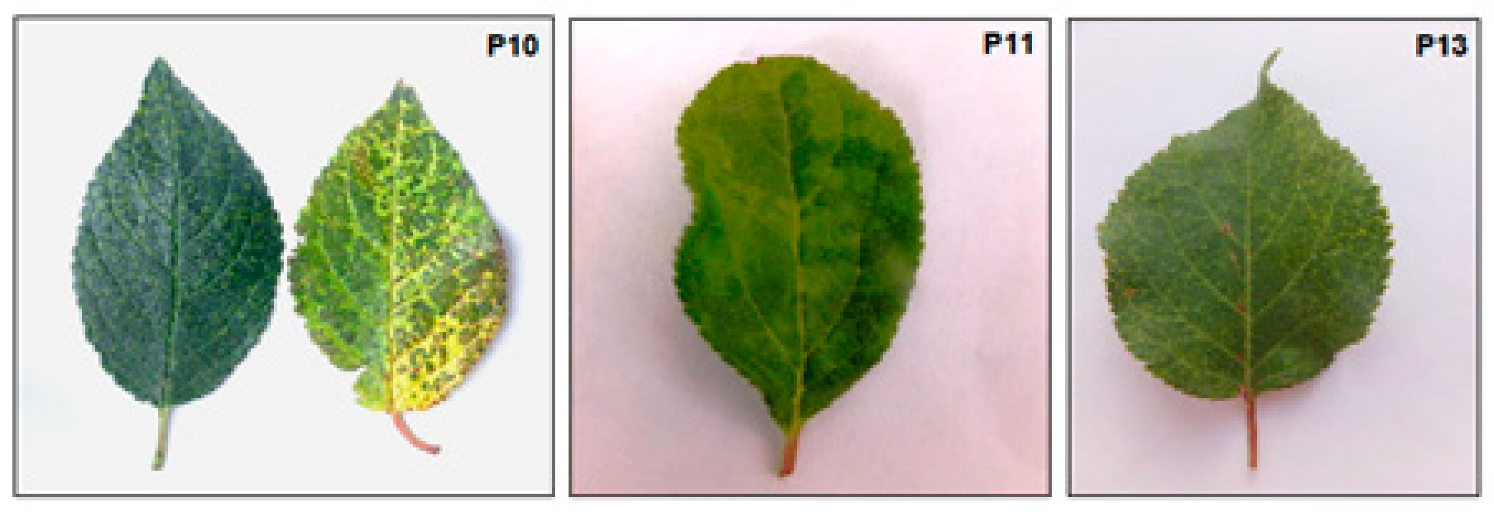
3.1. Plant Material and Virus Detection

Three ten-year-old trees with symptoms of suspected viral infection were selected for metatranscriptomic analysis. The samples taken from these trees were as follows: P10 from P. domestica cultivar “Čačanska beste” grafted on P. cerasifera; P11 from P. domestica seedling of unknown cultivar; and P13 from ((P. cerasifera Ehrh. x P. armeniaca L.) x P. brigantiaca Vill.) hybrid grafted on P. cerasifera. The symptoms observed on the leaves of these plants are presented in Figure 1.
Both P10 and P13, as well as the P11 samples, tested positive in ELISA for PNRSV and PDV, respectively (Table S1). The amplicons of the expected sizes, 380 bps, were obtained by RT-PCR assay (Figure S1). Their sequencing by the Sanger method confirmed PNRSV in samples P10 and P13 and PDV in sample P11. Thus, the HTS results were corroborated by ELISA and RT-PCR.

3.2. HTS Results

The reads related to PNRSV and PDV were generated from samples P10, P11, and P13 (Table 1). They covered the assembled ilarvirus-specific contigs with an average depth of coverage 145× to 900×. In addition, reads related to the fungal ourmia-like virus 1 (OuLV1) genome were found in sample P10.

3.3. Characterization of the PDV Genome

Three contigs related to PDV genomic RNAs were assembled in the P11 sample. According to BLASTn, the contig of 3341 nt was most closely related (95.2–95.5%) to RNA1 of Canadian PDV isolates 13C233 (MZ220979), 13C259 (MZ220983), and Salmo BC cherry (U57648) from sweet cherry and Australian isolate NS9R1 (KY883326) from Prunus. This contig covered genomic RNA1 of the related isolates nearly completely, except for seven to 26 3′-terminal and eight 5′-terminal nt. ORF1 3168 nt long encoding replicase of 1055 amino acid (aa) residues was identified in the contig. The methyltransferase (MET) and helicase (HEL) domains were predicted at positions 95–394 and 766–1025, respectively. ORF1 was flanked with 5′- and 3′-non-coding regions (NCR) of 29 and 144 nt, respectively.
Another contig, 2585 nt long, was most closely related (92.1–92.3% identity) to RNA2 of Canadian isolates Niagara D5 (MK522388), 13C226 (MZ220866), and 13C233 (MZ220870) from sweet cherry, encompassing RNA2 of these isolates nearly completely, except for four to 49 nt at their 5′ or 3′ ends. ORF2 2364 nt long encoding a protein of 787 aa was identified in the contig. Bromoviridae RdRp motif was found at positions 298–669 of this protein. ORF2 was flanked with 5′- and 3′-NCRs of 17 and 204 nt, respectively.
One more contig, of 2124 nt, was most closely related (95–95.9% identity) to RNA3 of PDV-SWM1 (GU181406) and PDV-PA63 (GU181405) Poland isolates from sweet cherry and PDV-SOF15P11 (GU181404) and PDV-SOF17P17 (GU181399) Italian isolates from sour cherry. ORF3a 882 nt and ORF3b 657 nt long were identified, which encode the MP and CP of 293 and 218 aa, respectively. ORF3a and ORF3b were separated by the intergenic region (IR) of 71 nt. The 5′- and 3′-NCRs consisted of 254 and 260 nt, respectively. The lysine (K) and arginine (R)-rich motif KxxxKxxRxxxxRxxxKxK and the crucial arginine residue, both of which are responsible for binding to the 3′-NCRs of viral RNAs [8], were detected in the N-termini of the MP and CP, respectively.
Thus, the nearly complete genome of the Russian PDV isolate from plum was sequenced. Three genomic RNAs of the virus were assembled and four ORFs typical of PDV were identified. The genome sequences of the P11 isolate were deposited in GenBank under accession numbers OQ995020 to OQ995022.

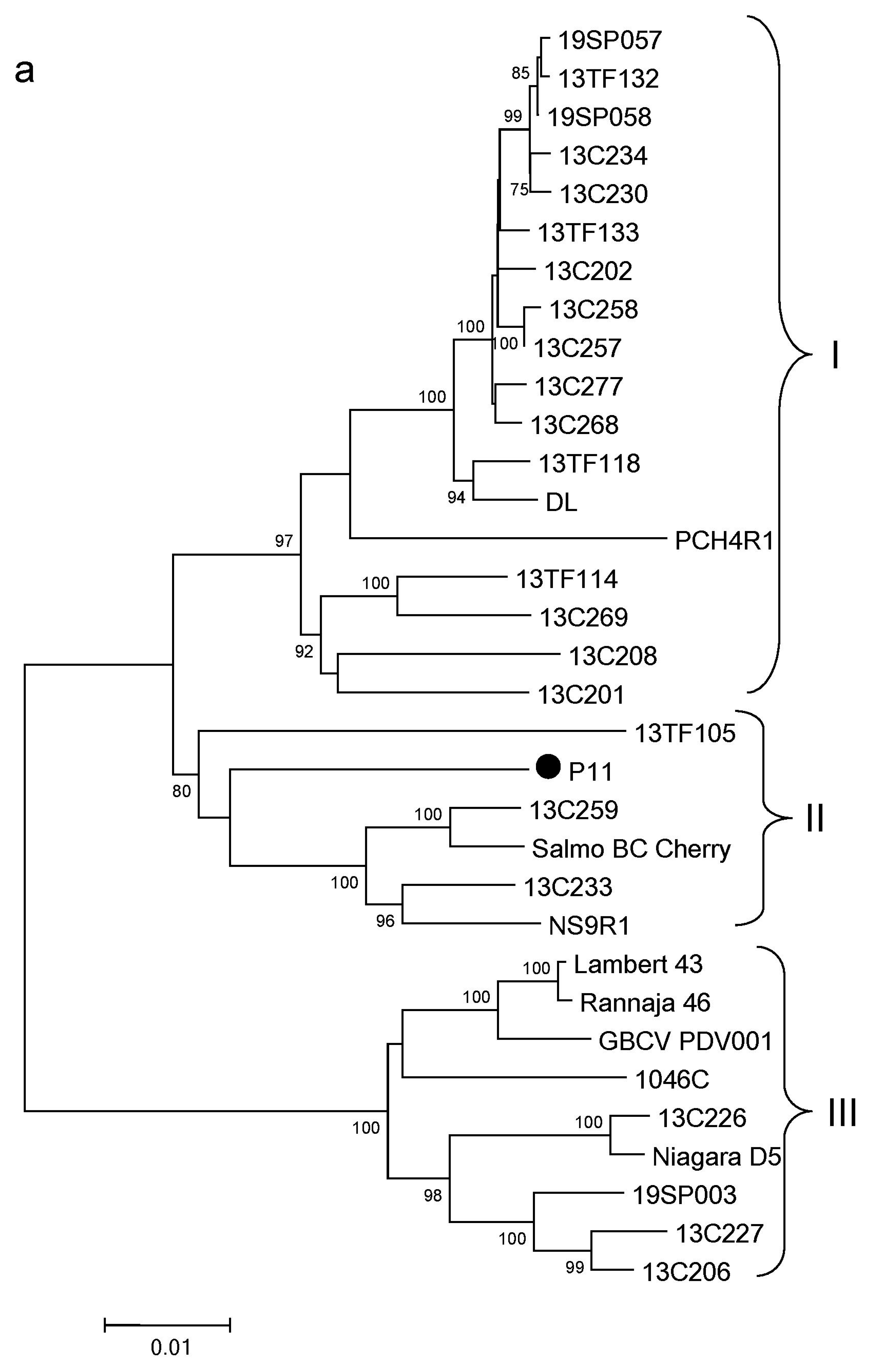
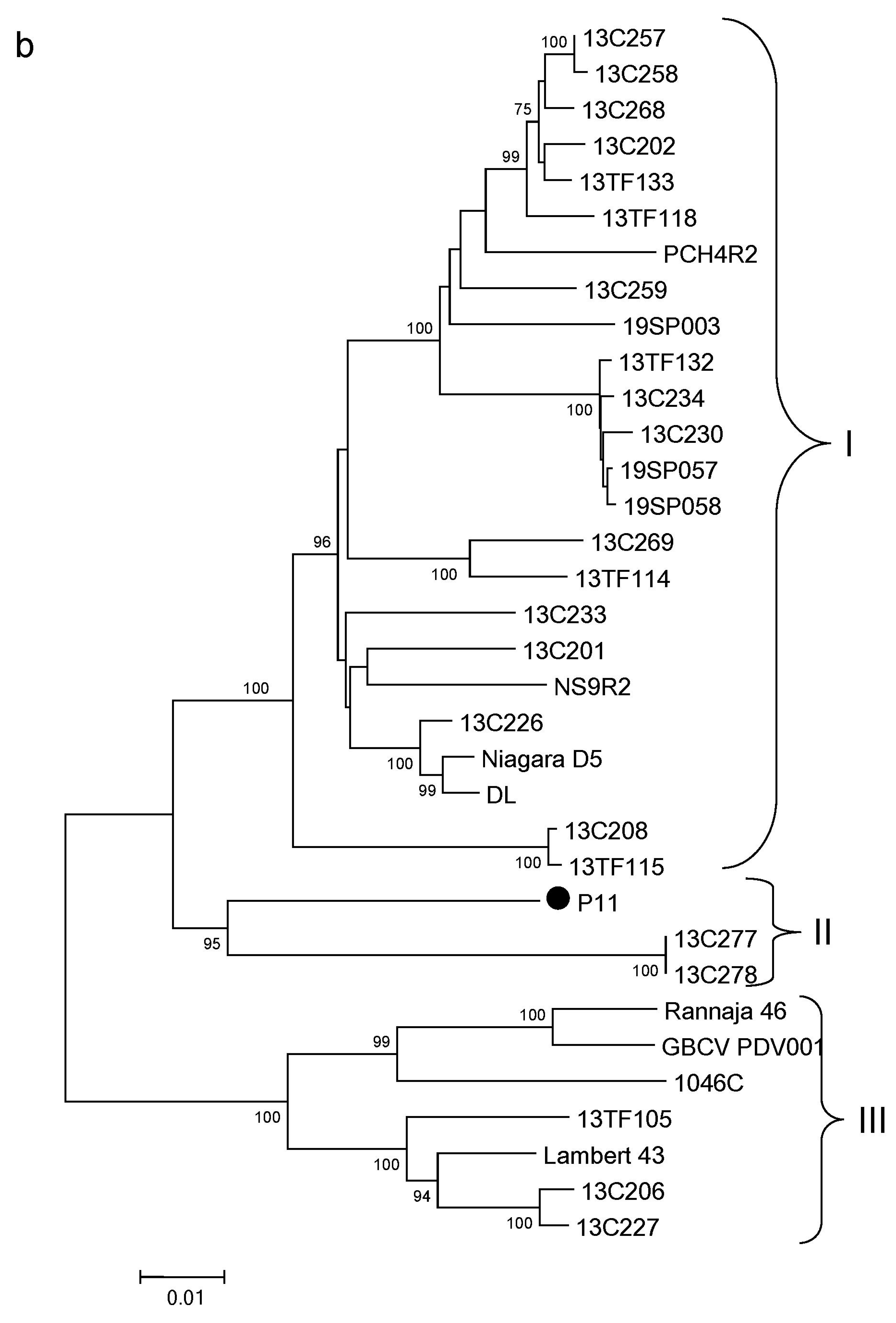
3.4. Phylogenetic Analysis of PDV

For phylogenetic analysis, all complete sequences of three PDV RNAs available in GenBank on May 2023 (Table S2) were used. However, their alignments showed that the 5′- and 3′-NCRs of different isolates were heterogeneous and varied significantly in length. Therefore, an ORF sequence-based phylogeny was generated. In the case of RNA3, the analyzed genome region also included the IR separating ORF3a and ORF3b.
ORF1, ORF2, and ORF3a-IR-ORF3b were divided into three main clusters (Figure 2a–c) that were in agreement with the previous phylogenies based on the CP, MP, or RNA3 sequences [21,22,23,24,25]. Most isolates were affiliated with the same cluster, irrespective of the analyzed genome segment. At the same time, a number of isolates (13C206, 13C226, 13C227, 13C233, 13C257, 13C258, 13C259, 13C277, 13TF105, Niagara D5, and 19SP003, all from Canada) relocated between clusters, depending on the genome segment studied. The genome segments of the isolate P11 were all assigned to cluster II (Figure 2a–c). However, the composition of this group differed from one tree to another, due to both the mentioned migration of some isolates among clusters and the presence of many new isolates in the tree reconstructed from RNA3-derived sequences. Thus, the immediate surroundings of P11 in the cluster varied from tree to tree. Nevertheless, the clustering of the P11 ORF1 and ORF3a-IR-ORF3b was consistent with the sequence identity analysis, while ORF2 clustering did not match the BLASTn results.

3.5. Characterization of the PNRSV Genomes

Three PNRSV-related contigs were assembled from the reads generated in both the P10 and P13 samples. In P10, the contig of 3314 nt was most closely related (over 99% identity) to RNA1 of the sour cherry isolates Cigany (MF069044) and PV-0962 (OP357942) from the Czech Republic and Germany, respectively. This contig covered RNA1 of these isolates nearly completely, except for several nt at the 5′- and 3′-termini. ORF1 of 3138 nt encoding replicase of 1045 aa was identified and MET and HEL domains were found at positions 51–387 and 753–1010 of the protein. ORF1 was flanked with the 5′- and 3′-NCRs of 16 and 160 nt, respectively. The 2568 nt long contig was most closely related (over 99% identity) to RNA2 of the Prunus isolates PV-0962 (OP357943), TNpeach5 (OL800570), Cigany (MF069045), and CNU1 (LC752755) from Germany, the USA, the Czech Republic, and South Korea, as well as from the Australian isolate SA from the honey bee Apis mellifera (MH427284). ORF2 of 2400 nt encoding a protein of 787 aa was identified in the contig. RdRp motif was found at positions 298–669 of this protein. ORF2 was flanked with 5′- and 3′-NCRs of 11 and 157 nt, respectively. The third contig of 1944 nt was most closely related (99% identity) to RNA3 of the Prunus isolates PV-0962 (OP357944) and Cigany (MF069046) from Germany and the Czech Republic. ORF3a 852 nt and ORF3b 675 nt long were identified, which encode the MP and CP of 283 and 224 aa, respectively. ORF3a and ORF3b were separated by the IR of 74 nt. The 5′- and 3′-NCRs consisted of 174 and 169 nt, respectively. The RNA-binding motif (KxxxRxxKKxxRxxKxKxKxR) and two RNA-binding arginine residues [8] were detected in the N-termini of the MP and CP, respectively.
Three PNRSV-related RNAs assembled in sample P13 were collinear to their counterparts from P10 and their structure was typical of PNRSV. However, RNA1, RNA2, and RNA3 of P13 were only 95.3%, 95.4%, and 96.5% identical to the corresponding P10 RNAs, respectively. Furthermore, BLASTn showed that they were closer to other PNRSV isolates. RNA1 of P13 was most closely related (97.5% identity) to the Canadian isolate 13C241 (MZ451020) from Prunus, RNA2—to the isolates Ruzyne (ON088601) from P. cerasifera (Czech Republic), and Pea1 (MH727228) from peach (China) (95.5% identity), while RNA3 was most closely related to the Czech isolate Ruzyne (ON088602) from P. cerasifera (97% identity). Apparently, two divergent PNRSV isolates were detected in the collection.
Nearly complete genomes of PNRSV isolates P10 and P13 were deposited in GenBank under accession numbers OR090897 to OR090902.

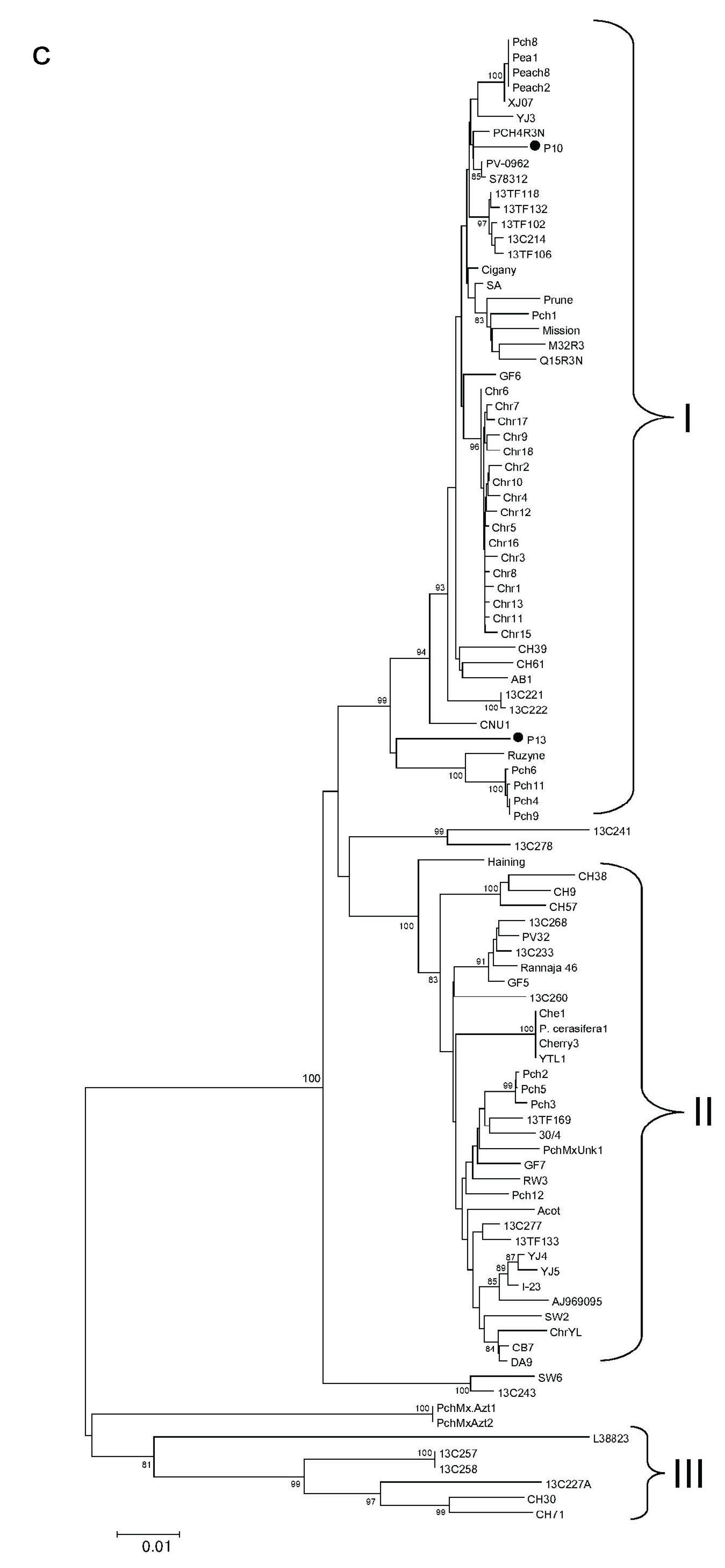
3.6. Phylogenetic Analysis of PNRSV

As for PDV, the phylogeny was based on ORFs and the analyzed genome region of RNA3 included the IR between ORF3a and ORF3b. All complete sequences of three PNRSV RNAs available in GenBank on May 2023 (Table S3) were used for the phylogenetic analysis.
Most PNRSV isolates grouped into two or three main clusters, while others occupied an intermediate position between them (Figure 3a–c). This result showed high PNRSV genetic diversity, in accordance with previous data [22,26,27,28,29]. ORF1, ORF2, and ORF3a-IR-ORF3b of P10 were assigned to cluster I, which consisted of a large number of isolates found on different stone fruits in many regions of the world. In contrast, there was no clear-cut position for P13 at the phylogenetic trees. At the same time, positions of the genomic segments of two Russian isolates on the phylogenetic trees corresponded to the sequence identity analysis. Like PDV, a few isolates (13TF133, CH57, Acot, P. cerasifera 1, Pch12, 13C227, 13C260, and Rannaja 46) migrated between clusters, depending on the analyzed genomic segment.

3.7. Recombination in the PDV and PNRSV Complete Genomes

Three genomic RNAs of some PNRSV and PDV isolates were assigned to different phylogenetic clusters (Figure 2a–c and Figure 3a–c). Recombination, which can be the reason for the incongruence of phylogenetic trees [30], was detected in RNA3 of a few Slovakian PDV isolates [21]. The ORF-based recombination analysis was employed using RDP4.101 program. Several recombination events were inferred in PDV and PNRSV isolates (Table 2). They were detected in each genomic RNAs of these viruses, except for PDV RNA2. For example, the PDV isolates 13C257 and 13C258, as well as PNRSV isolates 13C257 and 13C258, were recombinants, in which the MP gene was affected. Recombination could explain the relocation of these isolates to group III at the RNA3-based phylogenetic trees closer to potential major parents (Figure 2c and Figure 3c, respectively). However, no recombination was detected in the rest of the relocated PDV and PNRSV isolates or in the Russian isolates P10, P11, and P13.

3.8. Partial Characterization of Ourmia-like Virus Genome

Reads related to OuLV1 were assembled into a contig of 806 nt. This contig covered approximately one-third (positions 585 to 1392) of the genomic RNA of the OuLV1 isolates CREA-VE-6AS1 (NC_076380, 97.4% identity) and DMS10_23903 (MN532617, 96.4% identity) from the plant pathogenic fungi Cladosporum cladosporioides and Plasmopara viticola, respectively. The partial genome sequence of the fungal virus isolate named P10-Fun was deposited in GenBank under accession number OR183588. The detection of a mycovirus indicated that the tree was affected by a fungal infection, but the fungus species was not determined. Possibly, the symptoms observed on the plum leaves (Figure 1, sample P10) were of fungal origin.

4. Discussion

Virus diseases of stone fruits are of great concern due to their wide distribution and high economic significance. The timely and sensitive diagnosis of viruses is a clue in controlling virus diseases, tracing sources of infection, and studying the diseases’ impacts on infected trees. However, virus diagnosis in fruit trees can be difficult, due to the high genetic diversity of viruses, their presence in latent form, or uneven distribution in the host [31]. The HTS approach potentially allows the finding of all viruses infecting a plant and characterizing their genomes.
In this work, an attempt was made to identify viruses in the field Prunus germplasm collection using HTS. The virome of symptomatic Prunus trees was studied. Based on HTS results, PNRSV, PDV, and fungal ourmia-like virus were detected. Although PDV is observed mainly on cherries (Table S2), in this work it was found on plum. Both ilarviruses were confirmed by RT-PCR and ELISA tests. The foliar symptoms looked not quite common for these ilarviruses. However, ilarvirus symptoms on stone fruits are known to be variable and depend on the isolate, the host, and the environmental conditions, ranging from typical symptoms to the absence of visible manifestations of the disease. In addition, PNRSV symptoms in the P10 sample may be masked by a putative fungal infection. As the studied trees bloom and bear fruit every year, the viruses could infect them through pollen. Samples P10 and P13 could be also infected through myrobalan (P. cerasifera) rootstock, which is no longer available and cannot be tested for viruses. Considering the economic impact of PNRSV and PDV on stone fruit crops, a broader survey is needed to determine the prevalence and genetic diversity of these viruses in the collection and limit their further spread.
Complete genomes of two divergent PNRSV isolates and one PDV isolate were sequenced. Four ORFs and all motifs in the virus proteins, typical for these viruses, were identified in the new isolates. This is the first report and characterization of the PNRSV and PDV full-length genomes from Russia, expanding the information on their geographical distribution and genetic diversity. Multiple alignments of all genome segments of the Russian PNRSV and PDV isolates, together with other isolates of these viruses, available in GenBank, revealed no specific features in the Russian isolates, apart from point mutations randomly distributed along the genome.
According to BLASTn and phylogenetic analysis, the Russian isolate P10 was very close (over 99% identity, irrespective to the analyzed genome segment) to the PNRSV isolates Cigany, PV-0962, TNpeach5, and CNU1 found on various Prunus species in the Czech Republic, Germany, the USA, and South Korea. P13 was most closely related to other PNRSV isolates from Prunus spp., such as 13C241 (RNA1, 97.5% identity), Ruzyne and Pea1 (RNA2, 95.5% identity), and Ruzyne (RNA3, 96.9% identity), that originated from the Czech Republic, China, and Canada. Thus, BLASTn and phylogenetic analysis showed that RNA1, RNA2, and RNA3 of P10 were most similar to the same PNRSV isolates available in GenBank. Three genome segments of P13 were also most closely related to PNRSV isolates available in GenBank., although unlike those of the P10, they were known isolates of this virus. In contrast, each of three genome RNA of the P11 was closest to quite different PDV isolates, sharing 92 to 96% identity (Section 3.3, Figure 2a–c). In addition, the CP gene of the P11 was shown to be closest to the Turkish PDV isolates Sa12 (KF718675), PD8 (EF524271), and M27 (KF718668) from sweet cherry (98% identity). (They were not included in the alignments arranged from complete ORFs.) Thus, the position of the Russian PDV isolate, as well as others, remains uncertain. As the number of full-length PDV genomes increases, the position of the Russian PDV isolate on phylogenetic trees will possibly become clearer.
Taken together, this shows that the new Russian isolates from plums and ((P. cerasifera x P. armeniaca) x P. brigantiaca) trihybrids were the closest to the PNRSV and PDV isolates found on various stone fruit crops in different countries and continents. This suggests that PNRSV and PDV isolates from different hosts, and of different geographical origin, can be very similar.
Current PNRSV and PDV phylogeny is mainly based on the CP, MP, or RNA3 sequences [11]. In this work, all (nearly) complete RNA1, RNA2, and RNA3 of PNRSV and PDV available in GenBank were employed in phylogenetic analysis. For PDV, 33, 34, and 46 sequences of RNA1, RNA2, and RNA3, respectively, were retrieved from GenBank (Table S2). For PNRSV, 44, 40, and 98 RNA1, RNA2, and RNA3 sequences, respectively, were used for the analysis (Table S3).
Phylogenetic analysis of the ORF1, ORF2, and ORF3a-IR-ORF3b genome segments of PDV and PNRSV showed substantial genetic diversity of these viruses, consistent with previous phylogenies of their RNA3 derived sequences [21,22,23,24,25,26,27,28,29]. Despite this, no geographical clustering was detected for the full-length genomes of the PDV and PNRSV isolates compared in this work. One of the explanations may be that these viruses are currently continuing to spread, constantly mixing the existing populations.
In addition, phylogenetic analysis of genome-wide sequences revealed no host clustering among PNRSV and PDV isolates. This was perhaps due to easy hybridization between Prunus species [3], which might potentially contribute to the cross-species transmission of the viruses. On the other hand, the vast majority of full-length genome sequences deposited in GenBank are cherry isolates that can distort the results of the host clustering analysis. It cannot be ruled out that further study of PNRSV and PDV isolates from other Prunus species will enable us to explain the lack of the host clustering.
Comparison of the phylogenetic trees reconstructed from three genomic segments showed that some isolates moved from one phylogroup to another, depending on the exploring genome segment. For example, the PDV isolates Niagara D5 and 13C226 were assigned to group III (RNA1), I (RNA 2) or group II (RNA 3). The PDV isolates 13C259 and NS9R relocated from group II (RNA1) to group I (RNA2 and RNA3) (Figure 2a–c). The PNRSV isolate P. cerasifera-1 moved from group I (RNA1) to group II (RNA2 and RNA3), but the immediate surroundings of RNA2 and RNA3 of this isolate were quite different (Figure 3a–c). It should be noted that the mentioned clustering was supported by high bootstrap values (100%). Such relocations of the isolates change the branching order and lead to the incongruence of the trees reconstructed for each of three genome RNAs. Incongruence of the phylogenetic trees may stem from different evolutionary histories of the genome segments of some PDV and PNRSV isolates, due to recombination or reassortment in their ancestors.
Several recombination events were detected in PDV and PNRSV isolates (Table 2). However, these findings cannot explain the different branching order for those isolates that are unaffected by recombination. Reassortment may be another possible explanation. Since members of the genus Ilarvirus have a segmented genome, and each of their three genomic RNAs is encapsidated in a separate viral particle, mixed infection of a plant with different virus isolates can lead to the exchange of genome segments between them and the emergence of reassortants in offspring. In the genus Cucumovirus (which also belongs to the family Bromoviridae), viable reassortants of cucumber mosaic virus, peanut stunt virus, and tomato aspermy virus were obtained experimentally [32] and found in nature [33]. Intra-specific reassortment was not studied in ilarviruses, although it has been suggested in the genesis of this genus [33,34]. Further study of the putative reassortment in PNRSV and PDV isolates may enhance our understanding of the evolution and genetic diversity of ilarviruses.

Supplementary Materials

The following supporting information can be downloaded at: https://www.mdpi.com/article/10.3390/horticulturae9080941/s1, Text file S1: CTAB-based protocol for total RNA isolation; Figure S1: Agarose gel electrophoresis of PCR products generated by RT-PCR assay of ilarviruses using degenerate ilarvirus-specific primers [20]. Sample names are shown above the picture. NC—negative control (see Section 2.4 in the text). The arrow to the right to the picture indicates PCR products of the corresponding size. M—GeneRuler 100 bp Plus DNA Ladder (Thermo Scientific).; Table S1: Detection of prunus necrotic ringspot virus (PNRSV) and prune dwarf virus (PDV) by ELISA (A405); Table S2: Prune dwarf virus isolates employed in phylogenetic analysis; Table S3: Prunus necrotic ringspot virus isolates employed in phylogenetic analysis.

Author Contributions

Conceptualization, S.C. and I.M.; methodology, A.S., S.T. and N. S.; software, F.S. and N.S.; validation, A.S., S.T., N.S., F.S., K.P. and I.M.; investigation, S.C., A.S., N.S. and K.P.; writing—original draft preparation, S.C.; writing—review and editing, A.S., S.T., N.S., F.S., K.P. and I.M.; visualization, A.S. and F.S.; supervision, I.M.; funding acquisition, S.C. All authors have read and agreed to the published version of the manuscript.

Funding

This research was funded by the Russian Science Foundation, grant number 23-16-00032.

Data Availability Statement

Sequencing data were deposited in SRA and GenBank, and their accession numbers are provided within the article.

Acknowledgments

We thank Lubov Lukicheva and Valentina Gorina, the supervisors of the Nikita Botanical Garden Prunus germplasm collection, for their assistance during the collection survey.

Conflicts of Interest

The authors declare no conflict of interest.

References

  1. Hadidi, A.; Barba, M.; Candresse, T.; Jelkmann, W. Virus and Virus-Like Diseases of Pome and Stone Fruits; APS Press: St. Paul, MN, USA, 2011. [Google Scholar]
  2. Maliogka, V.; Minafra, A.; Saldarelli, P.; Ruiz-García, A.; Glasa, M.; Katis, N.; Olmos, A. Recent advances on detection and characterization of fruit tree viruses using high-throughput sequencing technologies. Viruses 2018, 10, 436. [Google Scholar] [CrossRef] [PubMed]
  3. Rubio, M.; Martinez-Gomez, P.; Marais, A.; Sanches-Navarro, J.A.; Pallas, V.; Candresse, T. Recent advances and prospect in Prunus virology. Ann. Appl. Biol. 2017, 171, 125–138. [Google Scholar] [CrossRef]
  4. Rott, M.; Xiang, Y.; Boyes, I.; Belton, M.; Saeed, H.; Kesanakurti, P.; Hayes, S.; Lawrence, T.; Birch, C.; Bhagwat, B.; et al. Application of next generation sequencing for diagnostic testing of tree fruit viruses and viroids. Plant Dis. 2017, 101, 1489–1499. [Google Scholar] [CrossRef]
  5. Martelli, G.; Flores, R.; Schneider, B. Classification of pome and stone fruit viruses, viroids and phytoplasmas. In Virus and Virus-Like Disease of Pome and Stone Fruits; Hadidi, A., Barba, M., Candresse, T., Jelkman, W., Eds.; APS Press: St. Paul, MN, USA, 2011; pp. 13–16. [Google Scholar]
  6. Hammond, R.W. Prunus necrotic ringspot virus. In Virus and Virus-Like Disease of Pome and Stone Fruits; Hadidi, A., Barba, M., Candresse, T., Jelkman, W., Eds.; APS Press: St. Paul, MN, USA, 2011; pp. 207–213. [Google Scholar]
  7. Caglayan, K.; Ulubas-Serce, C.; Gazel, M.; Varvery, C. Prune dwarf virus. In Virus and Virus-like Disease of Pome and Stone Fruits; Hadidi, A., Barba, M., Candresse, T., Jelkman, W., Eds.; APS Press: St. Paul, MN, USA, 2011; pp. 199–205. [Google Scholar]
  8. Pallas, V.; Aparicio, F.; Herranz, M.C.; Sanchez-Navarro, J.A.; Scott, S.W. The molecular biology of ilarviruses. Adv. Vir. Res. 2013, 87, 139–181. [Google Scholar]
  9. Tayal, M.; Wilson, C.; Cieniewicz, E. Bees and thrips carry virus-positive pollen in peach orchards in South Carolina, United States. J. Econ. Entomol. 2023, 116, 1091–1101. [Google Scholar] [CrossRef] [PubMed]
  10. Koziel, E.; Bujarski, J.J.; Otulak, K. Molecular biology of Prune dwarf virus—A lesser known member of the Bromoviridae but a vital component in the dynamic virus-host cell interaction network. Int. J. Mol. Sci. 2017, 18, 2733. [Google Scholar] [CrossRef]
  11. Pallas, V.; Aparicio, F.; Herranz, M.C.; Amari, K.; Sanches-Pina, M.A.; Myrta, A.; Sanchez-Navarro, J.A. Ilarviruses in Prunus spp.: A continued concern for fruit trees. Phytopathology 2012, 102, 1108–1120. [Google Scholar] [CrossRef]
  12. Mitrofanova, I.V.; Smykov, A.V.; Mitrofanova, O.V.; Lesnikova-Sedoshenko, N.P.; Chirkov, S.N.; Zhdanova, I.V. Using in vitro embryo culture for obtaining new breeding forms of peach. Acta Hortic. 2020, 1289, 159–165. [Google Scholar] [CrossRef]
  13. Prikhod’ko, Y.N.; Chirkov, S.N.; Metlitskaya, K.V.; Tsubera, L.V. Distribution of viral diseases of stone fruits in European part of Russia. Agric. Biol. 2008, 1, 26–32. (In Russian) [Google Scholar]
  14. Trifonova, A.A.; Boris, K.V.; Mesyats, N.V.; Tsiupka, V.A.; Smykov, A.V.; Mitrofanova, I.V. Genetic diversity of peach cultivars from the collection of the Nikita Botanical Garden based on SSR markers. Plants 2021, 10, 2609. [Google Scholar] [CrossRef] [PubMed]
  15. Gambino, G.; Perrone, I.; Gribaudo, I. A rapid and effective method for RNA extraction from different tissues of grapevine and other woody plants. Phytochem. Anal. 2008, 19, 520–525. [Google Scholar] [CrossRef] [PubMed]
  16. Nurk, S.; Meleshko, D.; Korobeynikov, A.; Pevzner, P.A. metaSPAdes: A new versatile metagenomic assembler. Genome Res. 2017, 27, 824–834. [Google Scholar] [CrossRef] [PubMed]
  17. Langmead, B.; Salzberg, S. Fast gapped-read alignment with Bowtie 2. Nat. Methods 2012, 9, 357–359. [Google Scholar] [CrossRef]
  18. Kumar, S.; Stecher, G.; Tamura, K. MEGA7: Molecular Evolutionary Genetics Analysis Version 7.0 for Bigger Datasets. Mol. Biol. Evol. 2016, 33, 1870–1874. [Google Scholar] [CrossRef]
  19. Martin, D.P.; Murrel, B.; Golden, M.; Khoosal, A.; Muhire, B. RDP4: Detection and analysis of recombinant pattern in virus genomes. Virus Evol. 2015, 1, vev003. [Google Scholar] [CrossRef]
  20. Untiveros, M.; Perez-Egusquiza, Z.; Clover, G. PCR assays for the detection of members of the genus Ilarvirus and family Bromoviridae. J. Virol. Methods 2010, 165, 97–104. [Google Scholar] [CrossRef] [PubMed]
  21. Predajna, L.; Shihelska, N.; Benedictova, D.; Soltys, K.; Candresse, T.; Glasa, M. Molecular characterization of Prune dwarf virus cherry isolates from Slovakia shows their substantial variability and reveals recombination events in PDV RNA3. Eur. J. Plant Pathol. 2017, 147, 877–885. [Google Scholar] [CrossRef]
  22. Bonilla, F.R.; Cieniewicz, E. Distribution and diversity of prunus necrotic ringspot virus, prune dwarf virus, and peach latent mosaic viroid in wild Prunus spp. in South Carolina and Georgia. PhytoFrontiers 2022, 2, 363. [Google Scholar] [CrossRef]
  23. Kamenova, I.L.; Borisova, A.Z. Biological and molecular properties of Prune dwarf virus cherry isolates from Bulgaria. J. Plant Dis. Prot. 2022, 129, 301–311. [Google Scholar] [CrossRef]
  24. Ulubas Serce, C.; Ertunc, F.; Ozturk, A. Identification and genomic variability of Prune dwarf virus variants infecting stone fruit trees in Turkey. J. Phytopathol. 2009, 157, 298–305. [Google Scholar] [CrossRef]
  25. Vaskova, D.; Petrzik, K.; Spak, J. Molecular variability of the capsid protein of the prune dwarf virus. Eur. J. Plant Pathol. 2000, 106, 573–580. [Google Scholar] [CrossRef]
  26. Noorani, M.S.; Baid, S.B.; Khan, J.A.; Pravej, A. Whole genome characterization and diagnostics of prunus necrotic ringspot virus (PNRSV) infecting apricot in India. Sci. Rep. 2023, 13, 4393. [Google Scholar] [CrossRef] [PubMed]
  27. Kamenova, I.L.; Borisova, A.Z. Molecular variability of the coat protein gene of prunus necrotic ringspot virus on sweet and sour cherry in Bulgaria. J. Plant Pathol. 2021, 103, 97–104. [Google Scholar] [CrossRef]
  28. Ulubas Serce, C.; Ertunc, F. RT-PCR detection and molecular characterization of Prunus necrotic ringspot virus isolates occurring in Turkey. J. Phytopathol. 2004, 152, 498–502. [Google Scholar] [CrossRef]
  29. Fiore, N.; Fajardo, T.V.; Prodan, S.; Herranz, M.C.; Aparicio, F.; Montealegre, J.; Elena, S.F.; Pallas, V.; Sanchez-Navarro, J. Genetic diversity of the movement and coat protein genes of South American isolates of Prunus necrotic ringspot virus. Arch. Virol. 2008, 153, 909–919. [Google Scholar] [CrossRef] [PubMed]
  30. Chare, E.R.; Holmes, E.C. A phylogenetic survey of recombination in plant viruses. Arch. Virol. 2006, 151, 933–946. [Google Scholar] [CrossRef]
  31. Ravelonandro, M. Reliable metodologies and impactful tools to control fruit tree viruses. Crops 2021, 1, 32–41. [Google Scholar] [CrossRef]
  32. Pita, J.S.; Roossink, M.J. Fixation of emerging interviral recombination in Cucumber mosaic virus populations. J. Virol. 2013, 87, 1264–1269. [Google Scholar] [CrossRef]
  33. Chen, Y.; Chen, J.; Zhang, H.; Tang, X.; Du, Z. Molecular evidence and sequence analysis of a natural reassortant between Cucumber mosaic virus subgroup IA and II strains. Virus Genes 2007, 35, 405–413. [Google Scholar] [CrossRef]
  34. Codoner, F.; Elena, S.F. The promiscuous evolutionary history of the family Bromoviridae. J. Gen. Virol. 2008, 89, 1739–1747. [Google Scholar] [CrossRef]
Figure 1. Virus-like symptoms on the leaves of Prunus plants infected with prunus necrotic ringspot virus (P10 and P13) and prune dwarf virus (P11).
Horticulturae 09 00941 g001
Figure 2. Phylogenetic analysis of ORF1 (a), ORF2 (b), and ORF3a-IR-ORF3b genome segment (c) of prune dwarf virus conducted in MEGA7 [18]. The names of isolates are shown next to the end of branches. The trees were reconstructed using the neighbor-joining method. The evolutionary distances were computed using the Kimura 2-parameter model. Bootstrap values (>75%) from 1000 replicates are shown next to the corresponding nodes. Curly brackets combine isolates belonging to the same cluster. The scale bar indicates the number of substitutions per nucleotide. The Russian isolate P11 is highlighted with a black circle (●). The numbers (I, II, III) belong to the same cluster.
Horticulturae 09 00941 g002aHorticulturae 09 00941 g002bHorticulturae 09 00941 g002c
Figure 3. Phylogenetic analysis of ORF1 (a), ORF2 (b), and ORF3a-IR-ORF3b genome region (c) of prunus necrotic ringspot virus conducted in MEGA7 [18]. The names of isolates are shown next to the end of branches. The trees were reconstructed using the neighbor-joining method and the Kimura 2-parameter model. Bootstrap values (>75%) from 1000 replicates are shown next to the corresponding nodes. Curly brackets combine isolates belonging to the same cluster. The scale bar indicates the number of substitutions per nucleotide. The Russian isolates P10 and P13 are highlighted with a black circle (●). The numbers (I, II, III) belong to the same cluster.
Horticulturae 09 00941 g003aHorticulturae 09 00941 g003bHorticulturae 09 00941 g003c
Table 1. Results of high-throughput sequencing of Prunus samples.
SampleNumber of Clean Reads a per SampleVirus DetectedNumber of Virus-
Specific Reads b
P1023657640Prunus necrotic ringspot virus 2710 (RNA1)
1956 (RNA2)
4368 (RNA3)
Ourmia-like virus 58
P1117497020Prune dwarf virus 3645 (RNA1)
3146 (RNA2)
7272 (RNA3)
P132903382Prunus necrotic
ringspot virus
7444 (RNA1)
3424 (RNA2)
1129 (RNA3)
a Pair-ended reads of 250 nt length; b determined using Bowtie2 v.2.4.4.
Table 2. Recombination events inferred in complete genomes of prune dwarf virus (PDV) and prunus necrotic ringspot virus (PNRSV) by the Recombination Detection Program v.4.101.
VirusGenome RegionRecombinantBeginning BreakpointEnding BreakpointMajor ParentMinor Parentp Values *
PDVORF1PCH4R1600110113TF118Salmo BC Cherry10−5
3118331713TF118Salmo BC Cherry10−7
ORF3a-IR-ORF3bPA789631614SWM113TF11410−8
PE2479631613Rannaja 46SOF17P1710−18
SOF15P111933SWM1SOF17P1710−7
13C2574756113C22613C27810−9
13C2584756113C22613C27810−9
PNRSVORF1Che112682090P. cerasifera 1Rannaja 4610−28
Che220903138CH57P. cerasifera 110−35
Q15R1N179013TF132CH5710−33
TNpeach5844154213C243Q15R1N10−15
TNpeach53262413C243SCh-Taian10−7
ORF2Che11243ORF2 end PV-0962P. cerasifera 110−14
Che21324ORF2 end PV-0962P. cerasifera 110−26
ORF3a-IR-ORF3b13C2574826413C227A13C26010−15
13C2584826413C227A13C26010−15
* Multiple comparison (MC) corrected probability of false-positive detection of recombination event.
Disclaimer/Publisher’s Note: The statements, opinions and data contained in all publications are solely those of the individual author(s) and contributor(s) and not of MDPI and/or the editor(s). MDPI and/or the editor(s) disclaim responsibility for any injury to people or property resulting from any ideas, methods, instructions or products referred to in the content.

