Article

Micromorphology of Barleria albostellata (Grey Barleria) Flower and Pollen Grains

by
Serisha Gangaram
1,*,
Yougasphree Naidoo
1,
Yaser Hassan Dewir
2,
Moganavelli Singh
1 and
Katalin Magyar-Tábori
3
1
School of Life Sciences, Westville Campus, University of KwaZulu-Natal, P.O. Box X54001, Durban 4000, South Africa
2
Plant Production Department, College of Food and Agriculture Sciences, King Saud University, Riyadh 11451, Saudi Arabia
3
Research Institute of Nyíregyháza, Institutes for Agricultural Research and Educational Farm (IAREF), University of Debrecen, P.O. Box 12, 4400 Nyíregyháza, Hungary
*
Author to whom correspondence should be addressed.
Horticulturae 2023, 9(7), 732; https://doi.org/10.3390/horticulturae9070732
Submission received: 17 May 2023 / Revised: 14 June 2023 / Accepted: 20 June 2023 / Published: 21 June 2023
(This article belongs to the Special Issue Morphology, Palynology and Phytochemicals of Medicinal Plants)

Abstract

:
Barleria albostellata C.B. Clarke (grey barleria, Acanthaceae) is an indigenous shrub to South Africa and has been relatively understudied. This shrub is a valuable medicinal plant with a wide spectrum of antibacterial and anti-inflammatory activities. Detailed studies on the floral and pollen morphology on B. albostellata are rare. This study was conducted to observe the morphology of the flower and pollen grains using stereomicroscopy and scanning electron microscopy (SEM). Morphological observations showed numerous non-glandular trichomes on the bracteoles and bracts of B. albostellata. Three types of trichomes were identified on these structures: I—unicellular, II—multangulate-dendritic branched non-glandular trichomes, and III—capitate glandular trichomes. A taxonomical description of the floral structures using stereo and SEM micrographs is provided. SEM micrographs revealed the pollen grains as globose tricolporate with a rough honeycomb exine, and small granules inside the lumina. The diameter of the pollen grains was 77.53 ± 5.63 μm, whereas the aperture of these grains was 14.31 ± 0.59 µm. This study provides insight into the floral biology of B. albostellata, and the results presented here will add to the body of knowledge and encourage further research on this species.

1. Introduction

Flower receptiveness plays an important role in pollination variability, reproductive success, and plant productivity [1,2]. These active traits include timing of the anther opening and pollen appearance, anther and stigma position, and flower receptiveness and morphology [3,4]. Pollination involves the transferal of pollen from the anther to the stigma of the flower [2,5], and the rate of success is highly dependent on pollen viability (the capability of pollen to induce seed set efficiently) [6,7]. Furthermore, the reproductive success of a plant may depend on its ability to attract flower visitors. These visitors that aid in pollination may exert selection on specific floral traits that are attractive to them [7,8,9]. The genus Barleria has approximately 300 species of shrubs and herbs that are distributed in the subtropical and tropical regions of the world [10,11,12,13]. Members of this genus originated from the Far East of Japan, through southern Asia, Arabia, India, Africa, Madagascar to as far west of Central America and Mexico [12,14]. Several species of Barleria are known for their floral diversity. Additionally, there are specialized structures on their surface for the synthesis, storage, and/or secretion of secondary metabolites with the ultimate goal of anti-herbivory tactics and protection against water loss [15,16]. These structures are known as trichomes and occur on the plant surface as hairs or external glands. Trichomes may be family or species-specific and may vary in their chemical composition [15]. Plant secretory structures can also be used as taxonomic characters, assisting in the identification of plant families [15].
Barleria albostellata, an evergreen shrub, thrives in semi-shade to full-sun woodland areas of South Africa, and under suitable conditions, grows up to 1.5 m in height (Figure 1). However, in colder regions, they can become deciduous to semi-deciduous [17]. In South Africa, B. albostellata, generally known as ‘grey barleria’ or in Afrikaans the ‘Bosviooltjie’, belongs to the family Acanthaceae [17]; and is widely distributed from Limpopo, Gauteng, and Mpumalanga to KwaZulu-Natal [17]. The genus name Barleria was derived from a French botanist and Dominican monk, Jacques Barrelier [17]. This shrub flourishes from September to May, with beautiful white flowers appearing sporadically (Figure 1). Flowers appear from a dense compound inflorescence and are surrounded by four leafy-bracts [18]. The blooming flowers are white in color and have a tinge of purple on the bracts. In contrast to the flowers, the leaves are grey-green and have an abundance of velvety hairs. This plant develops fairly quickly and reaches maturity in about three years [17]. Barleria albostellata contains medicinal properties which were verified by Amoo et al. [19]. It was found that several extracts from this plant exhibited excellent anti-inflammatory properties and a broad-spectrum of antibacterial activity. This plant has a relatively high flavonoid content, with an added effect from tannin and iridoid compounds [20]. Although B. albostellata has no recorded practice in traditional medicine, many species of Barleria have been used in traditional medicine and were confirmed to contain various compounds possessing biological effects such as analgesic, anti-inflammatory, antileukemic, antihyperglycemic, antitumor, anti-amoebic, antibiotic, and virucidal activities [21,22,23,24,25,26].
Acanthaceae is regarded as a eurypalynous family [27], with significant diversity in the pollen shape, size, exine structure, apertures, and ornamentation [28,29,30,31,32]. Plants in this family demonstrate a thriving diversity of ecological and morphological characteristics. These include a large range of pollinator relationships and floral morphologies [33,34,35]. Members of Barleria are easily recognized for their globose, tricolporate pollen with roughly reticulate (also referred to as the honeycomb-patterned) and inter-apertural exine [18,36,37]. Characterizing the morphology of pollen grains is useful in plant systematics and this can further add to the body of knowledge within the genus and family. Little is known on the floral and pollen morphology of B. albostellata, however a substantial amount of work has been done in other species within the family Acanthaceae [38,39,40] and in other species of Barleria. Previous studies have found that members of this genus are pollinated by moths [41,42], or attract various species of butterflies [17]. Additionally, it was noted that the flowers of B. albostellata were also pollinated by insects and butterflies [43]. On a regular basis, carpenter bees were also observed to visit the flowers of B. albostellata. Plants within this genus produce copious amounts of nectar which attract bumble bees [17]. Several morphological features of the B. albostelata flower and pollen grains have been largely unexplored. Secretory structures documented within Barleria include non-glandular and glandular peltate and capitate trichomes. Therefore, the present study aimed to describe the floral morphology and pollen of B. albostellata using stereo and scanning electron microscopy.

2. Materials and Methods

2.1. Plant Materials

Flowers of B. albostellata were collected from the University of KwaZulu-Natal, School of Life Sciences, Westville Campus (29°49′51.6″ S, 30°55′30″ E), Durban, South Africa. A voucher specimen (Accession no. 7973000) was deposited in the Ward Herbarium of the University of KwaZulu-Natal, Life Sciences, Westville Campus. Five replicates of flowers were analyzed using microscopy techniques.

2.2. Stereomicroscopy

Fresh flowers were examined using the Nikon AZ100 stereomicroscope (Nikon Corporation, Yokohama, Japan) equipped with a Nikon Fiber Illuminator and photographed using the NIS-Elements Software (NIS-elements D 3.00).

2.3. Scanning Electron Microscopy (SEM)

The micromorphology of chemically-fixed flowers of B. albostellata was examined in detail. The initial step of preparation involved dissecting the different parts of the flower: petal, stigma, style, anther, and filament and thereafter primary fixating the sections (±10 mm2) in 2.5% glutaraldehyde for 18–24 h. After primary fixation, samples were rinsed for 5 min each (thrice) with 0.1 M sodium phosphate buffer (pH 7.2) and then post-fixed in 0.5% osmium tetroxide for 3 h at room temperature. The samples were washed thrice (for 5 min each) with sodium phosphate buffer and dehydrated gradually with increasing concentrations of ethanol (30%, 50%, 75%, 100%) twice, for 5 min each, followed by exposure to 100% ethanol for two sessions, each of 10 min. Dehydrated samples were critically point-dried using the Quorum K850 Critical Point Dryer (Quorum Technologies Ltd., Laughton, East Sussex, UK) with a vertical chamber. Samples were mounted onto aluminum stubs using double-sided adhesive carbon tape and sputter coated with a layer of gold using the Quorum 150 RES (Quorum Technologies Ltd.), a combined system for carbon and sputter coating. The samples were then viewed and photographed using the LEO 1450 SEM at a working distance (WD) of 12–15 mm. Images were captured using the SmartSEM image software (Zeiss, Jena, Germany). (protocol was adapted from the microscopy and microanalysis unit, University of KwaZulu-Natal, Westville). With respect to the stigma of the flower, pollen grains were dusted from the stigma onto aluminum stubs using double-sided adhesive carbon tape and sputter coated with a layer of gold using the Quorum 150 RES (Quorum Technologies Ltd.), a combined system for carbon and sputter coating. Images of pollen grains were captured using the SmartSEM image software (Zeiss, Jena, Germany). Diameters of pollen grains were determined using ImageJ software Java 1.53e (Fiji, http://fiji.sc/Fiji, accessed on 10 June 2021) [44]

3. Results and Discussion

3.1. Analysis of Floral Structures via Stereomicroscopy

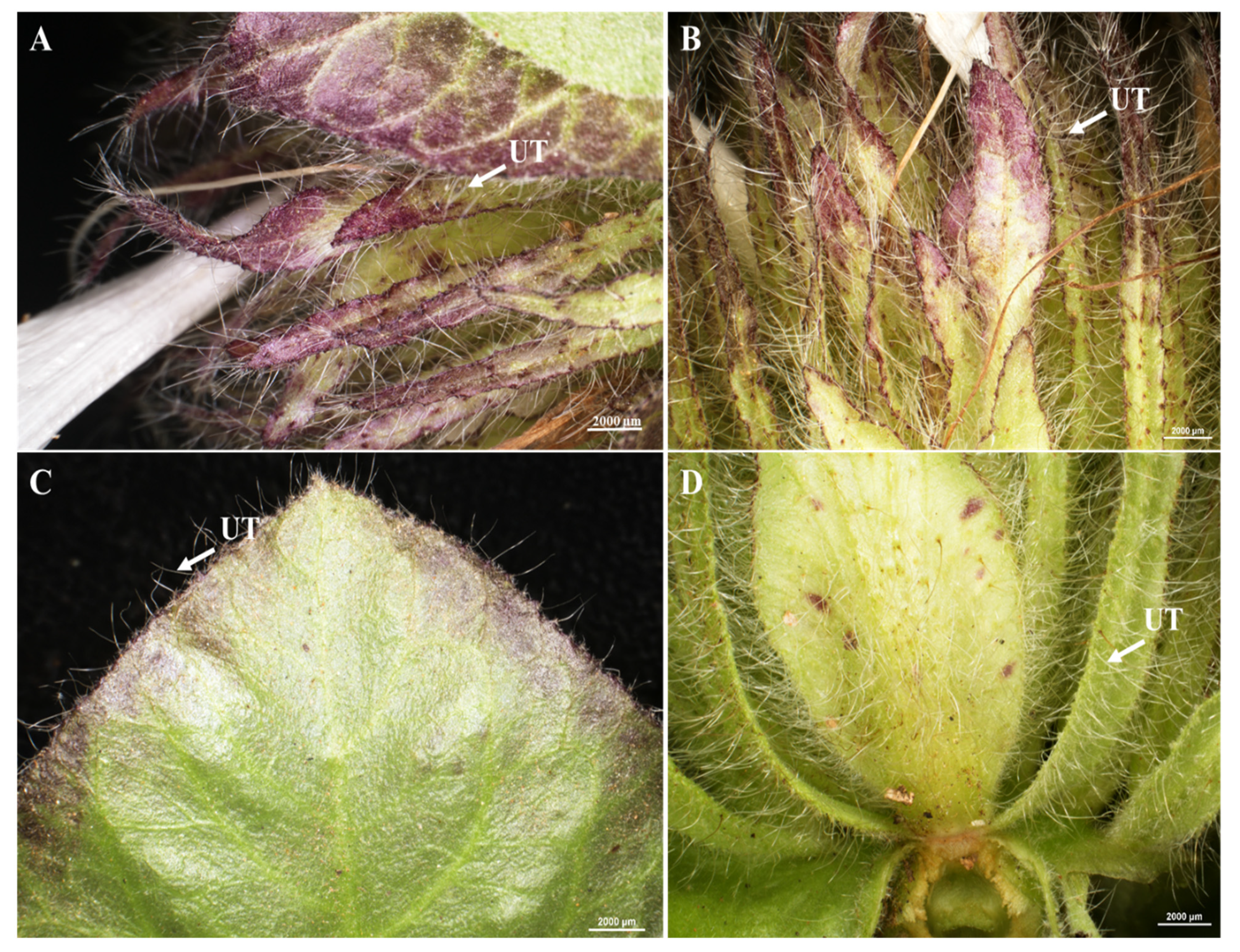
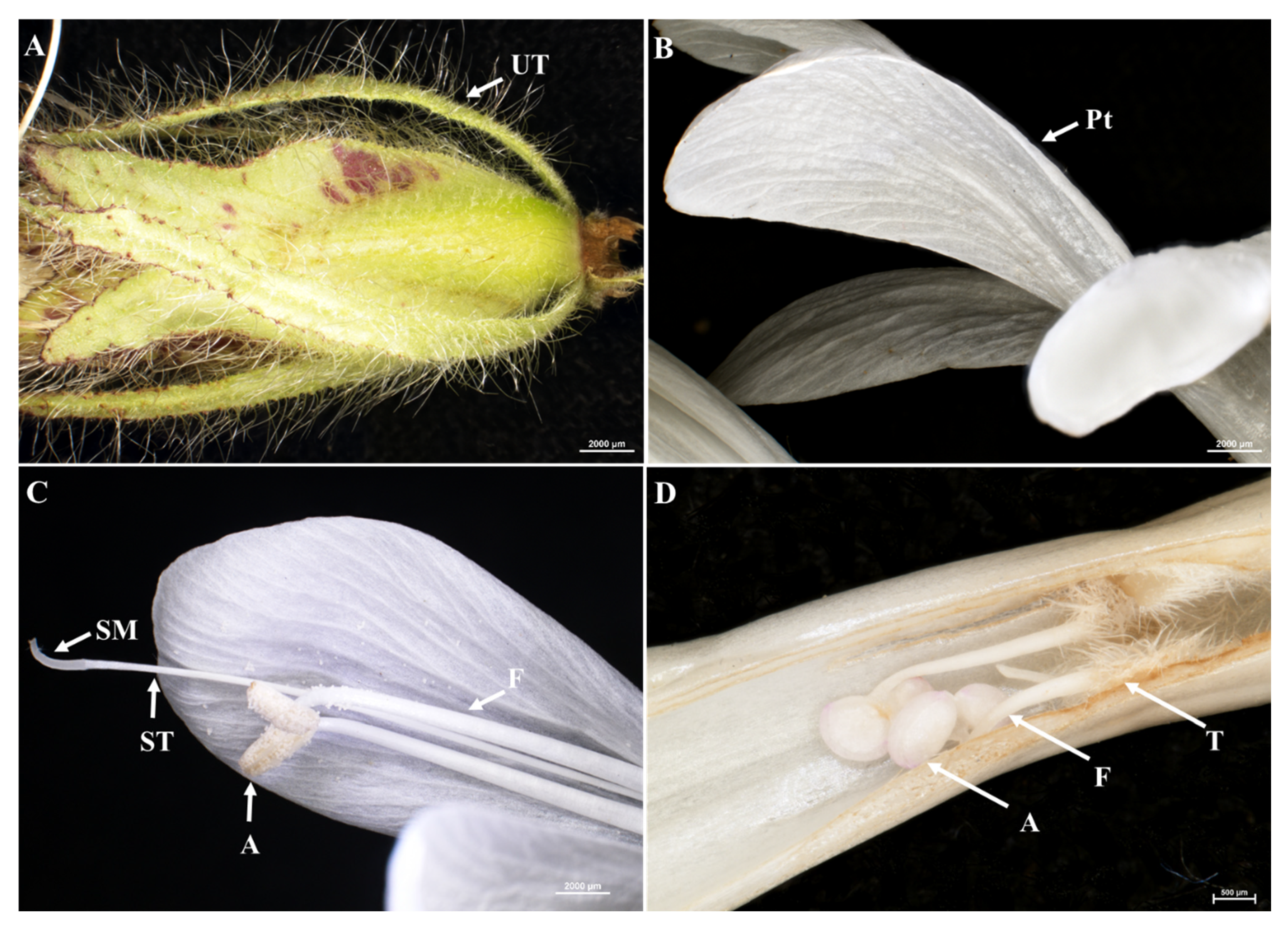
Bracteoles of B. albostellata vary from narrowly ovate to ovate, with glabrous or hairy surfaces and margins that are spiny, with scanty teeth (Figure 2A–D). Long white hairs (unicellular non-glandular trichomes) are prominent on the surface and margins of the floral bracts, (Figure 2A,B) upper, and lower bracteoles (Figure 2C,D), and a posterior lobe with sharp, curved apiculus (Figure 3A). Certain species of Barleria have characteristic non-glandular trichomes which are unicellular [45]. Non-glandular trichomes are recognized exclusively for their physical protection in plants against biotic and abiotic stresses [46,47], and to deter herbivores from feeding and ovipositing insects [48,49,50].
Bracts are highly modified, chartaceous, foliaceous, with reticulate venation being prominent; margins are entire, serrate, or irregularly dentate. The inflorescence is a compound, terminal synflorescence, capitate or strobilate with units of solitary flowers. Flowers of B. albostellata (2–4 flowers) are bisexual with a nectariferous disc, zygomorphic, and in cymes (a wide, flat-topped, distinct flower cluster in which the central flowers are opened first) [51,52]. Flowering is an important phenological event, which impacts the reproductive success of a species [53]. The flowers are enclosed by four leafy, hairy, purple-tinged bracts. Purple-tinged bracts are assumed to contain some sort of nectar (Figure 2A–D). The delimitation of the genus ‘Barleria’ and specifically the taxonomic description of the leaves and flowers of B. albostellata have been only described by Balkwill and Balkwill [18].
The corolla (petals, 1 + 4) is irregular, thin, tubular, and gamopetalous (Figure 3B). The scent of the flowers of B. albostellata is produced nocturnally, with the strongest smell produced by mature, unopened buds, than with the open flowers. The floral visitors observed to interact with the flowers of B. albostellata were butterflies and bees. Similar observations were reported by Balkwill et al. [54] in flowers of B. greenii. Generally, flowers of Barleria are pollinated by butterflies, and they were described as large, white, and tubular, comprising deep nectaries/nectariferous discs, which function as nectar guides [55,56]. Bumblebees were observed to frequently visit B. greenii and remove nectar from outside of the flower, by creating a narrow slit at the base of the corolla tube [54]). Several trichome-derived compounds are used as attractants for species-specific pollination [57]. Trichomes are also involved in specialized mechanisms of insect capture for pollination [58]. The fertile stamens (anther + filament), inserted on the corolla, are usually present in pairs (Figure 3C) and are not didynamous. Filaments are long and may appear as twisted, can cross over each other, and are usually hairy at the base (Figure 3C). Anthers are basifixed and longitudinally dehisce, whilst the style is terete. The stigma is filiform and is found beyond the level of the dehisced anthers, while the style arches upward and is terete (Figure 3C). Similar morphological characteristics were observed in flowers of B. greenii [54] and in B. saxatilis [59]. They suggested that the position of the stigma above the anthers promotes autonomous self-pollination. The slit along the lower corolla lobe revealed the growing stamen with hairy trichomes attached to the lower region of the filament (Figure 3D), a characteristic of species within Barleria [60].

3.2. Floral Structures Observed via Scanning Electron Microscopy

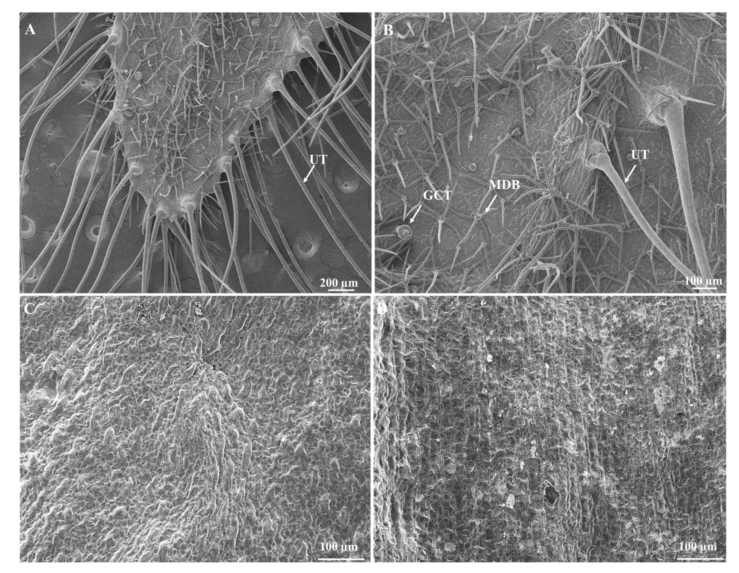
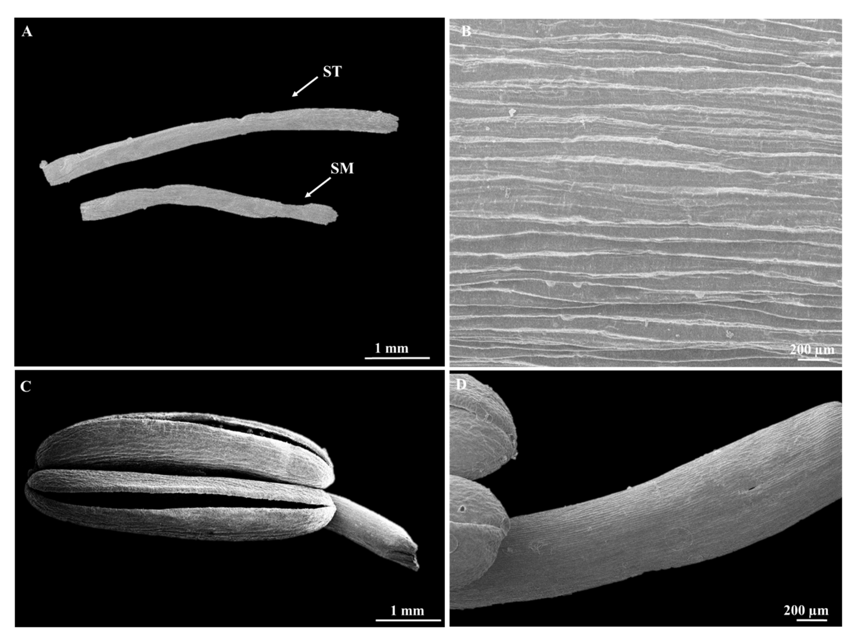
Floral bracts were heavily pubescent with non-glandular and glandular trichomes (Figure 4A,B). Unicellular non-glandular trichomes were highly dense, long, pointed, and located on the serrated edges of the floral bracts or occurring along the mid-region. Similar observations were reported for B. aristata floral bracts [61]. Perhaps the edges of the floral bracts might have responded to insect damage, therefore increasing the trichome density. There were only a few glandular capitate trichomes scattered all over the floral bracts, while the multangulate-dendritic branched (MDB) non-glandular trichomes were predominant (Figure 4A,B). In certain cases, the MDB non-glandular trichomes were found to ‘arch over’ the glandular trichomes. Due to its proximity, the MDB non-glandular trichomes may provide some sort of physical protection to these glandular tichomes. The adaxial and abaxial surfaces of a petal contained several grooves and appeared as coarse and pitted (Figure 4C,D), with epidermal cells in an irregular shape. With high magnification, parallel striations can be seen on a section of the surface of the stigma (Figure 5B). The cap of the anther is curved and round (protection of pollen), with a slit in the middle (Figure 5C).

3.3. Pollen Morphology

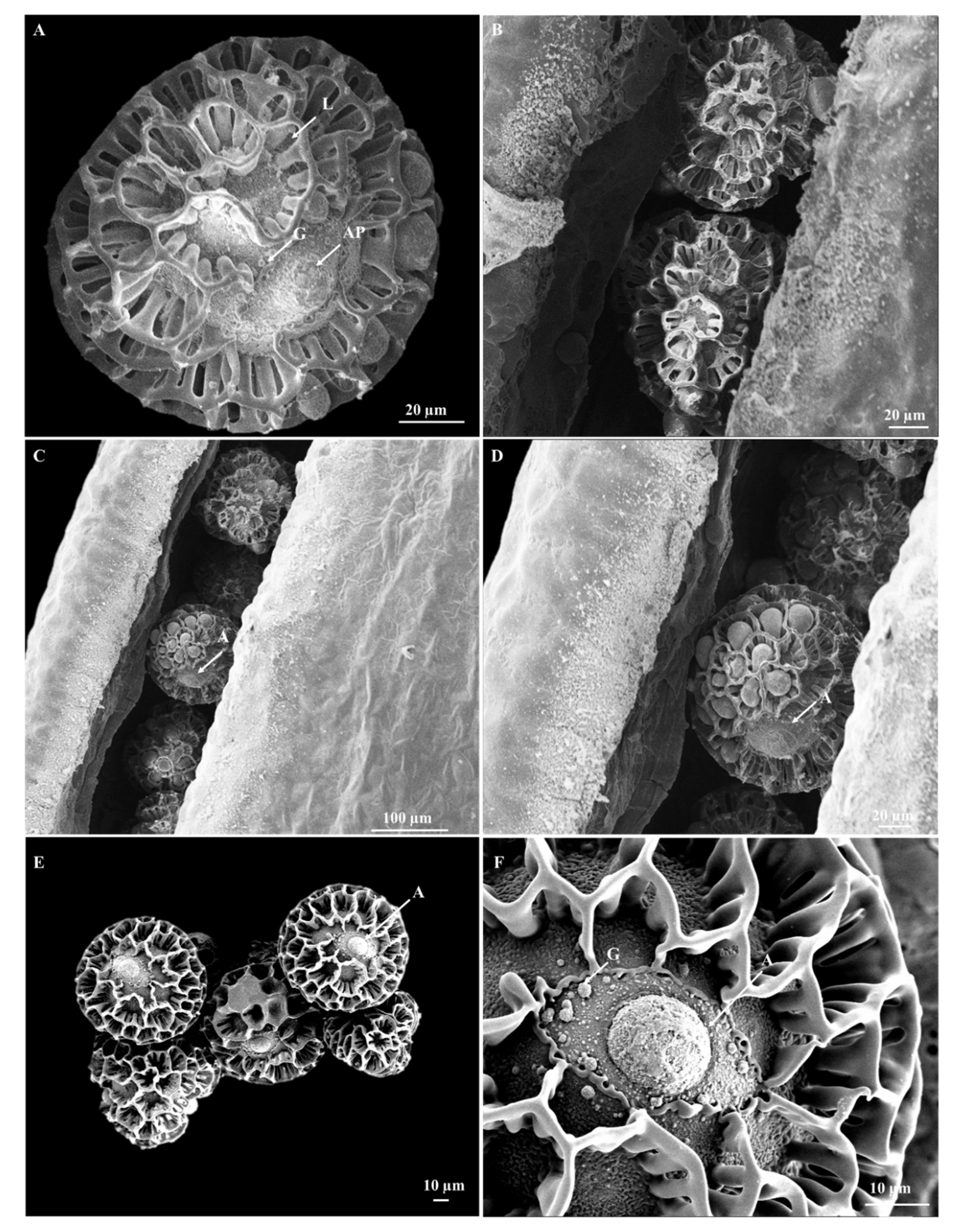
Pollen micromorphological features have contributed beneficial phylogenetic information in the accurate identification of species within Acanthaceae [13,39]. Scanning electron micrographs of pollen grains had an open reticulate tectum and appeared as globose tricolporate in equatorial view, honeycombed-shaped, with intense, coarse reticulation of the inter-apertural exine (Figure 6A); these characteristics are specific to pollen found in species of Barleria [11,18,36,37,60,62,63]. Pollen grains in B. albostellata had a diameter of 77.53 ± 5.63 μm (Figure 6).
This parameter varied from 60.5 ± 0.3 μm in B. parviflora to 81.5 ± 1 μm in B. orbicularis. Similar pollen grain sizes were found in B. albostellata, and almost the same diameters (74.9 ± 0.7 μm) were documented for B. ventricosa and B. proxima (79.1 ± 1 μm), respectively [13]. Before pollination at maturity, pollen grains are located inside the cap of the anther for protection (Figure 6B–D). Tiny granules are observed inside lumina of the pollen grain (Figure 6A). This was also noted in B. parviflora, B. ventricosa [13], B. prionitis, and B. hochstetteri [64]. The aperture of pollen grains in B. albostellata appeared circular in shape (Figure 6E,F), which was also noted in B. bispinosa [13]. The aperture width of pollen grains in B. albostellata was 14.31 ± 0.59 µm. The width varied from 9 μm in B. acanthoides and B. aculeata, 13 μm in B. tetracantha, 16 μm in B. ventricosa and B. bispinosa, to 23 μm in B. prionitis [13]. Pollen grains in various families are recognized by distinct morphological features represented in their exine [65]. Similar pollen grains characteristics to that of B. albostellata were found in B. grootbergensis [66] and B. durairajii [63]. Studies highlight that pollen viability is significantly reduced with increasing air humidity and temperature [67]. Barleria albostellata thrives in subtropical and tropical conditions, well-drained soils, and can grow under relatively cold conditions [43]. Pollen grains in Barleria are characteristic to the family Acanthaceae, however their reticulate ornamentation displays close resemblances with those found in their associated genera such as Lepidagathis, Crabbea, and Ruellia [68].
The morphology of the flower and pollen grains using various microscopic techniques showed numerous non-glandular trichomes on the bracteoles and bracts of B. albostellata. Three types of trichomes were identified on these structures and were found in other species of Barleria [54,59,60,61]. MDB non-glandular trichomes may provide some sort of physical protection to the glandular capitate tichomes. The pollen micromorphological features found are characteristic to species of Barleria [11,18,36,37,54,62,63].

4. Conclusions

The combination of stereo- and scanning electron microscopy facilitated the identification of the floral and pollen morphology of B. albostellata. Knowledge on the floral biology and pollen morphology of B. albostellata obtained by microscopy techniques has been very incomplete so far. Floral structures identified were compared to previously reported information in other species of Barleria. Pollen grains of B. albostellata are complex, intricate, and display reticulate sculpturing. Thus, the results presented in this study contribute significantly to our growing understanding of the floral and pollen biology of B. albostellata. In this regard, this study is novel, and results reported here will also assist taxonomists in identifying B. albostellata using SEM micrographs of their distinct pollen structures. Additional ultrastructural studies on the floral structures should be conducted to further examine the internal features of the cells and organelles. Further studies may also focus on evaluating the micromorphology of the seeds and roots of B. albostellata.

Author Contributions

Conceptualization, S.G. and Y.N.; methodology, S.G. and Y.N.; formal analysis, S.G., Y.N. and M.S.; investigation, S.G., Y.N. and M.S.; data curation, S.G., Y.N., Y.H.D. and M.S.; writing—original draft preparation, S.G. and Y.N.; writing—review and editing, Y.N., Y.H.D., M.S. and K.M.-T.; validation, M.S., Y.H.D. and K.M.-T.; visualization, M.S., Y.H.D. and K.M.-T.; supervision, Y.N., Y.H.D. and M.S.; project administration, Y.N.; funding acquisition, Y.N. and Y.H.D. All authors have read and agreed to the published version of the manuscript.

Funding

National Research Foundation (Grant No. 118897), South Africa and Researchers Supporting Project number (RSP2023R375), King Saud University, Riyadh, Saudi Arabia.

Data Availability Statement

All data are presented in the article.

Acknowledgments

Authors extend their appreciation to the National Research Foundation (Grant No. 118897), South Africa and the staff at the Microscopy and Microanalysis Unit at the University of KwaZulu-Natal for their assistance with the microscopy components of the research. The authors acknowledge Researchers Supporting Project number (RSP2023R375), King Saud University, Riyadh, Saudi Arabia.

Conflicts of Interest

The authors declare no conflict of interest.

References

  1. Zhang, J.; Lin, M.; Chen, H.; Zhu, Q.; Chen, X. Floral biology and pistil receptivity of the drumstick tree (Moringa oleifera Lam.). Arch. Biol. Sci. 2018, 70, 299–305. [Google Scholar] [CrossRef] [Green Version]
  2. Liu, F.; Gao, C.; Chen, M.; Tang, G.; Sun, Y.; Li, K. The impacts of flowering phenology on the reproductive success of the narrow endemic Nouelia insignis Franch (Asteraceae). Ecol. Evol. 2021, 11, 9396–9409. [Google Scholar] [CrossRef] [PubMed]
  3. Harder, L.D.; Williams, N.M.; Jordan, C.Y.; Nelson, W.A. The effects of floral design and display on pollinator economics and pollen dispersal. In Cognitive Ecology of Pollination: Animal Behaviour and Floral Evolution; Chittka, L., Thomson, J.D., Eds.; Cambridge University Press: Cambridge, UK, 2001; pp. 297–317. [Google Scholar]
  4. Budečević, S.; Hočevar, K.; Manitašević Jovanović, S.; Vuleta, A. Phenotypic Selection on Flower Traits in Food-Deceptive Plant Iris pumila L.: The Role of Pollinators. Symmetry 2023, 15, 1149. [Google Scholar] [CrossRef]
  5. McMullen, C.K.; Close, D.D. Wind pollination in the Galápagos Islands. Not. Galápagos 1993, 52, 12–17. [Google Scholar]
  6. Dafni, A.; Firmage, D. Pollen viability and longevity: Practical, ecological and evolutionary implications. Plant Syst. Evol. 2000, 222, 113–132. [Google Scholar] [CrossRef]
  7. Christopher, D.A.; Mitchell, R.J.; Karron, J.D. Pollination intensity and paternity in flowering plants. Ann. Bot. 2020, 125, 1–9. [Google Scholar] [CrossRef] [PubMed]
  8. Kay, K.M.; Sargent, R.D. The role of animal pollination in plant speciation: Integrating ecology, geography, and genetics. Annu. Rev. Ecol. Syst. 2009, 40, 637–656. [Google Scholar] [CrossRef] [Green Version]
  9. Sletvold, N.; Trunschke, J.; Smit, M.; Verbeek, J.; Ågren, J. Strong pollinator-mediated selection for increased flower brightness and contrast in a deceptive orchid. Evolution 2016, 70, 716–724. [Google Scholar] [CrossRef]
  10. Mabberley, J.B. The Plant—Book: A Portable Dictionary of the Vascular Plants, 2nd ed.; Cambridge University Press: Bath, UK, 1997. [Google Scholar]
  11. Darbyshire, I.; Vollesen, K.; Kelbessa, E. Acanthaceae, part 2. In Flora Zambesiaca; Timberlake, J.R., Martins, E.S., Eds.; Royal Botanic Gardens: London, UK, 2015; 8p. [Google Scholar]
  12. Kumari, R.; Kumar, S.; Kumar, A.; Goel, K.K.; Dubey, R.C. Antibacterial, antioxidant and immuno-modulatory properties in extracts of Barleria lupulina Lindl. BMC Complemen. Altern. Med. 2017, 17, 484. [Google Scholar] [CrossRef] [Green Version]
  13. Al-Hakimi, A.S.; Faridah, Q.; Abdulwahab, A.; Latiff, A. Pollen and seed morphology of Barleria L. (Barlerieae: Ruellioideae: Acanthaceae) of Yemen. S. Afr. J. Bot. 2018, 116, 185–191. [Google Scholar] [CrossRef]
  14. Balkwill, M.J.; Balkwill, K. A preliminary analysis of distribution patterns in a large, pantropical genus, Barleria L. (Acanthaceae). J. Biogeog. 1998, 25, 95–110. [Google Scholar] [CrossRef]
  15. Svoboda, K.P.; Svoboda, T.G.; Syred, A.D. Secretory Structures of Aromatic and Medicinal Plants: A Review and Atlas of Micrographs; Microscopix Publications: Knighton, UK, 2000; pp. 7–12. [Google Scholar]
  16. Schmid, N.B.; Giehl, R.F.; Döll, S.; Mock, H.P.; Strehmel, N.; Scheel, D.; Kong, X.; Hider, R.C.; Von Wirén, N. Feruloyl-CoA 6′-Hydroxylase1-dependent coumarins mediate iron acquisition from alkaline substrates in Arabidopsis. Plant Physiol. 2014, 164, 160–172. [Google Scholar] [CrossRef] [PubMed] [Green Version]
  17. Froneman, W.; Le Roux, L.N. Barleria albostellata. 2007. Available online: http://pza.sanbi.org/barleria-albostellata (accessed on 2 February 2019).
  18. Balkwill, M.J.; Balkwill, K. Delimitation and infra-generic classification of Barleria (Acanthaceae). Kew Bull. 1997, 52, 535–573. [Google Scholar] [CrossRef]
  19. Amoo, S.O.; Finnie, J.F.; Van Staden, J. In vitro pharmacological evaluation of three Barleria species. J. Ethnopharmacol. 2009, 121, 274–277. [Google Scholar] [CrossRef]
  20. Amoo, S.O.; Ndhlala, A.R.; Finnie, J.F.; Van Staden, J. Antifungal, acetylcholinesterase inhibition, antioxidant and phytochemical properties of three Barleria species. S. Afr. J. Bot. 2011, 77, 435–445. [Google Scholar] [CrossRef] [Green Version]
  21. Yosook, C.; Panpisutchai, Y.; Chaichana, S.; Santisuk, T.; Reutrakul, V. Evaluation of anti-HSV-2 activities of Barleria lupulina and Clinacanthus nutans. J. Ethnopharmacol. 1999, 67, 179–187. [Google Scholar] [CrossRef]
  22. Wang, B.U.; Wu, M.; Perchellet, E.M.; Mcilvain, C.J.; Sperfslage, B.J.; Huang, X.; Tamura, M.; Stephany, H.A.; Hua, D.H.; Perchellet, J.P. Asynthetic triptycene bisquinone which blocks nucleoside transport and induces DNA fragmentation, retains its cytotoxic efficacy in daunorubicin-resistant HL-60 cell lines. Int. J. Oncol. 2001, 19, 1169–1178. [Google Scholar]
  23. Jassim, S.A.A.; Naji, A.M. Novel antiviral agents: A medicinal plant perspective. J. App. Microbiol. 2003, 95, 412–427. [Google Scholar] [CrossRef] [Green Version]
  24. Suba, V.; Murugesan, T.; Arunachalam, G.; Mandal, S.C.; Saha, B.P. Anti-diabetic potential of Barleria lupulina extract in rats. Phytomedicine 2004, 11, 202–205. [Google Scholar] [CrossRef]
  25. Suba, V.; Murugesan, T.; Kumaravelrajan, R.; Mandal, S.C.; Saha, B.P. Antiinflammatory, analgesic and antiperoxidative efficacy of Barleria lupulina Lindl. extract. Phytother. Res. 2005, 19, 695–699. [Google Scholar] [CrossRef]
  26. Chomnawang, M.T.; Surassmo, S.; Nukoolkarn, V.S.; Gritsanapan, W. Antimicrobial effects of Thai medicinal plants against acne-inducing bacteria. J. Ethnopharma 2005, 101, 330–333. [Google Scholar] [CrossRef] [PubMed]
  27. Raj, B. Pollen morphological studies in the Acanthaceae. Grana Palynol. 1961, 3, 3–108. [Google Scholar]
  28. Graham, V.A.W. Delimitation and infra-generic classification of Justicia (Acanthaceae). Kew Bull. 1988, 43, 551–624. [Google Scholar] [CrossRef]
  29. Daniel, T.F. Pollen morphology of Mexican Acanthaceae: Diversity and systematic significance. Proc. Calif. Acad. Sci. 1998, 508, 217–256. [Google Scholar]
  30. Bhatt, A.; Naidoo, Y.; Nicholas, A. The foliar trichomes of Hypoestes aristata (Vahl) Sol. ex Roem. & Schult var aristata (Acanthaceae) a widespread medicinal plant species in tropical sub-Saharan Africa: With comments on its possible phylogenetic significance. Biol. Res. 2010, 43, 403–409. [Google Scholar]
  31. Choopan, T.; Grote, P.J. Cystoliths in the leaves of the genus Pseuderanthemum (Acanthaceae) in Thailand. Int. J. Sci. 2015, 12, 13–20. [Google Scholar]
  32. House, A.; Balkwill, K. FIB-SEM enhances the potential taxonomic significance of internal pollen wall structure at the generic level. Flora 2017, 236, 44–57. [Google Scholar] [CrossRef]
  33. McDade, L.A.; Masta, S.E.; Moody, M.L.; Waters, E. Phylogenetic relationships among Acanthaceae: Evidence from two genomes. Syst. Bot. 2000, 25, 106–121. [Google Scholar] [CrossRef]
  34. Daniel, T.F.; McDade, L.A.; Manktelow, M.; Kiel, C.K. The “Tetramerium Lineage” (Acanthaceae: Acanthoideae: Justicieae): Delimitation and intra-lineage relationships based on cp and nrlTS sequence data. Syst. Bot. 2008, 33, 416–436. [Google Scholar] [CrossRef]
  35. McDade, L.A.; Daniel, T.F.; Kiel, C.A. The Tetramerium Lineage (Acanthaceae, Justicieae) Revisited: Phylogenetic relationships reveal polyphyly of many new world genera accompanied by rampant evolution of floral morphology. Syst. Bot. 2018, 43, 97–116. [Google Scholar] [CrossRef]
  36. McDade, L.A.; Daniel, T.F.; Kiel, C.A. Toward a comprehensive understanding of phylogenetic relationships among lineages of Acanthaceae s.l. (Lamiales). Am. J. Bot. 2008, 95, 1136–1152. [Google Scholar] [CrossRef] [PubMed]
  37. Darbyshire, I.; Vollesen, K.; Kelbessa, E. Acanthaceae, part 2. In Flora of Tropical East Africa; Beentje, H., Ed.; Royal Botanic Gardens: London, UK, 2010. [Google Scholar]
  38. Scott, J. Dimorphism in Eranthemum. J. Bot. 1872, 10, 161–166. [Google Scholar]
  39. Raza, J.; Ahmad, M.; Zafar, M.; Athar, M.; Sultana, S.; Majeed, S.; Yaseen, G.; Imran, M.; Nazish, M.; Hussain, A. Comparative foliar anatomical and pollen morphological studies of Acanthaceae using light microscope and scanning electron microscope for effective microteaching in community. Microsc. Res. Techniq. 2020, 83, 103–1117. [Google Scholar] [CrossRef] [PubMed]
  40. Nilanthi, R.M.R.; Samarakoon, H.; Jayawardana, N.; Hathurusinghe, B.; Wijesundara, S.; Bandaranayake, P.C.G. Strobilanthes glandulata (Acanthaceae), a new species from Sri Lanka based on the morphological and molecular evidences. Phytotaxa 2022, 573, 1–14. [Google Scholar]
  41. Pooley, E. A Field Guide to Wild Flowers Kwazulu-Natal and the Eastern Region; Natal Flora Publications Trust: Durban, South Africa, 1998. [Google Scholar]
  42. Scott-Shaw, C.R.; Johnson, I.M.; Styles, D.; Makholela, T.; Von Staden, L. Barleria greenii (Balkwill, M., Balkwill, K.). National Assessment: Red List of South African Plants Version 2007, 1. Available online: http://redlist.sanbi.org/species.php?species=3909-26 (accessed on 10 October 2021).
  43. Froneman, W.; Plants of South Africa. South Africa: Lowveld National Botanical Garden. 2010. Available online: http://www.plantzafrica.com/plantab/barleriapriondel.htm (accessed on 2 February 2019).
  44. Schindelin, J.; Arganda-Carreras, I.; Frise, E.; Kaynig, V.; Longair, M.; Pietzsch, T.; Preibisch, S.; Rueden, C.; Saalfeld, S.; Schmid, B.; et al. Fiji: An open-source platform for biological-image analysis. Nat. Methods 2012, 9, 676–682. [Google Scholar] [CrossRef] [PubMed] [Green Version]
  45. Ahmad, K.J. Epidermal hairs of Acanthaceae. Blumea-Biodivers. Evol. Biogeogr. Plants 1978, 24, 101–117. [Google Scholar]
  46. Werker, E. Trichome diversity and development. Adv. Bot. Res. 2000, 31, 1–35. [Google Scholar]
  47. Wagner, G.J.; Wang, E.; Shepherd, R.W. New approaches for studying and exploiting an old protuberance, the plant trichome. Ann. Bot. 2004, 93, 3–11. [Google Scholar] [CrossRef] [Green Version]
  48. Levin, D.A. The role of trichomes in plant defense. Q. Rev. Biol. 1973, 48, 3–15. [Google Scholar] [CrossRef]
  49. Baur, R.; Binder, S.; Benz, G. Non-glandular leaf trichomes as short-term inducible defense of the grey alder, Alnus incana (L.), against the chrysomelid beetle, Agelastica alni L. Oecologia 1991, 87, 219–226. [Google Scholar] [CrossRef]
  50. Szyndler, M.W.; Haynes, K.F.; Potter, M.F.; Corn, R.M.; Loudon, C. Entrapment of bed bugs by leaf trichomes inspires microfabrication of biomimetic surfaces. J. R. Soc. Interface 2013, 10, 20130174. [Google Scholar] [CrossRef] [PubMed] [Green Version]
  51. Palmer, E.; Pitman, N. Trees of Southern Africa; Balkema: Amsterdam, The Netherlands; Cape Town, South Africa, 1972. [Google Scholar]
  52. Schmidt, S.; Lotter, M.; McCleland, W. Trees and Shrubs of Mpumalanga and the Kruger National Park; Jacana: Johannesburg, South Africa, 2002. [Google Scholar]
  53. Fenner, M. The phenology of growth and reproduction in plants. Perspec. Plant Ecol. Evol. Syst. 1998, 1, 78–91. [Google Scholar] [CrossRef] [Green Version]
  54. Balkwill, M.J.; Balkwill, K.; Vincent, P.L.D. Systematic studies in the Acanthaceae: A new species of Barleria from Natal. S. Afr. J. Bot. 1990, 56, 571–576. [Google Scholar] [CrossRef] [Green Version]
  55. Burkhardt, D. Colour discrimination in insects. Adv. Insect Physiol. 1964, 3, 131–173. [Google Scholar]
  56. Faegri, K.; Van der Pijl, L. A short history of the study of pollination ecology. In Principles of Pollination Ecology; Elsevier: Amsterdam, The Netherlands, 1979; pp. 1–77. [Google Scholar]
  57. Caissard, J.C.; Joly, C.; Bergougnoux, V.; Hugueney, P.; Mauriat, M.; Baudino, S. Secretion mechanisms of volatile organic compounds in specialized cells of aromatic plants. Recent Res. Dev. Cell Biol. 2004, 2, 1–15. [Google Scholar]
  58. Oelschlägel, B.; Gorb, S.; Wanke, S.; Neinhuis, C. Structure and biomechanics of trapping flower trichomes and their role in the pollination biology of Aristolochia plants (Aristolochiaceae). New Phytol. 2009, 184, 988–1002. [Google Scholar] [CrossRef]
  59. Makholela, T.M.; Van der Bank, F.H.; Balkwill, K.; Manning, J.C. Allozyme variation in Barleria saxatilis (Acanthaceae) is lower than in two congeneric endemics. S. Afr. J. Bot. 2004, 70, 515–520. [Google Scholar] [CrossRef] [Green Version]
  60. Obermeijer, A.A. A revision of the South African species of Barleria. Ann. Transvaal Mus. 1933, 15, 123–180. [Google Scholar]
  61. Darbyshire, I. New species in Barleria sect. Stellatohirta (Acanthaceae) from Africa. Kew Bull. 2008, 63, 261–268. [Google Scholar] [CrossRef]
  62. Gosavi, K.V.C.; Nalawade, A.D.; Yadav, S.R. Taxonomic identity, rediscovery and epitypification of Barleria sepalosa (Acanthaceae) from northern Western Ghats, India. Rheedea 2013, 24, 23–26. [Google Scholar]
  63. Ravikumar, K.; Narasimhan, D.; Devanathan, K.; Gnanasekaran, G. Barleria durairajii (Acanthaceae): A new species from Tamil Nadu, India. Rheedea 2016, 26, 136–141. [Google Scholar]
  64. Scotland, R.W.; Vollesen, K. Classification of Acanthaceae. Kew Bull. 2000, 3, 513–589. [Google Scholar] [CrossRef]
  65. Tripathi, S.; Singh, S.; Roy, R.K. Pollen morphology of Bougainvillea (Nyctaginaceae): A popular ornamental plant of tropical and subtropical gardens of the world. Rev. Palaeobot. Palynol. 2017, 239, 31–46. [Google Scholar] [CrossRef]
  66. Darbyshire, I.; Tripp, E.A.; Dexter, K.G. A new species and a revised record in Namibian Barleria (Acanthaceae). Kew Bull. 2012, 67, 759–766. [Google Scholar] [CrossRef]
  67. Aronne, G.; Buonanno, M.; De Micco, V. Reproducing under a warming climate: Long winter flowering and extended flower longevity in the only Mediterranean and maritime Primula. Plant Biol. 2014, 17, 535–544. [Google Scholar] [CrossRef]
  68. Shendage, S.M.; Yadav, S.R. Pollen Morphology of Barleria L. (Acanthaceae) from India. Phytomorphology 2009, 59, 121–126. [Google Scholar]
Figure 1. Barleria albostellata found along a pathway at the University of KwaZulu-Natal, Westville Campus. (A,B) White, tubular flowers emerge sporadically in spring and summer.
Horticulturae 09 00732 g001
Figure 2. Stereomicrographs of the floral bracts of B. albostellata. (A,B) Floral bracts abaxial surface; (C) upper bracteole abaxial surface; (D) lower bracteole abaxial surface. Abbreviations: UT = unicellular non-glandular trichome.
Horticulturae 09 00732 g002
Figure 3. Stereomicrographs of the bracts and petals of B. albostellata. (A) Posticous calyx lobes of bracts, outer surface; (B) petals of the flower; (C) stamen, stigma, and style; (D) slit along the lower corolla lobe. Abbreviations: UT = unicellular non-glandular trichome; T = trichome; SM = stigma; ST = style; A = anther; F = filament; Pt = petal.
Horticulturae 09 00732 g003
Figure 4. Scanning electron micrographs of the floral morphology of B. albostellata. (A) Floral bract; (B) glandular and non-glandular trichomes, on the floral bracts; (C) adaxial surface of a petal; (D) abaxial surface of a petal. Abbreviations: UT = unicellular non-glandular trichome; MDB = multangulate-dendritic branched non-glandular trichome; GCT = glandular capitate trichome.
Horticulturae 09 00732 g004
Figure 5. Scanning electron micrographs of the floral morphology of B. albostellata. (A) Low magnification image of a dissected section of the style and stigma; (B) high magnification image of a section of the stigma; (C) anther; (D) filament. Abbreviations: SM = stigma; ST = style.
Horticulturae 09 00732 g005
Figure 6. Scanning electron micrographs of the pollen micromorphology of B. albostellata. (A) Single pollen grain, equatorial view; (BE) pollen grains found within the anther; (F) aperture of pollen grain. Abbreviations: AP = aperture; G = granules; L = lumina.
Horticulturae 09 00732 g006
Disclaimer/Publisher’s Note: The statements, opinions and data contained in all publications are solely those of the individual author(s) and contributor(s) and not of MDPI and/or the editor(s). MDPI and/or the editor(s) disclaim responsibility for any injury to people or property resulting from any ideas, methods, instructions or products referred to in the content.