Article

Foliar Application of Chitosan Accelerates Wound Periderm Formation with an Intensified Deposition of Suberin Polyphenolic and Lignin in the Wounds of Potato Tubers

1
College of Food Science and Engineering, Gansu Agricultural University, Lanzhou 730070, China
2
Lanzhou Industrial Research Institute, Lanzhou 730050, China
3
Institute of Potato, Gansu Academy of Agricultural Sciences, Lanzhou 730070, China
*
Author to whom correspondence should be addressed.
Horticulturae 2023, 9(6), 663; https://doi.org/10.3390/horticulturae9060663
Submission received: 9 May 2023 / Revised: 31 May 2023 / Accepted: 1 June 2023 / Published: 3 June 2023

Abstract

:
Potato tubers are susceptible to wounding during post-harvest processes, leading to quality decline, perishability and large economic losses. In this study, the potato cultivar, ‘Longshu No.7′, was foliar-sprayed with 3% chitosan (w/v) three times during the pre-harvest period after flowering to evaluate the effect of foliar spraying with chitosan on suberization processing in the wounds of harvested potato tubers. Our results demonstrated that foliar sprayed with chitosan significantly reduced wound-induced fresh weight loss and dry rot disease index by 37.34% and 41.60% on day 28 after wounding, respectively. Foliar sprayed with chitosan accelerated the deposition of suberin polyphenolic and lignin at the wound sites of potato tubers with the formation of thicker cell layers. This occurred with increased localized activities of key enzymes in the suberin polyphenolic and lignin pathways, including phenylalanine ammonia lyase, 4-coumaryl-coenzyme A ligase, cinnamyl alcohol dehydrogenase and peroxidase (33.90–64.32%), as well as the contents of cinnamic acid, sinapic acid, flavonoids, lignins and total phenolics (19.70–23.46%) in the wounded sites of potato tubers on day 7 after wounding. Our results indicated that foliar application of chitosan accelerated wound-induced suberization of potato tubers and could mitigate post-harvest product damages.

1. Introduction

Potato (Solanum tuberosum L.) is the world’s fourth largest food crop and an important raw material in food industrial processing, with annual production reaching 18 million tons in China (potato grain production is converted to 20% of the fresh weight of potatoes as the standard yield; data from the 2021 statistics of the National Bureau of China). About 70–80% of the total production of potato tubers is stored after harvest for consumption over the following year [1]. However, the primary periderm of potato tubers can be fragile and susceptible to peeling, scraping and friction injuries during harvesting, post-harvest transportation and storage operations [2]. Epidermal wounds are the main channels for pathogen infection and water loss, leading to a decline in product quality and increased product decay during storage [3]. In China, the incidence of potato tuber rot during storage was reported to be on average between 10 and 30% and up to 60%, which represents huge economic losses [4]. Injury to the tuber periderm leads to secondary periderm formation as protection against pathogen infestations and water loss [5]. This healing process involves wound-induced suberization at the wound site, which occurs with the localized synthesis and deposition of suberin polyphenolic (SPP) and lignin. However, this process can take 2–3 weeks to complete, which is sufficient for substantial water loss, opportunistic pathogen entry, disease incidence and product loss during storage [5]. Therefore, the study of methods to accelerate wound-induced suberization is of economic importance [3,6].
Chitosan, a deacetylation product of chitin (poly-β-(1,4)-N-acetyl-D-glucosamine), is widely found in the exoskeletons of crustaceans, insects and the cell walls of fungi. Chitosan functions as an elicitor in the plant system for the induction of antimicrobial activities and innate disease resistance [7,8]. Chitosan is non-toxic, edible and biodegradable, and has been commercially registered for production in some countries for use in food preservation [9]. Reports have shown that soaking or coating treatments of plant products by chitosan can effectively control the occurrence of post-harvest decay in some fruits and vegetables [10,11,12]. Significantly, some studies have shown that pre-harvest foliar application of chitosan can also effectively enhance plant stress resistance and reduce the occurrence of field diseases in wheat and maize [13,14]. For fruit and vegetable preservation, Nia et al. (2021) found that foliar spraying of table grapes with chitosan improved fruit disease resistance and reduced the incidence of fruit during the post-harvest period [15]. Li et al. (2021) reported that pre-harvest spraying of chitosan promoted the accumulation of SPP in muskmelon fruits and enhanced the rate of wound healing at the fruit surface [16]. Chitosan treatment also induced lignin production in suspension cells of Pinus elliottii and callus of Oryza sativa [17,18]. However, the effect of foliar application of chitosan on the wound-induced suberization of potato tubers after harvest has not been reported. In this study, S. tuberosum L. cv. Longshu No.7 was used to explore the effects of pre-harvest foliar spraying of chitosan on the suberization of wounded tubers after harvest and the underlying biochemical mechanism involved and to provide a theoretical basis for the use of this treatment to maintain product quality during commercial potato tuber storage.

2. Materials and Methods

2.1. Materials

Chitosan (poly-β-(1,4)-2-amino-d-glucose) with a degree of deacetylation of ≥90% was purchased from WN Group of Publishers Ltd. (Mansouriah block1, Paris, France). 1,3,5-Trihydroxybenzene (No. Y93552), standards for cinnamic acid (No. B21082) and sinapic acid (No. B25310) were purchased from Yuanye Biotechnology Co., Ltd. (Shanghai, China).
Fusarium sambucinum was isolated from potato tissues exhibiting typical symptoms of dry rot. The identity of the pathogen was verified from its rDNA-ITS sequence and the pathogenicity was verified in potato tubers according to Koch’s Postulates [19]. The pathogen was preserved in Potato Dextrose Agar (PDA) medium at 4 °C and cultured on a PDA medium for 7 days at 28 °C before use.

2.2. Experimental Design

Virus-free seeds of S. tuberosum L. cv. Longshu No.7 potatoes, provided by the Potato Institute of the Gansu Academy of Agricultural Sciences (Lanzhou, China), were planted in an open field on 20 April 2019 in Nangou Village, Huichuan Town, Weiyuan County, Dingxi City, Gansu Province (35°06′30″ N, 103°58′15″ E, 2260 m above sea level). Potatoes were planted in six rows per plot, with an inter-row spacing of 60 cm and plant spacing of 30 cm, and 210 plants were sown in each plot. Potato plants were sprayed evenly (1 L/30 plants) using a hand sprayer with either water (control) or 3% (w/v) chitosan at the flowering stage, the tuber bulking stage and 2 weeks before harvesting, respectively. The experiment was conducted in a completely randomized group arrangement with three replications [20]. Tubers were developed to maturity and harvested on 10 October 2019, dried in the sun for 4 h, and then packed into standard corrugated cartons. The tubers used for physiological and biochemical analyses were stored refrigerated (5 ± 2 °C, RH 80–90%) until further use.

2.3. Methods

2.3.1. Artificial Wounding of Potato Tubers

Three batches of 300 potato tubers from each of the chitosan and control treatments of similar size, free of mechanical damage, pests and diseases were selected, rinsed under running water and soaked in 1% sodium hypochlorite for 10 min. After air drying, the periderm was cut along the equatorial plane with a sterile scalpel to a depth of 0.3–0.5 mm according to the method of Zheng et al. (2020) [6]. The wounded tubers were then stored in a polyethylene preservation box, which had moist sterilized filter paper inside. The box was wrapped with a perforated black bag in the dark at 20 ± 2 °C with an RH of 75–85%. The images of the artificial wounding of potato tuber and storing procedures after injury are shown in Figure 1.

2.3.2. Determination of Dry Rot Disease Index and Loss of Fresh Weight in Wounded Potato Tubers

The loss of tuber fresh weight and the disease index of dry rot were determined according to the method of Zheng et al. (2020) [6]. Using sterile water containing 0.01% (w/v) Tween 80, spore suspensions (1 × 10⁶ spores/mL) were prepared from 7-day-old F. sambucinum solid PDA cultures. On days 7, 14 and 28 after injury, 20 μL of the F. sambucinum spore suspension was evenly applied at the surface of wounded tubers, which were air-dried, placed in polyethylene preservation boxes wrapped with perforated black bags and stored in a ventilated storage room in the dark (20 ± 2 °C, RH 75–85%) for 7 days before determining the disease incidence [20]. Ninety tubers were used for each treatment and randomly divided into three groups of thirty categorized as three biological replicates. The severity of the dry rot was graded based on the percentage of tubers displaying visible fungi on the tuber surface [20]. The disease index was then calculated using the following equation with three biological replicates.
D i s e a s e   i n d e x = ( N u m b e r   o f   d i s e a s e d   t u b e r s × R e l a t i v e   l e v e l   v a l u e ) ( T o t a l   n u m b e r   o f   t u b e r s × H i g h   e s t r e p r e s e n t a t i v e   v a l u e ) × 100
To determine the fresh weight loss, ninety artificially wounded tubers were selected from each treatment group and randomly divided into three biological replicates of thirty tubers for storage under the conditions described above. The fresh weight loss was calculated according to the following equation on days 7, 14 and 28 after wounding. The experiment was repeated three times.
F r e s h   w e i g h t   l o s s ( % ) = F r e s h   w e i g h t   b e f o r e   w o u n d e d F r e s h   w e i g h t   a f t e r   w o u n d e d F r e s h   w e i g h t   b e f o r e   w o u n d e d × 100

2.3.3. Microscopic Observation of SPP and Lignin Deposition at the Wound Site of Potato Tubers

The vertical wound surface of the tuber was cut into 0.2–0.3 mm slices of ca. 1 cm2 using a sterile scalpel, which were washed three times with distilled water to remove starch particles. Lignin was stained by the phloroglucinol–hydrochloric acid method [6] and observed using a microscope (CX21FS1C Olympus, Tokyo, Japan). The deposition of SPP was monitored from its autofluorescence using a fluorescence microscope (Shimadzu RF-5301 PC, Tokyo, Japan), with the excitation wavelength and the emission wavelength at 340–390 nm and 420 nm, respectively [21]. The IS Capture software (Tucsen, Fujian, China) was used to determine the thickness of the cell layer of lignin and SPP [6].

2.3.4. Sample Collection

The suberized tissue sampling was collected from the wounded site after 0, 1, 3, 5 and 7 days of artificial injury according to the method of Ge et al. (2021) with some modifications [20]. Briefly, the sample was taken 3 mm around and below the wound site. Samples were snap-frozen in liquid nitrogen and ground into a powder with a grinding mill (IKA M20, IKA-Werke GmbH & Co., KG, Staufen im Breisgau, Germany), and then stored at −80 °C until further analysis.

2.3.5. Determination of Enzyme Activities in SPP and Lignin Anabolism

For the determination of the relative activities of phenylalanine aminolase (PAL), 4-coumaroyl-coenzyme A ligase (4CL), cinnamyl alcohol dehydrogenase (CAD), peroxidase (POD), and 0.5 g (FW) of sampled tissue were homogenized on ice with 2 mL of extraction reagents and centrifuged at 4 °C and 10,000× g for 10 min; the supernatant was used as the enzyme crude extract. The activities of the enzyme were analyzed using microplate assay kits as the manufacturer’s instructions (Comin Bio. Co., Ltd., Suzhou, China). Crude extracts were placed on ice and tested within 20 min. A total of 10 μL crude extract and 190 μL reaction reagent were added and mixed as a reaction system for measuring the enzyme activity. The extraction and reaction reagents were provided in the kits. Colorimetric determination was made by ultraviolet and visible spectrophotometers (Shimadzu UV-2450, Tokyo, Japan). Using BSA as a standard, the protein content of the tissue extracts (mg∙mL−1) was determined using the Bradford assay [22]. The activity of the enzyme was expressed as U activity mg−1 protein. One unit of PAL and POD enzymes was defined as that required to achieve an increase in the absorbance of 290 nm of 0.1 min−1∙mL−1. One unit of 4CL was defined as that required for the production of one nmoL of 4-coumaryl coenzyme A per minute and one unit of CAD enzyme activity was defined as that required for the generation of one nmol of Nicotinamide adenine dinucleotide phosphate per minute. All enzyme activities presented were determined from three biological replicates.

2.3.6. Determination of Metabolic Contents of Suberization

The content of phenolic acid monomers (cinnamic acid and sinapic acid) was determined according to the method of Gruz et al. (2008) with minor modifications [23]. Briefly, 1 g of the frozen tissue homogenate was extracted by ultrasonication in 3 mL of 70% (v/v) methanol for 30 min and centrifuged twice at 8000× g for 20 min. The supernatant was concentrated in a vacuum concentrator (EYELA UT2000, Tokyo, Japan) and dissolved in 1 mL of a 70:30:1 mixture of methanol, ultrapure water and glacial acetic acid, respectively, and filtered using a 0.22 μm nylon filter membrane (Biosharp, Hefei, China). Quaternary gradient ultrafast liquid chromatography (ACQUITY Arc, Waters, Milford, MA, USA) and Symmetry® C18 column (4.6 mm × 250 mm, 5 μm) were used to analyze the filtrate. The analysis conditions used were as described by Zhu et al. (2022) [21], where cinnamic and sinapic acids were detected at 276 nm and 325 nm, respectively, and identified from their retention times relative to those of their pure standards. The content of these phenolic acid monomers was calculated from their standard curves and expressed as μg∙g−1 (FW). The relative contents of lignin, total phenols and flavonoids were measured according to the method of Ge et al. (2021) [20], where the contents of lignin and total phenols were expressed as OD280∙g−1 (FW), and the content of flavonoid was expressed as OD325∙g−1 (FW)

2.4. Statistical Analysis

All data were presented as the average of at least three biological replicates and ± standard error. Significance testing was performed using Duncan’s multiple difference with SPSS 19.0 (Chicago, IL, USA), and with p < 0.05 as the threshold. All graphs were generated using Origin 2023 (OriginLab, Northampton, MA, USA).

3. Results

3.1. Foliar Spraying of Chitosan Reduces the Effects of Wounding on Tuber Fresh Weight Loss and Dry Rot Development

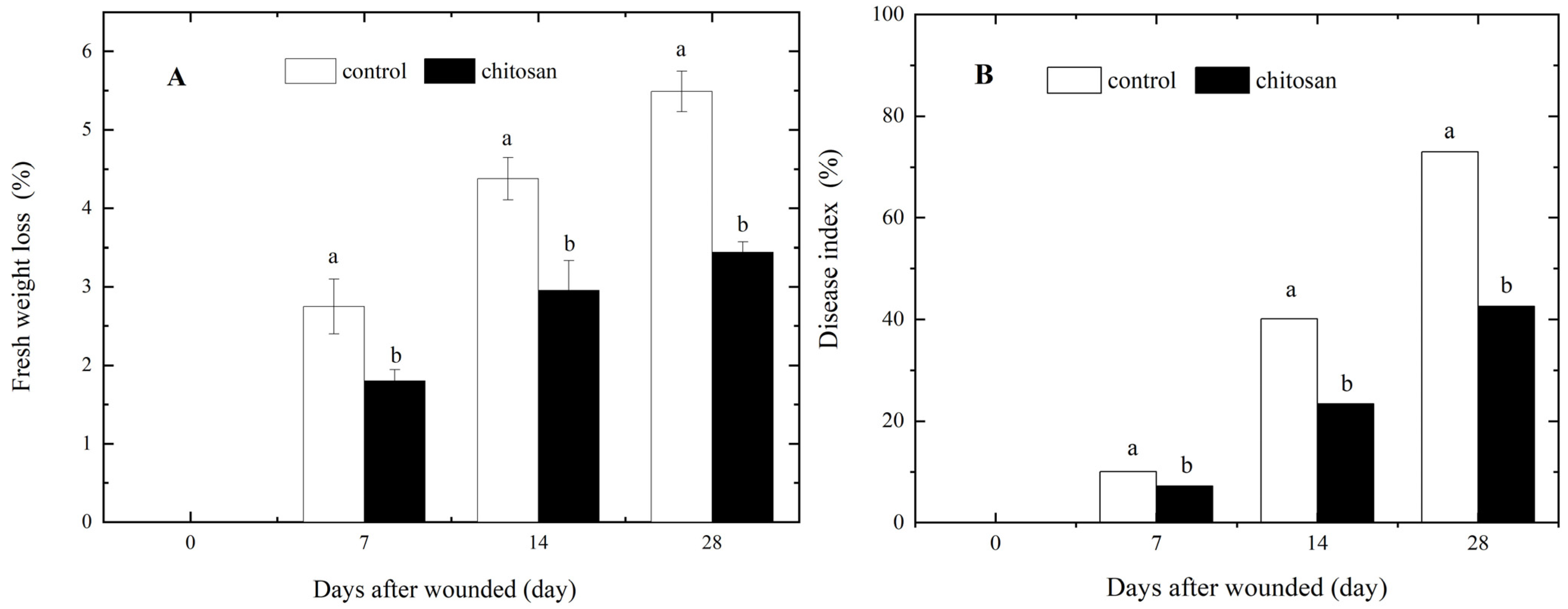
Both fresh weight loss and dry rot disease development were promoted by tuber wounding. The loss of fresh weight in the wounded tubers gradually increased with increasing healing time. However, the fresh weight loss in the wounded tubers of the control group was significantly higher (5.49%) than that in the chitosan-sprayed group (3.44%) after 28 days of storage, representing a ca. 37.34% reduction in fresh weight loss after wounding (p < 0.05; Figure 1A). Typical dark brown spots symptomatic of dry rot were observed on the surfaces of the injured tubers inoculated with F. sambucinum. In the control group, the disease index of dry rot increased rapidly from the 7th (ca. 10%) to the 28th day of incubation (ca. 73%). In contrast, the disease index in the wounded tubers of the chitosan-sprayed group was consistently and significantly lower (p < 0.05) than that in the control group by 28.80–41.60% (Figure 2B). These results demonstrate that the foliar spraying of chitosan significantly reduced fresh weight loss and the development of dry rot in wounded potato tubers after harvesting.

3.2. Effect of Foliar Spraying of Chitosan on SPP and Lignin Accumulation at the Wound Site of Potato Tuber

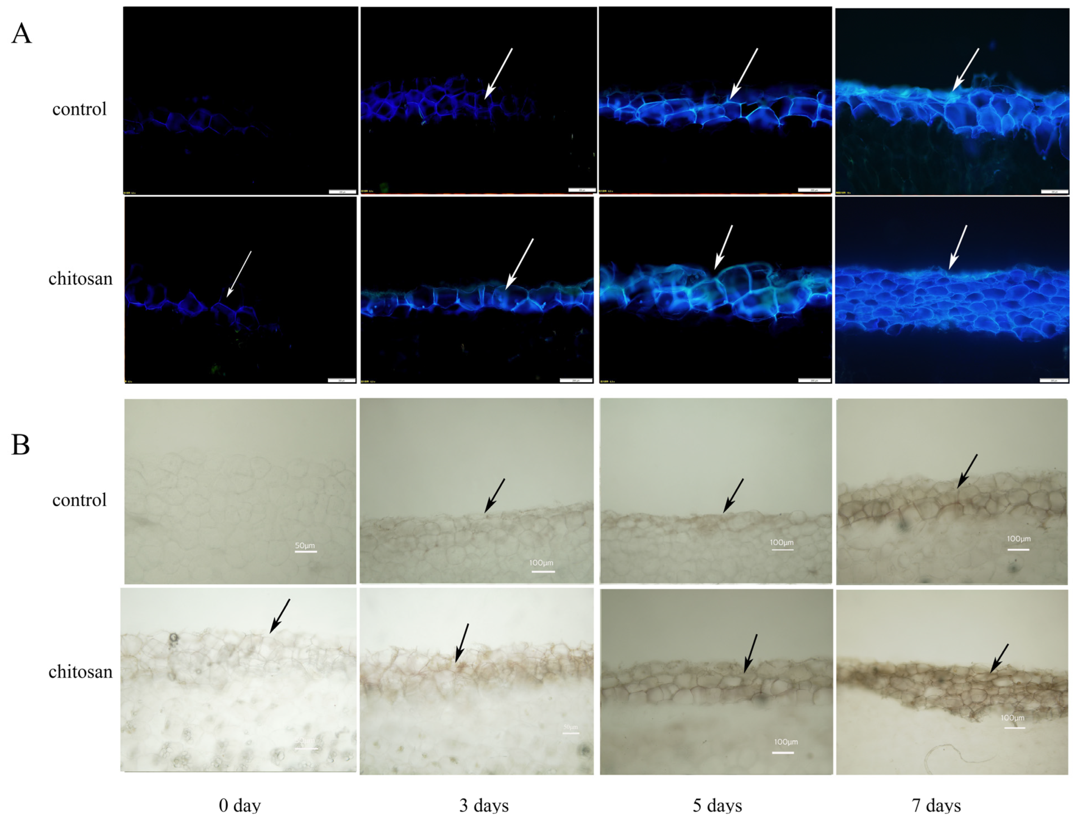
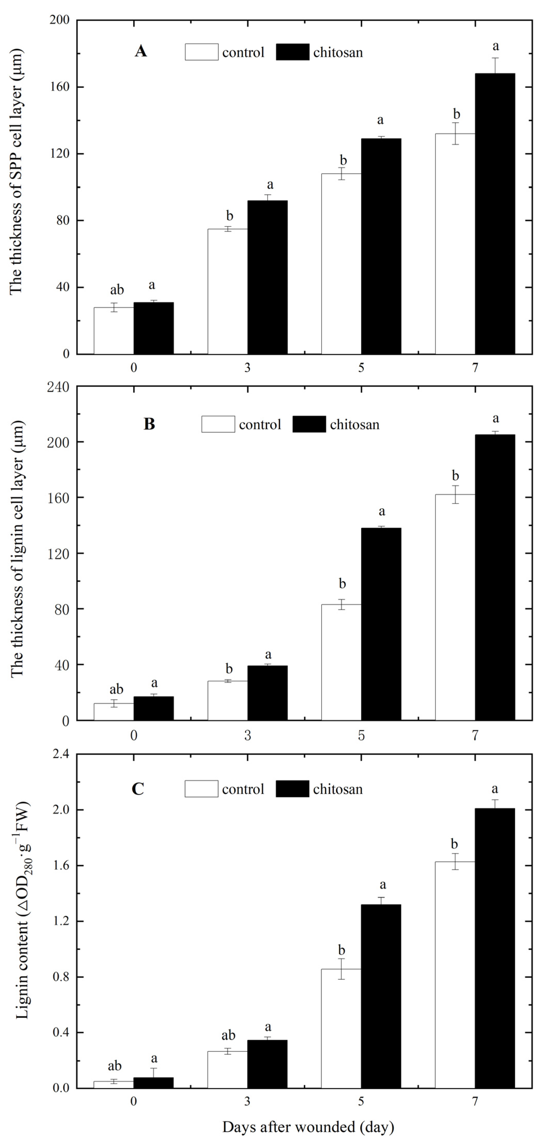
The SPP and lignin layers are important components of the wound periderm in wounded potato tubers. As shown in Figure 3A, tubers from the foliar spraying of the chitosan group showed an increased deposition of SPP at the wound site relative to the control group as early as 3 days after injury. After 7 days of injury, tubers from the chitosan treatment group showed larger increases in SPP fluorescence intensity and fluorescent cell layer thickness (27.27%; Figure 4A) at the wound site relative to the control tubers. Similarly, tuber wounding induced the localized deposition of lignin, which increased over 7 days (Figure 3B). After 7 days of injury, the lignin content of the treated group increased by 23.46% (Figure 4C) and the thickness of the cell layer by 26.54% compared to the control group (Figure 4B). These results indicated that the foliar spraying of chitosan significantly accelerated the deposition of SPP and lignin at the wounds of potato tubers.

3.3. Effect of Foliar Spraying of Chitosan on Key Enzyme Activities of SPP and Lignin Anabolism during Wound-Induced Suberization

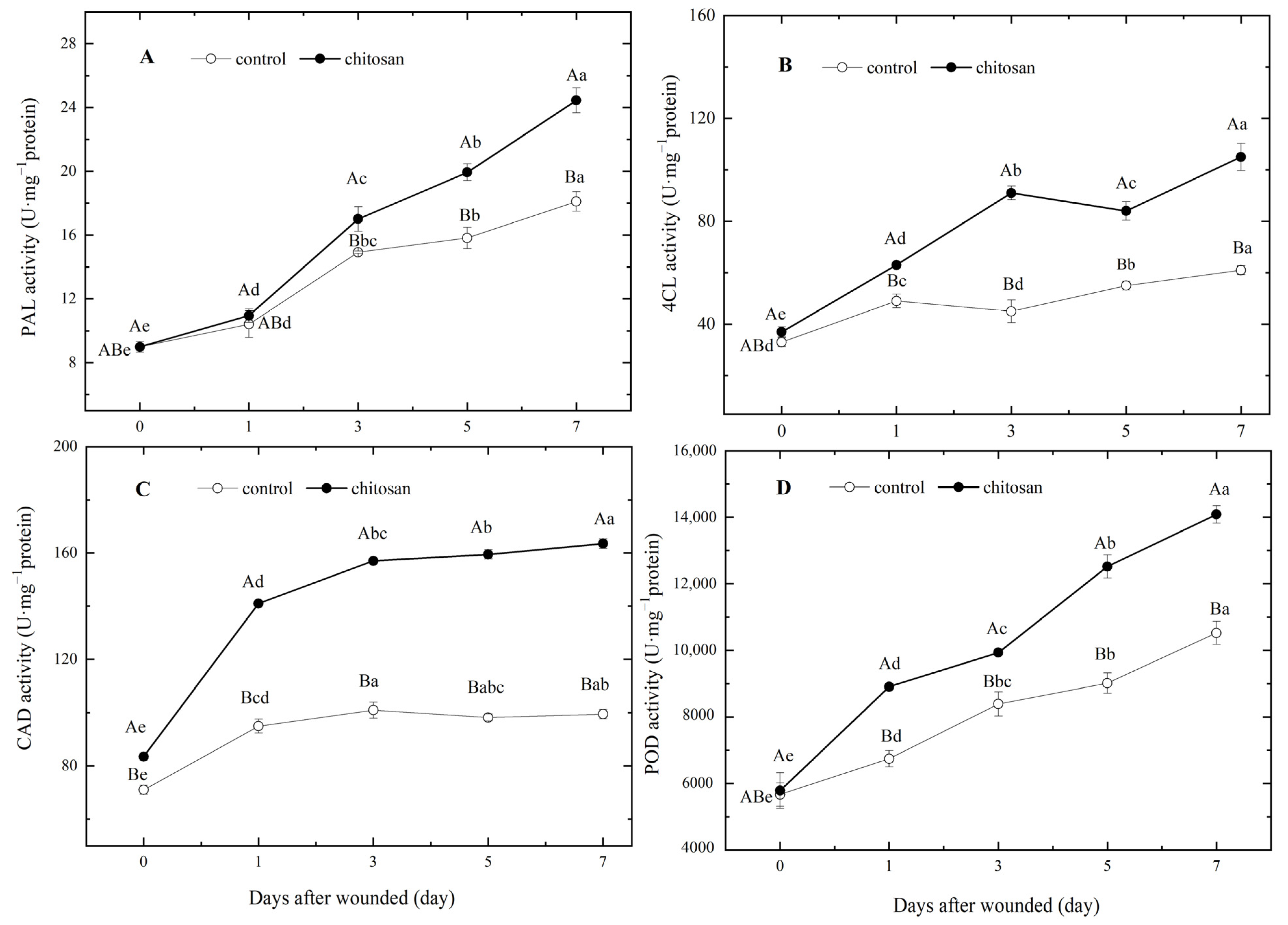
The activities of key enzymes in both the SPP and lignin synthesis pathways were determined. Prior to wounding, there were no significant differences (p ≥ 0.05) in the activities of PAL, 4CL or POD between the control and chitosan treatment groups. However, the activity of CAD was comparatively 17.61% higher in tubers of the chitosan treatment group at the time of harvest (Figure 5).
Following wounding, the activities of the four key enzymes showed a gradual increase in the control group. However, these enzyme activities in the tubers of the chitosan treatment group were all higher than those of the control group (Figure 5). Notable relative differences included a larger increase in PAL activity 0–3 days post-injury (Figure 5A) and two peaks of 4CL activity on days 3 and 7 after wounding (Figure 5B). The activity of CAD at the wound site was 48.42% higher than that of the control after 1 day of wounding and maintained higher activities over the following 6 days (Figure 5C). POD activities showed a continual increase after wounding, with the highest activity on day 7 (Figure 5D). Relative to the control group, the activities of PAL, 4CL, CAD and POD of the chitosan treatment group were 35.02%, 55.74%, 64.32% and 33.9% higher, respectively, after 7 days of wounding (Figure 5). These results indicated that the foliar spraying of chitosan significantly enhanced the localized mobilization of PAL, 4CL, CAD and POD activities in response to the wounding of potato tubers after harvest.

3.4. Effects of the Foliar Spraying of Chitosan on the Contents of Phenolic acid Monomers, Total Phenols and Flavonoids in Tubers during Wound-Induced Suberization

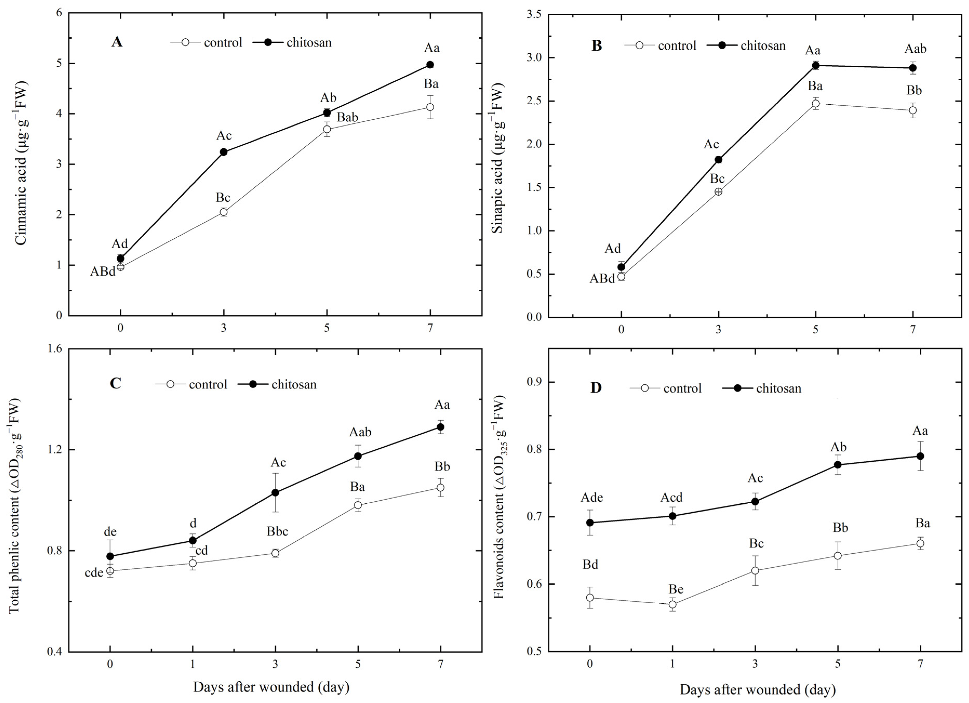
SPPs are polymerized from different phenolic acid monomers, of which cinnamic acid and sinapic acid are the main constituents. Total phenols and flavonoids are the sum of phenolic acid in tubers. The data on day 0 showed that the foliar spraying of chitosan had no significant effect on the contents of cinnamic acid, sinapic acid, or total phenols (Figure 6A–C), but resulted in a significantly higher flavonoid content relative to the control (p< 0.05; Figure 6D). Following tuber wounding, the contents of phenolic acids and flavonoids showed an increasing trend (Figure 6), and the contents of sinapic acid and cinnamic acid increased rapidly, reaching a maximum value on days 5 and day 7, respectively. In the chitosan-treated group, the contents of both phenolic acid monomers were significantly higher relative to the control group (Figure 6A,B). In tubers of the chitosan treatment group, the total phenolic content was significantly higher than that of the control group from days 3 to 7 after wounding (Figure 6C), whereas the flavonoid content was not only relatively higher directly after harvesting but was also induced to higher levels after wounding with further increases over time (Figure 6D). Compared to the control group, the content of cinnamic acid, sinapic acid, total phenols and flavonoids in the tubers from the chitosan-treated group increased by 20.34%, 20.50%, 22.86% and 19.70%, respectively, on day 7 after wounding (Figure 6). The above results indicated that the foliar spraying of chitosan promoted a higher rate of synthesis of cinnamic acid, sinapic acid, total phenols and flavonoids in potato tubers after harvest during wound-induced suberization.

4. Discussion

Pre-harvest treatments, including seed dipping or foliar spraying with selected agents, have been shown to enhance stress resistance of fruit and vegetable crops during plant cultivation, reduce damage to product quality from aspects of post-harvest processes, and improve post-harvest preservation of the freshness of fruits and vegetables, all of which have great potential for application in agricultural production [24,25,26,27,28]. Many agents have been reported to improve the post-harvest quality of fruits and vegetables, such as brassinosteroid, salicylic acid, sorbitol, sodium nitroprusside, sodium silicate, chitosan, etc., during the pre-harvest or post-harvest period [21,24,29,30,31,32]. Chitosan is generally recognized as safe (GRAS) and can be applied to many crops during pre-harvest [10]. It is known that potato tubers decay easily in water, so harvested tubers are not suitable for preservative treatment with water solution after harvest, as potato tubers are not suitable for post-harvest treatments involving their immersion, and there is a lack of fumigation treatments in current potato production practices. Therefore, the pre-harvest foliar application is a good strategy for improving potato tuber quality after harvest.
Our results showed that pre-harvest foliar spraying of chitosan three times during potato tuber development was effective in reducing fresh weight loss and dry rot disease development in wounded tubers during the post-harvest period (Figure 2). The reduced fresh weight loss is likely a direct result of the accelerated formation of the wound periderm [20]. Potato tubers are susceptible to Fusarium spp. and F. sambucinum, which are the dominant causal pathogens of dry rot during potato storage [4]. Pre-harvest foliar spraying of chitosan significantly impaired the development of dry rot in injured tubers, which can be related to the accelerated suberization of the wound periderm and its provision of an enhanced protective barrier against pathogen infection. Similar post-harvest protective effects have been reported for potatoes using a stroby (kresoxim-methyl) [20].
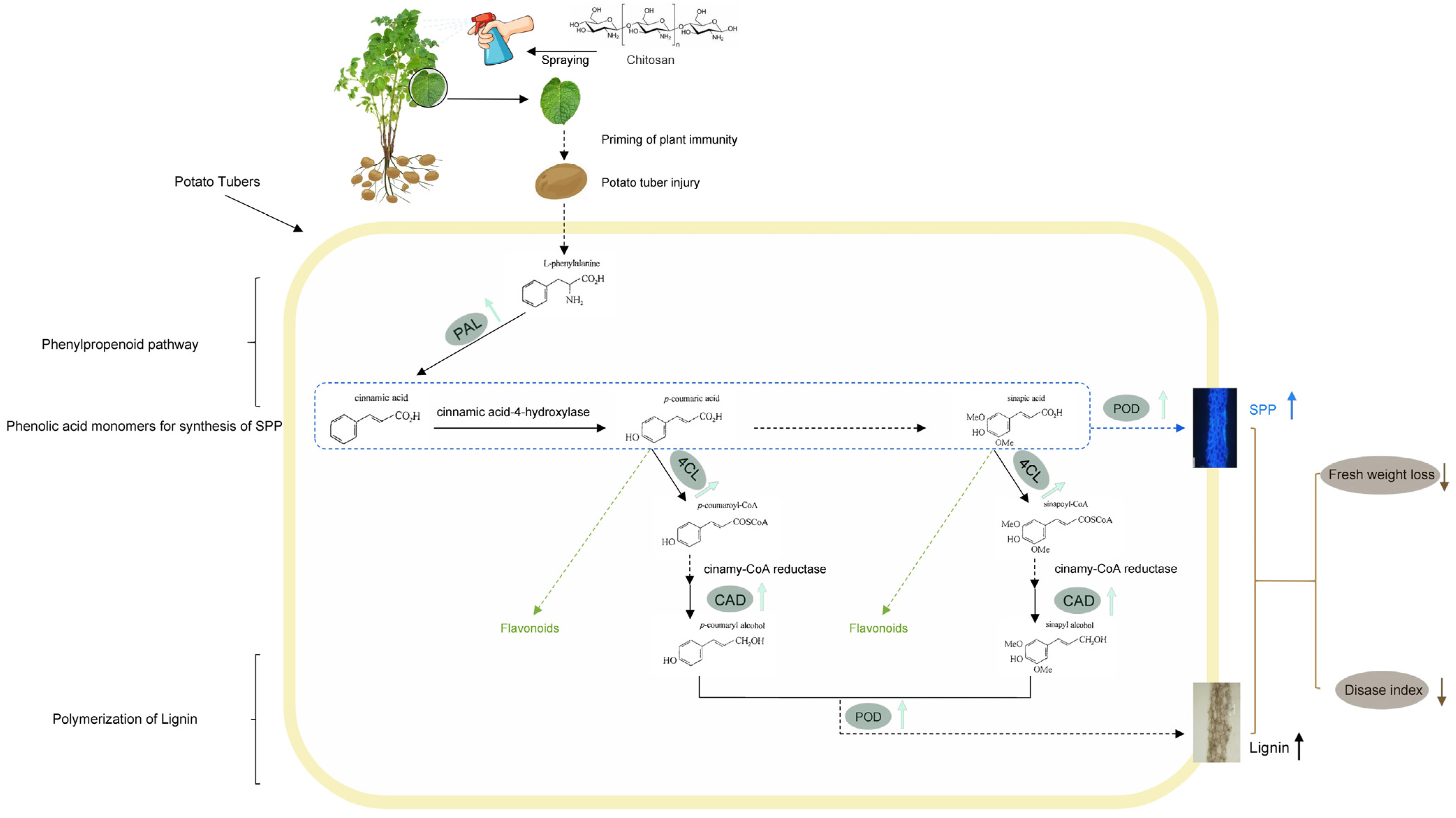
Wound-induced tissue suberization is a complex biological process involving an increase in precursor phenylpropanoid synthesis, SPP and lignin formation in potentially dedicated pathways [5,16,33]. The phenylpropanoid pathway is an important secondary metabolic pathway closely related to the plant immune system, and large amounts of phenolic acids are biosynthesized via this pathway [34]. PAL is the key rate-limiting enzyme in the phenylpropanoid pathway and catalyzes the deamination of phenylalanine to produce trans-cinnamic acid [35], which undergoes a series of catalytic transformations to generate many phenolic compounds, including p-coumarate, caffeic, ferulic acid and sinapic acid [33]. Phenolic acid monomers are catalyzed to hydroxylated phenolic acids, which are polymerized in the presence of POD to produce SPP [31]. Hydroxylated phenolic acids are also catalyzed by 4CL to acetylate and produce p-coumaroyl-CoA, feruloyl-CoA, sinapic-CoA, etc., which are further catalyzed in the presence of CAD to produce monolignols, including p-coumaryl, coniferyl and sinapyl alcohols, which are subsequently polymerized by POD into lignin [31,33,35]. The increased availability of phenolic substrates has been associated with an accelerated deposition of SPP and lignin during the wound-induced suberization process [4,21]. Phenolic acids provide precursor substrates for the synthesis of SPP and lignin, which provide important waterproofing properties and a protective barrier against pathogen infection in the periderm of potato tubers [36]. It has been shown that chitosan treatment can enhance fruit resistance by activating the activity of defense enzymes related to the lignin synthesis pathway [15,16,24], promote lignin synthesis and prolong fruit shelf life in citrus [37,38], pears [39], grapes [40], muskmelons [16] and bamboo shoots [41]. These effects occurred during the foliar spraying of chitosan in our study. Our results showed that pre-harvest foliar spraying of chitosan enhanced the wound-induction of PAL, 4CL, CAD and POD activities (Figure 5), and promoted increased contents of two phenolic acid monomers in the wounded sites of potato tubers (Figure 6A,B). Key enzyme activities of the phenylpropanoid pathway, SPP and lignin anabolism were activated, which were associated with an accelerated SPP and lignin deposition in the suberizing wound periderm (Figure 3 and Figure 4).
Many phenolic acids and the derivatives of the phenylpropanoid pathway contribute to the total phenolic content of plant tissues [31,35]. The total synthesized phenols and flavonoids can have antioxidant and antimicrobial activities, which inhibit pathogen expansion in the host [7,24,33,42]. The accumulation of total phenols and flavonoids can contribute to improved fruit and vegetable storage performance through potential antimicrobial and antioxidant activities. Li et al. (2021) found that the pre-harvest chitosan treatment increased the content of flavonoids during wound healing of post-harvest melon fruits and reduced the disease index of fruit [16]. Cui et al. (2020) reported that pre-harvest chitosan spray promoted the post-harvest synthesis of phenolic acids in apricot with the enhancement of the fruit’s antioxidant capacity [24]. Potato varieties with high contents of total phenols tend to have a higher disease resistance [6]. Pre-harvest chitosan spray induced the accumulation of total phenols and flavonoids in muskmelons and apricots, together with an enhanced fruit resistance to infection and extended shelf-life [16,24]. Flavonoids have high antioxidant capacity and antimicrobial activity in plant products, which can effectively improve plant resistance to infection by pathogens [43,44]. In this study, the foliar spraying of chitosan led to an increased content of flavonoid in tubers during harvest and a relatively larger increase in flavonoid levels after tuber wounding (Figure 6D), as well as an increased content of total phenols after wounding in potato tubers (Figure 6C), which are consistent with an improvement in antioxidant capacity, inhibition of pathogen infection and a reduction in disease index.
The pre-harvest chitosan treatment presented here involves foliar spraying of potato plants with 3% (w/v) chitosan during the flowering period, tuber enlargement period and two weeks before tuber harvest. The efficacy of this treatment in improving post-harvest potato tuber performance was successfully field-tested in 2017 and 2018. A similar method was developed for the pre-harvest spraying of muskmelon with chitosan in [16]. As for the regulatory mechanism underlying the beneficial effects of the foliar spraying of chitosan on the wound-induced suberization of potato tubers, further research at the gene level of priming is needed.

5. Conclusions

In summary, foliar spraying of chitosan on potato plants activated PAL and increased cinnamic acid, sinapic acid, total phenolics and flavonoids in tuber wounds. Foliar spraying of chitosan activated 4CL, CAD and POD and increased SPP and lignin on wounded sites of potato tubers. The accelerated deposition of SPP and lignin domains of the periderm was associated with an enhanced wound periderm formation in the wounds of potato tubers, resulting in alleviated wound-facilitated fresh weight loss and dry rot disease index.
Based on our findings, we hypothesize that the reduced fresh weight loss and disease index of dry rot result from the accelerated ability of wound periderm formation. Pre-harvest application of chitosan elicited immune activity in potato tubers after post-harvest and activated PAL, 4CL, CAD and POD, increased phenolic compounds, resulting in intensified deposition of SPP and lignin, and accelerated wound periderm formation in the wounds of potato tubers. A possible model of foliar spraying of chitosan to accelerate wound periderm formation of potato tubers is illustrated in Figure 7. The foliar spraying of chitosan is a good strategy to improve the potato tuber quality. Our study provides an alternative and eco-friendly treatment for the preservation of commercial potato tuber qualities after harvest.

Author Contributions

Conceptualization, Y.Z. (Yan Zhu) and Y.Z. (Yuanshou Zhao); methodology, W.Z., F.Z. and J.Y.; software, W.S. and W.G.; validation, X.G. and R.C.; formal analysis, F.C. and Y.Z. (Yuanshou Zhao); investigation, X.G. and W.G.; resources, F.C.; data curation, W.S. and J.Y.; writing—original draft preparation, Y.Z. (Yan Zhu) and W.Z.; writing—review and editing, Y.Z. (Yan Zhu) and W.S.; visualization, R.C. and F.Z.; project administration, Y.Z. (Yan Zhu); funding acquisition, Y.Z. (Yan Zhu) and F.C. All authors have read and agreed to the published version of the manuscript.

Funding

This research was funded by the Young Doctor Fund Project of the Gansu Provincial Education Department grant number 2022QB-077, the National Natural Science Foundation of China, grant number 31760473, and the Excellent Postgraduate Innovation Star Project of Gansu Province grant number 2023CXZX-687.

Data Availability Statement

No new data were created or analyzed in this study. Data sharing does not apply to this article.

Conflicts of Interest

The authors declare that they have no known competing financial interests or personal relationships that could have influenced the work reported in this paper.

References

  1. Sun, J.; Wang, X.; Huang, Z.; Sun, H.; Cheng, Q.; Zhu, M. Classification and integration of storage and transportation engineering technologies in potato producing areas of China. J. Agric. Sci. Technol. 2017, 18, 710–718. [Google Scholar]
  2. Campilho, A.; Nieminen, K.; Ragni, L. The development of the periderm: The final frontier between a plant and its environment. Curr. Opin. Plant Biol. 2020, 53, 10–14. [Google Scholar] [CrossRef] [PubMed]
  3. Singh, B.; Bhardwaj, V.; Goutam, U. Potato periderm is the first layer of defence against biotic and abiotic stresses: A Review. Potato Res. 2021, 64, 131–146. [Google Scholar] [CrossRef]
  4. Du, M.R.; Ren, X.Y.; Sun, Q.H.; Wang, Y.; Zhang, R.F. Characterization of Fusarium spp. causing potato dry rot in China and susceptibility evaluation of Chinese potato germplasm to the pathogen. Potato Res. 2012, 55, 175–184. [Google Scholar] [CrossRef] [Green Version]
  5. Lulai, E.C.; Neubauer, J.D. Wound-induced suberization genes are differentially expressed, spatially and temporally, during closing layer and wound periderm formation. Postharvest Biol. Technol. 2014, 90, 24–33. [Google Scholar] [CrossRef]
  6. Zheng, X.Y.; Jiang, H.; Bi, Y.; Wang, B.; Wang, T.L.; Li, Y.C.; Gong, D.; Wei, Y.N.; Li, Z.C.; Prusky, D. Comparison of wound healing abilities of four major cultivars of potato tubers in China. Postharvest Biol. Technol. 2020, 164, 111167. [Google Scholar] [CrossRef]
  7. Al-Mokadem, A.Z.; Alnaggar, A.E.-A.M.; Mancy, A.G.; Sofy, A.R.; Sofy, M.R.; Mohamed, A.K.S.H.; Abou Ghazala, M.M.A.; El-Zabalawy, K.M.; Salem, N.F.G.; Elnosary, M.E.; et al. Foliar application of chitosan and phosphorus alleviate the potato virus Y-induced resistance by modulation of the reactive oxygen species, antioxidant defense system activity and gene expression in potato. Agronomy 2022, 12, 3064. [Google Scholar] [CrossRef]
  8. Duan, C.; Meng, X.; Meng, J. Chitosan as a preservative for fruits and vegetables: A review on chemistry and antimicrobial properties. J. Bioresour. Bioprod. 2019, 4, 11–21. [Google Scholar] [CrossRef]
  9. Miranda-Castro, S.P. Application of chitosan in fresh and minimally processed fruits and vegetables. In Chitosan in the Preservation of Agricultural Commonities; Bautista-Baños, S., Romanazzi, G., Jiménez-Aparicio, A., Eds.; Academic Press: Cambridge, MA, USA, 2016; pp. 67–113. [Google Scholar]
  10. Romanazzi, G.; Sanzani, S.M.; Bi, Y.; Tian, S.; Martinez, P.G.; Alkan, N. Induced resistance to control postharvest decay of fruit and vegetables. Postharvest Biol. Technol. 2016, 122, 82–94. [Google Scholar] [CrossRef]
  11. Niu, X.D.; Zhu, L.; Xi, L.J.; Guo, L.; Wang, H.S. An antimicrobial agent prepared by N-succinyl chitosan immobilized lysozyme and its application in strawberry preservation. Food Control. 2020, 108, 106829. [Google Scholar] [CrossRef]
  12. Romanazzi, G.; Moumni, M. Chitosan and other edible coatings to extend shelf life, manage postharvest decay, and reduce loss and waste of fresh fruits and vegetables. Curr. Opin. Biotechnol. 2022, 78, 102834. [Google Scholar] [CrossRef] [PubMed]
  13. Deshaies, M.; Lamari, N.; Ng, C.K.Y.; Ward, P.; Doohan, F.M. The impact of chitosan on the early metabolomic response of wheat to infection by Fusarium graminearum. BMC Plant Biol. 2022, 22, 73. [Google Scholar] [CrossRef]
  14. Hassan, E.O.; Shoala, T.; Attia, A.M.F.; Badr, O.A.M.; Mahmoud, S.Y.M.; Farrag, E.S.H.; EL-Fiki, I.A.I. Chitosan and nano-chitosan for management of Harpophora maydis: Approaches for investigating antifungal activity, pathogenicity, maize-resistant lines, and molecular diagnosis of plant infection. J. Fungi 2022, 8, 509. [Google Scholar] [CrossRef]
  15. Nia, A.E.; Taghipour, S.; Siahmansour, S. Pre-harvest application of chitosan and postharvest Aloe vera gel coating enhances quality of table grape (Vitis vinifera L. cv. ‘Yaghouti’) during postharvest period. Food Chem. 2021, 347, e129012. [Google Scholar]
  16. Li, Z.C.; Xue, S.L.; Xu, X.Q.; Wang, B.; Zheng, X.Y.; Li, B.J.; Xie, P.D.; Bi, Y.; Prusky, D. Preharvest multiple sprays with chitosan accelerate the deposition of suberin poly phenolic at wound sites of harvested muskmelons. Postharvest Biol. Technol. 2021, 179, 111565. [Google Scholar] [CrossRef]
  17. Lesney, M.S. Growth responses and lignin production in cell suspensions of Pinuse elliottii ‘elicited’ by chitin, chitosan or mycelium of Cronartium quercum f.sp. fusiforme. Plant Cell Tissue Org. 1989, 19, 23–31. [Google Scholar] [CrossRef]
  18. Notsu, S.; Saito, N.; Kosaki, H.; Inui, H.; Hirano, S. Stimulation of phenylalanine ammonia-lyase activity and lignification in rice callus treated with chitin, chitosan, and their derivatives. Biosci. Biotechnol. Biochem. 1994, 58, 552–553. [Google Scholar] [CrossRef]
  19. Todd, O.A.; Peter, B.M. Candida albicans and Staphylococcus aureus pathogenicity and polymicrobial interactions: Lessons beyond Koch’s Postulates. J. Fungi 2019, 5, 81. [Google Scholar] [CrossRef] [Green Version]
  20. Ge, X.Z.; Zhu, Y.; Li, Z.C.; Bi, Y.; Yang, J.; Zhang, J.L.; Prusky, D. Preharvest multiple fungicide stroby sprays promote wound healing of harvested potato tubers by activating phenylpropanoid metabolism. Postharvest Biol. Technol. 2021, 171, 111328. [Google Scholar] [CrossRef]
  21. Zhu, Y.T.; Zong, Y.Y.; Liang, W.; Kong, R.; Gong, D.; Han, Y.; Li, Y.C.; Bi, Y.; Prusky, D. Sorbitol immersion accelerates the deposition of suberin polyphenolic and lignin at wounds of potato tubers by activating phenylpropanoid metabolism. Sci. Hortic. 2022, 297, 110971. [Google Scholar] [CrossRef]
  22. Bradford, M.M. A rapid and sensitive method for the quantitation of microgram quantities of protein utilizing the principle of protein-dye binding. Anal. Biochem. 1976, 72, 248–254. [Google Scholar] [CrossRef] [PubMed]
  23. Gruz, J.; Novak, O.; Strnad, M. Rapid analysis of phenolic acids in beverages by UPLC-MS/MS. Food Chem. 2008, 111, 789–794. [Google Scholar] [CrossRef]
  24. Cui, K.B.; Shu, C.; Zhao, H.D.; Fan, X.G.; Cao, J.K.; Jiang, W.B. Preharvest chitosan oligochitosan and salicylic acid treatments enhance phenol metabolism and maintain the postharvest quality of apricots (Prunus armeniaca L.). Sci. Hortic. 2020, 267, 109334. [Google Scholar] [CrossRef]
  25. Benkeblia, N.; Tennant, D.P.F.; Jawandha, S.K.; Gill, P.S. Preharvest and harvest factors influencing the postharvest quality of tropical and subtropical fruits. In Postharvest Biology and Technology of Tropical and Subtropical Fruits; Yahia, E.M., Ed.; Woodhead Publishing Limited: Cambridge, UK, 2011; pp. 112–141. [Google Scholar]
  26. Wójcik, P. Effects of preharvest sprays of iodine, selenium and calcium on apple biofortification and their quality and storability. PLoS ONE 2023, 18, e0282873. [Google Scholar] [CrossRef] [PubMed]
  27. Zheng, J.R.; Chen, H.; Wang, T.L.; Mustafa, G.; Liu, L.H.; Wang, Q.M.; Shao, Z.Y. Quality improvement of tomato fruits by preharvest application of chitosan oligosaccharide. Horticulturae 2023, 9, 300. [Google Scholar] [CrossRef]
  28. Fu, E.; Zhang, Y.; Li, H.; Wang, X.; Zhang, H.; Xiao, W.; Chen, X.; Li, L. Chitosan reduces damages of strawberry seedlings under high-temperature and high-light stress. Agronomy 2023, 13, 517. [Google Scholar] [CrossRef]
  29. Han, Y.; Yang, R.R.; Zhang, X.J.; Wang, Q.H.; Wang, B.; Zheng, X.Y.; Li, Y.C.; Prusky, D. Brassinosteroid accelerates wound healing of potato tubers by activation of reactive oxygen metabolism and phenylpropanoid metabolism. Foods 2022, 11, 906. [Google Scholar] [CrossRef]
  30. Ge, Y.H.; Chen, Y.R.; Li, C.Y.; Zhao, J.R.; Wei, M.L.; Li, X.H.; Yang, S.Q.; Mi, Y.T. Effect of sodium nitroprusside treatment on shikimate and phenylpropanoid pathways of apple fruit. Food Chem. 2019, 290, 263–269. [Google Scholar] [CrossRef]
  31. Han, Y.; Yang, R.R.; Wang, Q.H.; Wang, B.; Prusky, D. Sodium silicate promotes wound healing by inducing the deposition of suberin polyphenolic and lignin in potato tubers. Front. Plant Sci. 2022, 13, 942022. [Google Scholar] [CrossRef]
  32. Wang, B.; Bi, Y. The role of signal production and transduction in induced resistance of harvested fruits and vegetables. Food Qual. Saf. 2021, 5, 1–8. [Google Scholar] [CrossRef]
  33. Lulai, E.C.; Campbell, L.G.; Fugate, K.K.; Mccue, K.F. Biological differences that distinguish the 2 major stages of wound healing in potato tubers. Plant Signal. Behav. 2016, 11, 1256531. [Google Scholar] [CrossRef] [Green Version]
  34. Dong, N.Q.; Lin, H.X. Contribution of phenylpropanoid metabolism to plant development and plant-environment interactions. J. Integr. Plant Biol. 2021, 63, 180–209. [Google Scholar] [CrossRef] [PubMed]
  35. Vogt, T. Phenylpropanoid biosynthesis. Mol. Plant 2010, 3, 2–20. [Google Scholar] [CrossRef] [PubMed] [Green Version]
  36. Bernards, M.A.; Razem, F.A. The poly (phenolic) domain of potato suberin: A non-lignin cell wall bio-polymer. Phytochemistry 2000, 57, 1115–1122. [Google Scholar] [CrossRef] [PubMed]
  37. Zhao, Y.J.; Deng, L.L.; Zhou, Y.H.; Ming, J.; Yao, S.X.; Zeng, K.F. Wound healing in citrus fruit is promoted by chitosan and Pichia membranaefaciens as a resistance mechanism against Colletotrichum gloeosporioides. Postharvest Biol. Technol. 2018, 145, 134–143. [Google Scholar] [CrossRef]
  38. Gao, Y.; Kan, C.N.; Wan, C.P.; Chen, C.Y.; Chen, M.; Chen, J.Y. Quality and biochemical changes of navel orange fruits during storage as affected by cinnamaldehyde-chitosan coating. Sci. Hortic. 2018, 239, 80–86. [Google Scholar] [CrossRef]
  39. Kou, X.H.; Guo, W.L.; Guo, R.Z.; Li, Z.H. Effects of chitosan, calcium chloride, and pullulan coating treatments on antioxidant activity in pear cv.“Huang guan” during storage. Food Bioprocess Technol. 2014, 7, 671–681. [Google Scholar] [CrossRef]
  40. Hu, B.; Lin, L.; Fang, Y.J.; Zhou, M.; Zhou, X.Y. Application of chitosan-lignosulfonate composite coating film in grape preservation and study on the difference in metabolites in fruit wine. Coatings 2022, 12, 494. [Google Scholar] [CrossRef]
  41. Yang, H.Q.; Zheng, J.Y.; Huang, C.H.; Zhao, X.F.; Chen, H.Y.; Sun, Z.D. Effects of combined aqueous chlorine dioxide and chitosan coatings on microbial growth and quality maintenance of fresh-cut bamboo shoots (Phyllostachys praecox f. prevernalis.) during storage. Food Bioprocess Technol. 2015, 8, 1011–1019. [Google Scholar] [CrossRef]
  42. Hahlbrock, K.; Grisebach, H. Enzymic controls in the biosynthesis of lignin and flavonoids. Ann. Rev. Plant Physiol. 1976, 30, 105–130. [Google Scholar] [CrossRef]
  43. Cascaes, M.M.; Guilhon, G.M.S.P.; Zoghbi, M.d.G.; Andrade, E.H.A.; Santos, L.S.; Silva, J.K.R.; Uetanabaro, A.P.T.; Araújo, I.S. Flavonoids, antioxidant potential and antimicrobial activity of Myrcia rufipila mcvaugh leaves (myrtaceae). Nat. Prod. Res. 2021, 35, 1717–1721. [Google Scholar] [CrossRef] [PubMed]
  44. Górniak, I.; Bartoszewski, R.; Króliczewski, J. Comprehensive review of antimicrobial activities of plant flavonoids. Phytochem. Rev. 2019, 18, 241–272. [Google Scholar] [CrossRef] [Green Version]
Figure 1. Artificial wounding and storing procedures of potato tubers. (A) Wounded tubers and sterile scalpel; (B) polyethylene preservation box; (C) wrapped black bags.
Horticulturae 09 00663 g001
Figure 2. Effects of the foliar spraying of chitosan on (A) the fresh weight loss and (B) the development of dry rot disease in wounded potato tubers. Lowercase letters indicate a significant difference between the different treatment groups at the same time point after injury (p < 0.05).
Horticulturae 09 00663 g002
Figure 3. Effect of foliar spraying of chitosan on the localized accumulation of (A) SPP and (B) lignin in wounded potato tubers. White arrows indicate the deposition of SPP at the wound sites and black arrows indicate the deposition of lignin at the wound sites, respectively.
Horticulturae 09 00663 g003
Figure 4. Effect of foliar spraying of chitosan on (A) the thickness of the SPP cell layer at the wound site, (B) the thickness of the lignin cell layer at the wound site, and (C) the content of lignin. Lowercase letters indicate a significant difference between the different treatment groups (p < 0.05).
Horticulturae 09 00663 g004
Figure 5. Effects of the foliar spraying of chitosan on enzyme activities related to SPP and lignin synthesis at the wound site of potato tubers. The activities of (A) PAL, (B) 4CL, (C) CAD and (D) POD are shown. Capital letters indicate significant differences between the control and chitosan treatments (p < 0.05). Lowercase letters indicate significant differences between time points in the same treatment group (p < 0.05).
Horticulturae 09 00663 g005
Figure 6. Effect of foliar spraying of chitosan on the contents of (A) cinnamic acid, (B) sinapic acid, (C) total phenolics and (D) flavonoids at the wound site of potato tubers. Capitals letters indicate significant (p < 0.05) differences between chitosan and control treatments. Lowercase letters indicate significant (p < 0.05) differences between time points in the same treatment group.
Horticulturae 09 00663 g006
Figure 7. Foliar spraying of chitosan accelerated the deposition of suberin polyphenolic and lignin in the wounds of potato tubers by eliciting phenylpropanoid metabolism via the polymerization of SPP and lignin. Reactions denoted by solid lines are known, whereas those denoted by dashed lines are hypothetical metabolic steps catalyzed by multiple enzymes. Black arrows represent the flow of material that synthesizes lignin, blue dashed lines in the box and blue arrows represent the flow of material that synthesizes SPP, and green arrows represent the synthesis of flavonoids, respectively. The black brackets and words on the left side of the model diagram show different metabolic pathways, and the brown brackets and ellipses on the right side of the model diagram show the variation in fresh weight loss and disease index of potato tubers after wounding. PAL, phenylalanine ammonialyase; 4CL, 4-coumaric acid coenzyme A ligase; CAD, cinnamoyl alcohol dehydrogenase; POD, peroxidase; SPP, suberin polyphenolic.
Horticulturae 09 00663 g007
Disclaimer/Publisher’s Note: The statements, opinions and data contained in all publications are solely those of the individual author(s) and contributor(s) and not of MDPI and/or the editor(s). MDPI and/or the editor(s) disclaim responsibility for any injury to people or property resulting from any ideas, methods, instructions or products referred to in the content.