Article

Effects of Citric Acid and Humic-like Substances on Yield, Enzyme Activities, and Expression of Genes Involved in Iron Uptake in Tomato Plants

by
Fabián Pérez-Labrada
1,
Adalberto Benavides-Mendoza
2,
Antonio Juárez-Maldonado
1,
Susana Solís-Gaona
3 and
Susana González-Morales
4,*
1
Departamento de Botánica, Universidad Autónoma Agraria Antonio Narro, Saltillo 25315, Mexico
2
Departamento de Horticultura, Universidad Autónoma Agraria Antonio Narro, Saltillo 25315, Mexico
3
United Phosphorus Limited, UPL, Saltillo 25290, Mexico
4
Departamento de Horticultura, CONAHCyT-Universidad Autónoma Agraria Antonio Narro, Saltillo 25315, Mexico
*
Author to whom correspondence should be addressed.
Horticulturae 2023, 9(6), 630; https://doi.org/10.3390/horticulturae9060630
Submission received: 21 April 2023 / Revised: 24 May 2023 / Accepted: 25 May 2023 / Published: 27 May 2023
(This article belongs to the Special Issue Responses to Abiotic Stresses in Horticultural Crops)

Abstract

:
Iron (Fe) deficiency is a common abiotic stress on plants growing in calcareous soils where low organic matter content, high carbonate–bicarbonate concentration, and high pH precipitate Fe in unavailable forms. Enzymatic activity is a mechanism for plants to access soil nutrients; enzymes such as H+-ATPase, phosphoenolpyruvate carboxylase (PEPC), and the intracellular enzyme ferric reduction oxidase (FRO) are involved in Fe absorption. The effects of the application of citric acid (CA) and humic-like substances (HLS) on the yield, H+-ATPase, PEPC, and FRO enzyme activity, and expression of LeHA1, LePEPC1, and LeFRO1 genes in tomato plants grown under calcareous soil were studied. CA and HLS improved the SPAD units and increased the number of harvested fruits and yield per plant. Temporary alterations in enzyme activity, which reduced PEPC and FRO activity in roots, were documented. In leaf tissue, CA resulted in lower expression of LeHA1 and LePEPC1 and the induction of LeFRO1 expression, whereas HLS application resulted in higher expression of LePEPC1 and LeFRO1. In roots, LeHA1 expression increased with HLS, whereas LePEPC1 and LeFRO1 showed lower expression with CA and HLS, respectively. The application of CA and HLS through a nutrient solution in combination with Fe-chelate can improve Fe nutrition in tomato plants potted in calcareous soil by inducing temporal alterations in PEPC and FRO enzyme activity and LeFRO1 and LeHA1 gene expression.

1. Introduction

Iron (Fe) plays a crucial role in photosynthesis and mitochondrial respiration, and, as a cofactor of enzymes such as superoxide dismutase, acotinase, lipoxygenases, nitrogenase, carotenoid cleavage oxygenases/dioxygenase, lutein cleavage dioxygenase, and cupins, among others, it is generally found in high proportions in chloroplasts—up to 80% [1]. Therefore, plants require an average of ~10−9–10−4 M Fe ions for optimal development [2]. Fe is taken up from the soil; however, despite its abundance in soil, a high percentage of Fe exists as Fe3+, which is highly insoluble and has very low availability [3]. This subsequently generates abiotic stress in the form of Fe deficiency. An Fe-deficient environment is generated in calcareous soils, where Fe precipitates in forms unavailable to plants due to high pH (>7.5), high content of carbonates (CaCO3), and low content of organic matter [4]. Similarly, the high bicarbonate (HCO3) concentration (in conjunction with the high pH) decreases Fe mobility within the plant, triggering iron deficiency chlorosis (IDC), which is characterized by the loss of chlorophyll, interveinal chlorosis of young leaves, deformation of leaves, reduction of photosynthesis and growth, and decrease in plant production [1].
Because most of the Fe in the soil is present as Fe3+, plants use enzymatic mechanisms to access it, employing a reduction-based strategy (I) or a chelation-based strategy (II) [5]. Strategy II plants (with Oryza sativa as a model) secrete phytosiderophores using transporter of mugineic acid 1 (TOM1), which chelates Fe3+ [6]. The phytosiderophores-Fe3+ complex is subsequently absorbed by yellow stripe 1 (YS1) or yellow stripe-like 1 (YSL) [7]; Fe2+ uptake by iron-regulated transporter (IRT) 1/2 may also occur [8].
In contrast, strategy I plants (with Arabidopsis thaliana L. as a model) initially induce rhizospheric acidification through a membrane-associated proton pump, H+-ATPase [9], which releases Fe3+ from the soil, and through ferric reduction oxidase (FRO), which reduces Fe3+ to Fe2+. Fe2+ is captured and finally absorbed and transported into the plant by an iron-regulated transporter-like protein (ZIP) family transporter, IRT1, and natural resistance-associated macrophage protein (NRAMP1) [6,10]. The FRO, IRT1, and NRAMP1 genes are regulated transcriptionally by the central basic helix–loop–helix (bHLH) FER-like Fe deficiency-induced transcription factor (FIT) [11]. Similarly, phenolic compounds such as coumarins [12,13] and organic acids [6] are exuded by roots and are involved in the Fe-making machinery by facilitating the solubilization and reduction of unavailable Fe in the soil.
Under conditions of Fe deprivation, plants regulate the enzymatic activity associated with Fe access at the genic level. For example, strategy I plants, such as tomato (Solanum lycopersicum L.), substantially increase FRO enzymatic activity in the roots [14]. In pea plants (Pisum sativum L.), in addition to higher FRO activity, overexpression of FRO1, IRT1, and HA1 was observed in the roots [15]. Similarly, in cucumber (Cucumis sativus L.) an increase in CsHA1 transcripts was reported in a higher proportion in the roots than in the leaves [16], whereas overexpression of HA6, which allowed for the outflow of H+ from the root, was reported in a citrus species [17]. A key regulator in the activation of these mechanisms is FIT, which facilitates Fe uptake [18].
The application of synthetic chelates is commonly recommended for the prevention and reduction of Fe deficiencies in plants grown in calcareous soils. However, these products can be lost by leaching or adsorbing on the soil particles because of the high pH values and high carbonate content of the soil and because of their high affinity with calcium or magnesium [19]. In this sense, citric acid (2-hydroxy-propane-1,2,3-tricarboxylic acid) and leonardite-derived substances (humic, fulvic acid, and humin) improve Fe nutrition in calcareous soils by increasing Fe availability and optimizing synthetic chelate efficiency [20,21].
The exogenous application of citric acid in nutrient solution was found to increase leaf tissue Fe content in tomatoes potted in calcareous soils [21]. Citric acid improves Fe bioavailability by converting Fe to its plant-available form [22]. Similarly, the application of leonardite plus ferrous sulfate heptahydrate was found to improve the fertility of calcareous vineyard soils by increasing their Fe content [23]. The application of Fe complexed with extractable humic substances in water and Fe-citrate led to the overexpression of FRO, IRT1, and NRAMP in cucumber [24] as well as a modulation of the transcripts of the gene encoding H+-ATPase in tomato [25]. Moreover, this strategy also provides a long-term supply of Fe to plants grown in calcareous soils [26]. The aim of this study was to determine the yield, H+-ATPase, phosphoenolpyruvate carboxylase (PEPC), and FRO enzyme activity, and expression of LeHA1, LePEPC1, and LeFRO1 in tomato plants potted in calcareous soil under the application of citric acid (CA) and humic-like substances (HLS).

2. Materials and Methods

2.1. Plant Growth Conditions

Tomato (Solanum lycopersicum L. cv. ‘Río Grande’) (Crow Seed) plants were grown in a greenhouse with a polyethylene cover and 70% natural irradiance (with recorded averages of 50–60% relative humidity, 32 °C ambient temperature, 736.5 µM m−2 s−1 photosynthetically active radiation, and 430 ppm CO2 concentration). The tomato seeds were grown for 35 days in a tray containing a germination substrate, and then the seedlings were transplanted into pots containing 9 L of calcareous soil (pH 8.5, 0.2% organic matter, and 5 mg kg−1 Fe content). The plants were fertilized from transplantation to the end of the experiment with Steiner solution [27] at 100% concentration, which contained 4.5 mmol Ca(NO3)2·4H2O, 2.0 mmol MgSO4, 0.7 mmol KNO3, 2.0 mmol K2SO4, and 1.6 mmol KH2PO4. Micronutrients were applied from a stock solution containing H3BO3 (2.86 g L−1), MnSO4·H2O (2.15 g L−1, ZnSO4·7H2O (0.39 g L−1), and CuSO4·5H2O (0.078 g L−1), and Na2MoO4·5H2O (0.09 mg L−1). Finally, Fe was applied in the form of Fe gluconate-ethylenediaminetetraacetic acid (EDTA)-type chelate (at 3 mg L−1), except in the Fe-deprivation treatment, where it was not added. The pH of the nutrient solution was maintained at 6.3 with an electrical conductivity of 2.0 dS cm−1.
The treatments studied were CA (0.1 mM + Fe gluconate-EDTA), HLS (400 µL L−1 + Fe gluconate-EDTA), “without organic amendment” (WOA, only Fe gluconate-EDTA), and an Fe-deprivation control (ID, no organic acid and Fe in solution). The concentrations used were selected according to previous results cited by Pérez-Labrada et al. [21]. The citric acid used was food-grade (99.9% purity). The HLS (Arysta LifeScience) contained 10 g L−1 humic acid carbon, 90 g L−1 fulvic acid carbon, 6 g L−1 total nitrogen, 6 g L−1 urea nitrogen, and 38 g L−1 water soluble K2O with a density of 1.15 g mL−1 and at pH 9.2. The treatments CA and WOA were applied daily through localized irrigation during plant development. The HLS treatment was only applied weekly with the corresponding irrigation volume (15 total applications during crop development). The control (ID) was irrigated daily through localized irrigation during plant development. All treatment compositions were added to the nutrient solution. The nutrient solution was provided to the plants through localized irrigation from transplanting until the end of the experiment.
Considering the phenology of the tomato plant, the sampling dates of leaf-root tissue were chosen according to these stages: vegetative growth (27 days after transplantation, DAT), flowering-anthesis (49 DAT), and physiological maturity of fruits of the 2nd bunch of fruit (89 DAT). We collected the three youngest leaves that had completely expanded, placed them in aluminum bags, immediately froze them with liquid N2, and stored them at –80 °C. Root tissue was collected on the same leaf-sampling date; 5 cm of secondary roots (obtained at 10 cm depth and 5 cm stem distance) were collected, briefly washed with deionized water, placed in aluminum bags, immediately frozen with liquid N2, and stored at –80 °C. Plant growth parameters were determined at 27, 49, and 84 DAT. The stem diameter was measured with a sliding caliper 150 mm between the first and second pair of true leaves from the base. Plant length was measured with a flexometer from the base of the stem (soil surface) to the distal growing apex. On fully expanded young leaves, SPAD-unit readings were taken using a Minolta SPAD-502 chlorophyll meter (Konica Minolta, Inc., Osaka, Japan). SPAD values were taken as an average of three readings taken at different locations from the base to the apex of each leaf. Finally, the yield per plant was calculated as the sum of the total number of harvested fruits.

2.2. Enzymatic Activity

Briefly, the enzyme extract was obtained by homogenizing the tissue in liquid N2 and adding buffer at 4 °C (50 mM Tris-HCL, pH 7.5, 10% glycerol, 20% PVPP, 10 mM MgCl2, 1 mM EDTA, 14 mM β-mercaptoethanol, 1 mM PMSF, and 10 µg mL−1 leupeptin; Sigma-Aldrich, St. Louis, MO, USA). The extract was then filtered through a nylon micropore (0.45 µm pore size) and centrifuged at 10,000 rpm for 15 min. The supernatant was centrifuged again at 10,000 rpm for 30 min, and the precipitate was resuspended in the same buffer and stored at –80 °C [28] until use in the determination of H+-ATPase and PEPC activity. H+-ATPase activity (EC 7.1.2.1) was determined using a spectrophotometric method [29], coupling the hydrolysis of ATP to the oxidation of NADH [28]; 100 µL of extract was taken, and 25 mM MOPS-BTP buffer (pH 6.5) was added, containing 250 mM sucrose, 50 mM KCl, 1 mM ATP, 1 mM PEP, 0. 25 mM NADH, 15 µg mL−1 lactate dehydrogenase (EC 1.1.1.27), 30 µg mL−1 pyruvate kinase (EC 2.7.1.40), and 0.015% Brij® 58 (Sigma-Aldrich). Changes in absorbance were measured in a spectrophotometer at 340 nm. The H+-ATPase activity was calculated using the NADH standard curve. Phosphoenolpyruvate carboxylase activity (PEPC, EC 4.1.1.31) was determined by coupling its activity to the malate dehydrogenase catalyzed by the oxidation of NADH [30]; to initiate the reaction, 100 µL of extract was taken, and standard buffer containing Tris-HCl (100 mM, pH 8.0), MgCl2 (5 mM), PEP (2.5 mM), NADH (0.2 mM), NaHCO3 (10 mM), and MDH (15 µg mL−1) was added. Changes in absorbance were quantified at 340 nm. PEPC activity was calculated using the NADH standard curve.
Chelate ferric reductase activity (FRO, EC 1.16.1.7) was quantified spectrophotometrically by Fe (II)-BPDS concentration according to Romera et al. [31]; 20 mg of tissue was placed in an Eppendorf tube, and 2 mL of CaSO4-7H2O (0.2 mM) was added and allowed to stand for 5 min. Then, the sample was centrifuged (5 min at 10,000 rpm at 4 °C), and the precipitate was recovered and placed in a new tube, where 10 mL of fresh nutrient solution (without Fe) supplemented with 0.3 mM BPDS (Sigma-Aldrich) and 100 µM Fe(III)-EDTA (Sigma-Aldrich) was added. The pH of the solution was adjusted to 5.5 with 5 mM MES-NaOH (Sigma-Aldrich). It was incubated for 1 h in the dark at room temperature, and finally, the absorbance was measured at 535 nm. BPDS forms a red water-soluble complex with Fe2+ and only a weak complex with Fe3+. The amount of reduced iron was calculated by the concentration of the Fe2+-BPDS complex using an extinction coefficient of 22.14 mM cm−1. The protein content of the microsomal fraction membranes was determined using 5 µL of the enzyme extract and 250 µL of Bradford reagent, which were incubated at room temperature for 5 min. Afterward, the absorbance was measured at 630 nm in an ELISA plate reader (BioTek, ELx808 model, Winooski, VT, USA) with BSA as a protein standard [32].

2.3. Real-Time Reverse-Transcriptase PCR

The RNA of leaves and roots was extracted using TRIzol reagent [33]; the tissue was ground in liquid N2, 100 mg was taken and placed in an Eppendorf tube, and immediately after, 1 mL of TRI Reagent® (MRC, TR 118) was added; the mixture was homogenized gently and incubated for 5 min at room temperature. Subsequently, 200 μL of chloroform was added, shaken vigorously, and incubated for 15 min. Then, the samples were centrifuged at 12,000 rpm (15 min at 4 °C), and the supernatant was recovered and placed in a new Eppendorf tube. Then, 500 μL of isopropanol (4 °C) was added, mixed gently, and incubated for 10 min at room temperature. The sample was centrifuged for 10 min at 12,000 rpm and 4 °C, and the supernatant was removed by removing excess isopropanol from the formed RNA pellet. The RNA pellet was washed with 500 μL of 70% ethanol (4 °C), and excess was removed and allowed to dry. The pellet was suspended in 50 μL of water (dissolved at 60 °C). Finally, the RNA solution was treated with DNase I (Sigma Aldrich) and stored at 4 °C. The RNA quantity and quality were determined using a UV-Vis spectrophotometer (260/280 nm ratio) and via denaturing electrophoresis, respectively. An ImProm-IITM Reverse Transcription System Kit (Promega, Madison, WI, USA) was used for the synthesis of cDNA following the manufacturer’s instructions; cDNA was synthesized from 1 μg of RNA sample. The primers corresponded to the endogenous internal control gene (ACT) and study genes LeFRO1, LePEPC1, and LeHA1 (Table 1). For LeFRO1 and LePEPC1, the sequences cited by Paolacci et al. [34] and Diamantopoulous et al. [35], respectively, were considered. The primers were designed using AmplifiX 17.0 (CNRS by Nicolas Jullien, Marseille, France), OligoAnalyzer 3.1 (Integrated DNA Technologies IDT, Coralville, IA, USA), and Primer-BLAST (National Center for Biotechnology Information, Bethesda, MD, USA).
Real-time PCR was performed in a final volume of 20 µL. For ACT, 10 µL of SYBR® Select Master Mix (Applied Biosystems, Foster City, CA, USA), 0.10 µL of forward primer (72 nM), 0.08 µL of reverse primer (60 nM), 2 µL of cDNA, and 7.82 µL of nuclease-free water were added. For LeHA1, 10 µL of SYBR® Select Master Mix (Applied Biosystems), 0.13 µL of forward primer (100 nM), 0.13 µL of reverse primer (100 nM), 2 µL of cDNA diluted 1:5, and 7.74 µL of nuclease-free water were added. For LePEPC1, 10 µL of SYBR® Select Master Mix (Applied Biosystems), 0.13 µL of forward primer (100 nM), 0.27 µL of reverse primer (200 nM), 2 µL of cDNA, and 7.60 µL of nuclease-free water were added. For LeFRO1, 10 µL of SYBR® Select Master Mix (Applied Biosystems), 0.13 µL of forward primer (100 nM), 0.13 µL of reverse primer (100 nM), 2 µL of cDNA, and 7.74 µL of nuclease-free water were added. The ACT gene was used to normalize the expression ratio of each gene, and changes in expression were calculated using the standard relative curve method [36].

2.4. Data Analysis

A completely randomized design was used for the experimental development, with 15 biological replicates per treatment. The experimental unit was an individual plant in a pot. One-way analysis of variance (ANOVA) and Fisher’s least significant difference test (p < 0.05) were applied to the plant growth parameters. Data of enzymatic activity were analyzed using a two-way ANOVA and a multiple means comparison using Fisher’s least significant difference test (p < 0.05), in which one factor was the organ evaluated (root and leaf), and the other factor was the treatment applied. The gene expression was normalized compared to the internal reference gene (ACT). A one-way Kruskal–Wallis nonparametric test by ranks was applied to the gene expression data (p < 0.05). All statistical analyses were performed using IBM SPSS v. 19.0.

3. Results

3.1. Plant Growth

The application of CA and HLS resulted in statistically significant differences (p < 0.05) in the growth parameters evaluated (Table 2) compared to the control (ID). For example, the addition of CA to the fertilizer solution increased stem diameter (14% at 84 DAT), plant height (39% at 27 DAT), and leaf number (43% at 49 DAT). The weekly application of HLS increased SPAD units by 110% and 223% at 49 and 84 DAT, respectively, compared to ID. Similarly, the total fruit harvested and maximum production increased by 93% and 265%, respectively, in HLS-treated plants. In addition, the use of AC and HLS stimulated stem diameter at different sampling times up to 15% with respect to WOA. Plant height was lower under HLS at 27 and 49 DAT compared to CA and WOA. Plants treated with HLS and CA showed an improvement in leaf number at 27 and 84 DAT compared to WOA. The CA and WOA treatments increased this variable by 18% over HLS at 49 DAT. SPAD units were improved with CA and HLS with respect to WOA by up to 14.8%. Finally, HLS showed a higher number of fruits harvested and a higher yield per plant than the WOA and CA treatments.

3.2. Enzymatic Activity

Regardless of treatment, there was higher H+-ATPase and PEPC activity in leaf tissue, whereas the FRO activity was higher in root tissue. Except for H+-ATPase activity at 84 DAT, there were significant differences (p < 0.001) among the plant tissues studied.

3.2.1. H+-ATPase Activity

The highest H+-ATPase activity was documented at 27 DAT in leaf tissue. In this sampling, the CA and HLS treatments resulted in 61% and 51% reductions, respectively, compared to ID. At 49 DAT, the activity increased in root tissue, whereas it was reduced in leaf tissue compared to the first sampling; higher activity was documented under CA. During the fruit harvest stage (84 DAT), the activity of this enzyme increased in the leaf tissue, with 46% more activity in CA than in ID (Table 3).

3.2.2. PEPC Activity

In leaf tissue at 27 DAT, 84% higher PEPC activity was found in WOA, and 22% lower PEPC activity was found in plants receiving the HLS treatment relative to ID. On the other hand, at 49 DAT, the HLS and CA treatments promoted 55% and 50% increases in PEPC activity, respectively. Finally, at 84 DAT, plants treated with HLS showed a 150% increase in PEPC activity compared to ID. In root tissue, at 27 DAT, there was a PEPC-activity reduction of 39% and 67% in the HLS and CA treatments, respectively, whereas at 84 DAT, the CA and WOA treatments showed the lowest PEPC activity compared to ID (Table 3).

3.2.3. FRO Activity

In leaf tissue at 27 DAT, FRO activity was reduced by 12.5% in all treatments relative to ID, whereas at 49 and 84 DAT, the activity increased by 35.8% and 56.70% in the WOA treatment. On the other hand, similar FRO activity was found between treatments in root tissue, except at 27 DAT, where the CA treatment minimally reduced FRO activity compared to ID (Table 3).

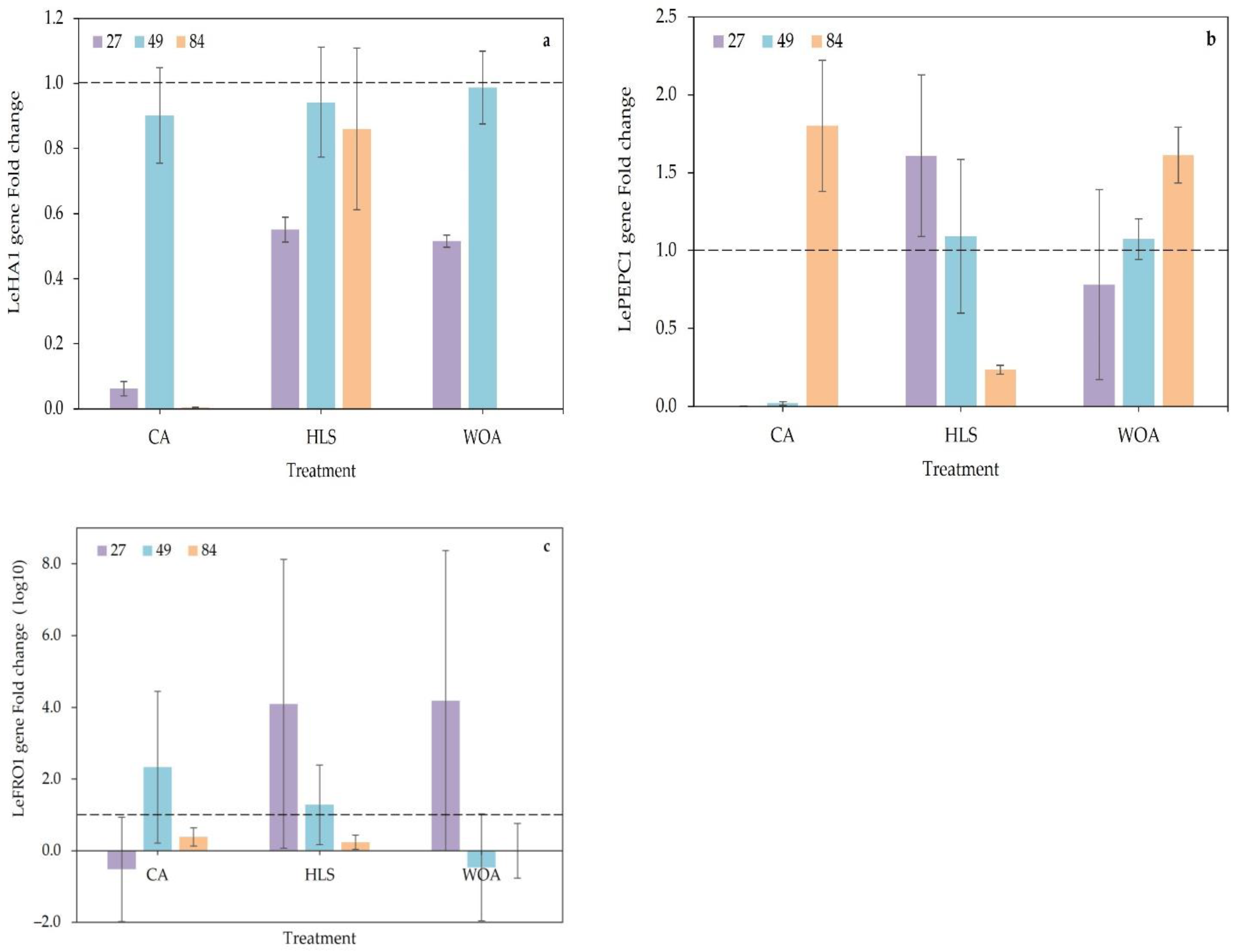
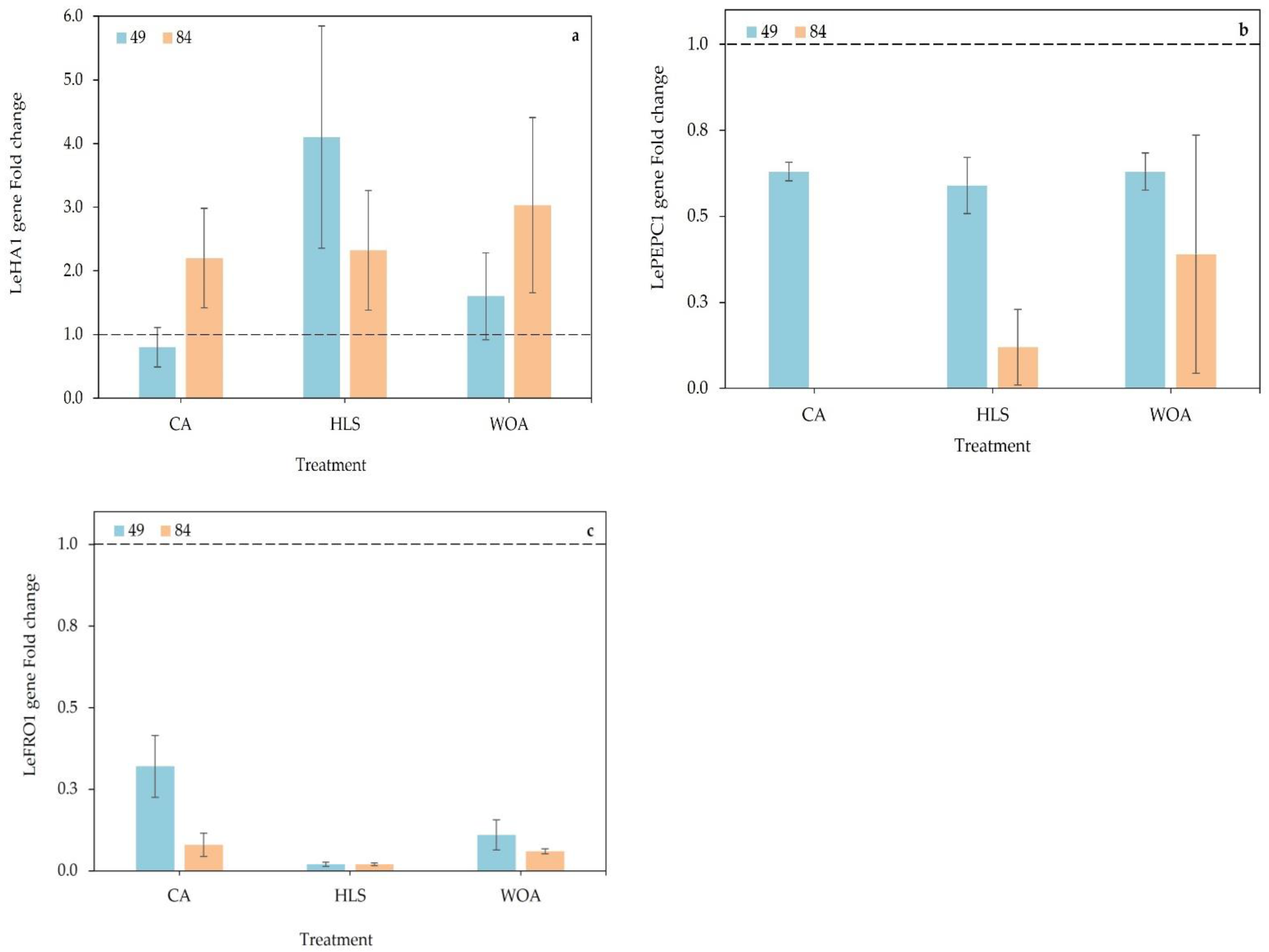
3.3. Gene Expression

The expression of the genes evaluated in leaf tissue is shown in Figure 1. The partial or complete overexpression and/or repression of a gene can be determined by comparing the expression of that gene against a critical threshold [37]. This threshold (value can be one) corresponds to the absolute value of a calibrator gene [38]. Based on this calibration, LeHA1 was repressed to the highest degree in CA at 27 (p < 0.01) and 84 DAT (0.9- and 1.0-fold change, respectively), followed by WOA (1.0-fold change at 84 DAT, Figure 1a), whereas LePEPC1 showed elevated expression in HLS at 27 DAT (0.6-fold change) compared to ID. However, LePEPC1 was repressed under CA treatment at 27 and 49 DAT (up to 0.8-fold change; p < 0.01), whereas it showed elevated expression (p < 0.01) at 84 DAT (up to 1.1-fold change, Figure 1b). In the case of LeFRO1, high expression was observed at 27 and 49 DAT under the HLS treatment (4.10- and 4.19-fold change), whereas the CA treatment resulted in high expression at 49 DAT (2.33-fold change) and repression (p < 0.01) at 84 DAT. This gene exhibited elevated expression with WOA at 27 DAT (Figure 1c).
Root gene expression was only evaluated at 49 and 84 DAT because increased expression of FIT (and probably FRO1, HA1, and PEPC1) has been associated with increased expression during the visible inflorescence stage (49 DAT) followed by a decrease during the fruit set-ripening stage (84 DAT) [18]. The behavior of gene expression in root tissue is presented in Figure 2. For LeHA1, elevated expression was documented at both sampling times; HLS at 49 DAT resulted in the greatest increase (3.8-fold), followed by WOA treatment (1.9-fold change), relative to ID. At 84 DAT, this gene showed 1.2- and 1.9-fold changes under HLS and WOA, respectively (Figure 2a). Regarding LePEPC1 (Figure 2b), repression was documented to a greater extent at 84 DAT in CA (1.0-fold change) and HLS (0.8-fold change) relative to ID. LeFRO1 exhibited repression; HLS application repressed (p < 0.01) LeFRO1 up to 1.2- and 0.9-fold relative to ID at 49 and 84 DAT, respectively (Figure 2c).

4. Discussion

Previous studies have found that the application of CA [39] and leonardite-derived compounds [21] improved the growth and yield of tomatoes grown in calcareous soil, probably via plant stimulation [40,41] and punctually by increasing photosynthetic parameters [42]. Similarly, the use of humic-Fe materials in soybean may be a suitable strategy for the management of Fe nutrition in calcareous soil [43], consequently leading to a reduction in Fe deficiency and an improvement in plant growth, development, and productivity. This is because the addition of Fe together with humic complexes act as “organic chelators” that boost plant growth [41]. CA and leonardite-derived complexes exhibit various levels of complexation with Fe, promoting plant growth by inducing a physiological response [44]. In addition, CA can reduce abiotic stress resulting from phosphorus deficiency in plants grown in calcareous soils [45,46,47]. Likewise, it has been described that the use of humic substances in calcareous soils promotes nutrient bioavailability by generating soluble complexes with minerals [23,48] and increasing nutrient uptake [40,41], and that their combination with chelate Fe (Fe-EDDHA) can increase the yield and nutrient content of plants [49].
Abiotic stress from Fe deprivation is signaled through the phloem from sinks in the root system, where there is an alteration in the absorption and transport of this nutrient [50]. In nongrass monocotyledons and in dicotyledons, such as tomato, that develop under conditions of Fe deprivation and/or in calcareous soils, strategy I presents an enzymatic mechanism for coping with Fe-deficient conditions; according to the results obtained, H+-ATPase and FRO enzymes may be involved in this mechanism and act together with enzymes of the antioxidant system in addition to the regulation of iron transporters (IRT1, ZIP and NRAMP1) and probably coumarin excretion [13]. Although our study does not show a situation of iron deficiency recovery, we can clarify the individual impact of CA and HLS on the enzymatic mechanisms related to iron metabolism in tomato plants grown in calcareous soil.
The H+-ATPase enzyme generates electrochemical potential gradients by energizing ion channels and transporter proteins in the plasma membrane via the extrusion of H+, with the consequent consumption of ATP [51]. Partial or total nutrient (e.g., Fe) deprivation in the growth medium, high carbonate–bicarbonate concentration, and/or high soil pH can induce an increase in root H+-ATPase activity, acidifying the rhizosphere, solubilizing Fe, and preventing Fe precipitation [52]. Our results show higher activity of this enzyme in leaf tissue, which is probably due to its physiological role in the regulation of intra- and extracellular pH, loading of assimilates in the phloem, and redistribution of nutrients [51].
H+-ATPase activity can be stimulated by humic substances [53]. Applications of humic acid purified from leonardite increased plasma membrane H+-ATPase activity in cucumber roots and shoots [54,55]. This contrasts with what was found in the present study, where a reduction in tomato root tissue H+-ATPase activity via HLS was documented; the reduction was probably derived from the promotion of soluble complexes that allowed Fe to exist in a bioavailable form in the soil [23,48] or via the acidifying effect of HLS [56]. This may also have been due to feedback control mechanisms and the concentration of NO3 in the medium [55]. In contrast, the increase in the activity of this enzyme in leaf tissue (at 49 DAT) could be a consequence of the biostimulation (promoting a redistribution of nutrients) induced by HLS and CA [40,41]. In addition, CA is associated with the energy metabolism of the plant [44]; thus, H+-ATPase activity would be necessary to generate gradients in the different cellular compartments of the leaf. The increase in root H+-ATPase activity at 84 DAT under CA application implies an alteration in the gradients of the root system, as in radical cells, this enzyme plays an active role in xylem loading [51].
Tomato plants are thought to implement both strategies I and II; thus, in addition to extruding protons, reducing Fe, and promoting its transport, tomato plants also induce the synthesis of organic compounds (e.g., citrate, malate, and phenols, among others) [57]. In particular, CA tends to accumulate in the root under moderate conditions of Fe deprivation [58,59], where it acts as a reserve for chelating Fe and can present absorption windows over time [13]. The PEPC enzyme, which catalyzes the fixation of bicarbonate (in the presence of Mg2+) to phosphoenolpyruvate, generating oxaloacetate and releasing Pi in C4 plants, could play a crucial role in this context. In nonphotosynthetic tissues and in C3 plants, PEPC enables anaplerotic reactions that provide intermediates in the Krebs cycle [30,58,60]. According to the pH-stat theory, PEPC would have to be forcibly activated to balance the pH change generated by the H+-ATPase activity [30]; in this sense, the addition of acidifying compounds (such as CA and HLS) may impact PEPC activity via the contribution of carbonaceous skeletons.
According to previous studies, PEPC activity increases in roots under Fe-deficient conditions [30,58]. However, severe conditions of Fe deprivation (without Fe in nutrient solution) cause a reduction in the PEPC activity of the root system [59]. However, in this study, it was observed that the addition of HLS (+Fe chelated) promoted an increase in this enzyme (49 and 84 DAT), whereas CA and WOA reduced its activity (27 and 84 DAT). In the case of leaf tissue, the addition of CA and HLS increased PEPC activity. The modification of PEPC activity in the root and leaf tissue of tomato plants in response to CA and HLS application may be due to an alteration of anaplerotic reactions via the contribution of exogenous carbon skeletons [44], via a reprogramming mechanism in carbon metabolism under inadequate Fe conditions that facilitate root exudation [58], or via the acidic environment that can promote its exogenous addition [56].
The intracellular enzyme FRO is associated with the cell membrane and is responsible for reducing Fe3+ to soluble, bioavailable Fe2+ [61]. In leaf tissue, FRO activity is highly dependent on the Fe content in the cytoplasmic solution [62]. In this case (leaf tissue), a reduction in FRO activity was found at 27 DAT (in all treatments), which may imply the contribution and presence of Fe in its assimilable state (Fe2+), whereas the increase in activity (49 and 84 DAT) may have been because Fe was present as Fe3+ in the apoplast because of the content of HCO3 and pH [60]; however, high bicarbonate content and pH apoplastic in plants may decrease FRO activity, causing Fe deficiency symptoms [63]. An increase in FRO enzyme activity has been documented in the roots of pea and tomato plants under Fe-deficient conditions [14,15,64], as was found in this work (27 and 84 DAT), which may suggest that the iron applied via nutrient solution is in its assimilable state (Fe2+) via complexation with HLS or CA. The increase in the activity of this enzyme may or may not be accompanied by a greater number of secondary roots, which would imply a greater number of reduction zones [14].
Humic substances can reduce Fe3+ to soluble forms due to their photocatalytic properties or their redox activity [65,66,67], contributing Fe to the chemical chelate and to the plant (as Fe-complex), which results in an increased concentration of soluble Fe in the soil and an enhanced translocation of Fe from the roots to the leaves [26]. However, the high HCO3 content could precipitate Fe2+ to Fe3+ again, raising its concentration in the rhizosphere and stimulating root FRO activity, as seen in our results (49 DAT). A humic soil environment can condition FRO activity in the root plant, which could partially explain the behavior of this enzyme under our HLS treatment. A study carried out on cucumber reported that the application of humic acid purified to leonardite enhanced FRO activity at 72 h [54]. Repeated application of leonardite Fe humates can precipitate in the root, blocking entry pores in the cell wall and reducing Fe transport [68] as well as probably reducing FRO activity. However, a previous study demonstrated that the repeated application of 400 µL L−1 of HLS on tomato plants potted in calcareous soils had a positive effect on Fe uptake [21]. Therefore, the reduction in the activity of this enzyme could not respond in the aforementioned sense. Despite this, caution should be exercised when applying humic substances. CA—a 6-carbon tricarboxylic compound [69]—is able to generate an acidic microenvironment in the rhizospheric zone, keeping Fe in its soluble and plant-available form and resulting in the low FRO activity (at least at 49 DAT) observed in our study.
The critical threshold of the calibrator gene values [38] allows us to determine the level of overexpression and/or total or partial repression of a gene [37]. In this context, LeHA1, LeFRO1, and probably LePEPC1 may act as indicators of metabolically active Fe in the plant and in the growth medium [70].
Considering that the most suitable pH for the development of tomato plants is 6.0 [50], plants developed in calcareous soils with a high pH (8.5) and high HCO3 content present a nutrient deficit response [13], triggering changes in the expression of specific genes [71]. Previous studies have documented increased expression of the H+-ATPase enzyme promoter gene in Fe-deprived tomato, pea, and cucumber plants [15,16,25,72]. The data obtained here suggest that the modification of this gene can be assumed, in addition to nutrient input, to be a response to the soil pH and rhizospheric HCO3 content because via these treatments, Fe (3 mg L−1) was made available to the plant. In that sense, the repression of LeHA1 (at 27 and 84 DAT) in leaf tissue under CA treatment and its high expression in root tissue under HLS treatment (at 49 DAT) suggest spatiotemporal expression patterns [14]. Such alterations would imply a modification of H+ extrusion from the root tissue into the rhizospheric medium or cellular compartments [17,52]. In addition, humic complexes can induce an acid reaction in the soil by way of their diversity of functional groups [56], affecting LeHA1 expression.
The application of purified humic acid from leonardite (2–250 mg L−1 C) on cucumber grown under Fe-deficient conditions led to higher expression of the CsHA2 gene (up to 4-fold over the control) and low expression of CsHA1 (downregulated up to 5-fold) in the apical root [54]. These results show similar trends to ours; however, the present work showed higher LeHA1 expression in tomato roots without Fe deficiency and with continuous application of HLS. This discrepancy may be due to the differential response of the gene isoform [54,55], which leads to transient changes in H+-ATPase activity.
Similarly, the gene encoding the PEPC protein showed increased expression in the root system under Fe-deficient environments [30,58]. Here, we found that LePEPC1 was repressed in the roots of plants treated with CA and HLS with an Fe supply (in the form of EDTA) (Figure 2b). This suggests a reduced requirement for anaplerotic reactions due to the Fe supply in the growth medium. In the case of elevated leaf expression under HLS at 27 DAT, this may be indicative of active Fe in the plant [70].
In tomatoes, the PEPC gene family may be associated with other types of stresses, such as salinity and cold [73], but its expression could also be a means of coping with nutrient deficits (e.g., Fe) [59]. Applications of a mixture of organic acids (succinic, citric, malic, and oxalic acids, 100 μM) on alfalfa were reported to regulate the expression of the PEPC gene in roots under conditions of abiotic stress via Al [74].
The expression of the gene encoding FRO is strongly influenced, in calcareous soils, by the Fe content in the rhizospheric solution, the exposure time, and the HCO3 content [14,75]. In plants, there is a higher expression of this gene in roots, shoots, and reproductive organs, whereas it is constitutively expressed in leaves [76]. In both cases, variations in the amino acid residues of FRO1 are vital to maintaining the stability and high activity of the FRO enzyme [61].
Tomato and pea plants deprived of Fe show increased expression of FRO1 in the root [15,64]. In this regard, several studies have shown that the application of leonardite humic acid in cucumber plants generates a higher expression of CsFRO1 (between two- and ninefold) after the first days of application. Likewise, leonardite-derived substances applied to tomato plants subjected to Fe deficiency increase LeFRO1 gene expression [70,77]. The use of an Fe complex with water-extractable humic substances (Fe-WEHS) or with citrate (Fe-citrate) generates a higher expression of FRO, IRT1, and NRAMP in cucumber [24], whereas in tomatoes under Fe deficiency, these compounds cause higher expression of LeFRO1, LIRT1, LIRT2, and Ferritin2 in leaves [70,77].
In our study, we found repression of LeFRO1 at 49 and 84 DAT (Figure 2c) in tomato roots with HLS application. This suggests that HLS enhances Fe bioavailability in addition to procuring soluble complexes in calcareous soil [23,48]. In the case of CA, its endogenous variations in tomato plants could be involved in the regulation of nuclear target gene expression [72]. In addition, the alteration in CA concentration affects the regulation of the tricarboxylic cycle [78]. In this sense, when applied exogenously, CA can act as a potential substrate of several metabolic pathways protecting the plant against abiotic stresses [69].
The discrepancy in transcriptional and post-transcriptional stimulation found in the present work may be due to the presence of a series of specific regulatory mechanisms for each level [54] or to associated feedback mechanisms prior to enzymatic activation or gene expression that increase Fe root concentration and Fe translocation [54]. However, the use of CA and/or HLS as an Fe chelate partner could supply stable Fe to the root, stabilize the chelated Fe, or generate an environment conducive to Fe absorption [70], thereby improving the growth and development of plants grown under calcareous soils. These mechanisms could reduce the power of the experimental design without affecting its validity, being necessary to establish unrestricted conditions to determine the precise effect on the supply of stable Fe of CA and HLS.
The enzymatic and gene expression responses observed in tomato plants (grown under calcareous soil) treated with CA and HLS offer a promising outlook in the prevention of Fe deficiency; however, as both compounds show complexation with Fe and stimulate the plant [44], it is necessary to verify possible synergistic effects when applied in combination.

5. Conclusions

Iron deprivation in tomato plants caused a reduction in growth, development, and yield. The application of CA or HLS (continuously or weekly, respectively) throughout the crop cycle through a fertilizer solution improved the SPAD units, number of fruits harvested, and yield per plant. These substances also induced temporary alterations in enzyme activity (p < 0.05), reducing PEPC and FRO activity in roots. In leaf tissue, the CA treatment resulted in lower expression of LeHA1 and LePEPC1 (p < 0.01) as well as induced overexpression of LeFRO1 (p < 0.01). In root tissue, HLS treatment resulted in LeHA1 overexpression and LePEPC1 and LeFRO1 repression (p < 0.01), whereas CA repressed LePEPC1 expression (p < 0.01). Thus, the use of CA and HLS may be a potential strategy in the management of ferric nutrition in tomato plants, as it can cause temporal alterations in enzyme activity (PEPC and FRO) and gene expression (LeHA1, LePEPC1, and LeFRO1) associated with iron uptake.

Author Contributions

Conceptualization, S.G.-M. and S.S.-G.; methodology, F.P.-L.; software, F.P.-L.; validation, A.B.-M. and A.J.-M.; formal analysis, F.P.-L.; writing—original draft preparation, F.P.-L.; writing—review and editing, F.P.-L. and S.G.-M.; funding acquisition, S.S.-G. All authors have read and agreed to the published version of the manuscript.

Funding

This research received no external funding.

Data Availability Statement

Not applicable.

Acknowledgments

The authors thank CONACyT and Arysta LifeScience for financial support.

Conflicts of Interest

The authors declare no conflict of interest.

References

  1. Murgia, I.; Marzorati, F.; Vigani, G.; Morandini, P. Plant iron nutrition: The long road from soil to seeds. J. Exp. Bot. 2022, 73, 1809–1824. [Google Scholar] [CrossRef] [PubMed]
  2. Mori, S. Iron acquisition by plants. Curr. Opin. Plant Biol. 1999, 2, 250–253. [Google Scholar] [CrossRef] [PubMed]
  3. Kobayashi, T.; Nozoye, T.; Nishizawa, N.K. Iron transport and its regulation in plants. Free Radic. Biol. Med. 2019, 133, 11–20. [Google Scholar] [CrossRef]
  4. Wahba, M.M.; Labib, F.; Zaghloul, A. Management of Calcareous Soils in Arid Region. Int. J. Environ. Pollut. Environ. Model. 2019, 2, 248–258. [Google Scholar]
  5. Marschner, H.; Römheld, V. Strategies of plants for acquisition of iron. Plant Soil 1994, 165, 261–274. [Google Scholar] [CrossRef]
  6. Martín-Barranco, A.; Thomine, S.; Vert, G.; Zelazny, E. A quick journey into the diversity of iron uptake strategies in photosynthetic organisms. Plant Signal. Behav. 2021, 16, 1975088. [Google Scholar] [CrossRef] [PubMed]
  7. Chao, Z.F.; Chao, D.Y. Similarities and differences in iron homeostasis strategies between graminaceous and nongraminaceous plants. New Phytol. 2022, 236, 1655–1660. [Google Scholar] [CrossRef]
  8. Li, S.; Song, Z.; Liu, X.; Zhou, X.; Yang, W.; Chen, J.; Chen, R. Mediation of Zinc and Iron Accumulation in Maize by ZmIRT2, a Novel Iron-Regulated Transporter. Plant Cell Physiol. 2022, 63, 521–534. [Google Scholar] [CrossRef]
  9. Santi, S.; Schmidt, W. Dissecting iron deficiency-induced proton extrusion in Arabidopsis roots. New Phytol. 2009, 183, 1072–1084. [Google Scholar] [CrossRef]
  10. Gupta, P.K.; Balyan, H.S.; Sharma, S.; Kumar, R. Biofortification and bioavailability of Zn, Fe and Se in wheat: Present status and future prospects. Theor. Appl. Genet. 2021, 134, 1–35. [Google Scholar] [CrossRef]
  11. Schwarz, B.; Bauer, P. FIT, a regulatory hub for iron deficiency and stress signaling in roots, and FIT-dependent and -independent gene signatures. J. Exp. Bot. 2020, 71, 1694–1705. [Google Scholar] [CrossRef] [PubMed]
  12. Rosenkranz, T.; Oburger, E.; Baune, M.; Weber, G.; Puschenreiter, M. Root exudation of coumarins from soil-grown Arabidopsis thaliana in response to iron deficiency. Rhizosphere 2021, 17, 100296. [Google Scholar] [CrossRef]
  13. Vélez-Bermúdez, I.C.; Schmidt, W. Plant strategies to mine iron from alkaline substrates. Plant Soil 2023, 483, 1–25. [Google Scholar] [CrossRef]
  14. Jiménez, M.R.; Casanova, L.; Saavedra, T.; Gama, F.; Suárez, M.P.; Correia, P.J.; Pestana, M. Responses of tomato (Solanum lycopersicum L.) plants to iron deficiency in the root zone. Folia Hortic. 2019, 31, 223–234. [Google Scholar] [CrossRef]
  15. Kabir, A.H.; Paltridge, N.G.; Able, A.J.; Paull, J.G.; Stangoulis, J.C.R. Natural variation for Fe-efficiency is associated with upregulation of Strategy I mechanisms and enhanced citrate and ethylene synthesis in Pisum sativum L. Planta 2012, 235, 1409–1419. [Google Scholar] [CrossRef] [PubMed]
  16. Santi, S.; Cesco, S.; Varanini, Z.; Pinton, R. Two plasma membrane H+-ATPase genes are differentially expressed in iron-deficient cucumber plants. Plant Physiol. Biochem. 2005, 43, 287–292. [Google Scholar] [CrossRef]
  17. Fan, Z.; Wu, Y.; Zhao, L.; Fu, L.; Deng, L.; Deng, J.; Ding, D.; Xiao, S.; Deng, X.; Peng, S.; et al. MYB308-mediated transcriptional activation of plasma membrane H+-ATPase 6 promotes iron uptake in citrus. Hortic. Res. 2022, 9, uhac088. [Google Scholar] [CrossRef]
  18. Filiz, E.; Kurt, F. FIT (Fer-like iron deficiency-induced transcription factor) in plant iron homeostasis: Genome-wide identification and bioinformatics analyses. J. Plant Biochem. Biotechnol. 2019, 28, 143–157. [Google Scholar] [CrossRef]
  19. Ferreira, C.M.H.; López-Rayo, S.; Lucena, J.J.; Soares, E.V.; Soares, H. Evaluation of the Efficacy of Two New Biotechnological-Based Freeze-Dried Fertilizers for Sustainable Fe Deficiency Correction of Soybean Plants Grown in Calcareous Soils. Front. Plant Sci. 2019, 10, 1335. [Google Scholar] [CrossRef]
  20. Zanin, L.; Tomasi, N.; Cesco, S.; Varanini, Z.; Pinton, R. Humic substances contribute to plant iron nutrition acting as chelators and biostimulants. Front. Plant Sci. 2019, 10, 675. [Google Scholar] [CrossRef]
  21. Pérez-Labrada, F.; Benavides-Mendoza, A.; Juárez-Maldonado, A.; Solís-Gaona, S.; González-Morales, S. Organic acids combined with Fe-chelate improves ferric nutrition in tomato grown in calcisol soil. J. Soil Sci. Plant Nutr. 2020, 20, 673–683. [Google Scholar] [CrossRef]
  22. Al-Balawna, Z.A.; Abu-Abdoun, I.I. Fate of Citric Acid Addition on Mineral Elements Availability in Calcareous Soils of Jordan Valley. Int. Res. J. Pure Appl. Chem. 2021, 22, 82–89. [Google Scholar] [CrossRef]
  23. Olego, M.Á.; Cuesta Lasso, M.; Quiroga, M.J.; Visconti, F.; López, R.; Garzón-Jimeno, E. Effects of Leonardite Amendments on Vineyard Calcareous Soil Fertility, Vine Nutrition and Grape Quality. Plants 2022, 11, 356. [Google Scholar] [CrossRef]
  24. Zanin, L.; Tomasi, N.; Rizzardo, C.; Gottardi, S.; Terzano, R.; Alfeld, M.; Janssens, K.; De Nobili, M.; Mimmo, T.; Cesco, S. Iron allocation in leaves of Fe-deficient cucumber plants fed with natural Fe complexes. Physiol. Plant. 2015, 154, 82–94. [Google Scholar] [CrossRef] [PubMed]
  25. Zamboni, A.; Zanin, L.; Tomasi, N.; Avesani, L.; Pinton, R.; Varanini, Z.; Cesco, S. Early transcriptomic response to Fe supply in Fe-deficient tomato plants is strongly influenced by the nature of the chelating agent. BMC Genom. 2016, 17, 35. [Google Scholar] [CrossRef] [PubMed]
  26. Cieschi, M.T.; Lucena, J.J. Leonardite iron humate and synthetic iron chelate mixtures in Glycine max nutrition. J. Sci. Food Agric. 2021, 101, 4207–4219. [Google Scholar] [CrossRef]
  27. Steiner, A.A. A universal method for preparing nutrient solutions of a certain desired composition. Plant Soil 1961, 15, 134–154. [Google Scholar] [CrossRef]
  28. Rabotti, G.; Zocchi, G. Plasma membrane-bound H+-ATPase and reductase activities in Fe-deficient cucumber roots. Physiol. Plant. 1994, 90, 779–785. [Google Scholar] [CrossRef]
  29. Palmgren, M.G.; Askerlund, P.; Fredrikson, K.; Widell, S.; Sommarin, M.; Larsson, C. Sealed Inside-Out and Right-Side-Out Plasma Membrane Vesicles. Plant Physiol. 1990, 92, 871–880. [Google Scholar] [CrossRef]
  30. Nisi, P.D.; Zochi, G. Phosphoenolpyruvate carboxylase in cucumber (Cucumis sativus L.) roots under iron deficiency: Activity and kinetic characterization. J. Exp. Bot. 2000, 51, 1903–1909. [Google Scholar] [CrossRef]
  31. Romera, F.; Welch, R.; Norvell, W.; Schaefer, S.; Kochian, L. Ethylene involvement in the over-expression of Fe(III)-chelate reductase by roots of E107 pea [Pisum sativum L. (brz, brz)] and chloronerva tomato (Lycopersicon esculentum L.) mutant genotypes. Biometals 1996, 9, 38–44. [Google Scholar] [CrossRef]
  32. Bradford, M.M. A rapid and sensitive method for the quantitation of microgram quantities of protein utilizing the principle of protein-dye binding. Anal. Biochem. 1976, 72, 248–254. [Google Scholar] [CrossRef] [PubMed]
  33. Rio, D.C.; Ares, M.; Hannon, G.J.; Nilsen, T.W. Purification of RNA Using TRIzol (TRI Reagent). Cold Spring Harb. Protoc. 2010, 2010, pdb-prot5439. [Google Scholar] [CrossRef] [PubMed]
  34. Paolacci, A.R.; Celletti, S.; Catarcione, G.; Hawkesford, M.J.; Astolfi, S.; Ciaffi, M. Iron deprivation results in a rapid but not sustained increase of the expression of genes involved in iron metabolism and sulfate uptake in tomato (Solanum lycopersicum L.) seedlings. J. Integr. Plant Biol. 2014, 56, 88–100. [Google Scholar] [CrossRef]
  35. Diamantopoulos, P.D.; Aivalakis, G.; Flemetakis, E.; Katinakis, P. Expression of three β-type carbonic anhydrases in tomato fruits. Mol. Biol. Rep. 2013, 40, 4189–4196. [Google Scholar] [CrossRef]
  36. Larionov, A.; Krause, A.; Miller, W. A standard curve based method for relative real time PCR data processing. BMC Bioinform. 2005, 6, 62. [Google Scholar] [CrossRef]
  37. Dembélé, D.; Kastner, P. Fold change rank ordering statistics: A new method for detecting differentially expressed genes. BMC Bioinform. 2014, 15, 14. [Google Scholar] [CrossRef]
  38. Love, M.I.; Huber, W.; Anders, S. Moderated estimation of fold change and dispersion for RNA-seq data with DESeq2. Genome Biol. 2014, 15, 550. [Google Scholar] [CrossRef]
  39. Pérez-Labrada, F.; Mendoza, A.B.; Valdez-Aguilar, L.A.; Robledo-Torres, V. Citric acid in the nutrient solution increases the mineral absorption in potted tomato grown in calcareous soil. Pakistan J. Bot. 2016, 48, 67–74. [Google Scholar]
  40. Massimi, M.; Radócz, L.; Csótó, A. Impact of Organic Acids and Biological Treatments in Foliar Nutrition on Tomato and Pepper Plants. Horticulturae 2023, 9, 413. [Google Scholar] [CrossRef]
  41. Sharma, S.; Anand, N.; Bindraban, P.S.; Pandey, R. Foliar Application of Humic Acid with Fe Supplement Improved Rice, Soybean, and Lettuce Iron Fortification. Agriculture 2023, 13, 132. [Google Scholar] [CrossRef]
  42. Zia-ur-Rehman, M.; Bani Mfarrej, M.F.; Usman, M.; Azhar, M.; Rizwan, M.; Alharby, H.F.; Bamagoos, A.A.; Alshamrani, R.; Ahmad, Z. Exogenous application of low and high molecular weight organic acids differentially affected the uptake of cadmium in wheat-rice cropping system in alkaline calcareous soil. Environ. Pollut. 2023, 329, 121682. [Google Scholar] [CrossRef] [PubMed]
  43. Cieschi, M.T.; Polyakov, A.Y.; Lebedev, V.A.; Volkov, D.S.; Pankratov, D.A.; Veligzhanin, A.A.; Perminova, I.V.; Lucena, J.J. Eco-friendly iron-humic nanofertilizers synthesis for the prevention of iron chlorosis in soybean (Glycine max) grown in calcareous soil. Front. Plant Sci. 2019, 10, 413. [Google Scholar] [CrossRef] [PubMed]
  44. Justi, M.; Silva, C.A.; Rosa, S.D. Organic acids as complexing agents for iron and their effects on the nutrition and growth of maize and soybean. Arch. Agron. Soil Sci. 2022, 68, 1369–1384. [Google Scholar] [CrossRef]
  45. Jalali, M.; Jalali, M. Effect of Low-Molecular-Weight Organic Acids on the Release of Phosphorus from Amended Calcareous Soils: Experimental and Modeling. J. Soil Sci. Plant Nutr. 2022, 22, 4179–4193. [Google Scholar] [CrossRef]
  46. Karadihalli Thammaiah, M.; Pandey, R.N.; Purakayastha, T.J.; Chobhe, K.A.; Vashisth, A.; Chandra, S.; Pawar, A.B.; Trivedi, A. Impact of Low Molecular Weight Organic Acids on Soil Phosphorus Release and Availability to Wheat. Commun. Soil Sci. Plant Anal. 2022, 53, 2497–2508. [Google Scholar] [CrossRef]
  47. Zhao, K.; Wang, C.; Xiao, X.; Li, M.; Zhao, W.; Wang, Y.; Yang, Y. The Hormetic Response of Soil P Extraction Induced by Low-Molecular-Weight Organic Acids. Processes 2023, 11, 216. [Google Scholar] [CrossRef]
  48. Sun, Q.; Liu, J.; Huo, L.; Li, Y.C.; Li, X.; Xia, L.; Zhou, Z.; Zhang, M.; Li, B. Humic acids derived from Leonardite to improve enzymatic activities and bioavailability of nutrients in a calcareous soil. Int. J. Agric. Biol. Eng. 2020, 13, 200–205. [Google Scholar] [CrossRef]
  49. Abdulla, A.A.; Esmai, A.O.; Yaseen, H.S. Combination Influence of Humic Acid and Chelated Iron on yield and quality of Broccoli (Brassica oleracea L.) in Erbil, Iraqi Kurdistan Region. ZANCO J. Pure Appl. Sci. 2023, 35, 126–135. [Google Scholar] [CrossRef]
  50. Gayomba, S.R.; Zhai, Z.; Jung, H.; Vatamaniuk, O.K. Local and systemic signaling of iron status and its interactions with homeostasis of other essential elements. Front. Plant Sci. 2015, 6, 716. [Google Scholar] [CrossRef]
  51. Palmgren, M.G. Plant plasma membrane H+-ATPases: Powerhouses for Nutrient Uptake. Annu. Rev. Plant Physiol. Plant Mol. Biol. 2001, 52, 817–845. [Google Scholar] [CrossRef] [PubMed]
  52. Chen, L.; Zhao, R.; Yu, J.; Gu, J.; Li, Y.; Chen, W.; Guo, W. Functional analysis of plasma membrane H+-ATPases in response to alkaline stress in blueberry. Sci. Hortic. 2022, 306, 111453. [Google Scholar] [CrossRef]
  53. Canellas, L.P.; Olivares, F.L.; Aguiar, N.O.; Jones, D.L.; Nebbioso, A.; Mazzei, P.; Piccolo, A. Humic and fulvic acids as biostimulants in horticulture. Sci. Hortic. 2015, 196, 15–27. [Google Scholar] [CrossRef]
  54. Elena, A.; Diane, L.; Eva, B.; Marta, F.; Roberto, B.; Zamarreño, A.M.; García-Mina, J.M. The root application of a purified leonardite humic acid modifies the transcriptional regulation of the main physiological root responses to Fe deficiency in Fe-sufficient cucumber plants. Plant Physiol. Biochem. 2009, 47, 215–223. [Google Scholar] [CrossRef]
  55. Mora, V.; Bacaicoa, E.; Zamarreño, A.-M.; Aguirre, E.; Garnica, M.; Fuentes, M.; García-Mina, J.-M. Action of humic acid on promotion of cucumber shoot growth involves nitrate-related changes associated with the root-to-shoot distribution of cytokinins, polyamines and mineral nutrients. J. Plant Physiol. 2010, 167, 633–642. [Google Scholar] [CrossRef]
  56. Alghamdi, S.A.; Al-Ghamdi, F.A.M.; El-Zohri, M.; Al-Ghamdi, A.A.M. Modifying of calcareous soil with some acidifying materials and its effect on Helianthus annuus (L.) growth. Saudi J. Biol. Sci. 2023, 30, 103568. [Google Scholar] [CrossRef]
  57. Astolfi, S.; Pii, Y.; Mimmo, T.; Lucini, L.; Miras-Moreno, M.B.; Coppa, E.; Violino, S.; Celletti, S.; Cesco, S. Single and Combined Fe and S Deficiency Differentially Modulate Root Exudate Composition in Tomato: A Double Strategy for Fe Acquisition? Int. J. Mol. Sci. 2020, 21, 4038. [Google Scholar] [CrossRef]
  58. Martinez-Cuenca, M.-R.; Iglesias, D.J.; Talon, M.; Abadia, J.; Lopez-Millan, A.-F.; Primo-Millo, E.; Legaz, F. Metabolic responses to iron deficiency in roots of Carrizo citrange [Citrus sinensis (L.) Osbeck. × Poncirus trifoliata (L.) Raf.]. Tree Physiol. 2013, 33, 320–329. [Google Scholar] [CrossRef]
  59. Covarrubias, J.I.; Rombolà, A.D. Organic acids metabolism in roots of grapevine rootstocks under severe iron deficiency. Plant Soil 2015, 394, 165–175. [Google Scholar] [CrossRef]
  60. Alhendawi, R.A.M.; Mohamed, A.A.M. The influence of high pH on maize growth and utilization of micronutrients under various concentrations of bicarbonates. Am. J. Agric. Environ. Sci. 2015, 15, 259–264. [Google Scholar] [CrossRef]
  61. Kong, D.; Chen, C.; Wu, H.; Li, Y.; Li, J.; Ling, H.-Q. Sequence Diversity and Enzyme Activity of Ferric-Chelate Reductase LeFRO1 in Tomato. J. Genet. Genomics 2013, 40, 565–573. [Google Scholar] [CrossRef]
  62. Larbi, A.; Morales, F.; Abadía, A.; Abadía, J. Changes in iron and organic acid concentrations in xylem sap and apoplastic fluid of iron-deficient Beta vulgaris plants in response to iron resupply. J. Plant Physiol. 2010, 167, 255–260. [Google Scholar] [CrossRef] [PubMed]
  63. Zhao, Y.; Liu, S.; Li, F.; Sun, M.; Liang, Z.; Sun, Z.; Yu, F.; Li, H. The low ferric chelate reductase activity and high apoplastic pH in leaves cause iron deficiency chlorosis in ‘Huangguan’ pears grafted onto quince A grown in calcareous soil. Sci. Hortic. 2023, 310, 111754. [Google Scholar] [CrossRef]
  64. Zamboni, A.; Zanin, L.; Tomasi, N.; Pezzotti, M.; Pinton, R.; Varanini, Z.; Cesco, S. Genome-wide microarray analysis of tomato roots showed defined responses to iron deficiency. BMC Genom. 2012, 13, 101. [Google Scholar] [CrossRef] [PubMed]
  65. Skogerboe, R.K.; Wilson, S.A. Reduction of ionic species by fulvic acid. Anal. Chem. 1981, 53, 228–232. [Google Scholar] [CrossRef]
  66. Struyk, Z.; Sposito, G. Redox properties of standard humic acids. Geoderma 2001, 102, 329–346. [Google Scholar] [CrossRef]
  67. Yang, F.; Tang, C.; Antonietti, M. Natural and artificial humic substances to manage minerals, ions, water, and soil microorganisms. Chem. Soc. Rev. 2021, 50, 6221–6239. [Google Scholar] [CrossRef]
  68. Cieschi, M.T.; Lucena, J.J. Iron and Humic Acid Accumulation on Soybean Roots Fertilized with Leonardite Iron Humates under Calcareous Conditions. J. Agric. Food Chem. 2018, 66, 13386–13396. [Google Scholar] [CrossRef]
  69. Tahjib-Ul-Arif, M.; Zahan, M.I.; Karim, M.M.; Imran, S.; Hunter, C.T.; Islam, M.S.; Mia, M.A.; Hannan, M.A.; Rhaman, M.S.; Hossain, M.A.; et al. Citric Acid-Mediated Abiotic Stress Tolerance in Plants. Int. J. Mol. Sci. 2021, 22, 7235. [Google Scholar] [CrossRef]
  70. Tomasi, N.; De Nobili, M.; Gottardi, S.; Zanin, L.; Mimmo, T.; Varanini, Z.; Römheld, V.; Pinton, R.; Cesco, S. Physiological and molecular characterization of Fe acquisition by tomato plants from natural Fe complexes. Biol. Fertil. Soils 2013, 49, 187–200. [Google Scholar] [CrossRef]
  71. Lee, S.; Rahman, M.M.; Nakanishi, H.; Nishizawa, N.K.; An, G.; Nam, H.G.; Jeon, J.-S. Concomitant Activation of OsNAS2 and OsNAS3 Contributes to the Enhanced Accumulation of Iron and Zinc in Rice. Int. J. Mol. Sci. 2023, 24, 6568. [Google Scholar] [CrossRef] [PubMed]
  72. Vigani, G.; Pii, Y.; Celletti, S.; Maver, M.; Mimmo, T.; Cesco, S.; Astolfi, S. Mitochondria dysfunctions under Fe and S deficiency: Is citric acid involved in the regulation of adaptive responses? Plant Physiol. Biochem. 2018, 126, 86–96. [Google Scholar] [CrossRef] [PubMed]
  73. Waseem, M.; Ahmad, F. The phosphoenolpyruvate carboxylase gene family identification and expression analysis under abiotic and phytohormone stresses in Solanum lycopersicum L. Gene 2019, 690, 11–20. [Google Scholar] [CrossRef]
  74. An, Y.; Zhou, P.; Xiao, Q.; Shi, D. Effects of foliar application of organic acids on alleviation of aluminum toxicity in alfalfa. J. Plant Nutr. Soil Sci. 2014, 177, 421–430. [Google Scholar] [CrossRef]
  75. Hsieh, E.-J.; Waters, B.M. Alkaline stress and iron deficiency regulate iron uptake and riboflavin synthesis gene expression differently in root and leaf tissue: Implications for iron deficiency chlorosis. J. Exp. Bot. 2016, 67, 5671–5685. [Google Scholar] [CrossRef]
  76. Li, L.; Cheng, X.; Ling, H.-Q. Isolation and characterization of Fe(III)-chelate reductase gene LeFRO1 in tomato. Plant Mol. Biol. 2004, 54, 125–136. [Google Scholar] [CrossRef]
  77. Tomasi, N.; Rizzardo, C.; Monte, R.; Gottardi, S.; Jelali, N.; Terzano, R.; Vekemans, B.; De Nobili, M.; Varanini, Z.; Pinton, R.; et al. Micro-analytical, physiological and molecular aspects of Fe acquisition in leaves of Fe-deficient tomato plants re-supplied with natural Fe-complexes in nutrient solution. Plant Soil 2009, 325, 25–38. [Google Scholar] [CrossRef]
  78. Drincovich, M.F.; Voll, L.M.; Maurino, V.G. Editorial: On the Diversity of Roles of Organic Acids. Front. Plant Sci. 2016, 7, 1592. [Google Scholar] [CrossRef]
Figure 1. Expression of the LeHA1 (a), LePEPC1 (b), and LeFRO1 (c) genes in tomato leaves of plants treated with CA and HLS. Values are means ± standard error (n = 5). The dashed line represents the constant value of the absolute control (ID).
Horticulturae 09 00630 g001
Figure 2. Expression of the LeHA1 (a), LePEPC1 (b), and LeFRO1 (c) genes in tomato roots of plants treated with CA and HLS. Values are means ± standard error (n = 5). The dashed line represents the constant value of the absolute control (ID).
Horticulturae 09 00630 g002
Table 1. Sequences of primers used for gene analysis.
Name GeneNomenclatureForward Primer 5′-3′Reverse Primer 5′-3′Tm (°C)
ActinACTINCCCAGGCACACAGGTGTTATCAGGAGCAACTCGAAGCTCA60
H+-ATPaseLeHA1GAACCCTTCATGGGCTCCAAGCAACTCACGTAGCCTAGCA60
PEPCLePEPC1TGCTGCATTGTTCGACAAGCCAAAAGTTCGCCGAAAGACAAC60
FROLeFRO1GCGGTGTTGAATATGCTAATCAAACTTTCCATCTCCCTATCG60
Table 2. Plant growth parameters of tomato plants treated with CA and HLS.
Sampling TreatmentStem Diameter (mm)Plant Height (cm)Number of LeavesSPAD-UnitTotal of Fruit HarvestProduction per Plant (kg)
27 DATCA10.80 ± 0.52 a45.70 ± 3.38 a13.20 ± 0.45 a54.19 ± 2.78 a--
HLS10.24 ± 0.96 a39.40 ± 4.39 b13.40 ± 0.89 a53.65 ± 2.38 a--
WOA10.00 ± 0.29 a45.50 ± 3.87 a12.60 ± 0.89 a53.85 ± 2.95 a--
ID8.22 ± 1.90 b32.80 ± 5.54 c10.80 ± 2.17 b33.55 ± 5.64 b--
49 DAT CA11.66 ± 1.21 a71.20 ± 3.63 a18.00 ± 2.24 a55.43 ± 3.42 a--
HLS12.54 ± 1.03 a68.20 ± 8.32 a15.20 ± 2.17 b57.91 ± 3.51 a--
WOA12.02 ± 0.45 a73.40 ± 5.94 a18.00 ± 0.71 a54.85 ± 2.17 a--
ID9.74 ± 0.56 b53.40 ± 2.30 b12.60 ± 1.67 c27.51 ± 5.13 b--
84 DATCA12.98 ± 1.28 ab 101.60 ± 11.04 a23.00 ± 2.35 a54.21 ± 4.12 a57.75 ± 6.44 ab2.76 ± 0.28 a
HLS13.84 ± 1.23 a102.60 ± 13.45 a23.80 ± 1.92 a56.03 ± 4.30 a61.95 ± 6.73 a2.82 ± 0.27 a
WOA11.98 ± 0.67 bc103.40 ± 4.10 a21.20 ± 1.48 a48.77 ± 5.91 a53.40 ± 4.38 b2.67 ± 0.08 a
ID11.40 ± 1.18 c64.60 ± 9.42 b17.00 ± 3.46 b17.34 ± 16.92 b32.05 ± 3.09 c0.77 ± 0.15 b
DAT = days after transplant. CA = citric acid. HLS = humic-like substances. WOA = without organic amendment. ID = Fe deprivation. Values are means ± standard deviations, n = 5. Within a row, values not sharing a letter are significantly different (p < 0.05).
Table 3. Enzymatic activity in tomato tissue treated with CA and HLS.
TissueSamplingTreatmentH+-ATPasePEPCFRO
Leaf27 DATCA1.46 ± 0.18 b482.3 ± 64.0 ab7.2 ± 0.2 b
HLS1.84 ± 0.07 ab267.1 ± 65.7 cd7.4 ± 0.1 b
WOA1.57 ± 0.05 ab633.6 ± 154.2 a7.2 ± 0.3 b
ID3.76 ± 2.22 a343.5 ± 56.9 bc9.6 ± 0.3 b
49 DATCA0.92 ± 0.11 b834.2 ± 151.4 a7.7 ± 0.2 b
HLS0.89 ± 0.04 b856.5 ± 140.6 a7.5 ± 0.3 b
WOA0.89 ± 0.05 b784.9 ± 194.0 a9.1 ± 0.4 b
ID0.65 ± 0.05 c552.7 ± 113.0 a6.7 ± 0.3 b
84 DATCA2.65 ± 1.33 a465.6 ± 221.8 b10.7 ± 1.2 b
HLS1.58 ± 0.06 a759.6 ± 136.5 a8.2 ± 0.2 bc
WOA1.25 ± 0.13 a141.0 ± 27.0 cd10.5 ± 1.1 b
ID1.81 ± 0.63 a304.0 ± 99.5 bc6.7 ± 1.1 c
Root27 DATCA0.016 ± 0.0005 b51.5 ± 29.7 e15.8 ± 2.1 a
HLS0.017 ± 0.0007 b95.2 ± 43.1 de17.2 ± 1.5 a
WOA0.017 ± 0.0004 b191.8 ± 16.0 cde17.3 ± 2.4 a
ID0.017 ± 0.0009 b156.9 ± 48.3 cde14.5 ± 1.3 a
49 DATCA1.35 ± 0.05 a26.2 ± 21.5 b22.0 ± 1.2 a
HLS1.28 ± 0.02 a11.6 ± 7.0 b23.2 ± 0.7 a
WOA1.26 ± 0.13 a16.6 ± 12.2 b22.2 ± 2.9 a
ID1.32 ± 0.05 a4.6 ± 0.1 b25.3 ± 1.5 a
84 DATCA1.50 ± 0.05 a4.5 ± 0.0 d21.6 ± 0.9 a
HLS1.27 ± 0.11 a205.6 ± 21.1 bcd20.1 ± 1.7 a
WOA1.29 ± 0.03 a11.8 ± 7.6 d21.4 ± 1.1 a
ID1.48 ± 0.04 a261.9 ± 16.5 bcd20.1 ± 1.8 a
DAT = days after transplant. CA = citric acid. HLS = humic-like substances. WOA = without organic amendment. ID = Fe deprivation. H+-ATPase activity expressed as: µmol NADH min−1 mg−1 prot. PEPC activity expressed as: nmol NADH min−1 mg−1 prot. FRO activity expressed as: nmol Fe2+ reduced (g FW tissue) −1 h−1. Values are means ± standard deviations, n = 5. Within a row, values not sharing a letter are significantly different (p < 0.05).
Disclaimer/Publisher’s Note: The statements, opinions and data contained in all publications are solely those of the individual author(s) and contributor(s) and not of MDPI and/or the editor(s). MDPI and/or the editor(s) disclaim responsibility for any injury to people or property resulting from any ideas, methods, instructions or products referred to in the content.

