Article

Molecular Characterization, Population Structure Analysis, and Association Mapping of Turkish Parsley Genotypes Using iPBS Markers

by
Ömer Faruk Coşkun
Department of Horticulture, Faculty of Agriculture, Hatay Mustafa Kemal University, Antakya 31060, Turkey
Horticulturae 2023, 9(3), 336; https://doi.org/10.3390/horticulturae9030336
Submission received: 26 January 2023 / Revised: 26 February 2023 / Accepted: 27 February 2023 / Published: 3 March 2023

Abstract

:
Parsley (Petroselinum crispum Mill.) is a vegetable that has many benefits for human health, and its cultivation is increasing. In order to carry out breeding studies in parsley, genotypes should be characterized in terms of some characteristics. In this study, some phytochemical properties of 18 different parsley genotypes were revealed, and their genetic diversity was determined with the iPBS (Inter Primary Binding Site) marker system. In the study, the polymorphism rate was 31.9%, the mean PIC (Polymorphic Information Content) was 0.17, and the similarity coefficients were between 0.87 and 0.99. The number of subpopulations was determined as two, and 10 markers were detected at expression levels of 19–33% related to phytochemical properties. The results of this study show that there is a phytochemical and genetic variation in parsley. Parsley genotypes with certain phytochemical properties and genetic structures can be used more effectively in breeding programs.

1. Introduction

Parsley is an important vegetable belonging to the Apiaceae family, which has been widely consumed in many countries for over 2000 years. Parsley is a biennial herb but is grown commercially as an annual herb in many parts of the world due to its edible and aromatic leaves [1]. Apart from being consumed as food, parsley is also used in the pharmaceutical, cosmetic, and perfume industries [2,3]. Parsley contains phenolic acids and flavonoids that prevent oxidation stress significantly. It also has high apigenin, high luteolin and, caffeic acid content, which may be beneficial for human health [4]. Parsley is also rich in potassium, iron, ascorbic acid, vitamins, and essential oils [5]. Due to these substances, the high antioxidant capacity of parsley is beneficial for human health by delaying or preventing many diseases [2,6,7,8,9]. It has been reported that parsley can be used to help treat people with Alzheimer’s, cardiovascular diseases, and thrombosis [10,11]. In addition, it is stated that regular consumption of parsley as a food or supplement can reduce the effect of cancer [12,13].
There are three most common parsley subspecies: Petroselinum crispum ssp. tuberosum, Petroselinum crispum ssp. crispum and Petroselinum crispum ssp. neapolitanum [14]. The parts that can be dried, frozen, or consumed raw are usually the leaves. Therefore, flat leaf (Petroselinum crispum ssp. neapolitanum) is the most widely cultivated parsley type. There is considerable variation among parsley genotypes in characteristics such as morphology, growth routine, flower color, stems, leaves, and chemical configuration [15]. Parsley is a strong source of income, providing its producers with a steady income throughout the year. Commercial parsley production is increasing day by day all over the world. Approximately 105.574 tons/year of parsley are grown in Turkey [16].
Genetic differences in plants can affect phytochemical properties [17]. Commercialization of agricultural products, the widespread use of hybrid seeds, and monoculture farming can cause genetic erosion and loss of genetic resources that may be beneficial in future demands. Generally, uncommercialized low–yield genotypes can better adapt to unfavorable environmental conditions [18]. For these reasons, it is necessary to protect and improve agricultural biodiversity. In recent years, considering global warming and other abiotic stress factors, the necessity of protecting local genotypes has increased. Determining the genetic relationships in the germplasm collection is of great importance for effective breeding and germplasm maintenance. For this reason, it is important to determine the genetic structures of plants. In general, studies on parsley have focused on its medicinal and bioactive components. Genetic characterization studies that can lead to breeding studies are insufficient.
Phytochemical and molecular markers can be used to determine genetic characterization. Phytochemical parameters and DNA band profiles can show the variation between genotypes at a sensitive level. DNA molecular markers can be obtained from plants at any growth stage and are not affected by environmental or seasonal factors [19,20]. Different marker techniques have been used successfully in vegetables to determine molecular characterization [21,22,23]. Retrotransposons are an important source of plant genetic variation and have large genomes [24]. Primers obtained using different retrotransposon sites are used as a marker technique. One of these techniques, PCR–based iPBS (inter primary binding site), is a technique with high reproducibility and polymorphism rate. DNA sequence information is not required for the iPBS marker technique. There are studies on genetic diversity with iPBS–retrotransposon markers [25,26]. Additionally, to our knowledge, molecular genetic studies to detect genetic relationships between different parsley genotypes are scarce. ISSR, SRAP, RAPD and, AFLP techniques were used in some studies [27,28,29,30]. The iPBS technique has not been studied in parsley genotypes before. Our aim in this study is to evaluate the genetic and phytochemical diversity of 18 local parsley genotypes in Turkey. The data obtained in this study can be used in future breeding programs.

2. Materials and Methods

2.1. Plant Materials

The study was carried out in Hatay Mustafa Kemal University, Faculty of Agriculture, Department of Horticulture, trial station and Genetics laboratory in 2022. A total of 18 parsley (P. crispum) genotypes (Table 1) were grown in pots under controlled greenhouse conditions. Each genotype was grown in 3 pots and 4 replications, and according to widely accepted recommendations. Seedlings were harvested at the stage with 10–15 cm length and 5–8 leaves, and analyzes were carried out.

2.2. Phytochemical Analysis

A total of seven phytochemical content analyzes were performed on parsley genotypes. Total soluble solids (TSS, %) content was measured using a digital refractometer. Total acid content (TA, % oxalic acid) was determined by potentiometric titration, and pH value was determined by standard methods [31] with a digital pH meter. Photosynthetic parameters were determined according to the method described by Holm [32] and Wettstein [33]. On 2 g of fresh sample mass, 15 mL of acetone was added in three steps; the samples were homogenized and filtered. The pigment extract was diluted with acetone to a final volume of 25 mL. Chlorophyll and carotenoid amounts were calculated according to the Holm–Wettstein equations, and the final results were expressed as mg/g.
Chlorophyll a = 9.784 × A662 − 0.990 × A644 (mg/L)
Chlorophyll b = 21.426 × A644 − 4.65 × A662 (mg/L)
Total chlorophyll = 5.134 × A662 + 20.436 × A644 (mg/L)
Total carotenoids = 4.695 × A440 − 0.268 × (chlorophyll a + b)(mg/L)
Statistical analysis of experimental data was performed using SPSS (Statistical Package for the Social Sciences). Before the ANOVA analysis, all samples were tested for normal distribution according to the Shapiro–Wilk test and Levene’s test. Tukey’s HSD test was used to determine the significant difference between the means (p < 0.05). The results were expressed as mean ± standard errors.

2.3. Genetic Analysis

Ten DNA samples were bulked for each application. Genomic DNA was extracted from plants following the protocol of the cetyltrithylammnonium bromide (CTAB) method. The amount and purity of DNA were measured using a 1% agarose gel in 1X TAE (Tris, Acetic Acid, EDTA) buffer. Polymerase chain reaction (PCR) optimized 15 µL reactions contained 50 ng template DNA, 10 nmol dNTPs, 10 nmol iPBS primer, 5 U Taq DNA polymerase, and 1.5 µL of 10X polymerase chain reaction (PCR) buffer (50 mM KCl, 10 mM Tris–HCl, 2.5 mM MgCl2 and pH 8.3). The PCR thermal cycling profile is as follows; initial denaturation for 3 min at 95 °C, 38 cycles of 95 °C for 60 s, 50–60 °C for 60 s, 72 °C for 120 s, and final extension at 72 °C for 10 min. PCR products were separated on 1.5% agarose gel at 110 V for 5 h and visualized under UV light (Figure S1).
By using band data, some parameters of iPBS primers (number of bands, polymorphism ratio, polymorphic information content, etc.) were obtained. Using the GenAlEx 6.5 software, the number of different alleles, number of effective alleles, Shannon’s information index, haploid genetic diversity (H), and unbiased diversity were calculated. Genetic similarity values between eighteen parsley genotypes were determined using the Numerical Taxonomy Multivariate Analysis System (NTSYS) package program. The genetic similarity coefficient was calculated according to the Dice method [34], and the dendrogram showing the similarities between the individuals was obtained by the UPGMA (Unweighted Pair Group Method with Arithmetic Mean) method. Principle Component Analysis (PCA) was also conducted with the use of this matrix in NTSYS software.
Population structure was analyzed in STRUCTURE V2.3 for K values ranging from 1 to 10 [35,36]. Each running was repeated 5 times with 50,000 burn–in length. The most likely population ancestor was determined by Evanno’s correction [37].
In addition, association mapping studies were performed on 18 parsley genotypes using phytochemical and DNA data. The general linear model (General Linear Model–GLM + (Q)), in which the Q matrix showing the population structure is used, was used in the relationship mapping. The significance level between the marker and phenotypic traits was calculated with the TASSEL 5.0 program [38] based on the p values and the F test. The Q matrix showing the population structure used in the mapping was obtained from the STRUCTURE program.

3. Results

3.1. Phytochemical Variations

TSS, TA, pH, and photosynthetic parameters were analyzed. In this study, TSS contents were determined in the range of 2.32–5.34%. The mean TSS value in all genotypes was determined as 4.43 ± 0.36. The highest value was determined in genotype 10 (5.34 ± 0.29), and the lowest value was determined in genotype 13 (2.32 ± 0.41). TSS values obtained in genotype 10 are statistically significantly higher than in genotypes 3, 8, 9, 11, 12, 13, 14, 15, and 18. It can be said that parsley genotypes originating from “Osmaniye” have the lowest TSS values. In this study, the total amount of acid was determined in the range of 1.04–2.59%. TA value was determined as 1.76 ± 0.02 in all genotypes. The lowest value was determined in genotype 15 (1.04 ± 0.03), and the highest value was determined in genotype number 1 (2.59 ± 0.01). TA value determined in genotype 1 originating from “Hatay–Samandag” is significantly higher than other genotypes, and TA values determined in genotype 15 originating from “Osmaniye” are significantly lower than other genotypes. It can be said that parsley genotypes originating from “Osmaniye” have the lowest TA values. The pH was determined between 5.67 and 5.87 and a mean of 5.79 ± 0.02 in all genotypes. The smallest value was obtained in genotype 2 (5.67 ± 0.02), and the highest value was obtained in genotype 8 (5.87 ± 0.02). Among the genotypes, pH values obtained from genotypes 8, 1, 6, and 7 are significantly higher than other genotypes. The pH values of genotype number 2 are significantly lower than all other genotypes (Table 2).
Chlorophyll a values were determined between 0.48 ± 0.02 mg/g and 0.31–0.61 mg/g on average. The highest value was determined in genotypes 1 and 11 (0.61), and the lowest value was determined in genotype 3 (0.31 ± 0.01). Chlorophyll a values determined in genotypes 1 and 11 were significantly higher than other genotypes except for genotypes 4 and 7. Chlorophyll a values determined in genotype number 6 are significantly lower than other genotypes except 2, 10, and 18. Chlorophyll b values were determined between 0.28 ± 0.01 mg/g and 0.18–0.37 mg/g on average. The highest value was determined in genotype 11 (0.37 ± 0.01), and the lowest value was determined in genotype number 3 (0.18 ± 0.01). The chlorophyll b value determined in genotype 11 is significantly higher than the other genotypes. The chlorophyll b value determined in genotype number 3 is significantly lower than other genotypes. Total chlorophyll values were determined between 0.76 ± 0.03 mg/g and 0.50–0.98 mg/g on average. The highest value was determined in genotype 11 (0.98 ± 0.04), and the lowest value was determined in genotype number 3 (0.50 ± 0.02). Total chlorophyll value determined in genotype 11 is significantly higher than other genotypes except for genotype 1. The total chlorophyll value determined in genotype number 3 is significantly lower than in other genotypes. The total carotenoid values were determined between 0.14 ± 0.01 mg/g and 0.10–0.18 mg/g on average. The highest value was determined in genotypes 1 and 11 (0.18), and the lowest value was determined in genotype 3 (0.10 ± 0.01). Total carotenoid values determined in genotypes 1 and 11 are significantly higher than other genotypes, and total carotenoid values determined in genotype 3 are significantly higher than other genotypes except for genotypes 2, 13, 17, and 18 (Table 3).

3.2. Genetic Diversity Analysis

In this study, 16 iPBS primers with the best amplification were used for genetic characterization. A total of 181 bands and an average of 11.3 bands per primer were obtained. There are a total of 65 polymorphic bands and an average of 4.1 polymorphic bands per primer. The polymorphism rates of the primers ranged between 10 and 75%, and the average polymorphism rate was 31.9%. The highest number of bands was obtained from primers iPBS–2272 (17) and iPBS–2249 (17), while the lowest number of bands was obtained from primer iPBS–2251 (7). The highest number of polymorphic bands was detected in primer iPBS–2272 (11), and the lowest number of polymorphic bands in primer iPBS–2246 (1). The highest polymorphism rate was obtained from iPBS–2239 (75%), and the lowest polymorphism rate was obtained from iPBS–2246 (10%) primer (Table 4).
The band frequency was calculated as the highest in the iPBS–2219 primer (0.96) and the mean 0.89. The number of distinct alleles was highest in the iPBS–2226 primer (1.82), with an average of 1.51. The average number of effective alleles was 1.19, and the highest was determined in the iPBS–2272 primer. The number of effective alleles was highest in the iPBS–2272 primer (1.39), with a mean of 1.19. Shannon’s Information Index values are 0.20 on average and were found in the range of 0.05–0.36. Diversity and Unbiased diversity mean values were determined as 0.12 and 0.13, respectively. Polymorphic information content was determined between 0.03 and 0.41 with an average of 0.17. The highest PIC value was obtained from iPBS–2272 primer (0.41), and the lowest PIC was obtained from iPBS–2246 primer (0.03) (Table 5).
Similarity coefficients were calculated using DNA data in the NTSYS package program. According to the DICE matrix, the similarity coefficient between the genotypes was determined between 0.87 and 0.99. The highest similarity values (0.9913) were obtained between genotypes 5 and 6. The most distant genotypes are genotypes 3 to 17, with a similarity coefficient of 0.87. In addition, the similarity value between the 3–15, 8–18, 10–17, 11–17, 12–15, 12–17, 14–17, 15–18, and 17–18 genotypes was determined under 0.9. Two main clusters occurred in the UPGMA dendrogram determined according to the Dice similarity index. The similarity coefficient between the two main clusters is 0.92. There were 13 genotypes in the first main cluster and five genotypes (8, 9, 13, 15, 17) in the second main cluster. In the first main cluster, genotype 14 was separated from other genotypes. The remaining genotypes consisted of three subsets. There are six genotypes (1, 2, 5, 6, 7, and 3) in the first subset, four genotypes (4, 10, 11, and 12) in the second subset, and three genotypes (16, 18, and 14) in the third subset. The closest clustering genotypes are genotypes 5 to 6. Secondly, the genotypes closest to each other are genotypes 10 and 11 (Figure 1).
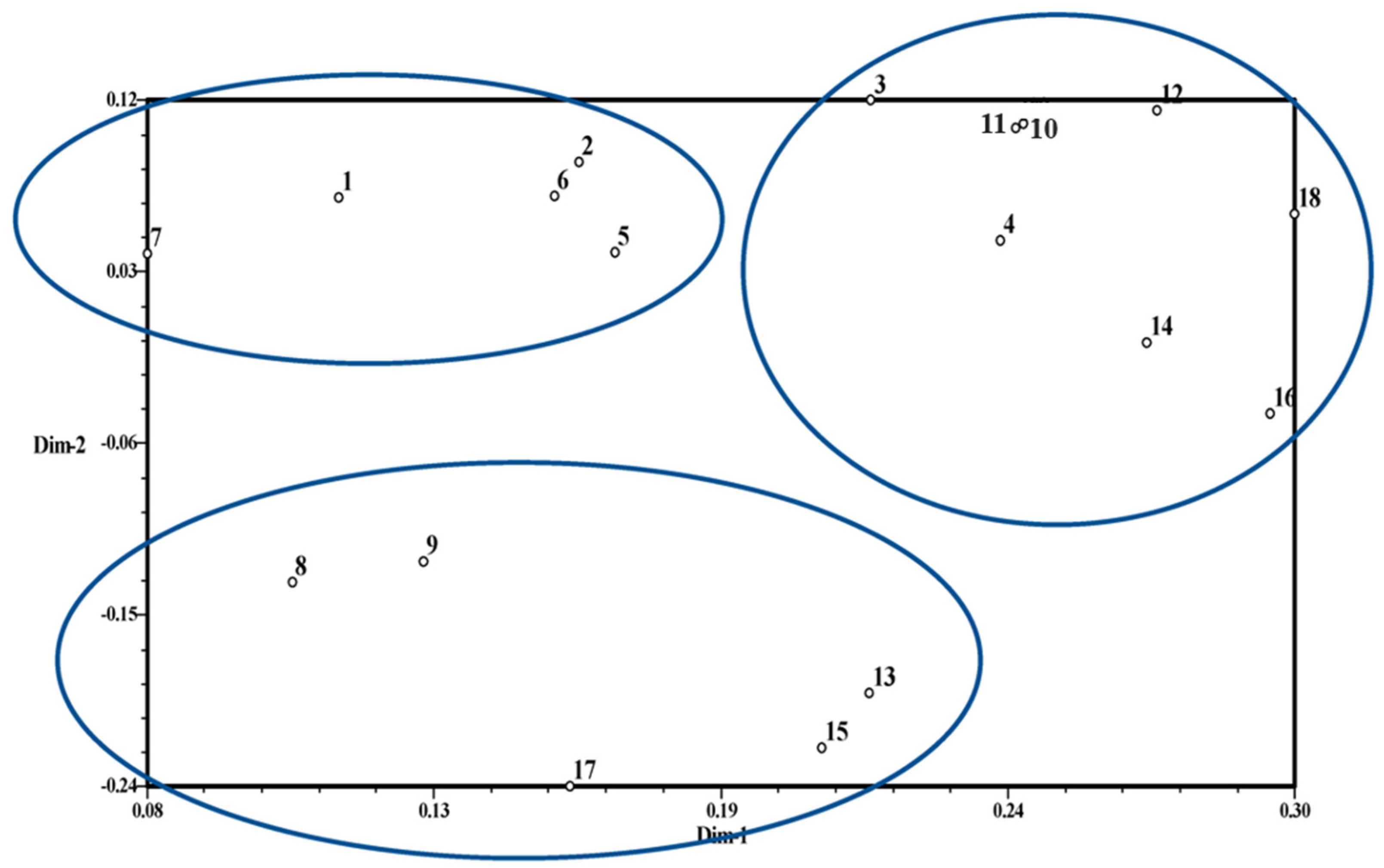
Two and three-dimensional graphics were obtained by performing PCA (Principal Components Analysis) in the NTSYS program. In the two–dimensional PCA graph, it was determined that the genotypes were grouped in three different clusters. There were six genotypes in the first cluster, seven genotypes in the second cluster, and five genotypes in the third cluster. Genotypes 10 and 12 are closest to each other. In the three–dimensional PCA graph, genotypes are generally divided into two parts. The most closely located genotypes are 10 and 11 (Figure 2 and Figure 3). Genotypes 3 and 17 were located at the farthest positions in the two–dimensional PCA (Figure 2), three–dimensional PCA and UPGMA dendrograms (Figure 3).

3.3. Population Structure

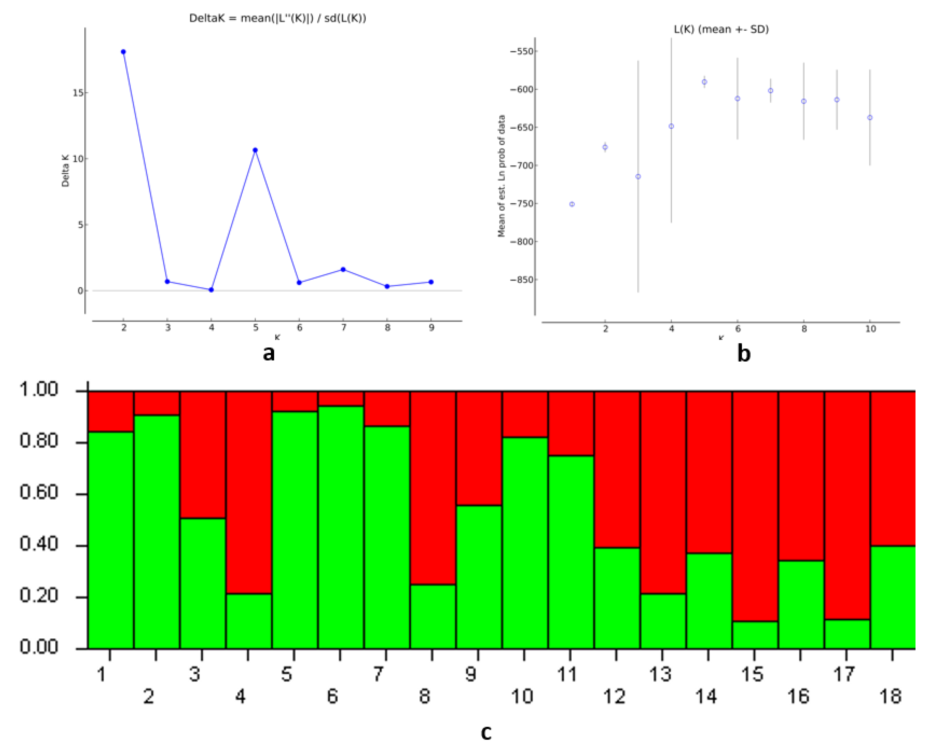
Analysis of population structure was performed using the Bayes method in Structure software [35]. According to the Delta K values obtained using the Structure Harvester program, the number of subpopulations is two (K = 2). In the data with two subpopulation values, Mean LnP(K) value was calculated as –676.05, Standard deviation LnP(K) value was 6.26, Ln′(K) value was 74.91, |Ln″(K)| value was 113.47 and Delta K value was 18.10. In the first subpopulation, the belonging values were determined between 0.056 and 0.889, and in the second subpopulation, the belonging values were between 0.111 and 0.944. A genotype can be unequivocally assigned to a subpopulation when the mixing coefficient (Qi) for that subpopulation is >0.8 [39,40]. Genotypes with intermediate mixing coefficients (i.e., with Qi < 0.8) are considered mixed. Among the genotypes, there are nine genotypes with a belonging value above 0.8. Genotypes assigned to the first subpopulation are genotypes 15 and 17. Genotypes assigned to the second subpopulation are genotypes 1, 2, 5, 6, 7, and 10. The remaining nine genotypes are considered admixed (Figure 4).

3.4. Associating Mapping

In this study, the relationship between phytochemical data and 181 iPBS markers in parsley was examined using the Tassel 5.0 program to create relationship mapping. In GLM (Q) (General Linear Model) analysis, three markers associated with TCC, one associated with TA, one associated with pH, two associated with chlorophyll a, two associated with chlorophyll b, and three markers associated with total carotenoid were found to be associated at the p < 0.05 level. The correlation level (R) of TA with the iPBS–2226–1140 marker was 33%, and the correlation level (R) of pH with the iPBS–2272–770 marker was determined as 24%. The level of association (R) of TCC ranged from 19% to 26%, and the highest associated marker is iPBS–2239–420. The level of association (R) in chlorophyll a ranged from 25% to 26%, and the highest associated marker is iPBS–2244–200. The level of association (R) in chlorophyll b ranged from 27% to 28%, and the highest associated marker is iPBS–2244–200. The same markers in chlorophyll a and chlorophyll b have similar levels of association. The level of association (R) in total carotenoids ranged from 21% to 23%, and the highest associated marker is iPBS–2249–1080 (Table 6). The number of markers with significant associations with six phytochemical properties is 10 out of a total of 181 markers. The markers iPBS–2228–450 and iPBS–2244–200 were associated with two traits (chlorophyll a and chlorophyll b).

4. Discussion

As far as we know, phytochemical characterization studies on local parsley genotypes are very few. iPBS marker studies and related marker identification studies have not been performed before. Seven different parameters that can show the genetic characterization of phytochemicals were examined. Total Soluble Solids contents are generally low in Apiaceae family members [41]. TSS values in the range of 2.32–5.34% determined in this study are low, similar to some other studies [5,42,43]. Similar to previous studies [5], in this study, a significant variation in TSS values were detected between parsley genotypes. In this study, TA values were determined between 1.04 and 2.59, while pH values were determined between 5.67 and 5.87 values. The pH value variation determined between parsley genotypes is lower than TCC and TA. Chlorophyll a, chlorophyll b, and total carotenoid values calculated in genotypes 1 and 11 in germplasm are higher than other genotypes. The significant variation in chlorophyll and carotenoid values among parsley genotypes grown under the same conditions may indicate that the genetic effect on these traits may be important.
Sixteen primers were used in this study. This number of primers is higher than the number of primers used in other studies [29,30]. In addition, the number of bands per primer was determined as 11.3 in this study. This value is higher than those previously determined in different marker techniques. Previously, the number of bands per primer for ISSR techniques was calculated as 11 [27], 7.83 [30], and 8.12 [44]. Although the number of bands per primer obtained in this study was higher than in the previous ones, the number of polymorphic bands per primer was found to be lower than in some studies [28,29]. However, a higher value (4.1) was obtained than the number of bands per 2.56 primers determined in the previous study examining Turkey genotypes [44]. These results may indicate that iPBS primers can give more polymorphic bands than ISSR primers in parsley genotypes. The lowest similarity coefficient was determined as 0.92 in the UPGMA dendrogram obtained by using the Dice coefficient. This value is similar to the similarity coefficient determined with ISSR primers by Coskun et al. [44]. While some studies found less genetic similarity [28], genetic distance was greater in studies using genotypes from different geographical areas [29,30].
The population structure of parsley was determined by Coskun et al. [44] with the Structure program. In the previous study, it was determined that parsley genotypes consisted of five subpopulations. In this study, two subpopulations were determined in Structure analysis using data obtained with iPBS primers. Although more genotypes were used than the number of genotypes used in the study of Coskun et al. [44], the low number of subpopulations may be related to the primer technique used. No previous association mapping studies have been performed on parsley. In this study, markers associated with some physiological characteristics were determined, and associating rates could be determined.

5. Conclusions

As a result of our study, it was determined that there was variation in the phytochemical parameters and genetic structure of the eighteen 18 parsley cultivars. However, genetic diversity was found to be low. It has been concluded that iPBS markers can be used to determine the genetic characterization of parsley genotypes. The data obtained indicate that the Turkish parsley germplasm can serve as an important source of genetic material for plant breeding and selection for the development of new varieties.

Supplementary Materials

The following supporting information can be downloaded at: https://www.mdpi.com/article/10.3390/horticulturae9030336/s1, Figure S1: Gel electrophoresis pattern of iPBS amplification using primers iPBS-2222 and iPBS-2243.

Funding

This research received no external funding.

Institutional Review Board Statement

Not applicable.

Informed Consent Statement

Not applicable.

Data Availability Statement

Not applicable.

Conflicts of Interest

The author declares no conflict of interest.

References

  1. Dadan, M.; Rybak, K.; Wiktor, A.; Nowacka, M.; Zubernik, J.; Witrowa–Rajchert, D. Selected chemical composition changes in microwave–convective dried parsley leaves affected by ultrasound and steaming pre–treatments–An optimization approach. Food Chem. 2018, 239, 242–251. [Google Scholar] [CrossRef] [PubMed]
  2. de Oliveira, V.S.; Chávez, D.W.H.; Paiva, P.R.F.; Gamallo, O.D.; Castro, R.N.; Sawaya, A.C.H.F.; Sampaio, G.R.; Torres, E.A.F.D.S.; Saldanha, T. Parsley (Petroselinum crispum Mill.): A source of bioactive compounds as a domestic strategy to minimize cholesterol oxidation during the thermal preparation of omelets. Food Res. Int. 2022, 156, 111199. [Google Scholar] [CrossRef] [PubMed]
  3. Dziki, D.; Hassoon, W.H.; Biernacka, B.; Gawlik–Dziki, U. Dried and powdered leaves of parsley as a functional additive to wheat bread. Appl. Sci. 2022, 12, 7930. [Google Scholar] [CrossRef]
  4. Michalaki, A.; Karantonis, H.C.; Kritikou, A.S.; Thomaidis, N.S.; Dasenaki, M.E. Ultrasound–assisted extraction of specific phenolic compounds from Petroselinum crispum leaves using response surface methodology and HPLC–PDA and Q–TOF–MS/MS identification. Appl. Sci. 2023, 13, 798. [Google Scholar] [CrossRef]
  5. Dobričević, N.; Šic Žlabur, J.; Voća, S.; Pliestić, S.; Galić, A.; Delić, A.; Fabek Uher, S. Bioactive compounds content and nutritional potential of different parsley parts (Petroselinum crispum Mill.). J. Cent. Eur. Agr. 2019, 20, 900–910. [Google Scholar] [CrossRef] [Green Version]
  6. Mouhoubi, K.; Boulekbache–Makhlouf, L.; Madani, K.; Palatzidi, A.; Perez–Jimenez, J.; Mateos–Aparicio, I.; Garcia–Alonso, A. Phenolic compounds and antioxidant activity are differentially affected by drying processes in celery, coriander and parsley leaves. Int. J. Food Sci. Technol. 2022, 57, 3467–3476. [Google Scholar] [CrossRef]
  7. Sener, G.; Karakadıoglu, G.; Ozbeyli, D.; Ede, S.; Yanardag, R.; Sacan, O.; Aykac, A. Petroselinum crispum extract ameliorates scopolamine–induced cognitive dysfunction: Role on apoptosis, inflammation and oxidative stress. Food Sci. Hum. Wellness 2022, 11, 1290–1298. [Google Scholar] [CrossRef]
  8. Petropoulos, S.A.; Fernandes, Â.; Finimundy, T.C.; Polyzos, N.; Pinela, J.; Ivanov, M.; Soković, M.; Ferreira, I.C.F.R.; Barros, L. The bioactivities and chemical profile of turnip–rooted parsley germplasm. Horticulturae 2022, 8, 639. [Google Scholar] [CrossRef]
  9. Arsenov, D.; Župunski, M.; Pajević, S.; Nemeš, I.; Simin, N.; Alnuqaydan, A.M.; Watson, M.; Aloliqi, A.A.; Mimica–Dukić, N. Roots of Apium graveolens and Petroselinum crispum—Insight into phenolic status against toxicity level of trace elements. Plants 2021, 10, 1785. [Google Scholar] [CrossRef]
  10. Gadi, D.; Bnouham, M.; Aziz, M.; Ziyyat, A.; Legssyer, A.; Legrand, C.; Lafeve, F.F.; Mekhfi, H. Parsley extract inhibits in vitro and ex vivo platelet aggregation and prolongs bleeding time in rats. J. Ethnopharmacol. 2009, 125, 170–174. [Google Scholar] [CrossRef]
  11. Adsersen, A.; Gauguin, B.; Gudiksen, L.; Jäger, A.K. Screening of plants used in Danish folk medicine to treat memory dysfunction for acetylcholinesterase inhibitory activity. J. Ethnopharmacol. 2006, 104, 418–422. [Google Scholar] [CrossRef]
  12. Danciu, C.; Cioanca, O.; Watz–Farcaș, C.; Hancianu, M.; Racoviceanu, R.; Muntean, D.; Zupko, I.; Oprean, C.; Tatu, C.; Paunescu, V.; et al. Botanical Therapeutics (Part II): Antimicrobial and In vitro anticancer activity against mcf7 human breast cancer cells of chamomile, parsley and celery alcoholic extracts. Anticancer. Agents Med. Chem. 2020, 21, 187–200. [Google Scholar] [CrossRef]
  13. Ashry, M.; Atia, I.; Morsy, F. Elmashad, wael protective efficiency of parsley (Petroselinum crispum) against oxidative stress, DNA damage and nephrotoxicity induced with anti–tuberculosis drugs. Int. J. Cancer Biomed. Res. 2021, 5, 27–36. [Google Scholar] [CrossRef]
  14. Petropoulos, S.A.; Daferera, D.; Akoumianakis, C.; Passam, H.; Polissiou, M. The effect of sowing date and growth stage on the essential oil composition of three types of parsley (Petroselinum crispum). J. Sci. Food Agric. 2004, 1610, 1606–1610. [Google Scholar] [CrossRef]
  15. Sarwar, S.; Ayyub, M.A.; Rezgui, M.; Nisar, S.; Jilani, I. Parsley: A review of habitat, phytochemistry, ethnopharmacology and biological activities. Int. J. Chem. Biochem. Sci. 2019, 9, 49–55. [Google Scholar]
  16. Turkish Statistical Institute (TUIK). 2022. Available online: https://data.tuik.gov.tr/Bulten/Index?p=Bitkisel–Uretim–Istatistikleri–2022–45504 (accessed on 10 January 2023).
  17. Simon, J.E.; Quinn, J. Characterization of essential oil of parsley. J. Agric. Food Chem. 1988, 36, 467–472. [Google Scholar] [CrossRef]
  18. Bekhradi, F.; Delshad, M.; Marín, A.; Luna, M.; Garrido, Y.; Kashi, A.; Babalar, M.; Gil, M. Effects of salt stress on physiological and postharvest quality characteristics of different Iranian genotypes of basil. Hortic. Environ. Biotechnol. 2015, 56, 777–785. [Google Scholar] [CrossRef]
  19. Jones, N.; Ougham, H.; Thomas, H.; Pasakinskiene, I. Markers and mapping revisited: Finding your gene. New Phytol. 2009, 183, 935–966. [Google Scholar] [CrossRef]
  20. Agarwal, M.; Shrivastava, N.; Padh, H. Advances in molecular marker techniques and their applications in plant sciences. Plant Cell Rep. 2008, 27, 617–631. [Google Scholar] [CrossRef] [PubMed]
  21. Karaman, K.; Dalda–Sekerci, A.; Yetisir, H.; Gulsen, O.; Coskun, O.F. Molecular, morphological and biochemical characterization of some turkish bitter melon (Momordica charantia L.) genotypes. Ind. Crops Prod. 2008, 123, 93–99. [Google Scholar] [CrossRef]
  22. Kirac, H.; Dalda–Sekerci, A.; Coskun, O.F.; Gulsen, O. Morphological and molecular characterization of garlic (Allium sativum L.) genotypes sampled from Turkey. Gen. Res. Crop Evol. 2022, 69, 1833–1841. [Google Scholar] [CrossRef] [PubMed]
  23. Coskun, O.F. Determination of genetic diversity in some pumpkin genotypes using SSR marker technique. Erzincan Univ. J. Sci. Tech. 2022, 15, 942–952. [Google Scholar]
  24. Bennetzen, J.L. Transposable element contributions to plant gene and genome evolution. Plant Mol. Biol. 2000, 42, 251–269. [Google Scholar] [CrossRef] [PubMed]
  25. Nadeem, M.A. Deciphering the genetic diversity and population structure of Turkish bread wheat germplasm using iPBS–retrotransposons markers. Mol. Biol. Rep. 2021, 48, 6739–6748. [Google Scholar] [CrossRef]
  26. Öztürk, H.İ.; Dursun, A.; Hosseinpour, A.; Haliloğlu, K. Genetic diversity of pinto and fresh bean (Phaseolus vulgaris L.) germplasm collected from Erzincan province of Turkey by inter–primer binding site (iPBS) retrotransposon markers. Turk. J. Agric. For. 2020, 44, 417–427. [Google Scholar] [CrossRef]
  27. Domblides, A.S.; Domblides, E.A.; Kharchenko, V.A.; Potekhin, G.A. Study of genetic variation among parsley (Petroselinum crispum Mill. Nym.) samples using RAPD and ISSR markers. Moscow Univ. Biol. Sci. Bull. 2010, 65, 152–154. [Google Scholar] [CrossRef]
  28. Nasiri, K.; Shojaeiyan, A.; Yadollahi, A.; Mirshekari, A.; Ghanbari, K. Genetic diversity assessment of some Iranian parsley (Petroselinum crispum Mill.) accessions using SRAP molecular marker. J. Veg. Sci. 2015, 1, 1–10. [Google Scholar]
  29. Ibrahim, H.M.M.; El–Leel, O.F.A.; Emam, K.A. Molecular profiling for genetic variability in Petroselinum crispum based on ISSR and RAPD markers. Middle East J. Agric. Res. 2017, 6, 67–75. [Google Scholar]
  30. Boutsika, A.; Sarrou, E.; Cook, C.M.; Mellidou, I.; Avramidou, E.; Angeli, A.; Martens, S.; Ralli, P.; Letsiou, S.; Selini, A.; et al. Evaluation of parsley (Petroselinum crispum) germplasm diversity from the Greek Gene Bank using morphological, molecular and metabolic markers. Ind. Crops Prod. 2021, 170, 113767. [Google Scholar] [CrossRef]
  31. AOAC. Official Methods of Analysis, 16th ed.; Association of Official Analytical Chemists: Washington, DC, USA, 1995. [Google Scholar]
  32. Holm, G. Chlorophyll mutations in barley. Acta Agric. Scan. 1954, 4, 457–471. [Google Scholar] [CrossRef]
  33. Wettstein, D. Chlorophyll letale und der submikroskopische formwechsel der plastiden. Exp. Cell Res. 1957, 12, 427–434. [Google Scholar] [CrossRef] [PubMed]
  34. Dice, L.R. Measures of the amount of ecologic association between species. Ecology 1945, 26, 297–302. [Google Scholar] [CrossRef]
  35. Pritchard, J.K.; Stephens, M.; Donnelly, P. Inference of population Structure using multilocus genotype data. Genetics 2000, 155, 945–959. [Google Scholar] [CrossRef] [PubMed]
  36. Falush, D.; Stephens, M.; Pritchard, J.K. Inference of population structure using multilocus genotype data: Linked loci and correlated allele frequencies. Genetics 2003, 164, 1567–1587. [Google Scholar] [CrossRef]
  37. Evanno, G.; Regnaut, S.; Goudet, J. Detecting the number of clusters of individuals using the software Structure: A simulation study. Mol. Ecol. 2005, 14, 2611–2620. [Google Scholar] [CrossRef] [Green Version]
  38. Bradbury, P.J.; Zhang, Z.; Kroon, D.E.; Casstevens, T.M.; Ramdoss, Y.; Buckler, E.S. TASSEL: Software for association mapping of complex traits in diverse samples. Bioinformatics 2007, 23, 2633–2635. [Google Scholar] [CrossRef]
  39. Vigouroux, Y.; Glaubitz, J.C.; Matsuoka, Y.; Major, M.; Doebley, J. Population structure and genetic diversity of the new world maize races assessed by microsatellites. Am. J. Bot. 2008, 95, 1240–1253. [Google Scholar] [CrossRef]
  40. Castillo, A.; Dorado, G.; Feuillet, C.; Sourdille, P.; Hernandez, P. Genetic structure and ecogeographical adaptation in wild barley (Hordeum chilense Roemer et Schultes) as revealed by microsatellite markers. BMC Plant Biol. 2010, 10, 266. [Google Scholar] [CrossRef] [Green Version]
  41. Parthasarathy, V.A.; Chempakam, B.; Zachariah, T.J. Chemistry of Spices; CAB International: London, UK, 2008. [Google Scholar]
  42. Karklelienė, R.; Dambrauskienė, E.; Juškevičienė, D.; Radzevičius, A.; Rubinskienė, M.; Viškelis, P. Productivity and nutritional value of dill and parsley. Hort. Sci. 2014, 41, 131–137. [Google Scholar] [CrossRef] [Green Version]
  43. Santos, J.; Herrero, M.; Mendiola, J.A.; Oliva–Teles, M.T.; Ibanez, E.; Delerue–Matos, C.; Oliveira, M.B.P.P. Fresh–cut aromatic herbs: Nutritional quality stability during shelf–life. LWT Food Sci. Tehnol. 2014, 59, 101–107. [Google Scholar] [CrossRef]
  44. Coskun, O.F.; Gunduz, Y.F.; Toprak, S.; Mavi, K. Molecular characterization of some parsley (Petroselinum crispum Mill.) genotypes. Mustafa Kemal Univ. J. Agri. Sci. 2023. Accepted. [Google Scholar]
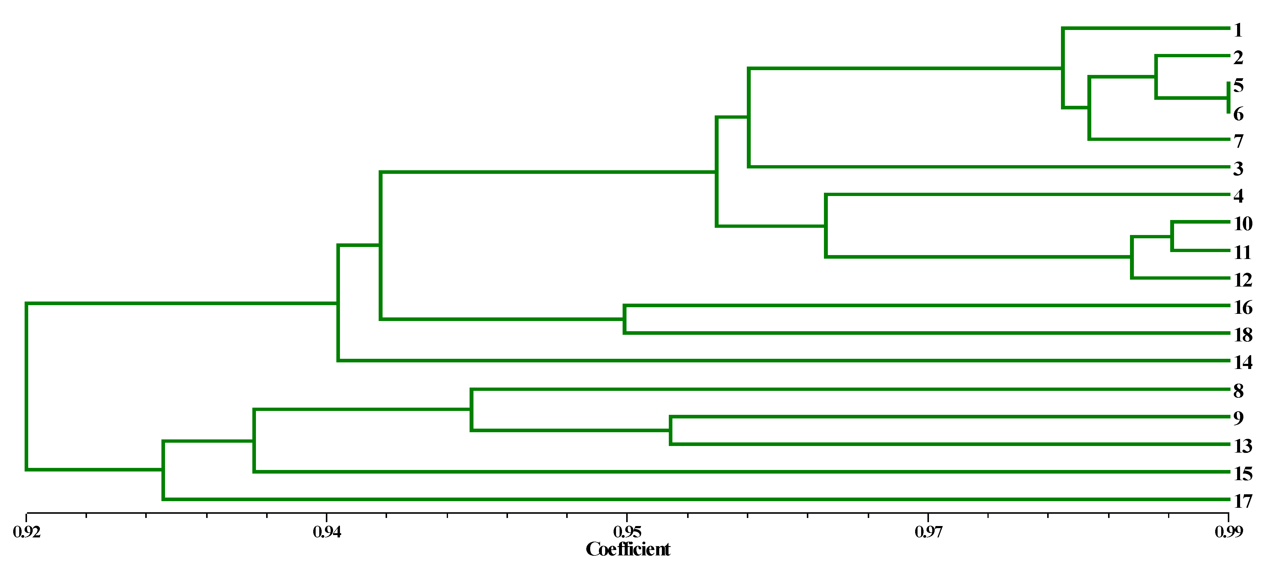
Figure 1. Dendrogram obtained from UPGMA (Unweighted Pair-Group Method with Arithmetic mean) cluster analysis using the Dice coefficient.
Horticulturae 09 00336 g001
Figure 2. PCA (Principal Components Analysis) two−dimensional plot for 18 parsley genotypes using 181 iPBS markers.
Horticulturae 09 00336 g002
Figure 3. PCA (Principal Components Analysis) three−dimensional plot for 18 parsley genotypes using 181 iPBS markers.
Horticulturae 09 00336 g003
Figure 4. (a) ΔK values for parsley genotypes. (b) L(K) values for parsley genotypes. (c) Proportions of the ancestry of 18 parsley accessions based on K = 2 (where K is the number of initial subpopulations, each indicated with a different color).
Horticulturae 09 00336 g004
Table 1. Genotype codes and locations.
NoCodeLocationNoCodeLocation
1HMKU–MA1Hatay–Samandag10HMKU–MA10Hatay–Arsuz
2HMKU–MA2Hatay–Samandag11HMKU–MA11Hatay–Antakya
3HMKU–MA3Hatay–Samandag12HMKU–MA12Hatay–Antakya
4HMKU–MA4Hatay–Samandag13HMKU–MA13Osmaniye
5HMKU–MA5Hatay–Arsuz14HMKU–MA14Osmaniye
6HMKU–MA6Hatay–Arsuz15HMKU–MA15Osmaniye
7HMKU–MA7Hatay–Arsuz16HMKU–MA16Kilis
8HMKU–MA8Hatay–Arsuz17HMKU–MA17Osmaniye
9HMKU–MA9Hatay–Arsuz18HMKU–MA18Hatay–İskenderun
Table 2. Total soluble solids, total acid content, and pH of 18 parsley genotypes.
GenotypeTotal Soluble Solids (%)Total Acid Content (%)pH–Value
14.67 ± 0.34 ab2.59 ± 0.01 a5.86 ± 0.01 ab
24.31 ± 0.31 a–c1.87 ± 0.01 f5.67 ± 0.02 h
33.67 ± 0.32 b–d2.21 ± 0.03 c5.75 ± 0.01 ef
44.32 ± 0.33 a–c2.11 ± 0.02 d5.74 ± 0.03 ef
54.67 ± 0.28 ab1.95 ± 0.01 ef5.84 ± 0.02 bc
64.63 ± 0.41 ab1.94 ± 0.03 ef5.86 ± 0.01 ab
74.28 ± 0.34 a–c1.97 ± 0.04 e5.85 ± 0.01 ab
83.33 ± 0.30 c–e1.54 ± 0.02 hi5.87 ± 0.02 a
93.67 ± 0.34 b–d1.62 ± 0.02 h5.77 ± 0.01 e
105.34 ± 0.29 a1.88 ± 0.02 ef5.71 ± 0.02 g
113.00 ± 0.01 de1.28 ± 0.02 jk5.81 ± 0.02 cd
123.33 ± 0.32 c–e1.34 ± 0.03 j5.81 ± 0.02 cd
132.32 ± 0.41 e1.53 ± 0.02 i5.85 ± 0.02 ab
143.67 ± 0.27 b–d1.22 ± 0.01 k5.73 ± 0.02 fg
152.33 ± 0.35 e1.04 ± 0.03 l5.75 ± 0.02 ef
164.55 ± 0.53 ab2.44 ± 0.02 b5.80 ± 0.02 d
174.31 ± 0.56 a–c1.74 ± 0.02 g5.80 ± 0.02 d
184.01 ± 0.86 b–d1.33 ± 0.01 j5.81 ± 0.01 cd
Average4.43 ± 0.361.76 ± 0.025.79 ± 0.02
Values followed by different lowercase letters in the column are significantly different based on the Tukey test (5%).
Table 3. Chlorophyll a/b, total chlorophyll and total carotenoid content of 18 parsley genotypes.
GenotypeChlorophyll a Content (mg/g)Chlorophyll b Content (mg/g)Total Chlorophyll Content (mg/g)Total Carotenoid Content (mg/g)
10.61 ± 0.01 a0.33 ± 0.02 b0.94 ± 0.03 ab0.18 ± 0.02 a
20.42 ± 0.02 e–g0.22 ± 0.01 f0.65 ± 0.03 i0.12 ± 0.01 de
30.31 ± 0.01 h0.18 ± 0.01 g0.50 ± 0.02 j0.10 ± 0.01 e
40.57 ± 0.02 ab0.31 ± 0.02 bc0.88 ± 0.04 bc0.14 ± 0.02 b–d
50.49 ± 0.03 cd0.29 ± 0.02 b–d0.78 ± 0.02 d–g0.14 ± 0.02 b–d
60.37 ± 0.04 g0.29 ± 0.02 b–d0.66 ± 0.05 hi0.13 ± 0.01 b–d
70.58 ± 0.03 ab0.24 ± 0.01 ef0.82 ± 0.04 c–f0.15 ± 0.01 b
80.46 ± 0.02 de0.28 ± 0.01 cd0.74 ± 0.03 f–h0.14 ± 0.01 b–d
90.54 ± 0.02 bc0.30 ± 0.01 bc0.84 ± 0.03 c–e0.14 ± 0.02 b–d
100.40 ± 0.01 fg0.24 ± 0.01 ef0.64 ± 0.03 i0.13 ± 0.01 b–d
110.61 ± 0.02 a0.37 ± 0.01 a0.98 ± 0.04 a0.18 ± 0.01 a
120.45 ± 0.02 d–f0.26 ± 0.02 d–f0.71 ± 0.03 g–i0.13 ± 0.01 b–d
130.45 ± 0.01 d–f0.25 ± 0.01 d–f0.71 ± 0.02 g–i0.12 ± 0.01 c–e
140.54 ± 0.02 bc0.32 ± 0.02 b0.86 ± 0.02 cd0.15 ± 0.02 bc
150.50 ± 0.03 cd0.30 ± 0.01 bc0.80 ± 0.03 c–f0.14 ± 0.01 b–d
160.48 ± 0.02 cd0.26 ± 0.03 de0.75 ± 0.01 fg0.14 ± 0.01 b–d
170.47 ± 0.02 de0.30 ± 0.02 bc0.77 ± 0.02 e–g0.12 ± 0.01 de
180.40 ± 0.03 fg0.23 ± 0.01 ef0.63 ± 0.03 i0.12 ± 0.01 de
Average0.48 ± 0.020.28 ± 0.010.76 ± 0.030.14 ± 0.01
Values followed by different lowercase letters in the column are significantly different based on the Tukey test (5%).
Table 4. Primer name, primer sequence, numbers of bands, and percentage of polymorphism detected by iPBS.
Primer NamePrimer Sequence 5′–3′Tm
°C
Number of Bands% Rate of Polym.
Polym.Total
iPBS–2272GGCTCAGATGCCA46111764.7
iPBS–2277GGCGATGATACCA4841233.3
iPBS–2217ACTTGGATGTCGATACCA513837.5
iPBS–2219GAACTTATGCCGATACCA5131030
iPBS–2222ACTTGGATGCCGATACCA5441136.4
iPBS–2226CGGTGACCTTTGATACCA5451145.5
iPBS–2228CATTGGCTCTTGATACCA5141233.3
iPBS–2230TCTAGGCGTCTGATACCA5431323.1
iPBS–2232AGAGAGGCTCGGATACCA5631030
iPBS–2239ACCTAGGCTCGGATGCCA586875
iPBS–2243AGTCAGGCTCTGTTACCA5421414.3
iPBS–2244GGAAGGCTCTGATTACCA546966.7
iPBS–2246ACTAGGCTCTGTATACCA5111010
iPBS–2249AACCGACCTCTGATACCA5441723.5
iPBS–2251GAACAGGCGATGATACCA542728.6
iPBS–2252TCATGGCTCATGATACCA5141233.3
Total 65181574.6
Average 4.111.331.9
Table 5. Band frequency, number of different alleles (Na), number of effective alleles (Ne), Shannon’s information index (I), diversity (h), unbiased diversity (uh), and polymorphic information content (PIC) detected by iPBS.
PrimerBand Freq.pqNaNeIhuhPIC
iPBS–22720.710.710.291.711.390.340.220.240.41
iPBS–22770.870.870.131.331.220.190.130.140.19
iPBS–22170.920.920.081.381.150.140.090.090.13
iPBS–22190.960.960.041.301.090.110.070.070.07
iPBS–22220.910.910.091.361.200.200.130.140.15
iPBS–22260.750.750.251.821.370.360.230.240.37
iPBS–22280.930.930.071.751.150.220.120.130.13
iPBS–22300.910.910.091.311.100.120.070.080.13
iPBS–22320.880.880.121.801.280.290.180.190.21
iPBS–22390.910.910.091.751.200.230.140.140.16
iPBS–22430.970.970.031.141.080.080.050.050.06
iPBS–22440.930.930.071.781.170.230.130.140.14
iPBS–22460.980.980.021.101.040.050.030.030.03
iPBS–22490.900.900.101.241.090.090.050.060.12
iPBS–22510.900.900.101.571.220.230.140.150.17
iPBS–22520.890.890.111.751.240.260.160.170.19
Average0.890.890.111.511.190.200.120.130.17
Table 6. Associating mapping results for some characters using the GLM (General Linear Model).
TraitsMarkerpR
Total Soluble SolidsiPBS–2239–4200.0089640.26
Total Soluble SolidsiPBS–2243–4000.0288650.19
Total Soluble SolidsiPBS–2251–3300.0288650.19
Total AcidiPBS–2226–11400.0081910.33
pHiPBS–2272–7700.0453470.24
Chlorophyll aiPBS–2228–4500.0373630.25
Chlorophyll aiPBS–2244–2000.0373630.26
Chlorophyll biPBS–2228–4500.0291880.27
Chlorophyll biPBS–2244–2000.0291880.28
Total CarotenoidiPBS–2249–10800.0412170.23
Total CarotenoidiPBS–2244–5400.044480.22
Total CarotenoidiPBS–2272–8500.0486880.21
Disclaimer/Publisher’s Note: The statements, opinions and data contained in all publications are solely those of the individual author(s) and contributor(s) and not of MDPI and/or the editor(s). MDPI and/or the editor(s) disclaim responsibility for any injury to people or property resulting from any ideas, methods, instructions or products referred to in the content.