Article

Effect of Osmolytes on Photoassembly of Functionally Active Mn4CaO5 Cluster in Mn-Depleted Photosystem II Preparations Isolated from Spinach Leaves

by
Denis V. Yanykin
1,2,*,
Dina V. Kazantseva
1 and
Andrey A. Khorobrykh
2
1
Prokhorov General Physics Institute of the Russian Academy of Sciences, 38 Vavilova St., Moscow 119991, Russia
2
Institute of Basic Biological Problems, FRC PSCBR, Russian Academy of Sciences, 2 Institutskaya St., Pushchino 142290, Russia
*
Author to whom correspondence should be addressed.
Horticulturae 2023, 9(12), 1339; https://doi.org/10.3390/horticulturae9121339
Submission received: 10 November 2023 / Revised: 6 December 2023 / Accepted: 13 December 2023 / Published: 14 December 2023

Abstract

:
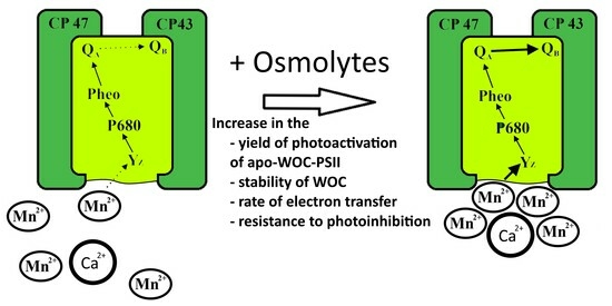
The effect of osmolytes (trehalose, sucrose, betaine, D-glucose and hydroxyectoine) on the photoassembly of the functionally active inorganic core of the water-oxidizing complex (Mn4CaO5 cluster) in Mn-depleted PSII preparations (apo-WOC-PSII) was investigated. It was revealed that the efficiency of the photoassembly of the Mn4CaO5 cluster was considerably (three times) increased in the presence of 1 M disaccharides (trehalose or sucrose) in contrast to other osmolytes. It was found that the osmolytes (especially trehalose or sucrose) improved the redox interaction of exogenous Mn2+ with apo-WOC-PSII, enhanced the protective effect of Mn2+ against the photoinhibition of apo-WOC-PSII, protected CaCl2-treated PSII preparations against thermoinactivation, and stabilized the water-oxidizing complex and electron transport from QA to QB in native PSII preparations during heat treatment. It is suggested that the ability of osmolytes to enhance the efficiency of the photoassembly of a Mn4CaO5 cluster depends on their effect on the following key processes: the redox interaction of Mn2+ with apo-WOC-PSII; the stability of apo-WOC-PSII to photoinhibition during the photoactivation procedure; and the stabilization of both the newly assembled functionally active Mn4CaO5 cluster and the electron transfer from QA to QB.

Graphical Abstract

1. Introduction

Photosystem II (PSII) is a pigment–protein complex located in the thylakoid membrane. PSII catalyzes the light-induced oxidation of water to O2 and the reduction of plastoquinone. The photochemical reaction center (RC) of PSII converts the excitation energy of chlorophyll into the energy of separated charges and produces the strongest biological oxidant, a dimer of chlorophyll a designated as P680+•, with a redox potential of 1.1–1.27 V [1,2,3]. To oxidize water, the water-oxidizing complex (WOC) containing an enzymatic center with the inorganic core (Mn4CaO5 cluster) is oxidized via the sequential absorption of photons and charge separation in the RC to form a series of intermediate states called S-states (S0, S1, S2, S3 and S4), with the transition from S4 to S0 accompanied by the oxidation of two molecules of water and the production of O2.
Leaves absorb light energy, which can damage the photosynthetic apparatus and lead to photoinhibition. The development of photoinhibition depends on environmental conditions such as water stress and temperature [4,5,6,7,8,9]. PSII is especially sensitive to photoinhibition [10]. Photoinhibition leads ultimately to the degradation of the D1 protein, which is synthesized in vivo and re-built into the PSII (see reviews [11,12]). So, PSII reparation includes the replacement of a damaged D1 protein with a newly synthesized D1. Since the majority of the ligands of the Mn4CaO5 cluster are located on the D1 protein, the photoassembly of the inorganic core of the WOC is also required after the replacement of the damaged D1 protein with a new one. The process of photoassembly of Mn4CaO5 cluster is called the photoactivation of PSII. Briefly, the photoassembly of the Mn4CaO5 cluster can be described by the following steps: photooxidation of the first bound Mn2+ to Mn3+ and then formation of a Mn3+–Mn2+ complex. The following light-induced charge separation in the RC leads to the oxidation of this complex with the formation of a metastable Mn3+–Mn3+ complex. The coordination of two other Mn2+ ions (which are also oxidized) leads to the formation of a functionally active Mn4CaO5 cluster. This kinetic model has been designated as a “two-quantum series model” [13]. The molecular mechanisms that cause these kinetic features remain unresolved.
The experiments on the photoassembly of the Mn4CaO5 cluster were mainly carried out on isolated PSII membranes lacking a Mn4CaO5 cluster. Although the photoassembly of the Mn4CaO5 cluster in these cases may be different from that in vivo, in vitro studies provided a wide range of information regarding the cofactors involved in the photoassembly of the Mn4CaO5 cluster [14,15,16,17,18,19,20,21,22,23,24,25,26,27]. A detailed description of the PSII photoactivation is presented in the reviews in [28,29,30]. It is known that the donor side of PSII is selectively inactivated under the illumination of a PSII-deprived Mn4CaO5 cluster, and the damage to PSII components is ascribed to the formation of the long-lived forms of P680+• or TyrZ [31,32,33,34]. So, the photoactivation of oxygen-evolving activity in apo-WOC-PSII preparations is carried out with low-intensity light and at high concentrations of Mn2+ ions to exclude the formation of the long-lived forms of P680+• or TyrZ. In addition to the light-induced oxidation of Mn2+ ions, the stabilization of an assembled functionally active Mn4CaO5 cluster seems to be required.
Plants often produce osmolytes, which are membrane-impermeable solutes that accumulate in the cytoplasm to very high concentrations [35]. One of the best-known and important mechanisms of abiotic stress response adopted by plants is the biosynthesis and accumulation of osmolytes. Osmolytes remain non-toxic, even at molar concentrations. The most common osmolytes include sugars (trehalose and sucrose), amino acids (proline, glutamate, glutamine and alanine) and their derivatives (ectoine and hydroxyectoine), quaternary amines (betaine, polyamines and dimethyl sulfonioproprionate) and polyols including sugar alcohols (mannitol, sorbitol, pinitol, glycerol and galactinol) [36,37,38].
These osmoprotectors perform many functions during stress events, including the scavenging of ROS; balancing cell redox; and the stabilization of pH, proteins, enzymes and membranes [38,39,40,41]. The accumulation of sucrose [42], trehalose [43] and raffinose [44] is observed in dehydrating resurrection plants. Saccharides (such as sucrose) are known membrane protectants that can stabilize cellular processes [45]. It is known that glucose and sucrose accumulate in specific locations in resurrection plant tissue during dehydration and probably function to protect specific cellular structures (e.g., chloroplasts and tonoplast membranes) from desiccation [46]. Sugars, such as trehalose, are involved in preventing protein aggregation during desiccation [47]. Unlike other sugars, trehalose is distinguished by its unique physicochemical properties such as inertness, thermal stability, high glass transition temperature, and stability over a wide pH range [48,49,50,51]. Studies of trehalose effect on biological objects found that trehalose increases the resistance of plants to drought and salinization [52,53,54], the action of heavy metals [55], and the effects of low [56] and high [57] temperatures.
It is known that different osmolytes can stabilize the PSII complex, which is the most sensitive component of the photosynthetic membrane to the action of various stress factors (temperature and light) [58,59,60,61,62,63,64], and also increases the PSII photochemical activity [65]. It was shown that betaine stabilizes the WOC during the incubation of PSII membranes at room temperature, alkaline pH and high salt concentrations by preventing the dissociation of extrinsic proteins and manganese ions from the WOC [58,61,66,67]. It was shown that partial electron transfer reactions that are more intimate with the reaction center complex are also stabilized by glycinebetaine and sucrose [61]. Another osmolyte, hydroxyectoine, is among the most studied osmolytes in relation to the stabilization of biomolecules. Recently, it was shown that hydroxyectoine stimulates the oxygen-evolving activity of PSII and enhances the protective effect of exogenous electron donors against the donor-side photoinhibition of apo-WOC-PSII preparations [68]. One of the carbohydrates that stand out among others is trehalose. It was shown that trehalose protects PSII during thawing [69,70] and prevents the aggregation and inactivation of PSII membrane fragments during long-term storage [71], although it does not exhibit a protective effect during the thermal inactivation of isolated PSII D1/D2/cyt b559 reaction center complexes of PSII [62]. It has been demonstrated that the addition of trehalose stabilizes electron transport in oxygen-evolving PSII preparations [72,73]. In addition to the protective properties, trehalose acts on electronic transport in bacterial type 2 reaction centers [74] and apo-WOC-PSII [75]. It has been shown that the Mn2+-induced protection of apo-WOC-PSII against photoinactivation is greatly increased in the presence of trehalose [64]. It may indicate that trehalose reorganizes the donor side of apo-WOC-PSII, and that, in turn, contributes to better electron transfer from Mn2+ to RC of PSII. Trehalose increases the rate of oxygen evolution in PSII core complexes and PSII membrane fragments [71] that, according to the author’s assumption, is associated with the transition of PSII to a more optimal conformation caused by changes in the hydration of PSII. In addition, the investigation of the protein dynamics associated with the S1→S2 transition of WOC in PSII core complexes by the use of FTIR spectroscopy in the presence of a substrate-based inhibitor, ammonia, allowed for trehalose to exclude water molecules from the solvation layer of the WOC [76]. However, later, additional data disproved this hypothesis while strongly supporting the water entrapment and anchorage models [77]. Moreover, the addition of trehalose induces the transformation of QB-non-reducing PSII RC into QB-reducing PSII RC [75], which decreased the probability of charge recombination in RCs.
In this work, we investigate the effect of osmolytes sucrose, trehalose, betaine, D-glucose and hydroxyectoine (Ect-OH) on the restoration of oxygen-evolving activity in the apo-WOC-PSII related to the photoassembly of the functionally active Mn4CaO5 cluster.

2. Materials and Methods

2.1. Isolation and Treatment of PSII Preparations

Spinach plants were grown in a greenhouse (Institute of Basic Biological Problems, FRC PSCBR, Russian Academy of Sciences, Pushchino, Russia) at a temperature of 20/15 °C (day/night) and under natural illumination from 20 February 2023 to 25 April 2023. Oxygen-evolving PSII membranes were isolated from spinach leaves [78]. The preparations were suspended (2 mg Chl/mL) in a medium containing 50 mM Mes-NaOH (pH 6.5), 35 mM NaCl, 0.33 M sucrose and 10% glycerol and stored at −76 °C. The concentration of chlorophyll was determined as described previously [79]. PSII membranes deprived of the Mn cluster (apo-WOC-PSII) were obtained by NH2OH treatment as described previously [15]. In order to obtain PSII preparations with WOC-depleted extrinsic proteins without the extraction of a Mn4CaO5 cluster from the WOC. The oxygen-evolving PSII preparations were treated by 1 M CaCl2 (CaCl2-PSII preparations) [80]. In order to obtain PSII preparations with the two WOC-depleted extrinsic proteins PsbP and PsbQ without the removal of the PsbO protein and the Mn cluster, the oxygen-evolving PSII preparations were treated with 1 M NaCl (NaCl-PSII preparations) [81].

2.2. Procedure for Photoassembly of Functionally Active Mn4CaO5 Cluster in apo-WOC-PSII Preparations and Measurement of Oxygen-Evolving Activity

The photoassembly of functionally active Mn4CaO5 cluster in apo-WOC-PSII preparations was performed at 25 °C at a Chl concentration of 200 μg Chl/mL in the medium containing 50 mM Mes-NaOH (pH 6.5), 35 mM NaCl, MnCl2 (0.01–1 mM), 50 mM CaCl2 and 50 µM DCPIP in the absence or in the presence of 1 M osmolytes. After that, the apo-WOC-PSII preparations were illuminated for a designated time with red light-emitting diodes (λ = 625 nm, 35 μmol photon m−2 s−1). The efficiency of the photoassembly of a Mn4CaO5 cluster in apo-WOC-PSII preparations was determined based on the ability of the samples to produce photosynthetic oxygen. For all the samples, the rate of light-induced evolution of O2 was measured by monitoring the concentration of O2 with a Clark-type oxygen electrode for 60 s after starting the continuous saturating actinic illumination (λ > 650 nm, 1500 μmol photon m−2 s−1).
To standardize the measurement of oxygen-evolving activity, photoactivated apo-WOC-PSII preparations were diluted ten times with the medium containing 50 mM Mes-NaOH (pH 6.5), 35 mM NaCl, 0.33 M sucrose and 20 mM CaCl2 to a final concentration of 20 μg Chl/mL (in this case, the concentration of the additions used during photoactivation of the samples decreased 10 times); then, the electron acceptors (1 mM ferricyanide and 100 μM DCBQ) were added. In preliminary experiments, it was shown that the addition of any osmolytes in the concentration of 0.1 M to the measurement medium did not affect the oxygen-evolving activity of the photoactivated samples.

2.3. Mn2+ Photooxidation in apo-WOC-PSII and Mn-Dependent Protection of apo-WOC-PSII Preparations against Photoinhibition

According to previous publications [82,83], it was shown that the removal of manganese from the WOC led to a significant decrease in photoinduced changes in the yield of chlorophyll a fluorescence (Fv) associated with the photoreduction of the primary quinone electron acceptor QA. This is due to the loss of electron donation from the WOC to the PSII RC. The addition of Mn2+ or another electron donor for PSII restores the electron transport in the PSII reaction center, which causes an increase in photoinduced Fv. Therefore, the efficiency of Mn2+ photooxidation in apo-WOC-PSII was determined based on the degree of Fv restoration. The kinetics of Fv were measured in the medium containing 50 mM Mes (pH 6.5) and 35 mM NaCl in the absence of osmolytes or in the presence of 1 M osmolytes at a chlorophyll concentration of 10 μg/mL in a 10 mm cuvette at room temperature using a MULTI-COLOR PAM fluorometer (Waltz, Eichenring, Effeltrich, Germany).
The effect of osmolytes on the Mn-dependent protection of apo-WOC-PSII preparations against photoinhibition was studied as described earlier [64]. The samples were re-suspended at a concentration of 20 μg Chl/mL in a medium containing 50 mM Mes (pH 6.5) and 35 mM NaCl without or with 1 M osmolytes. Then, the samples were illuminated (λ = 625 nm, 15 μmol photon m−2 s−1) in the absence or in the presence of various concentrations of Mn2+. After 5 min of illumination, the samples were diluted twice and incubated in the dark for 10 min; then, an electron donor, 2 mM sodium ascorbate, was added, and Fv was measured. The degree of photoinhibition was determined based on the loss of the ability of apo-WOC-PSII preparations to reactivate the Fv with ascorbate. Ascorbate was chosen because its ability to restore Fv did not depend on the presence of osmolytes. The Fv amplitude measured in the apo-WOC-PSII preparations before photoinhibition in the presence of 2 mM sodium ascorbate was taken as 100%.

2.4. Determination of Stability of CaCl2-PSII Preparations

The stability of CaCl2-PSII preparations during their incubation at 25 °C was determined based on the loss of oxygen-evolving activity of the samples. The incubation of CaCl2-PSII preparations was performed in the dark at 110 μg Chl/mL in a medium containing 50 mM Mes (pH 6.5), 35 mM NaCl and 20 mM Ca2+ (medium A) in the absence or in the presence of 1 M osmolytes. After that, the preparations were diluted 11 times with medium A containing 0.01 M osmolytes, and then, the electron acceptors (1 mM ferricyanide and 100 μM DCBQ) were added. As a result of this dilution, the concentration of osmolytes during the measurement of oxygen-evolving activity was equal to 0.1 M. So, in order for the measurement conditions to be the same, the samples incubated in the absence of osmolyte were diluted with media containing 0.11 M osmolytes.

2.5. Estimation of Heat-Induced Inhibition of Electron Transfer in PSII

The study of the degree of heat-induced inhibition of electron transfer in PSII in the absence and in the presence of osmolytes was studied via fast chlorophyll a fluorescence (ChlF) kinetics with a high time resolution using a MULTI-COLOR PAM fluorometer. PSII preparations with an intact WOC before and after heat treatment were illuminated with saturating 880 ms flashes (λ = 625 nm, 2360 μmol photons m−2 s−1), and fluorescence data were recorded.
The heat treatment (incubation of the samples for 40 min at 30 °C) was performed in darkness both in the absence and in the presence of the osmolytes in a medium containing 50 mM Mes (pH 6.5) and 35 mM NaCl at 10 μg Chl/mL. The measurements of the ChlF kinetics were carried out in a medium containing 50 mM Mes (pH 6.5) and 35 mM NaCl at 10 μg Chl/mL at room temperature 25 °C. The double normalization of ChlF transients between 20 µs and 3 ms (WOJ = (Ft − FO)/(FJ − FO)) and between 20 µs and 30 ms (WOI = (Ft − FO)/(FI − FO)) in combination with the generation of difference kinetics (∆WOJ and ∆WOI; “after heat treatment” − “before heat treatment”) allowed for the visualization of so-called K-bands and J-bands, respectively.

2.6. Ultrasonic Interferometry and Refractometry

The stability of PSII preparations with an intact WOC, NaCl-PSII preparations or CaCl2-PSII preparations during their incubation at 25 °C was determined based on the change in relative velocity of an ultrasound using a fixed-length ultrasonic spectrometer [84]. The preparations were dissolved in a medium containing 50 mM Mes (pH 6.5) and 35 mM NaCl in the absence and in the presence of osmolytes at 200 μg Chl/mL.
Refractive index measurements were performed using a Multiwavelengths Refractometer Abbemat MW (Anton Paar, Graz, Austria) at 589.3 nm at 25 °C.

2.7. Statistical Analysis

In all experiments, data were collected from at least three replicates to ensure reliability of measurements. After data collection, the standard deviation of the measurements was calculated. The presence of the statistically significant differences between experimental groups were tested using one-way analysis of variance (ANOVA), followed by post hoc comparisons by Tukey’s test and Student’s t-test for independent means were performed. The normality requirements were checked using Shapiro–Wilk test. The difference was considered significant if p ≤ 0.05. Origin software was used to carry out these calculations.

3. Results

3.1. Effect of Osmolytes on Photoassembly of Mn4CaO5 Cluster in apo-WOC-PSII Preparations

Figure 1 shows the dependence of the efficiency of photoassembly of a Mn4CaO5 cluster in apo-WOC-PSII preparations on the duration of the photoactivation procedure. As seen in Figure 1, the efficiency of the photoassembly of a Mn4CaO5 cluster increased with increasing exposure of the samples to photoactivating light.
The rate of photosynthetic oxygen evolution in photoactivated PSII preparations after 30 min in the absence of osmolyte was about 17 µmol O2 (mg Chl h)−1. The presence of trehalose or sucrose during the photoactivation strongly stimulated the photoassembly so that the rate of oxygen evolution in the samples photoactivated in the presence of the disaccharides reached 55 µmol O2 (mg Chl h)−1. Betaine as well as Ect-OH showed weak stimulating effects (22 µmol O2 (mg Chl h)−1). D-glucose, in contrast to other osmolytes, had a negative effect on the photoactivation of the apo-WOC-PSII (12 µmol O2 (mg Chl h)−1). As can be seen, the efficiency of the photoassembly of Mn4CaO5 cluster in apo-WOC-PSII was about three times higher in the presence of the disaccharides, while the effect of betaine and Ect-OH was about 30%.
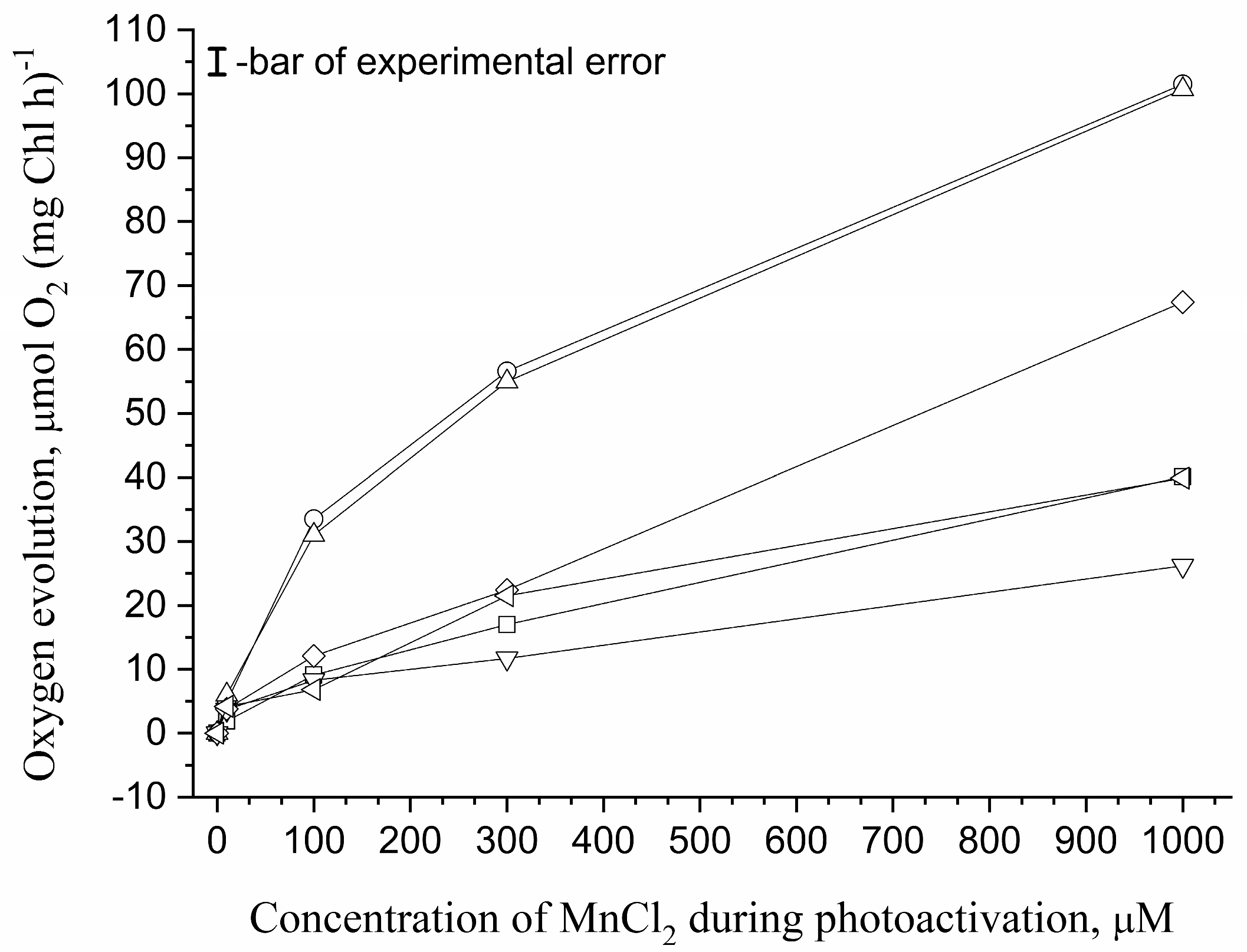
Figure 2 shows the dependence of the photoactivation of apo-WOC-PSII preparations on the concentration of added Mn2+. As previously shown, the maximum yield of the photoactivation of apo-WOC-PSII preparations was observed at 1 mM Mn2+ [15] and a further increase in Mn2+ concentration led to a decrease in the yield of photoactivation. Thus, the osmolyte effect on the photoassembly of Mn4CaO5 cluster was studied within a Mn2+ concentration of 10 μM to 1000 μM. The apo-WOC-PSII preparations photoactivated at 10 μM Mn2+ (corresponding to 10 Mn per reaction center of PSII) showed low rates of oxygen evolution (3–6 μmol O2 (mg Chl h)−1) without statistically significant differences between the effects of osmolytes. An increase in the concentration of added Mn2+ led to a significant increase in the yield of the photoactivation of apo-WOC-PSII, and the photoactivation was more efficient in the presence of the disaccharides than in the presence of other osmolytes or in their absence (Figure 2).
The disaccharides enhance the efficiency of the photoassembly of a Mn4CaO5 cluster by 3.5 times if the photoactivation of apo-WOC-PSII was performed in the presence of 100–300 μM Mn2+ and 2.5 times at 1 mM Mn2+. The decreased stimulatory effect of disaccharides at 1 mM Mn2+ may be due to the fact that the ability of apo-WOC-PSII to photooxidize Mn2+ improves with increasing Mn2+ concentration. Betaine and Ect-OH slightly stimulated the photoactivation of apo-WOC-PSII, except betaine stimulated the photoactivation by 70% at 1 mM added Mn2+. D-glucose did not stimulate the photoassembly of the Mn4CaO5 cluster but suppressed the photoactivation of apo-WOC-PSII preparations.
The first event of the photoassembly of the Mn4CaO5 cluster is the photooxidation of Mn2+ to Mn3+ at the high affinity manganese binding site. This process requires charge separation in the PSII reaction center with the formation of strong oxidants P680+• and TyrZ. However, the formation of the oxidants can also lead to damage to PSII by the donor mechanism. In addition, it is necessary to stabilize the intermediate formed during the photoassembly of the inorganic core of the WOC as well as the newly assembled Mn4CaO5 cluster. It seems that the efficiency of the photoassembly of the Mn4CaO5 cluster in apo-WOC-PSII depends on the following factors: (1) the ability of RC PSII to oxidize Mn2+; (2) the prevention of the formation of long-lived forms of P680+• and TyrZ; and (3) the stabilization of both intermediate products and the newly formed functionally active Mn4CaO5 cluster. Therefore, we studied the effect of osmolytes on Mn2+ photooxidation in apo-WOC-PSII, the photoinhibition of the samples, and the stability of CaCl2-PSII preparations as an analogue of the apo-WOC-PSII with the reassembled functionally active Mn4CaO5 cluster and heat-induced inhibition of electron transfer in PSII.

3.2. Effect of Osmolytes on Mn2+ Photooxidation in apo-WOC-PSII Preparations

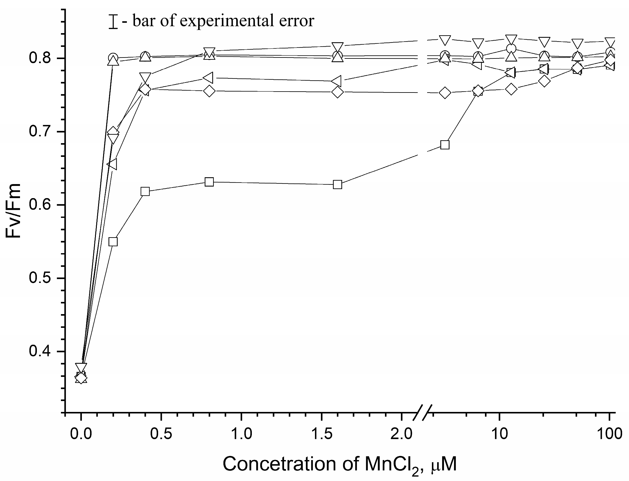
Figure 3 demonstrates the dependence of the reactivation of Fv (as a ratio in the Fv/Fm) on the concentration of added Mn2+ in the absence and in the presence of osmolytes.
As can be seen from this figure, the degree of reactivation of Fv increased with an increase in the concentration of Mn2+. However, the ability of exogenous Mn2+ to reactivate Fv strongly depended on the presence of osmolytes. The maximum reactivation of Fv was reached at 0.2 μM Mn2+ in the presence of 1 M sucrose or 1 M trehalose, about 0.4 μM Mn2+ in the presence of 1 M D-glucose, 1 M Ect-OH or 1 M betaine, and 12 μM Mn2+ in the absence of osmolyte. The results show that trehalose and sucrose more effectively stimulate Mn2+ photooxidation via the apo-WOC-PSII reaction centers.

3.3. Effect of Osmolytes on Mn-Induced Protection of apo-WOC-PSII against Photoinhibition

Figure 4 shows the effect of osmolytes on the photoinhibition of apo-WOC-PSII preparations dependent on the concentration of added Mn2+. The degree of photoinhibition of apo-WOC-PSII preparations was determined based on the loss of the capability of PSII to be reactivated by sodium ascorbate.
As shown in Figure 4, the 5 min exposition of apo-WOC-PSII preparations to light with an intensity of 15 μmol photon m−2 s−1 in the absence of added Mn2+ led to 60% photoinhibition. This is consistent with previous results showing the high sensitivity of apo-WOC-PSII to photoinhibition [31,85]. The addition of Mn2+ led to a decrease in the degree of photoinhibition of apo-WOC-PSII. The presence of osmolytes significantly enhanced the protective effect of Mn2+ against photoinhibition. In the absence of osmolytes, 50% protection of apo-WOC-PSII was observed at 10 µM Mn2+, as well as in the presence of 1 M betaine and Ect-OH at 1 μM Mn2+, 1 M D-glucose at 0.16 μM Mn2+, 1 M sucrose at 0.2 μM Mn2+ and 1 M trehalose at 0.1 µM Mn2+.

3.4. Osmolyte Effect on Stability of Mn4CaO5 Cluster in CaCl2-PSII Preparations

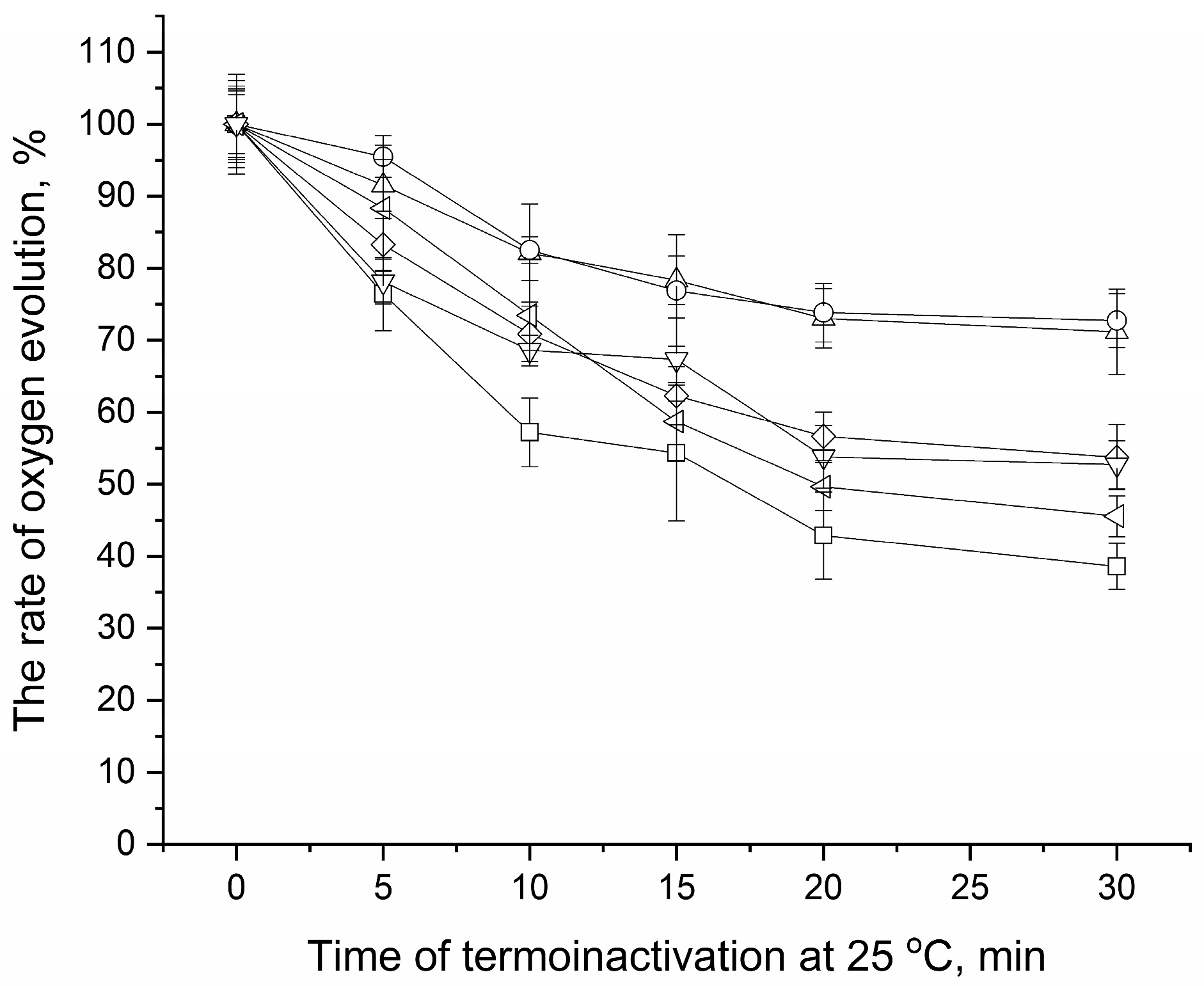
To elucidate whether the effect of osmolytes on the photoactivation of apo-WOC-PSII is related to the stabilization of the newly assembled Mn4CaO5 cluster, we studied the effect of osmolytes on the stability of CaCl2-PSII preparations (Figure 5). The incubation of CaCl2-PSII preparations at 25 °C (the temperature at which photoactivation was performed) resulted in the suppression of oxygen-evolving activity in the samples, which before the incubation, was 56 μmol O2 (mg Chl h)−1. In the absence of osmolytes, the samples lost about 50% of their oxygen-evolving activity after 15 min of incubation. The presence of osmolytes decreased the loss of oxygen-evolving activity in the samples. However, osmolytes differed in the protective efficiency. In the presence of Ect-OH, a 50% decrease in oxygen-evolving activity was observed after 20 min of incubation, while in the presence of D-glucose and betaine, the samples lost about 45% of their oxygen-evolving activity only after 30 min of incubation. Trehalose and sucrose showed the greatest protective effect: the oxygen-evolving activity of the samples after the 30 min incubation was reduced by only 30%.

3.5. Effect of Osmolytes on Heat-Induced Inhibition of Electron Transfer in PSII

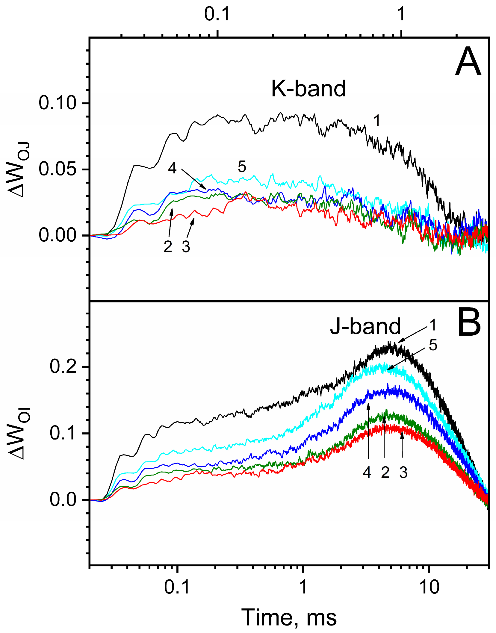
Using PSII particles with intact WOC, we employed an analysis of a fast (within one second) polyphasic rise in chlorophyll a fluorescence registered with a high time resolution to evaluate the degree of heat-induced inhibition of electron transfer in PSII. The double normalization of ChlF transients between 20 µs and 3 ms (WOJ = (Ft − FO)/(FJ − FO)) and between 20 µs and 30 ms (WOI = (Ft − FO)/(FI − FO)) in combination with the generation of difference kinetics (∆WOJ and ∆WOI; “after heat treatment” – “before heat treatment”) allowed for the visualization of so-called K-bands and J-bands, respectively. The K-band is believed to be associated with the disturbance of the water oxidation complex, and the J-band arises from the inhibition of electron transfer from QA to QB [86,87,88]. In the absence of osmolytes, an appearance of positive K-bands in ∆WOJ and J-bands in ∆WOI is observed after the incubation of PSII preparations for 40 min at 30 °C (Figure 6). The amplitude of the K-band became 75% lower when the incubation of the samples was performed in the presence of the osmolytes. The osmolytes also decreased the J-band, but in contrast to the K-band, their effects were different: the presence of trehalose or sucrose reduced the J-band by 50–55%, that of betaine reduced the J-band by 30% and that of Ect-OH reduced the J-band by 10%.
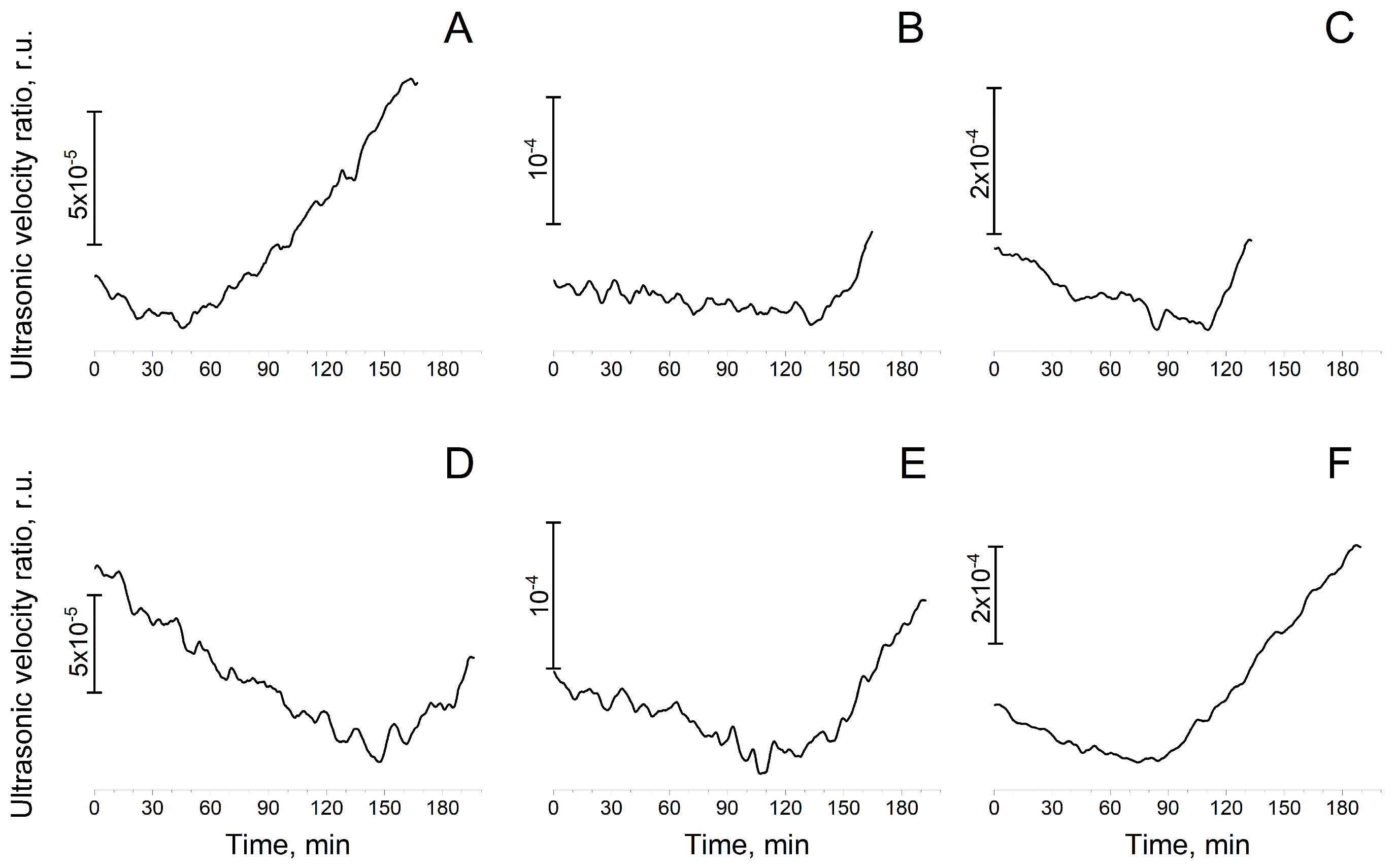
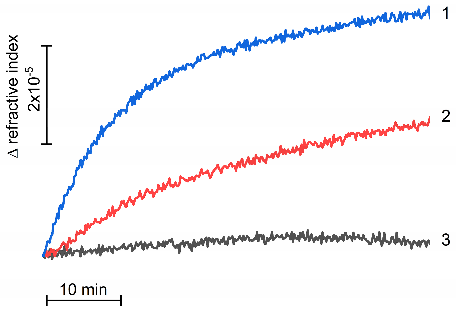
Fixed-length ultrasonic interferometry was used to determine the kinetics of damage to PSII preparations with an intact WOC during their incubation at 25 °C. Figure 7A demonstrates that the incubation of a suspension containing the PSII preparations at 25 °C led to a gradual decrease in the relative velocity of the ultrasound, probably due to the aggregation of PSII complexes. Refractometric measurements confirmed the aggregation of the preparations [89], which was more intense in the absence of osmolytes (Figure 8). After 40–45 min of incubation, a sharp increase in the ultrasound velocity was observed, which reflects the dissociation of the proteins of the water-oxidizing complex [84]. The absence of such an increase in ultrasound velocity during the incubation of NaCl-PSII and CaCl2-PSII preparations (Figure 9) may indicate that in native preparations, incubation leads to the dissociation of PsbP and PsbQ proteins from the WOC. The addition of osmolytes delayed the onset of the increase in ultrasound velocity (Figure 7B–F), which may reflect an increase in the WOC stability.

4. Discussion

One of the most sensitive sites of the photosynthetic electron transport chain of plants to various stress factors is photosystem II [10], which is capable of photogenerating the strongest biological oxidant [1,2,3] and contains a relatively unstable water-oxidizing complex. As a result, plants are forced to constantly restore damaged PSII complexes through the activation of the photoassembly process, which includes the process of photoformation of the water-oxidizing complex. This is most pronounced when plants grow under conditions of high solar insolation, elevated temperatures and lack of water. The synthesis and accumulation of osmolytes is one of the main strategies used by plants in response to various stress factors [90,91,92,93,94,95,96,97]. Our results for the first time show that the presence of trehalose and sucrose during photoactivation of the apo-WOC-PSII significantly increases the efficiency of the photoassembly of a Mn4CaO5 cluster. Samples photoactivated in the presence of these osmolytes showed much higher rates of light-induced oxygen evolution than samples photoactivated in the absence of osmolytes (Figure 1 and Figure 2). Other osmolytes either slightly stimulated the photoassembly of the Mn4CaO5 cluster (betaine and Ect-OH) or had a negative effect (D-glucose). This is probably due to their effect on factors influencing the photoassembly of the Mn4CaO5 cluster. The effectiveness of osmolytes will depend on their ability (i) to improve the photooxidation of Mn2+ by PSII RC; (ii) to reduce the photoinhibition of PSII; (iii) to stabilize the WOC and the electron transport; and (iv) to be inert towards the intermediates formed during the photoactivation.
Firstly, Mn2+ photooxidation improved due to the reaction centers of apo-WOC-PSII. The reactivation of Fv in apo-WOC-PSII by added Mn2+ (which may indicate the ability of apo-WOC-PSII to photooxidize Mn2+) significantly increases in the presence of osmolytes. Maximum Fv restoration required 60 times less concentration of added Mn2+ in the presence of trehalose or sucrose and about 30 times less in the presence of betaine, Ect-OH or D-glucose (Figure 3). Thus, osmolytes, especially trehalose and sucrose, significantly stimulate electron donation from exogenous Mn2+ to the apo-WOC-PSII reaction center. This may indicate that structural changes in apo-WOC-PSII induced by osmolytes are important for the redox interaction of Mn2+ with the donor-side PSII. When studying the effect of sucrose on PSII, it was suggested that sucrose leads to structural alterations at the active site of the WOC via changes in the hydration of the protein complex and induces shifts in the pKa at the aspartic or glutamic acid side [65]. It is likely that these osmolyte-induced changes increase the binding of Mn2+ to apo-WOC-PSII and the ability of PSII reaction centers to photooxidize Mn2+.
Secondly, the photoactivation of apo-WOC-PSII will depend on the protection of PSII from damage related to the donor side, since these preparations are very sensitive to photoinhibition induced by the formation of long-lived forms of P680+• or TyrZ [31] and so the addition of Mn2+ suppresses photoinhibition. In the absence of osmolytes, the maximum protection of apo-WOC-PSII against photoinhibition was observed at 100 μM MnCl2 (Figure 4). Osmolytes enhanced the protective effect of the added Mn2+, although with varying efficiency. The maximum protection of apo-WOC-PSII against photoinhibition required 500 times less Mn2+ in the presence of trehalose and 250 times less Mn2+ in the presence of sucrose or D-glucose compared to that required in the absence of osmolytes. Betaine and Ect-OH showed the weakest effect on the Mn-dependent protection of apo-WOC-PSII against photoinhibition among the osmolytes: in the presence of betaine and Ect-OH, only 20 and 10 times less Mn2+ was required, respectively, to achieve the maximum protective effect. The results demonstrate that the protective effect of trehalose and sucrose against the photoinhibition of apo-WOC-PSII is manifested at lower concentrations of exogenous Mn2+ than the effect of betaine and Ect-OH that correlates with the effect of osmolytes on the reactivation of Fv in apo-WOC-PSII by added Mn2+. This also may explain why betaine remarkably stimulated the photoactivation of apo-WOC-PSII only at high concentrations of added Mn2+. Some osmolytes (especially trehalose) also showed a protective effect against the photoinhibition of apo-WOC-PSII preparations without added Mn2+ (Figure 4) that is in agreement with previous data [64]. This is probably due to the fact that apo-WOC-PSII can contain trace amounts of manganese. The influence of osmolytes on the effects of manganese described in Section 3.2 and Section 3.3. has been partially investigated previously [64,68,75], and similar results were obtained. The use of apo-WOC-PSII preparations obtained by a different method (hydroxylamine treatment) in this study, which is slightly different from those used previously [15], makes it necessary to repeat these experiments.
Thirdly, it is known that osmolytes protect the water-oxidizing complex of PSII from termoinactivation [61,63]. This property of osmolytes can be responsible for the increase in the yield of photoactivation of apo-WOC-PSII due to the long photoactivation procedure (30 min, 25 °C) possibly being accompanied by the destruction of the photoassembled Mn4CaO5 cluster. By using CaCl2-PSII preparations (as an analogue of the apo-WOC-PSII with the photoassembled Mn4CaO5 cluster), it was revealed that the presence of trehalose and sucrose significantly slowed down the loss in the oxygen-evolving activity of CaCl2-PSII during their incubation at 25 °C (Figure 5). The effect of other osmolytes was not as striking as those of trehalose and sucrose. Thus, a significant enhancement in the efficiency of the photoactivation of apo-WOC-PSII by trehalose and sucrose can be associated with changes in apo-WOC-PSII that are important for manganese cluster photoassembly: improvement in the photooxidation of Mn2+ to Mn3+ by reaction centers of PSII, protection of PSII from damage induced by the formation of long-lived forms of P680+• or TyrZ, and stabilization of either a metastable intermediate complex or the formed functionally active Mn4CaO5 cluster. In addition, osmolytes stabilize the water-oxidizing complex, preventing the dissociation of the extrinsic proteins of the complex (Figure 7). This property of osmolytes may be manifested in the function and photoassembly of the WOC during the photorecovery of PSII in vivo. In our experiments (Figure 8) and early works [47,71], it was shown that osmolytes can prevent the aggregation of PSII preparations during long-term storage. This property of osmolytes may in turn improve the efficiency of Mn cluster photoassembly.
Weak stimulation of the photoactivation of apo-WOC-PSII by betaine and Ect-OH correlates with their effects on the above-listed factors. However, D-glucose, which improved the restoration of Fv by Mn2+, enhanced the protective effect of added Mn2+ against the photoinhibition of apo-WOC-PSII and increased the stability of CaCl2-PSII preparations, at the same time decreasing the efficiency of the photoassembly of the Mn4CaO5 cluster. It was shown that Mn(III) is capable of oxidizing D-glucose [98]. Therefore, the negative effect of D-glucose on the photoactivation of apo-WOC-PSII may be due to its redox interaction with Mn(III), formed as a result of the oxidation of Mn2+ by the RC of apo-WOC-PSII. This interaction may eliminate the formation of metastable products necessary for the formation of a Mn4CaO5 cluster.
The effective electron transfer within reaction centers of apo-WOC-PSII is necessary for the photoassembly of a Mn4CaO5 cluster, so blocking of the electron transfer from QA to QB may reduce the yield of photoactivation of the apo-WOC-PSII. The incubation of PSII preparations with an intact WOC for 40 min at 30 °C led to the appearance of J-bands, which were detected after subtracting the double-normalized kinetics obtained after and before incubation of the samples at 30 °C (Figure 6B). A J-band is believed to appear as a result of the inhibition of electron transfer from QA to QB [86,87,88]. Trehalose and sucrose, in comparison with other osmolytes (betaine and Ect-OH), significantly reduced the amplitude of J-bands (Figure 6B). Recently, it was shown that the addition of trehalose led to a significant increase in the rate of electron transfer between QA and QB as well as to the almost complete disappearance of “closed RCs of PSII”, where the electron transfer between QA and QB was blocked [75]. It seems that sucrose (like trehalose) may also stabilize electron transport from QA to QB in apo-WOC-PSII preparations during the photoactivation procedure. The stabilization of electron transport in QA – QB sites may also be important for the increase in the yield of photoactivation of apo-WOC-PSII.
Thus, the stimulation of the photoassembly of Mn4CaO5 clusters in apo-WOC-PSII by osmolytes will appear if, on the one hand, the osmolytes improve the photooxidation of Mn2+ by RC, reduce the photoinhibition of PSII and stabilize the WOC after the photoassembly of a functionally active Mn4CaO5 cluster as well as electron transport from QA to QB and, on the other hand, they do not interact with intermediate products formed during the photooxidation of bound Mn2+ in apo-WOC-PSII. The different influences of osmolytes on the photoassembly of a Mn4CaO5 cluster apparently depends on their effect on the protein structures of PSII in terms of the interactions with water molecules.
It is suggested that trehalose may change protein packing to the more optimal conformation and/or cause protein–protein interactions in the WOC, which increase the rate of turnover of S-state transitions and, consequently, the efficiency of water photooxidation and oxygen evolution [99]. It seems that such changes induced by trehalose also may promote the efficiency of Mn4CaO5 cluster photoassembly. So, the significant effect of trehalose and sucrose may be due to the fact that trehalose and sucrose are classified as osmolytes, which stabilize proteins that increase the free energy of both native and denatured states, and therefore, their role in PSII photoactivation can be compared to chaperones [100]. Betaine, which also stimulated the photoactivation of apo-WOC-PSII at high Mn2+ concentrations (but almost two times less than sucrose and trehalose), is attributed to the moderate alteration of osmolyte proteins [100].
The stimulating effect of osmolytes (especially of disaccharides) on the photoactivation of apo-WOC-PSII revealed in the present study may occur in vivo during the photo-driven formation and reparation of PSII. These processes include the WOC photoformation step, during which photosystem II is most susceptible to photodamage. The concentration of disaccharides in plant chloroplasts is strongly dependent on the plant species. For example, the amount of sucrose in chloroplasts can reach 15–30% of the amount of sucrose in protoplasts [101,102,103,104]. Chloroplasts are able to import sugars (including sucrose) across their membranes using specific transport proteins [104,105,106,107]. For example, plastidic sugar transporter (pSuT) has been reported to transport sucrose across the chloroplast membrane [108]. It has also been proposed that the chloroplast might act as a sucrose reservoir [108].

Author Contributions

Conceptualization, methodology A.A.K. and D.V.Y.; investigation A.A.K., D.V.K. and D.V.Y.; writing—original draft preparation A.A.K.; funding acquisition D.V.K. All authors have read and agreed to the published version of the manuscript.

Funding

The work was supported by the Russian Science Foundation Grant No. 22-24-01179, https://rscf.ru/project/22-24-01179/ (accessed on 19 September 2023).

Data Availability Statement

Data are contained within the article.

Acknowledgments

PAM fluorometry was carried out using the MULTI-COLOR PAM fluorometer of the Pushchino Center of Biological Research RAS (https://ckp-rf.ru/ckp/670266/ (accessed on 12 December 2023)).

Conflicts of Interest

The authors declare no conflict of interest.

References

  1. Klimov, V.; Allakhverdiev, S.; Demeter, S.; Krasnovsky, A. Photoreduction of pheophytin in chloroplast photosystem II as a function of the redox potential of the medium. Dokl. Akad. Nauk SSSR 1979, 249, 227–230. [Google Scholar]
  2. Ishikita, H.; Loll, B.; Biesiadka, J.; Saenger, W.; Knapp, E.-W. Redox potentials of chlorophylls in the photosystem II reaction center. Biochemistry 2005, 44, 4118–4124. [Google Scholar] [CrossRef] [PubMed]
  3. Allakhverdiev, S.I.; Tomo, T.; Shimada, Y.; Kindo, H.; Nagao, R.; Klimov, V.V.; Mimuro, M. Redox potential of pheophytin a in photosystem II of two cyanobacteria having the different special pair chlorophylls. Proc. Natl. Acad. Sci. USA 2010, 107, 3924–3929. [Google Scholar] [CrossRef] [PubMed]
  4. Ludlow, M.M. Light Stress at High Temperature; Elsevier: New York, NY, USA, 1987. [Google Scholar]
  5. Masojídek, J.; Trivedi, S.; Halshaw, L.; Alexiou, A.; Hall, D.O. The synergistic effect of drought and light stresses in sorghum and pearl millet. Plant Physiol. 1991, 96, 198–207. [Google Scholar] [CrossRef] [PubMed]
  6. Ögren, E. The significance of photoinhibition for photosynthetic productivity. In Photoinhibition of Photosynthesis from Molecular Mechanisms to the Field; Bios Scientific Publishers: Oxford, UK, 1994; pp. 433–447. [Google Scholar]
  7. Goh, C.-H.; Ko, S.-M.; Koh, S.; Kim, Y.-J.; Bae, H.-J. Photosynthesis and environments: Photoinhibition and repair mechanisms in plants. J. Plant Biol. 2012, 55, 93–101. [Google Scholar] [CrossRef]
  8. Long, S.P.; Humphries, S.; Falkowski, P.G. Photoinhibition of photosynthesis in nature. Annu. Rev. Plant Biol. 1994, 45, 633–662. [Google Scholar] [CrossRef]
  9. Tikkanen, M.; Grieco, M.; Nurmi, M.; Rantala, M.; Suorsa, M.; Aro, E.-M. Regulation of the photosynthetic apparatus under fluctuating growth light. Philos. Trans. R. Soc. B Biol. Sci. 2012, 367, 3486–3493. [Google Scholar] [CrossRef]
  10. Powles, S.B. Photoinhibition of photosynthesis induced by visible light. Annu. Rev. Plant Physiol. 1984, 35, 15–44. [Google Scholar] [CrossRef]
  11. Aro, E.-M.; Virgin, I.; Andersson, B. Photoinhibition of photosystem II. Inactivation, protein damage and turnover. Biochim. Biophys. Acta (BBA)—Bioenerg. 1993, 1143, 113–134. [Google Scholar] [CrossRef]
  12. Tyystjärvi, E. Photoinhibition of photosystem II. Int. Rev. Cell Mol. Biol. 2013, 300, 243–303. [Google Scholar]
  13. Radmer, R.; Cheniae, G.M. Photoactivation of the manganese catalyst of O2 evolution. II. A two-quantum mechanism. Biochim. Biophys. Acta (BBA)—Bioenerg. 1971, 253, 182–186. [Google Scholar] [CrossRef]
  14. Tamura, N.; Cheniae, G. Requirements for the photoligation of Mn2+ in PS II membranes and the expression of water-oxidizing activity of the polynuclear Mn-catalyst. FEBS Lett. 1986, 200, 231–236. [Google Scholar] [CrossRef]
  15. Tamura, N.; Cheniae, G. Photoactivation of the water-oxidizing complex in photosystem II membranes depleted of Mn and extrinsic proteins. I. Biochemical and kinetic characterization. Biochim. Biophys. Acta (BBA)—Bioenerg. 1987, 890, 179–194. [Google Scholar] [CrossRef]
  16. Miller, A.F.; Brudvig, G.W. Manganese and calcium requirements for reconstitution of oxygen-evolution activity in manganese-depleted photosystem II membranes. Biochemistry 1989, 28, 8181–8190. [Google Scholar] [CrossRef] [PubMed]
  17. Tamura, N.; Inoue, Y.; Cheniae, G.M. Photoactivation of the water-oxidizing complex in Photosystem II membranes depleted of Mn, Ca and extrinsic proteins: II. Studies on the functions of Ca2+. Biochim. Biophys. Acta (BBA)—Bioenerg. 1989, 976, 173–181. [Google Scholar] [CrossRef]
  18. Miyao, M.; Inoue, Y. An improved procedure for photoactivation of photosynthetic oxygen evolution: Effect of artificial electron acceptors on the photoactivation yield of NH2OH-treated wheat photosystem II membranes. Biochim. Biophys. Acta (BBA)—Bioenerg. 1991, 1056, 47–56. [Google Scholar] [CrossRef]
  19. Ananyev, G.M.; Dismukes, G.C. Assembly of the tetra-Mn site of photosynthetic water oxidation by photoactivation: Mn stoichiometry and detection of a new intermediate. Biochemistry 1996, 35, 4102–4109. [Google Scholar] [CrossRef]
  20. Ananyev, G.M.; Dismukes, G.C. High-resolution kinetic studies of the reassembly of the tetra-manganese cluster of photosynthetic water oxidation: Proton equilibrium, cations, and electrostatics. Biochemistry 1996, 35, 14608–14617. [Google Scholar] [CrossRef]
  21. Ananyev, G.M.; Dismukes, G.C. Calcium induces binding and formation of a spin-coupled dimanganese (II,II) center in the apo-water oxidation complex of photosystem II as precursor to the functional tetra-Mn/Ca cluster. Biochemistry 1997, 36, 11342–11350. [Google Scholar] [CrossRef]
  22. Zaltsman, L.; Ananyev, G.M.; Bruntrager, E.; Dismukes, G.C. Quantitative kinetic model for photoassembly of the photosynthetic water oxidase from its inorganic constituents: Requirements for manganese and calcium in the kinetically resolved steps. Biochemistry 1997, 36, 8914–8922. [Google Scholar] [CrossRef]
  23. Baranov, S.V.; Ananyev, G.M.; Klimov, V.V.; Dismukes, G.C. Bicarbonate accelerates assembly of the inorganic core of the water-oxidizing complex in manganese-depleted photosystem II: A proposed biogeochemical role for atmospheric carbon dioxide in oxygenic photosynthesis. Biochemistry 2000, 39, 6060–6065. [Google Scholar] [CrossRef] [PubMed]
  24. Baranov, S.; Tyryshkin, A.; Katz, D.; Dismukes, G.; Ananyev, G.; Klimov, V. Bicarbonate is a native cofactor for assembly of the manganese cluster of the photosynthetic water oxidizing complex. Kinetics of reconstitution of O2 evolution by photoactivation. Biochemistry 2004, 43, 2070–2079. [Google Scholar] [CrossRef] [PubMed]
  25. Miyao-Tokutomi, M.; Inoue, Y. Improvement by benzoquinones of the quantum yield of photoactivation of photosynthetic oxygen evolution: Direct evidence for the two-quantum mechanism. Biochemistry 1992, 31, 526–532. [Google Scholar] [CrossRef] [PubMed]
  26. Khorobrykh, A. A possible relationship between the effect of factors on photoactivation of photosystem II depleted of functional Mn and cytochrome b559. Biochim. Biophys. Acta (BBA)—Bioenerg. 2023, 1864, 148997. [Google Scholar] [CrossRef] [PubMed]
  27. Khorobrykh, A.A.; Yanykin, D.V.; Klimov, V.V. Enhancement of photoassembly of the functionally active water-oxidizing complex in Mn-depleted photosystem II membranes upon transition to anaerobic conditions. J. Photochem. Photobiol. B Biol. 2016, 163, 211–215. [Google Scholar] [CrossRef] [PubMed]
  28. Dasgupta, J.; Ananyev, G.M.; Dismukes, G.C. Photoassembly of the water-oxidizing complex in photosystem II. Coord. Chem. Rev. 2008, 252, 347–360. [Google Scholar] [CrossRef] [PubMed]
  29. Bao, H.; Burnap, R.L. Photoactivation: The light-driven assembly of the water oxidation complex of photosystem II. Front. Plant Sci. 2016, 7, 578. [Google Scholar] [CrossRef] [PubMed]
  30. Zhang, M.; Bommer, M.; Chatterjee, R.; Hussein, R.; Yano, J.; Dau, H.; Kern, J.; Dobbek, H.; Zouni, A. Structural insights into the light-driven auto-assembly process of the water-oxidizing Mn4CaO5-cluster in photosystem II. eLife 2017, 6, e26933. [Google Scholar] [CrossRef]
  31. Klimov, V.; Shafiev, M.; Allakhverdiev, S. Photoinactivation of the reactivation capacity of photosystem II in pea subchloroplast particles after a complete removal of manganese. Photosynth. Res. 1990, 23, 59–65. [Google Scholar] [CrossRef]
  32. Jegerschoeld, C.; Virgin, I.; Styring, S. Light-dependent degradation of the D1 protein in photosystem II is accelerated after inhibition of the water splitting reaction. Biochemistry 1990, 29, 6179–6186. [Google Scholar] [CrossRef]
  33. Telfer, A.; De Las Rivas, J.; Barber, J. β-Carotene within the isolated Photosystem II reaction centre: Photooxidation and irreversible bleaching of this chromophore by oxidised P680. Biochim. Biophys. Acta (BBA)—Bioenerg. 1991, 1060, 106–114. [Google Scholar] [CrossRef]
  34. Wang, W.-Q.; Chapman, D.J.; Barber, J. Inhibition of water splitting increases the susceptibility of photosystem II to photoinhibition. Plant Physiol. 1992, 99, 16–20. [Google Scholar] [CrossRef] [PubMed]
  35. Kurepin, L.V.; Ivanov, A.G.; Zaman, M.; Pharis, R.P.; Allakhverdiev, S.I.; Hurry, V.; Hüner, N.P. Stress-related hormones and glycinebetaine interplay in protection of photosynthesis under abiotic stress conditions. Photosynth. Res. 2015, 126, 221–235. [Google Scholar] [CrossRef] [PubMed]
  36. Khan, M.S.; Yu, X.; Kikuchi, A.; Asahina, M.; Watanabe, K.N. Genetic engineering of glycine betaine biosynthesis to enhance abiotic stress tolerance in plants. Plant Biotechnol. 2009, 26, 125–134. [Google Scholar] [CrossRef]
  37. Jewell, M.C.; Campbell, B.C.; Godwin, I.D. Transgenic plants for abiotic stress resistance. In Transgenic Crop Plants; Springer: Berlin/Heidelberg, Germany, 2010; pp. 67–132. [Google Scholar]
  38. Kumar, V.; Khare, T. Individual and additive effects of Na+ and Cl ions on rice under salinity stress. Arch. Agron. Soil Sci. 2015, 61, 381–395. [Google Scholar] [CrossRef]
  39. Ashraf, M.; Foolad, M.R. Roles of glycine betaine and proline in improving plant abiotic stress resistance. Environ. Exp. Bot. 2007, 59, 206–216. [Google Scholar] [CrossRef]
  40. Sicher, R.C.; Timlin, D.; Bailey, B. Responses of growth and primary metabolism of water-stressed barley roots to rehydration. J. Plant Physiol. 2012, 169, 686–695. [Google Scholar] [CrossRef] [PubMed]
  41. Keunen, E.; Peshev, D.; Vangronsveld, J.; Van Den Ende, W.; Cuypers, A. Plant sugars are crucial players in the oxidative challenge during abiotic stress: Extending the traditional concept. Plant Cell Environ. 2013, 36, 1242–1255. [Google Scholar] [CrossRef]
  42. Whittaker, A.; Bochicchio, A.; Vazzana, C.; Lindsey, G.; Farrant, J. Changes in leaf hexokinase activity and metabolite levels in response to drying in the desiccation-tolerant species Sporobolus stapfianus and Xerophyta viscosa. J. Exp. Bot. 2001, 52, 961–969. [Google Scholar] [CrossRef]
  43. Moore, J.P.; Hearshaw, M.; Ravenscroft, N.; Lindsey, G.G.; Farrant, J.M.; Brandt, W.F. Desiccation-induced ultrastructural and biochemical changes in the leaves of the resurrection plant Myrothamnus flabellifolia. Aust. J. Bot. 2007, 55, 482–491. [Google Scholar] [CrossRef]
  44. Peters, S.; Mundree, S.G.; Thomson, J.A.; Farrant, J.M.; Keller, F. Protection mechanisms in the resurrection plant Xerophyta viscosa (Baker): Both sucrose and raffinose family oligosaccharides (RFOs) accumulate in leaves in response to water deficit. J. Exp. Bot. 2007, 58, 1947–1956. [Google Scholar] [CrossRef] [PubMed]
  45. Hoekstra, F.A.; Golovina, E.A.; Buitink, J. Mechanisms of plant desiccation tolerance. Trends Plant Sci. 2001, 6, 431–438. [Google Scholar] [CrossRef] [PubMed]
  46. Martinelli, T. In situ localization of glucose and sucrose in dehydrating leaves of Sporobolus stapfianus. J. Plant Physiol. 2008, 165, 580–587. [Google Scholar] [CrossRef] [PubMed]
  47. Goyal, K.; Walton, L.J.; Tunnacliffe, A. LEA proteins prevent protein aggregation due to water stress. Biochem. J. 2005, 388, 151–157. [Google Scholar] [CrossRef] [PubMed]
  48. Crowe, L.M. Lessons from nature: The role of sugars in anhydrobiosis. Comp. Biochem. Physiol. Part A Mol. Integr. Physiol. 2002, 131, 505–513. [Google Scholar] [CrossRef] [PubMed]
  49. Francia, F.; Malferrari, M.; Sacquin-Mora, S.; Venturoli, G. Charge recombination kinetics and protein dynamics in wild type and carotenoid-less bacterial reaction centers: Studies in trehalose glasses. J. Phys. Chem. B 2009, 113, 10389–10398. [Google Scholar] [CrossRef] [PubMed]
  50. Fernandez, O.; Béthencourt, L.; Quero, A.; Sangwan, R.S.; Clément, C. Trehalose and plant stress responses: Friend or foe? Trends Plant Sci. 2010, 15, 409–417. [Google Scholar] [CrossRef] [PubMed]
  51. Lunn, J.E.; Delorge, I.; Figueroa, C.M.; Van Dijck, P.; Stitt, M. Trehalose metabolism in plants. Plant J. 2014, 79, 544–567. [Google Scholar] [CrossRef]
  52. Crowe, J.H.; Hoekstra, F.A.; Crowe, L.M. Anhydrobiosis. Annu. Rev. Physiol. 1992, 54, 579–599. [Google Scholar] [CrossRef]
  53. Jun, S.-S.; Choi, H.J.; Lee, H.Y.; Hong, Y.-N. Differential protection of photosynthetic capacity in trehalose-and LEA protein-producing transgenic plants under abiotic stresses. J. Plant Biol. 2008, 51, 327–336. [Google Scholar] [CrossRef]
  54. Williams, B.; Njaci, I.; Moghaddam, L.; Long, H.; Dickman, M.B.; Zhang, X.; Mundree, S. Trehalose accumulation triggers autophagy during plant desiccation. PLoS Genet. 2015, 11, e1005705. [Google Scholar] [CrossRef] [PubMed]
  55. Mostofa, M.G.; Hossain, M.A.; Fujita, M.; Tran, L.-S.P. Physiological and biochemical mechanisms associated with trehalose-induced copper-stress tolerance in rice. Sci. Rep. 2015, 5, 11433. [Google Scholar] [CrossRef] [PubMed]
  56. Nunes, C.; Schluepmann, H.; Delatte, T.L.; Wingler, A.; Silva, A.B.; Fevereiro, P.S.; Jansen, M.; Fiorani, F.; Wiese-Klinkenberg, A.; Paul, M.J. Regulation of growth by the trehalose pathway: Relationship to temperature and sucrose. Plant Signal. Behav. 2013, 8, e26626. [Google Scholar] [CrossRef] [PubMed]
  57. Zentella, R.; Mascorro-Gallardo, J.O.; Van Dijck, P.; Folch-Mallol, J.; Bonini, B.; Van Vaeck, C.; Gaxiola, R.; Covarrubias, A.A.; Nieto-Sotelo, J.; Thevelein, J.M. A Selaginella lepidophylla trehalose-6-phosphate synthase complements growth and stress-tolerance defects in a yeast tps1 mutant. Plant Physiol. 1999, 119, 1473–1482. [Google Scholar] [CrossRef] [PubMed]
  58. Papageorgiou, G.C.; Fujimura, Y.; Murata, N. Protection of the oxygen-evolving photosystem II complex by glycinebetaine. Biochim. Biophys. Acta (BBA)—Bioenerg. 1991, 1057, 361–366. [Google Scholar] [CrossRef]
  59. Papageorgiou, G.C.; Murata, N. The unusually strong stabilizing effects of glycine betaine on the structure and function of the oxygen-evolving photosystem II complex. Photosynth. Res. 1995, 44, 243–252. [Google Scholar] [CrossRef] [PubMed]
  60. Mohanty, P.; Hayashi, H.; Papageorgiou, G.; Murata, N. Stabilization of the Mn-cluster of the oxygen-evolving complex by glycinebetaine. Biochim. Biophys. Acta (BBA)—Bioenerg. 1993, 1144, 92–96. [Google Scholar] [CrossRef]
  61. Allakhverdiev, S.; Feyziev, Y.M.; Ahmed, A.; Hayashi, H.; Aliev, J.A.; Klimov, V.; Murata, N.; Carpentier, R. Stabilization of oxygen evolution and primary electron transport reactions in photosystem II against heat stress with glycinebetaine and sucrose. J. Photochem. Photobiol. B Biol. 1996, 34, 149–157. [Google Scholar] [CrossRef]
  62. Allakhverdiev, S.I.; Hayashi, H.; Nishiyama, Y.; Ivanov, A.G.; Aliev, J.A.; Klimov, V.V.; Murata, N.; Carpentier, R. Glycinebetaine protects the D1/D2/Cytb559 complex of photosystem II against photo-induced and heat-induced inactivation. J. Plant Physiol. 2003, 160, 41–49. [Google Scholar] [CrossRef]
  63. Klimov, V.V.; Allakhverdiev, S.I.; Nishiyama, Y.; Khorobrykh, A.A.; Murata, N. Stabilization of the oxygen-evolving complex of photosystem II by bicarbonate and glycinebetaine in thylakoid and subthylakoid preparations. Funct. Plant Biol. 2003, 30, 797–803. [Google Scholar] [CrossRef]
  64. Yanykin, D.; Khorobrykh, A.; Mamedov, M.; Klimov, V. Trehalose protects Mn-depleted photosystem 2 preparations against the donor-side photoinhibition. J. Photochem. Photobiol. B Biol. 2016, 164, 236–243. [Google Scholar] [CrossRef] [PubMed]
  65. Halverson, K.M.; Barry, B.A. Sucrose and glycerol effects on photosystem II. Biophys. J. 2003, 85, 1317–1325. [Google Scholar] [CrossRef] [PubMed]
  66. Mamedov, M.; Hayashi, H.; Wada, H.; Mohanty, P.; Papageorgiou, G.; Murata, N. Glycinebetaine enhances and stabilizes the evolution of oxygen and the synthesis of ATP by cyanobacterial thylakoid membranes. FEBS Lett. 1991, 294, 271–274. [Google Scholar] [CrossRef] [PubMed]
  67. Murata, N.; Mohanty, P.; Hayashi, H.; Papageorgiou, G. Glycinebetaine stabilizes the association of extrinsic proteins with the photosynthetic oxygen-evolving complex. FEBS Lett. 1992, 296, 187–189. [Google Scholar] [CrossRef] [PubMed]
  68. Yanykin, D.; Malferrari, M.; Rapino, S.; Venturoli, G.; Semenov, A.Y.; Mamedov, M. Hydroxyectoine protects Mn-depleted photosystem II against photoinhibition acting as a source of electrons. Photosynth. Res. 2019, 141, 165–179. [Google Scholar] [CrossRef] [PubMed]
  69. Hincha, D.K. Low concentrations of trehalose protect isolated thylakoids against mechanical freeze-thaw damage. Biochim. Biophys. Acta (BBA)—Biomembr. 1989, 987, 231–234. [Google Scholar] [CrossRef]
  70. Apostolova, E.; Bushova, M.; Tenchov, B.; Murata, N. Freezing damage and protective of photosystem 2 by sucrose and trehalose. In Research Photosynthesis; Kluwer Academic Publishers: Dordrecht, The Netherlands, 2005; pp. 165–168. [Google Scholar]
  71. Mamedov, M.; Petrova, I.; Yanykin, D.; Zaspa, A.; Semenov, A.Y. Effect of trehalose on oxygen evolution and electron transfer in photosystem 2 complexes. Biochemistry 2015, 80, 61–66. [Google Scholar] [CrossRef]
  72. Williams, W.; Gounaris, K. Stabilisation of PS-II-mediated electron transport in oxygen-evolving PS II core preparations by the addition of compatible co-solutes. Biochim. Biophys. Acta (BBA)—Bioenerg. 1992, 1100, 92–97. [Google Scholar] [CrossRef]
  73. Mamedov, M.; Nosikova, E.; Vitukhnovskaya, L.; Zaspa, A.; Semenov, A.Y. Influence of the disaccharide trehalose on the oxidizing side of photosystem II. Photosynthetica 2018, 56, 236–243. [Google Scholar] [CrossRef]
  74. Francia, F.; Palazzo, G.; Mallardi, A.; Cordone, L.; Venturoli, G. Residual water modulates QA−-to-QB electron transfer in bacterial reaction centers embedded in trehalose amorphous matrices. Biophys. J. 2003, 85, 2760–2775. [Google Scholar] [CrossRef]
  75. Yanykin, D.; Khorobrykh, A.; Mamedov, M.; Klimov, V. Trehalose stimulation of photoinduced electron transfer and oxygen photoconsumption in Mn-depleted photosystem 2 membrane fragments. J. Photochem. Photobiol. B Biol. 2015, 152, 279–285. [Google Scholar] [CrossRef] [PubMed]
  76. Polander, B.C.; Barry, B.A. A hydrogen-bonding network plays a catalytic role in photosynthetic oxygen evolution. Proc. Natl. Acad. Sci. USA 2012, 109, 6112–6117. [Google Scholar] [CrossRef] [PubMed]
  77. Mamedov, M.D.; Milanovsky, G.E.; Malferrari, M.; Vitukhnovskaya, L.A.; Francia, F.; Semenov, A.Y.; Venturoli, G. Trehalose matrix effects on electron transfer in Mn-depleted protein-pigment complexes of Photosystem II. Biochim. Biophys. Acta (BBA)—Bioenerg. 2021, 1862, 148413. [Google Scholar] [CrossRef] [PubMed]
  78. Ford, R.; Evans, M. Isolation of a photosystem 2 preparation from higher plants with highly enriched oxygen evolution activity. FEBS Lett. 1983, 160, 159–164. [Google Scholar] [CrossRef]
  79. Lichtenthaler, H.K. [34] Chlorophylls and carotenoids: Pigments of photosynthetic biomembranes. In Methods in Enzymology; Elsevier: Amsterdam, The Netherlands, 1987; Volume 148, pp. 350–382. [Google Scholar]
  80. Ono, T.-A.; Inoue, Y. Mn-preserving extraction of 33-, 24-and 16-kDa proteins from O2-evolving PS II particles by divalent salt-washing. FEBS Lett. 1983, 164, 255–260. [Google Scholar] [CrossRef]
  81. Miyao, M.; Murata, N. Partial disintegration and reconstitution of the photosynthetic oxygen evolution system. Binding of 24 kilodalton and 18 kilodalton polypeptides. Biochim. Biophys. Acta (BBA)—Bioenerg. 1983, 725, 87–93. [Google Scholar] [CrossRef]
  82. Klimov, V.V.; Allakhverdiev, S.I.; Shuvalov, V.A.; Krasnovsky, A.A. Effect of extraction and re-addition of manganese on light reactions of photosystem-II preparations. FEBS Lett. 1982, 148, 307–312. [Google Scholar] [CrossRef] [PubMed]
  83. Klimov, V.; Allakhverdiev, S.; Feyziev, Y.M.; Baranov, S. Bicarbonate requirement for the donor side of photosystem II. FEBS Lett. 1995, 363, 251–255. [Google Scholar] [CrossRef]
  84. Yanykin, D.V.; Astashev, M.E.; Khorobrykh, A.A.; Paskhin, M.O.; Serov, D.A.; Gudkov, S.V. Application of Fixed-Length Ultrasonic Interferometry to Determine the Kinetics of Light-/Heat-Induced Damage to Biological Membranes and Protein Complexes. Inventions 2022, 7, 87. [Google Scholar] [CrossRef]
  85. Klimov, V.; Hulsebosch, R.; Allakhverdiev, S.; Wincencjusz, H.; Van Gorkom, H.; Hoff, A. Bicarbonate may be required for ligation of manganese in the oxygen-evolving complex of photosystem II. Biochemistry 1997, 36, 16277–16281. [Google Scholar] [CrossRef]
  86. Haldimann, P.; Strasser, R.J. Effects of anaerobiosis as probed by the polyphasic chlorophyll a fluorescence rise kinetic in pea (Pisum sativum L.). Photosynth. Res. 1999, 62, 67–83. [Google Scholar] [CrossRef]
  87. Redillas, M.C.; Strasser, R.J.; Jeong, J.S.; Kim, Y.S.; Kim, J.-K. The use of JIP test to evaluate drought-tolerance of transgenic rice overexpressing OsNAC10. Plant Biotechnol. Rep. 2011, 5, 169–175. [Google Scholar] [CrossRef]
  88. Schansker, G.; Strasser, R.J. Quantification of non-Q B-reducing centers in leaves using a far-red pre-illumination. Photosynth. Res. 2005, 84, 145–151. [Google Scholar] [CrossRef] [PubMed]
  89. Tan, C.-Y.; Huang, Y.-X. Dependence of Refractive Index on Concentration and Temperature in Electrolyte Solution, Polar Solution, Nonpolar Solution, and Protein Solution. J. Chem. Eng. Data 2015, 60, 2827–2833. [Google Scholar] [CrossRef]
  90. Anjum, S.A.; Ashraf, U.; Tanveer, M.; Khan, I.; Hussain, S.; Shahzad, B.; Zohaib, A.; Abbas, F.; Saleem, M.F.; Ali, I. Drought induced changes in growth, osmolyte accumulation and antioxidant metabolism of three maize hybrids. Front. Plant Sci. 2017, 8, 69. [Google Scholar] [CrossRef] [PubMed]
  91. Yancey, P. Cellular and molecular physiology of cell volume regulation. In Competable and Counteracting Solutes; CRC Press (Taylor & Francis Group): Boca Raton, FL, USA, 1994; pp. 81–110. [Google Scholar]
  92. Ajithkumar, I.P.; Panneerselvam, R. ROS scavenging system, osmotic maintenance, pigment and growth status of Panicum sumatrense roth. under drought stress. Cell Biochem. Biophys. 2014, 68, 587–595. [Google Scholar] [CrossRef] [PubMed]
  93. Anjum, N.A.; Aref, I.M.; Duarte, A.C.; Pereira, E.; Ahmad, I.; Iqbal, M. Glutathione and proline can coordinately make plants withstand the joint attack of metal (loid) and salinity stresses. Front. Plant Sci. 2014, 5, 662. [Google Scholar] [CrossRef] [PubMed]
  94. Wang, Y.-M.; Meng, Y.-L.; Nii, N. Changes in glycine betaine and related enzyme contents in Amaranthus tricolor under salt stress. Zhi Wu Sheng Li Yu Fen Zi Sheng Wu Xue Xue Bao—J. Plant Physiol. Mol. Biol. 2004, 30, 496–502. [Google Scholar]
  95. Conde, A.; Silva, P.; Agasse, A.; Conde, C.; Gerós, H. Mannitol transport and mannitol dehydrogenase activities are coordinated in Olea europaea under salt and osmotic stresses. Plant Cell Physiol. 2011, 52, 1766–1775. [Google Scholar] [CrossRef]
  96. Sharma, S.S.; Dietz, K.-J. The significance of amino acids and amino acid-derived molecules in plant responses and adaptation to heavy metal stress. J. Exp. Bot. 2006, 57, 711–726. [Google Scholar] [CrossRef]
  97. Ningthoujam, M.; Habib, K.; Bano, F.; Zutshi, S.; Fatma, T. Exogenous osmolytes suppresses the toxic effects of malathion on Anabaena variabilis. Ecotoxicol. Environ. Saf. 2013, 94, 21–27. [Google Scholar] [CrossRef]
  98. Bakore, G.; Bararia, M. Kinetics of the Oxidation of D-Glucose by Manganese (III). Z. Phys. Chem. 1965, 229, 245–249. [Google Scholar] [CrossRef]
  99. Yanykin, D.V.; Khorobrykh, A.A.; Semenov, A.Y.; Mamedov, M.D. Effect of Trehalose on the Functional Properties of Photosystem II. In Photosynthesis: Molecular Approaches to Solar Energy Conversion; Shen, J.-R., Satoh, K., Allakhverdiev, S.I., Eds.; Springer International Publishing: Cham, Switzerland, 2021; pp. 447–464. [Google Scholar]
  100. Judy, E.; Kishore, N. Biological wonders of osmolytes: The need to know more. Biochem. Anal. Biochem 2016, 5, 1–5. [Google Scholar] [CrossRef]
  101. Voitsekhovskaja, O.V.; Koroleva, O.A.; Batashev, D.R.; Knop, C.; Tomos, A.D.; Gamalei, Y.V.; Heldt, H.-W.; Lohaus, G. Phloem loading in two Scrophulariaceae species. What can drive symplastic flow via plasmodesmata? Plant Physiol. 2006, 140, 383–395. [Google Scholar] [CrossRef] [PubMed]
  102. Santarius, K.; Milde, H. Sugar compartmentation in frost-hardy and partially dehardened cabbage leaf cells. Planta 1977, 136, 163–166. [Google Scholar] [CrossRef] [PubMed]
  103. Leidreiter, K.; Kruse, A.; Heineke, D.; Robinson, D.; Heldt, H.W. Subcellular volumes and metabolite concentrations in potato (Solanum tuberosum cv. Désirée) leaves 1. Bot. Acta 1995, 108, 439–444. [Google Scholar] [CrossRef]
  104. Schneider, T.; Keller, F. Raffinose in chloroplasts is synthesized in the cytosol and transported across the chloroplast envelope. Plant Cell Physiol. 2009, 50, 2174–2182. [Google Scholar] [CrossRef] [PubMed]
  105. Wang, C.-T.; Nobel, P.S. Permeability of pea chloroplasts to alcohols and aldoses as measured by reflection coefficients. Biochim. Biophys. Acta (BBA)—Biomembr. 1971, 241, 200–212. [Google Scholar] [CrossRef]
  106. Schäfer, G.; Heber, U. Glucose transport into spinach chloroplasts. Plant Physiol. 1977, 60, 286–289. [Google Scholar] [CrossRef]
  107. Nägele, T.; Heyer, A.G. Approximating subcellular organisation of carbohydrate metabolism during cold acclimation in different natural accessions of Arabidopsis thaliana. New Phytol. 2013, 198, 777–787. [Google Scholar] [CrossRef]
  108. Patzke, K.; Prananingrum, P.; Klemens, P.A.W.; Trentmann, O.; Rodrigues, C.M.; Keller, I.; Fernie, A.R.; Geigenberger, P.; Bölter, B.; Lehmann, M.; et al. The Plastidic Sugar Transporter pSuT Influences Flowering and Affects Cold Responses. Plant Physiol. 2019, 179, 569–587. [Google Scholar] [CrossRef]
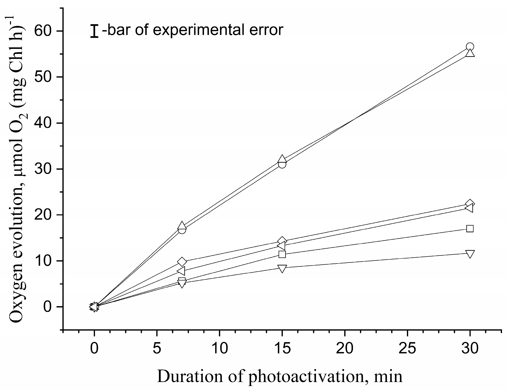
Figure 1. Dependence of the photoactivation of the O2-evolving activity in apo-WOC-PSII preparations on duration of photoactivation procedure. The procedure was performed in the absence (☐) and in the presence of 1 M sucrose (◯), 1 M trehalose (△), 1 M D-glucose (▽), 1 M betaine (◇) and 1 M Ect-OH (◁) in a medium containing 50 mM Mes-NaOH (pH 6.5), 35 mM NaCl, 50 mM CaCl2, 300 μM MnCl2 and 50 μM DCPIP at a chlorophyll concentration of 200 μg/mL. The light intensity (λ = 625 nm) during photoactivation was 35 μmol photon m−2 s−1. The measurements of the oxygen evolution rate in the samples were carried out in 50 mM Mes-NaOH (pH 6.5), 35 mM NaCl and 50 mM CaCl2 in the presence of exogenous electron acceptors −1 mM K3[Fe(CN)6] and 100 μM DCBQ at 20 μg Chl/mL under continuous illumination of the samples (λ > 650 nm, 1500 photon m−2 s−1). All experiments were repeated three times. Data are presented as mean values ± standard deviation.
Horticulturae 09 01339 g001
Figure 2. Dependence of the photoactivation of the O2-evolving activity in apo-WOC-PSII preparations on the concentration of MnCl2. Photoactivation was performed at different concentrations of MnCl2 in the absence (☐) and in the presence of 1 M sucrose (◯), 1 M trehalose (△), 1 M D-glucose (▽), 1 M betaine (◇) and 1 M Ect-OH (◁) in a medium containing 50 mM Mes-NaOH (pH 6.5), 35 mM NaCl, 50 mM CaCl2 and 50 μM DCPIP at a chlorophyll concentration of 200 μg/mL. The light intensity (λ = 625 nm) during 30 min photoactivation was 35 μmol photon m−2 s−1. The measurement of the rate of oxygen evolution in the samples was carried out in a medium containing 50 mM Mes-NaOH (pH 6.5), 35 mM NaCl and 50 mM CaCl2 in the presence of exogenous electron acceptors −1 mM K3[Fe(CN)6] and 100 μM DCBQ at 20 μg Chl/mL under continuous illumination of the samples (λ > 650 nm, 1500 μmol photon m−2 s−1). All experiments were repeated three times. Data are presented as mean values ± standard deviation.
Horticulturae 09 01339 g002
Figure 3. Dependence of the reactivation of photoinduced changes in the chlorophyll a fluorescence yield (Fv) on concentration of exogenous MnCl2. Fv reactivation was performed in the medium containing 50 mM Mes (pH 6.5), 35 mM NaCl in the absence of osmolyte (☐) and in the presence of 1 M sucrose (◯), 1 M trehalose (△), 1 M D-glucose (▽), 1 M betaine (◇) and 1 M Ect-OH (◁) at chlorophyll concentration of 10 μg/mL. All experiments were repeated three times. Data are presented as mean values ± standard deviation.
Horticulturae 09 01339 g003
Figure 4. Dependence of photoinduced Fv measured in the presence of 2 mM sodium ascorbate in apo-WOC-PSII preparations which were exposed to inhibitory light (λ = 625 nm, 15 μmol photon m−2 s−1) for 5 min in the presence of various concentration of MnCl2. Photoinhibition was performed in the medium containing 50 mM Mes (pH 6.5) and 35 mM NaCl at 20 μg Chl/mL in the absence (☐) and in the presence of 1 M sucrose (◯), 1 M trehalose (△), 1 M D-glucose (▽), 1 M betaine (◇), and 1 M Ect-OH (◁). The measurement of Fv was carried out in the medium containing 50 mM Mes (pH 6.5), 35 mM NaCl and 2 mM sodium ascorbate at 10 μg Chl/mL. The amplitude Fv measured in apo-WOC-PSII preparations in the presence of 2 mM sodium ascorbate before photoinhibition was taken as 100%. All experiments were repeated three times. Data are presented as mean values ± standard deviation.
Horticulturae 09 01339 g004
Figure 5. Dependence of O2-evolving activity in CaCl2-PSII preparations on time of incubation of preparation at 25 °C. The incubation was performed in the dark at 110 μg Chl/mL in the medium containing 50 mM Mes (pH 6.5), 35 mM NaCl and 20 mM Ca2+ (medium A) in the absence (☐) or in the presence of 1 M sucrose (◯), 1 M trehalose (△), 1 M D-glucose (▽), 1 M betaine (◇) and 1 M Ect-OH (◁). Light-induced oxygen evolution in the CaCl2-PSII preparations was measured in medium A in the presence of the electron acceptors (1 mM ferricyanide and 100 μM DCBQ) after 11 times dilutions of the samples. The rate of oxygen evolution in CaCl2-treated PSII preparations before the incubation (56 μmol O2 (mg Chl h)−1) was taken as 100%. All experiments were repeated three times. Data are presented as mean values ± standard deviation.
Horticulturae 09 01339 g005
Figure 6. Difference kinetics ∆WOJ and ∆WOI showing K-band (A) and J-band (B) formation in ChlF transients double-normalized between F0 and FJ phases, WOJ = (Ft − F0)/(FJ − F0), and between F0 and FI phases, WOI = (Ft − F0)/(FI − F0), induced by heat treatment of PSII membrane fragments. Heat treatment (HT, 30 °C, 40 min) was performed in darkness in the absence of osmolytes (1) and in the presence of 1 M sucrose (2), 1 M trehalose (3), 1 M betaine (4) and 1 M Ect-OH (5) in a medium containing 50 mM Mes (pH 6.5) and 35 mM NaCl at 10 μg Chl/mL. The presented kinetics are the differences ∆WOJ = [WOJ after HT − WOJ before HT] and ∆WOI = [WOI after HT − WOI before HT] of the average for 4 “before HT” and 5 “after HT” independent curves. In all experiments, Fv was induced by a single 880 ms saturating flash (λ = 625 nm, 2360 µmol photons m−2 s−1). Measurements were performed in a medium containing 50 mM Mes (pH 6.5) and 35 mM NaCl at 10 μg Chl/mL at room temperature, 25 °C.
Horticulturae 09 01339 g006
Figure 7. Changes in the relative velocity of ultrasound in the suspension containing photosystem II preparations with the native water-oxidizing complex during their incubation at 25 °C. Measurements were performed in the medium containing 50 mM Mes (pH 6.5) and 35 mM NaCl in the absence (A) and in the presence of 1 M sucrose (B), 1 M trehalose (C), 1 M D-glucose (D), 1 M betaine (E) and 1 M Ect-OH (F) at 200 μg Chl/mL and were repeated at least three times.
Horticulturae 09 01339 g007
Figure 8. Changes in the refractive index in the suspension containing photosystem II preparations with the native water-oxidizing complex in the absence (1) and in the presence of 1 M trehalose (2) during their incubation at 25 °C. Curve 3 reflects changes in the refractive index in 1 M trehalose solution without PSII preparations. Measurements were performed in the medium containing 50 mM Mes (pH 6.5) and 35 mM NaCl at 200 μg Chl/mL and were repeated at least three times.
Horticulturae 09 01339 g008
Figure 9. Changes in the relative velocity of ultrasound in the suspension containing photosystem II preparations with the native water-oxidizing complex (1), NaCl-PSII preparations (2) and CaCl2-PSII preparations (3) during their incubation at 25 °C. Measurements were performed in the medium containing 50 mM Mes (pH 6.5) and 35 mM NaCl at 200 μg Chl/mL and were repeated at least three times.
Horticulturae 09 01339 g009
Disclaimer/Publisher’s Note: The statements, opinions and data contained in all publications are solely those of the individual author(s) and contributor(s) and not of MDPI and/or the editor(s). MDPI and/or the editor(s) disclaim responsibility for any injury to people or property resulting from any ideas, methods, instructions or products referred to in the content.