Article

Establishment of an Efficient In Vitro Propagation Method for a Sustainable Supply of Plectranthus amboinicus (Lour.) and Genetic Homogeneity Using Flow Cytometry and SPAR Markers

by
Mohammad Faisal
* and
Abdulrahman A. Alatar
Department of Botany and Microbiology, College of Science, King Saud University, P.O. Box 2455, Riyadh 11451, Saudi Arabia
*
Author to whom correspondence should be addressed.
Horticulturae 2022, 8(8), 693; https://doi.org/10.3390/horticulturae8080693
Submission received: 23 June 2022 / Revised: 26 July 2022 / Accepted: 27 July 2022 / Published: 1 August 2022

Abstract

:
Plectranthus amboinicus (Lour.) Spreng is a medicinally important aromatic perennial herb used for the treatment of skin diseases, constipation, asthma, flu, fever, cough, and headache as well as a flavoring ingredient in traditional drinks, food, and meat stuffing. In this study, a high-performance in vitro propagation system of P. amboinicus through direct shoot organogenesis was developed using axillary node explants cultured on MS (Murashige and Skoog) medium augmented with 0.5, 2.5, 5.0, 7.5, and 10.0 µM of 6-benzyladenine (BA) or kinetin (Kin), alone or with 0.1, 0.5, 2.5, and 5.0 µM of indole-3-acetic acid (IAA) or α-naphthalene acetic acid (NAA). To optimize the regeneration potential of node explants, the effects of basal media strength and pH were also investigated. After 8 weeks of culture, explants cultured in full strength MS basal medium (pH 5.7) with 5.0 µM BA and 2.5 µM NAA exhibited the highest percentage (97.1%) of regeneration and the maximum number (19.3) of shoots per explant. Individual elongated shoots were rooted on half strength MS basal medium containing 0.25 µM indole 3-butyric acid (IBA) after 4 weeks of culture, producing 5.3 roots/shootlets with a root induction frequency of 93.7%. First time genetic stability of in vitro raised P. amboinicus plants was determined using SPAR markers, such as DAMD and ISSR, as well as flow cytometric tests, assuring the availability of authenticated raw materials for commercial production of the plant and its bioactive components.

1. Introduction

The Plectranthus genus is among the most prominent members of the Lamiaceae family. Plectranthus amboinicus (Lour.) Spreng, native to eastern and southern Africa, is an important medicinal succulent perennial herb which tends to be creeping or climbing; it has highly aromatic leaves with short erect hairs [1]. In folk medicine of different countries, it is used for the treatment of skin diseases, constipation, asthma, flu, fever, cough, and headache [2,3,4]. The high concentration of active compounds that are found in P. amboinicus is predominantly responsible for its varied therapeutic potential as well as its culinary uses. The plant’s raw leaves are eaten or added as a flavoring ingredient in traditional drinks, food, and meat stuffing [1,5]. Biochemical studies revealed that the essential oil extracted from this plant has high amounts of thymol [6], carvacrol [7], α-humulene, α-terpineol, β-caryophyllene, β-selinene, γ-terpinene, and p-cymene [4,8]. Previous literature revealed that this plant has several pharmacological effects, including antimicrobial [9], antibacterial [10,11], antifungal [8,12], and antiviral [13,14,15,16] activities. In addition to its antiepileptic [17], antitumorigenic [18,19], anti-inflammatory [20,21], and antioxidant [22,23] effects, P. amboinicus shows antagonistic activities against respiratory [24], cardiovascular [25], oral [26], digestive [27], and genitourinary [28] disorders. Furthermore, the leaf paste of this plant is effective in the treatment of wounds [29] and skin diseases [30,31].
Since 1960, tissue culture has been used as an alternative method for conventional propagation in many plant species, where a large number of plants can be produced in a relatively short time and small space from a small piece of plant tissue [32]. Culturing of plant cells and tissues in vitro has become a reliable and necessary technique for propagation, improvement, and mass-multiplication of several plant species [33]. Micropropagation has been used to propagate many species of Plectranthus, such as P. bourneae [34,35,36], P. edulis [37,38], P. amboinicus [39,40], P. barbatus [41], P. zeylanicus [42], and P. esculentus [43].
One of the major problems facing the micropropagation system is the occurrence of morphological, physiological, and molecular changes in in vitro regenerated plants [44]. Therefore, it is necessary to evaluate the genetic stability of the micropropagated plants to ensure their conformity with the donor plants. Polymerase chain reaction (PCR) based on SPAR (single primer amplification reaction) markers is one of the techniques used to analyze the genetic stability of micropropagated plants. Among SPAR markers, inter-simple sequence repeat (ISSR) and directed amplification of minisatellite-region DNA (DAMD) are successfully used to reveal the genetic homogeneity between micropropagated and donor plants in many plant species [33,45,46,47,48,49,50,51]. ISSR and DAMD techniques are cost-effective, quick, simple, avoid the use of radioactivity and DNA blotting, and are susceptible to automation [33,51,52]. In addition, genetic changes in regenerated plants can be detected by flow cytometry, which has been shown to be an efficient and reliable tool for estimation of ploidy level and genome size by calculating the nuclear DNA content [53]. The flow cytometry system has been used to evaluate the genetic fidelity of regenerated plants in many medicinal plants, including Bacopa monnieri [44], Ficus carica [54], Salix lapponum [55], and Brassica juncea [53].
The objective of this research was to develop an efficient and reproducible method for in vitro propagation of P. amboinicus from nodal explants for a sustainable large-scale production. Genetic homogeneity of micropropagated plantlets was assessed for the first time through DNA-based SPAR markers and flow cytometry to ensure the propagation and supply of true-to-type plantlets.

2. Materials and Methods

2.1. Shoot Materials and Explants Preparation

Shoots of the Plectranthus amboinicus were harvested during March–April 2021 from a plant maintained in the growth chamber at the Botany & Microbiology Department, King Saud University, which was full of healthy growth. The plant samples were identified with the help of a taxonomist in the department and verified at the department’s herbarium, where vouchers were deposited. The excised shoots were thoroughly cleaned in water with Tween-20 (2-3 drops) for a period of twenty minutes in order to eliminate any dust and dirt. After that, they were cut into pieces that were 2–3 cm in size and placed under running tap water for a period of 30 min. All the materials were moved into a biosafety laminar-air flow hood, where they were sterilized for three minutes with a freshly prepared 0.1 percent (w/v) mercury chloride (HgCl2) solution (Riedel-de Haan AG, Seelze, Germany) while being gently stirred. The sterilized shoots (nodal segments) were given a thorough washing in deionized ultra-pure water to eliminate any residues of the sterilant before being cut into pieces measuring 0.5–0.7 cm in length.

2.2. Culture Media and Growth Condition

The sterile shoot segments were grown in nutritional medium that included various concentrations and combinations of growth regulators. Murashige and Skoog [56] plant cell culture basal medium (MS; M5519, Sigma-Aldrich, Inc., St. Louis, MO, USA) was used in all assays; it was composed of macro and micro-salts, vitamins, 3 percent (w/v) sucrose (AppliChem GmbH, Darmstadt, Germany), and 0.8 percent (w/v) agar-agar (Avonchem Ltd., Wellington House, Cheshire, UK) and had a pH of 5.7. The pH of the MS basal was adjusted with 1 N aqueous solution of NaOH or HCl before steam sterilization in an ALP-autoclave (CLG-32L, ALP Co., Tokyo, Japan) for 20 min at 121 °C (15 psi). In a growth chamber (Conviron Adaptis-CMP6010, USA), all the cultured vials were incubated under 50 µmol m−2 s−1 l light illuminance provided by Philips 39-Watt T5 linear fluorescent tubes (F39T5/841/HO/ALTO, Philips, Amsterdam, The Netherlands) with a day–night photoperiod of 16/8 h and a temperature of 24 ± 2 °C.

2.3. Growth Regulators, Shoot Induction, and Proliferation

For shoot induction and proliferation, the sterilized nodal sections of P. amboinicus were cultivated on MS medium supplemented with 0, 0.5, 2.5, 5.0, 7.5, and 10.0 µM of 6-benzyladenine (BA; Duchefa Biochemie B.V., Haarlem, The Netherlands) or kinetin (Kin; Sigma-Aldrich Chemicals Co., MO, USA) at different concentrations, either individually or in combination with 0, 0.1, 0.5, 2.5., and 5.0 µM of auxins such as indole-3-acetic acid (IAA; Duchefa Biochemie B.V., Haarlem, The Netherlands) or α-naphthalene acetic acid (NAA; Duchefa Biochemie B.V., Haarlem, The Netherlands). The effects of the strength of the MS basal medium (one-quarter; one-third; half; and full strength) and various pH levels (4.7, 5.2, 5.7, and 6.2) on the in vitro morphogenic response of P. amboinicus nodal sections were also evaluated using optimum phytohormonal combinations and concentrations of BA (5.0 µM) and NAA (2.5 µM). In order to achieve a higher number of shoots from each nodal section, the responding plant materials were sub-cultured onto the fresh media every three weeks. After 8 weeks of in vitro growth and proliferation, data on the percentage of the explants’ regeneration as well as the number of axillary shoots per explant were recorded.

2.4. Rooting of Shootlets and Acclimation

Individually harvested, the in vitro proliferating shootlets of P. amboinicus with 2–3 pairs of substantially developed leaves were transplanted to a half strength MS basal medium supplemented with 0, 0.25, 0.5, 1.0, and 2.0 µM of indole 3-butyric acid (IBA) solidified with 0.8% (w/v) agar-agar and having a pH of 5.7. Data on the frequency of shootlets producing roots and the number of roots per shootlets were recorded after 4 weeks of transfer in rooting media.
For ex vitro acclimation, well-rooted plantlets were gently washed under the laboratory running water to remove the medium and agar residues. Then, the plantlets were transferred to pots containing sterile planting soil substrates (Planta-GuardTM, Germany). Maintaining the plantlets at high humidity for the first few days following transplanting is a critical factor for the survival of micropropagated plants. To retain humidity, the ex vitro transplanted plants were covered with clear polybags, which were removed after 4 weeks and irrigated with half strength MS basal medium containing macro and micro-salts devoid of vitamins. After 1 month, the percent survival of acclimated plants was calculated as follows:
Total   number   of   plants   survived Total   number   of   plants   trasnferred × 100  

2.5. Flow Cytometric Profile of Plants

Nuclei isolated from leaf samples (approximately 100 mg) of regenerated plants and donor plants of P. amboinicus were used to generate flow cytometric profiles. As previously reported by Galbraith et al. [57], the nuclei were isolated by cutting leaf samples with a new surgical blade in micro-Petri dishes (35 × 15 mm2) containing 2.0 mL of precooled isolation buffer (20 mM MOPS, 45 mM MgCl2, 30 mM sodium citrate, 0.1% (v/v) Triton X-100; pH 7.0). Using a double-layered nylon membrane of 30-micron thickness, the homogenates were filtered with the help of a micro syringe to eliminate any remaining leaf debris from the mixtures. The filtered suspensions were transferred to labelled Eppendorf tubes containing 2.5 μL of 10 mg/mL DNAse-free RNAse and incubated for 10 min. After 30 min of staining with 50 μgml−1 of propidium iodide (Sigma-Aldrich, Inc., St. Louis, MO, USA), the nuclei samples were passed and analyzed using a flow cytometry machine (Muse™ Cell Analyzer, Merck KGaA, Darmstadt, Germany).

2.6. DNA Extraction and Molecular Characterization

Genomic DNA was isolated from young leaves of the donor plant as well as micropropagated plants of P. amboinicus using the CTAB (cetyltrimethylammonium bromide) method [58]. Quantification and DNA purity was determined by a NanoDrop 200c Spectrophotometer (Thermo Fisher Scientific, Waltham, MA, USA). The DNA samples were diluted in Ultrapure Milli-Q water to a final concertation of 25 ng/µL and stored in a laboratory refrigerator at 4 °C until further use. Five DAMD (Directed amplifications of minisatellite-region DNA; Table 1) primers (Metabion International AG, Planegg, Germany) and nine ISSR (Inter-simple sequence repeat; Table 2) primers (GeneLink, Inc., Orlando, FL, USA) were used for PCR.
PCR amplification was carried out in 20 µL volumes containing 2.0 µL of 10× PCR buffer, 1.2 µL MgCl2 (25 mM), 0.4 µL dNTPs (10 mM), 1 µL (10 pmole) primers, 0.2 µL Taq polymerase (3 Unit), and 1.2 µL Template DNA (25/µL ng). Both DAMD and ISSR reactions were carried out with a Bio-Rad thermal cycler (T100; Bio-Rad Laboratories, Inc., Hercules, CL, USA) with initial denaturation for 2 min (1 cycle) at 94 °C, denaturation for 5 min (35 cycles) at 94 °C, annealing for 2 min at 47–57 °C, extension for 1 min at 72 °C, and a final extension for 7 min at 72 °C. The amplified DNA fragments were separated and resolved on 1.5% (w/v) agarose gel (Sigma Aldrich, Inc., St. Louis, MO, USA) containing 4 µL ethidium bromide (Sigma Aldrich, USA) in a horizontal electrophoresis system (Biometra, Göttingen, Germany) with 1× TBE (Tris-Boric acid-EDTA). The electrophoresis was run for 2 h and the DNA bands were visualized on a UV Gel-Documentation System (G:BOX F3, Syngene, Cambridge, UK). Each sample were run thrice and only intense and reproducible bands were scored.

2.7. Statistical Analysis

The experiments in this study were repeated thrice using a completely randomized design (CRD) with 20 explants per treatment, and the results were expressed as mean value and standard error (SE). One-way analysis of variance (ANOVA, San Francisco, CA, USA) with Tukey’s honestly significant difference test (Tukey’s HSD) analysis was performed for comparisons of significant differences between treatments at a p-value of less than 0.05. The data were processed and analyzed using IBM SPSS version 24 (SPSS Inc., Chicago, IL, USA) and the graphs were created using the Microsoft Excel by Microsoft for Windows for Mac.

3. Results

3.1. Effects of Cytokinin on Shoot Induction

Only 1–2 shoots were formed by the sterile nodal segments of P. amboinicus cultured on cytokinin-free medium, which were used as a control. Adding BA or Kin to the basal medium, on the other hand, significantly increased the number of shoots that sprouted from the cultivated shoot segments. Nodal segments of P. amboinicus respond quite differently in vitro depending on the cytokinin type and concentration used (Table 3). The cultures were carefully investigated at periodic intervals (sub-culture) to see if any morphogenic alterations in the nodal sections had occurred. It was determined that all tested concentrations of both BA and Kin cytokinins, i.e., 0.5, 2.5, 5.0, or 10 µM, enhanced the induction and proliferation of shoots. Within ten days of culture, the latent axillary bud swelled, and after four weeks, it differentiated into numerous microshoots (Figure 1A,B). In general, of the different concentrations of BA and Kin tested, the number of shootlets increased with increasing the concentration from 0.5 to 5.0 µM. In contrast, concentrations of BA and Kin in the media more than 5.0 µM resulted in fewer shoots with non-degenerative callusing (slight) from the cut ends of each explant. When compared to Kin, BA was shown to be more effective for the induction and proliferation of shootlets. The MS basal medium combined with 5.0 µM of BA exhibited the greatest number of 7.5 shootlets per explant in 79.3 percent of cultures.

3.2. Effects of Auxin with Optimal Concentration of BA

In another set of experiments, the effectiveness of the optimum concentration of BA in combination with concentrations of 0, 0.1, 0.5, 2.5, or 5.0 µM of auxins (i.e., IAA or NAA) for the induction and proliferation of shootlets was assessed (Table 4). The better multiplication response was obtained from a shoot section of P. amboinicus grown on medium with 5.0 µM BA and 2.5 µM NAA (Figure 1C). On this media, 97.1 percent of the explants generated 19.3 shootlets (Table 4). However, NAA concentrations over 2.5 µM, along with optimal BA concentration, reduced the frequency of shoots with non-degenerative callusing from the explants. When IAA was coupled with BA, it improved the morphogenic response but produced fewer shootlets than BA with NAA in the growth medium. The maximum number of shootlets, 15.3 and 12.2, was observed from each shoot section on 5.0 µM BA or Kin supplied medium containing 2.5 µM of IAA, respectively (Table 4).

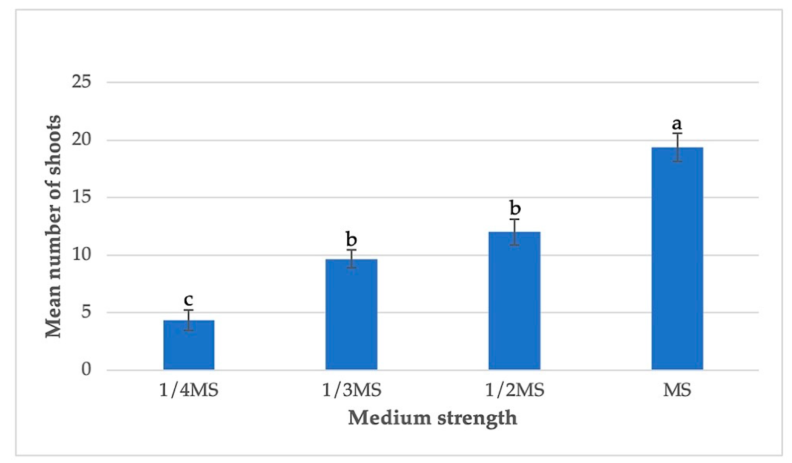
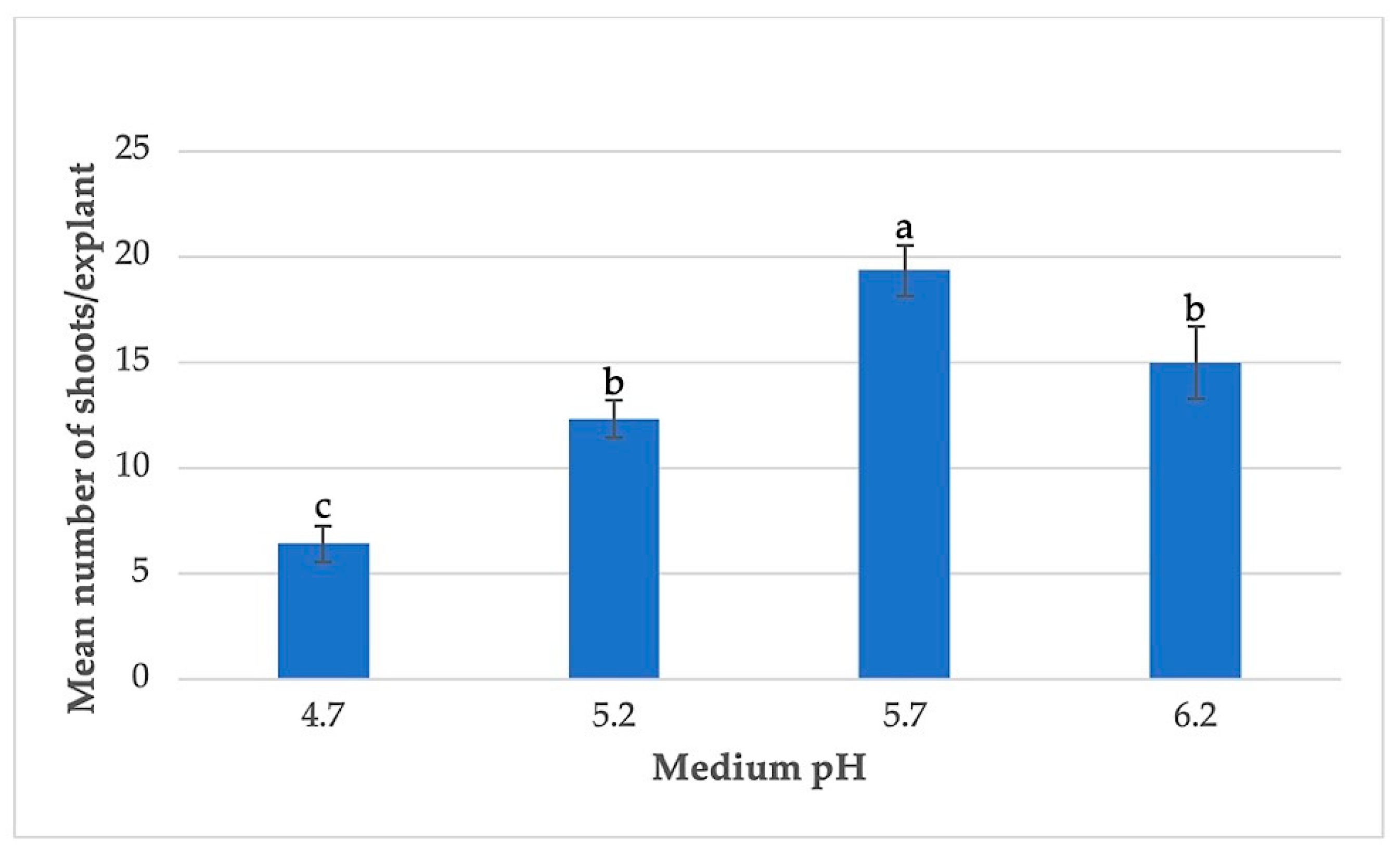
3.3. Effects of Basal Medium Strength and pH

In addition, varying strengths (one-quarter; one-third; half; and full MS) of basal medium were evaluated when combined with 5 µM BA and 2.5 µM NAA in order to get the best response for in vitro propagation and mass multiplication of P. amboinicus from nodal sections (Figure 2). Substantial differences were observed between the strengths of the medium used in this set of experiments. From the results, it was evidenced that the explants responded better in the MS basal medium supplied with the full strength of macro and micro-salts and vitamins. Shoot sections grown on MS basal media containing one-quarter macro and micro-salts as well as vitamins generated fewer shootlets. In subsequent studies, different pH values of MS basal medium (4.7, 5.2, 5.7, and 6.2) were employed to examine their influence on in vitro morphogenesis (Figure 3).
The pH 5.7 basal medium was shown to be optimal for the induction and proliferation of shoots, but the highly and severely acidic basal medium had a detrimental effect on in vitro shoot proliferation. The MS basal medium, which was supplied with full strength macro and micro-salts, vitamins, 5 µM BA, 2.5 µM NAA, and pH 5.7, provided the best response in terms of percent shoot induction (97.1) and number of shoots (19.3), according to the data collected throughout the investigation (Figure 3). The clusters of shootlets that formed were either sub-cultured onto new medium for further proliferation or individually transplanted into auxin-containing media for rhizogenesis.

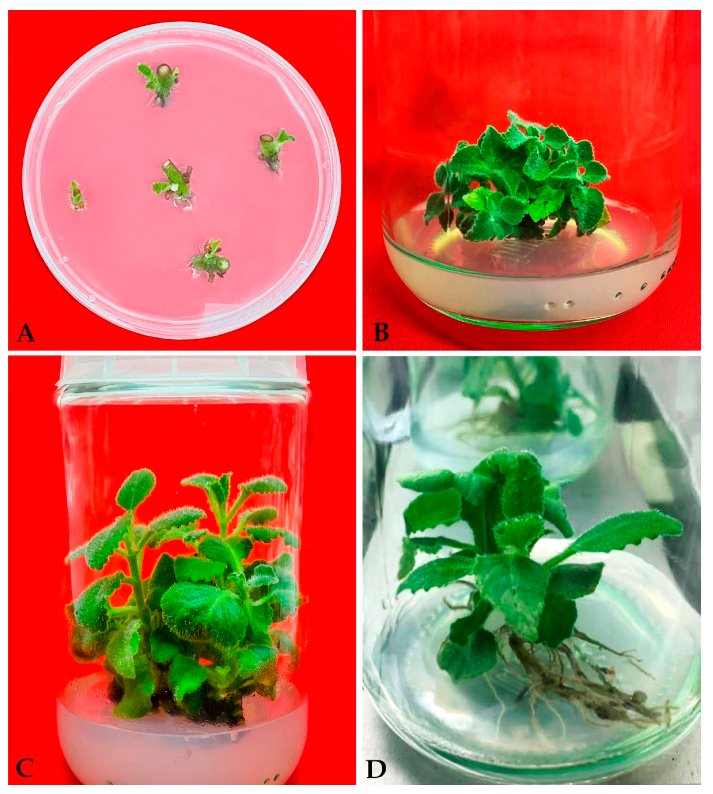
3.4. Rooting of Microshoots and Acclimation

For rooting, the individually harvested shootlets were transferred to MS basal medium containing an auxin such as IAA at concentration of 0, 0.25, 0.5, 1.0, and 2.0 µM (Table 5). Adding IBA to the basal medium was found to be effective for root induction from in vitro regenerated shootlets of P. amboinicus (Figure 1D). On medium containing 0.5 IBA, the shootlets responded best, yielding 5.3 roots/shootlets and with a root induction frequency of 93.7% in 4 weeks. For acclimatization, the individual plantlets with a complete root system were removed from semi-solid agar rooting media, shifted to pots containing sterile planting materials, and grown for weeks inside a growth chamber. After four weeks, the surviving pants were moved to field condition. Out of 50 in vitro-produced plantlets, 47 survived, resulting in a survival rate of 94 percent with no phenotypic changes between the plants.

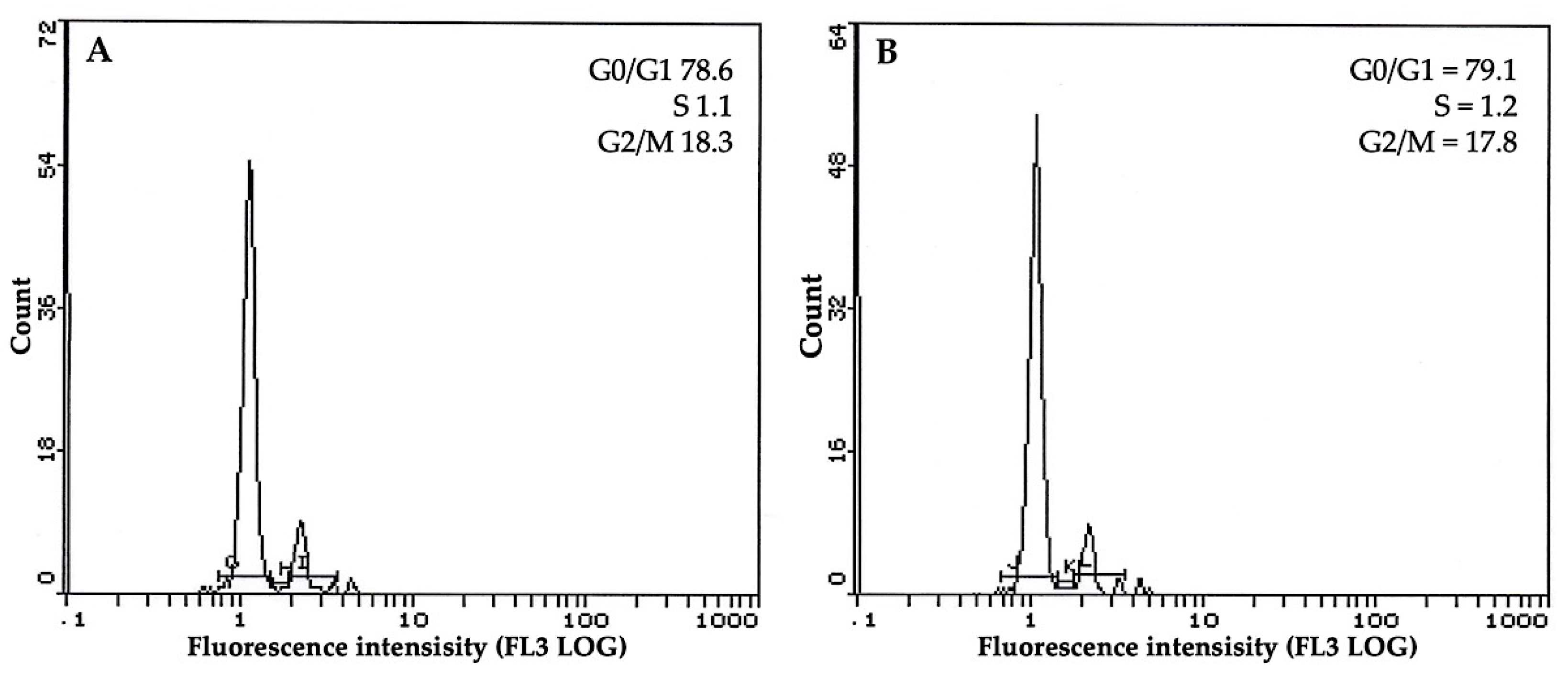
3.5. Flow Cytometric Profile of Plants

Flow cytometric examination of in vitro grown plants is a considerably faster and more reliable technique of ensuring ploidy genetic uniformity. Thus, comparative ploidy levels were determined in this study using a Muse Cell Analyzer in isolated nuclei from P. amboinicus micropropagated plants and a parent plant. The histograms produced from the nuclei of all plant sources, as previously stated, reveal a unimodal fluorescence peak of nuclear DNA. The cytometric profile indicated that all plant sources had nearly identical G0/G1 positions (Figure 4). As a result of the experimental findings, it was discovered that there were no differences between their usual fluorescence peak in the histogram of all plant sources, indicating that in vitro propagated P. amboinicus plants-maintained homogeneity of ploidy level, which ensures the successful clonal multiplication of this plant without compromising the genetic integrity.

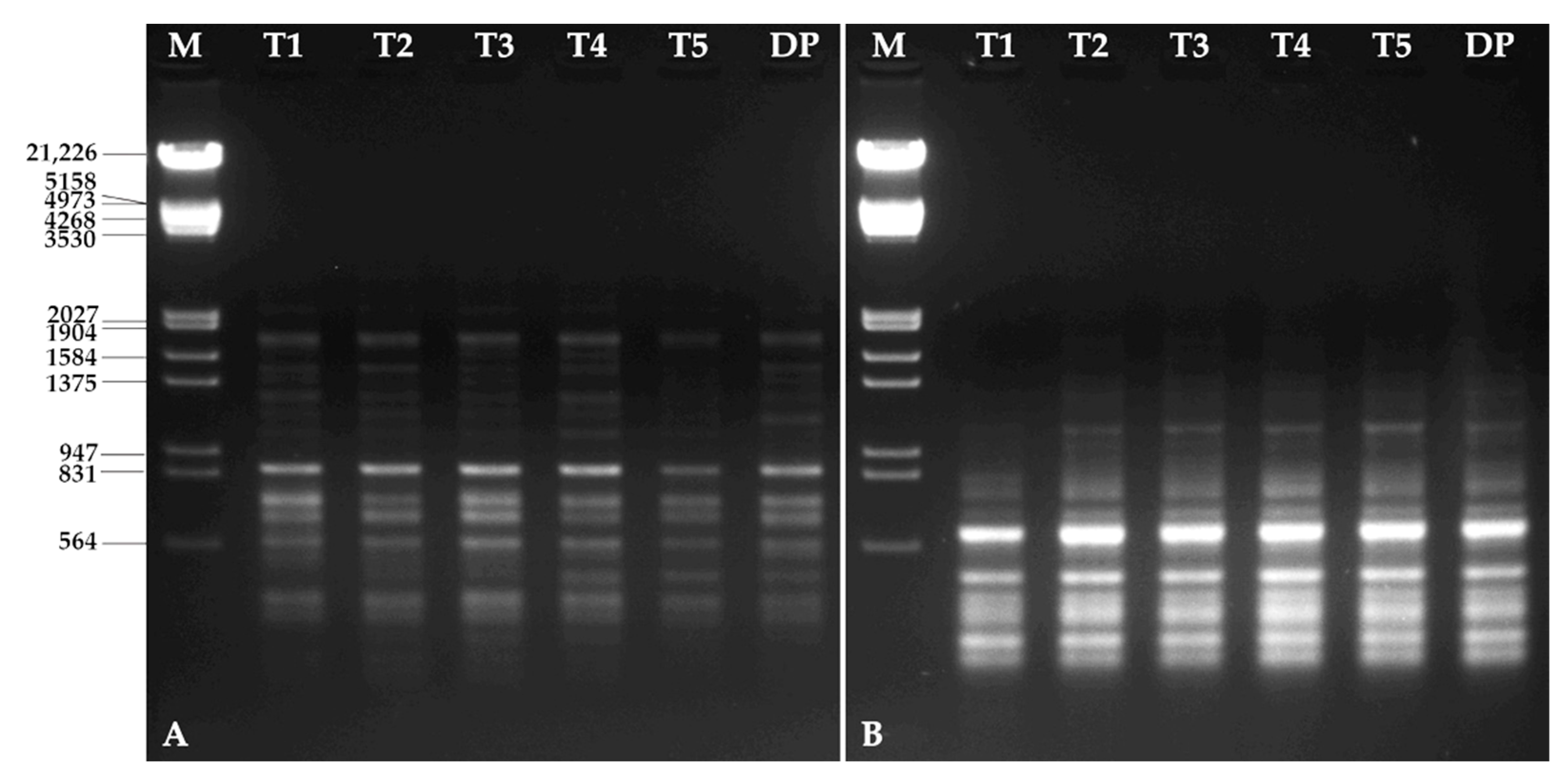
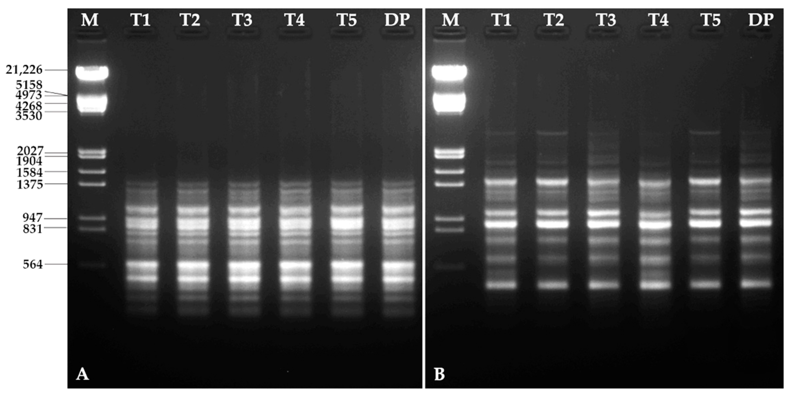
3.6. Molecular Characterization

DAMD and ISSR primers were used for molecular characterization of in vitro propagated plants growing in the greenhouse and compared with the DNA profile of the donor plant. Five DAMD primers were selected and screened out, where all the primers generated a total of 48 bands after PCR amplification with an average of 9.6 bands per DAMD primer (Table 1). The bands were monomorphic across all in vitro propagated plants of P. amboinicus and 100% similar to that of donor plants. The DNA banding profiles of the P. amboinicus genotypes using DAMD primers HVR and 33.6 are presented in Figure 5. Similarly, all nine ISSR primers tested also produced very clear, monomorphic bands within all in vitro propagated plants with a total of 150 scorable bands. The number of bands for each ISSR primer varied from 9 to 17 with an average of 11.4 bands per primer (Table 2, Figure 6).

4. Discussion

The most widely used approach for multiple shoot induction and plant regeneration is in vitro axillary bud proliferation, which is also thought to be the best way to ensure that the regenerated plants will have the same genetic makeup as the donor plants. Normally, dormant axillary buds are forced to develop into multiple shoots by proper application of growth regulators. Addition of cytokinins, viz., BA, Kin, TDZ, 2iP, and m-Topolin, in the medium led to the successful formation of multiple shoots in many plant species, including P. edulis [37,38], P. amboinicus [39,40], Ruta graveolens [33], Pongamia pinnata [59], Rumex pictus [60], Mentha × piperita [61], Atropa acuminata [62], Cannabis sativa [63], Campomanesia xanthocarpa [64], Zingiber officinale [65], Cicer arietinum [66,67], and Humulus lupulus [68].
The type of cytokinin used in the current research had a significant influence on the induction of multiple buds from nodal sections, with BA being shown to be more efficient than Kin. Apart from that, until BA concentration was optimized, both the regeneration frequency and the mean number of shoots generated per nodal sections continued to increase. Similar findings have been made for shoot organogenesis in a number of therapeutic plants in the past [69,70,71,72,73]. From the nodal explants of P. amboinicus, the concentration of BA at 5.0 µM produced the greatest number of shoots, compared to the other concentrations and PGR tested in this investigation. The current findings are comparable with those of Arumugam et al. [40] and Belete and Balcha [37], where 0.5 mg/L and 1.5 µM BA alone was proved to be more effective than kinetin for shoot induction in P. amboinicus and P. edulis, respectively. The presence of high BA in the culture medium, on the other hand, showed inhibitory effects on both shoot response and proliferation, which was consistent with the findings in Bambusa ventricose [74] and Sapium sebiferum [72].
The balance between auxin and cytokinin is critical during the whole micropropagation process, since the combination of the two regulates growth and development in a plant species. As a relatively high ratio of cytokinins is combined with a low ratio of auxins, they have a synergistic effect on cell division, resulting in a higher frequency of shoot bud induction and a greater number of shoots/explants when compared to cytokinin alone in the same experiment [75,76]. It is important to maintain a proper balance of plant growth regulators in the culture medium in combination with phytohormones produced by the plant when regenerating tissues in vitro, as this is one of the primary factors responsible for the induction, differentiation, and proliferation of shoots in the growth media. Similarly, in the present study, low auxin and high cytokinin concentrations also showed the greatest potential for inducing and multiplying shoots in P. amboinicus, and MS medium augmented with 5.0 µM BA in conjunction with 2.5 µM NAA was found to be the most effective treatments for direct shoot regeneration and multiplication in the microenvironment. In contrast, BA in combination with TDZ promoted a high number of shoots in P. bourneae [35]. The role of auxin and cytokinins in increasing axillary buds proliferation and ending apical dominance is thought to be the cause of the rise in the number of shoots observed in this investigation. The combination of auxin with cytokinin was also found to be effective for shoot multiplication and elongation in many plant species, including P. amboinicus [39,40], Cassia alata [77], Artemisia abrotanum [78], Syzygium cumini [79], Manihot esculenta [80], Hildegardia populifolia [81], Asparagus cochinchinensis [82], Basella rubra [83], and Sapium sebiferum [72].
The ability of microshoots to induce roots is critical because it has a direct impact on their survival in the greenhouse and their ability to adapt to their new environment. In the great majority of species, auxins are significant factors in root production because they stimulate the development of adventitious roots, which are essential for rooting [84]. The micropropagated shootlets of P. amboinicus were effectively rooted with IBA supplied in MS agar medium. The effects of IBA containing agar rooting media are in agreement with earlier approaches for inducing roots in P. amboinicus [39,40] and in several medicinal plants, including Ruta graveolens [33], Hemidesmus indicus [85], Rauvolfia tetraphylla [86], Sapium sebiferum [72], Artemisia vulgaris [87], and Asystasia gangetica [88].
Preserving the genetic consistency in tissue culture plants is critical for any micropropagation system prior to commercialization or in germplasm conservation. The PCR-based SPAR markers technique is one of the most significant approaches with increased use in the assessment of the genetic stability of tissue culture plants, since it uses only a small amount of genomic DNA, avoids the use of radioactivity and DNA blotting, and is susceptible to automation. In this study, DAMD and ISSR markers, as well flow cytometry, were used to confirm the genetic integrity of P. amboinicus micropropagated plants. Because micropropagation is known to induce somaclonal variation in micropropagated plants, the use of multiple markers has long been advocated for a better evaluation of genetic uniformity of in vitro plantlets [47]. Both DMAD and ISSR analyses showed a 100% monomorphic banding pattern, indicating the absence of variability among tissue culture plantlets of P. amboinicus. The appraisal of genetic constancy in a variety of micropropagated medicinal plants such as Mentha arvensis [46], Pittosporum eriocarpum [47], Bacopa monnieri [44], Curcuma zedoaria [48], Ruta chalepensis [50], Rauvolfia serpentina [89], Nepenthes khasiana [90] and Ficus carica [91] has been documented.

5. Conclusions

The present study describes a comprehensive and dependable approach for in vitro shoot regeneration, multiplication, and ex vitro establishment from axillary node explants of P. amboinicus, which will provide an alternative method for its mass multiplication and conservation as well as a source of material for commercial use and medicinal demands. For the first time, genetic homogeneity and true-to-type character of in vitro raised plants were confirmed by SPAR markers such as DAMD and ISSR, as well as flow cytometric analyses, ensuring the availability of legitimate raw materials for commercial production of the plant and its biologically active molecules.

Author Contributions

Conceptualization, M.F.; methodology, M.F. and A.A.A.; validation, M.F. and A.A.A.; formal analysis, M.F. and A.A.A.; investigation, M.F. and A.A.A.; resources, M.F. and A.A.A.; data curation, M.F.; writing—original draft preparation, M.F.; writing—review and editing, M.F. and A.A.A.; project administration, M.F. and A.A.A.; funding acquisition, M.F. and A.A.A. All authors have read and agreed to the published version of the manuscript.

Funding

This study was supported by the Researchers Supporting Project (RSP-2021/86), King Saud University, Riyadh, Saudi Arabia.

Institutional Review Board Statement

Not applicable.

Informed Consent Statement

Not applicable.

Data Availability Statement

Not applicable.

Acknowledgments

The authors are thankful to the Researchers Supporting Project (RSP-2021/86), King Saud University, Riyadh, Saudi Arabia for financial support.

Conflicts of Interest

The authors declare no conflict of interest.

References

  1. Arumugam, G.; Swamy, M.K.; Sinniah, U.R. Plectranthus amboinicus (Lour.) Spreng: Botanical, Phytochemical, Pharmacological and Nutritional Significance. Molecules 2016, 21, 369. [Google Scholar] [CrossRef]
  2. Lukhoba, C.W.; Simmonds, M.S.J.; Paton, A.J. Plectranthus: A review of ethnobotanical uses. J. Ethnopharmacol. 2006, 103, 1–24. [Google Scholar] [CrossRef]
  3. Hsu, K.-P.; Ho, C.-L. Antimildew Effects of Plectranthus amboinicus Leaf Essential Oil on Paper. Nat. Prod. Commun. 2019, 14, 1–6. [Google Scholar] [CrossRef]
  4. Senthilkumar, A.; Venkatesalu, V. Chemical composition and larvicidal activity of the essential oil of Plectranthus amboinicus (Lour.) Spreng against Anopheles stephensi: A malarial vector mosquito. Parasitol. Res. 2010, 107, 1275–1278. [Google Scholar] [CrossRef]
  5. Khare, R.S.; Kundu, S. Coleus aromaticus Benth-A Nutritive Medicinal Plant Of Potential Therapeutic Value. Int. J. Pharma Bio. Sci. 2011, 2, 488–500. [Google Scholar]
  6. Menéndez Castillo, R.A.; Pavón González, V. Plecthranthus amboinicus (Lour.) spreng. Rev. Cuba. Plantas Med. 1999, 4, 110–115. [Google Scholar]
  7. Singh, G.; Singh, O.P.; Prasad, Y.; De Lampasona, M.; Catalan, C. Studies on essential oils, Part 33: Chemical and insecticidal investigations on leaf oil of Coleus amboinicus Lour. Flavour Fragr. J. 2002, 17, 440–442. [Google Scholar] [CrossRef]
  8. Murthy, P.S.; Ramalakshmi, K.; Srinivas, P. Fungitoxic activity of Indian borage (Plectranthus amboinicus) volatiles. Food Chem. 2009, 114, 1014–1018. [Google Scholar] [CrossRef]
  9. Negi, P.S. Plant extracts for the control of bacterial growth: Efficacy, stability and safety issues for food application. Int. J. Food Microbiol. 2012, 156, 7–17. [Google Scholar] [CrossRef]
  10. Aguiar, J.J.S.; Sousa, C.P.B.; Araruna, M.K.A.; Silva, M.K.N.; Portelo, A.C.; Lopes, J.C.; Carvalho, V.R.A.; Figueredo, F.G.; Bitu, V.C.N.; Coutinho, H.D.M.; et al. Antibacterial and modifying-antibiotic activities of the essential oils of Ocimum gratissimum L. and Plectranthus amboinicus L. Eur. J. Integr. Med. 2015, 7, 151–156. [Google Scholar] [CrossRef]
  11. Vijayakumar, S.; Vinoj, G.; Malaikozhundan, B.; Shanthi, S.; Vaseeharan, B. Plectranthus amboinicus leaf extract mediated synthesis of zinc oxide nanoparticles and its control of methicillin resistant Staphylococcus aureus biofilm and blood sucking mosquito larvae. Spectrochim. Acta Part A Mol. Biomol. Spectrosc. 2015, 137, 886–891. [Google Scholar] [CrossRef]
  12. Oliveira, R.d.A.G.d.; Lima, E.d.O.; Souza, E.L.d.; Vieira, W.L.; Freire, K.R.; Trajano, V.N.; Lima, I.O.; Silva-Filho, R.N. Interference of Plectranthus amboinicus (Lour.) Spreng essential oil on the anti-Candida activity of some clinically used antifungals. Rev. Bras. Farmacogn. 2007, 17, 186–190. [Google Scholar] [CrossRef]
  13. Asiimwe, S.; Borg-Karlsson, A.-K.; Azeem, M.; Mugisha, K.M.; Namutebi, A.; Gakunga, N.J. Chemical composition and toxicological evaluation of the aqueous leaf extracts of Plectranthus amboinicus Lour. Spreng. Int. J. Pharm. Sci. Invent. 2014, 3, 19–27. [Google Scholar]
  14. Hattori, M.; Nakabayashi, T.; Lim, Y.A.; Miyashiro, H.; Kurokawa, M.; Shiraki, K.; Gupta, M.P.; Correa, M.; Pilapitiya, U. Inhibitory effects of various ayurvedic and Panamanian medicinal plants on the infection of herpes simplex virus-1 in vitro and in vivo. Phytother. Res. 1995, 9, 270–276. [Google Scholar] [CrossRef]
  15. Kusumoto, I.T.; Nakabayashi, T.; Kida, H.; Miyashiro, H.; Hattori, M.; Namba, T.; Shimotohno, K. Screening of various plant extracts used in ayurvedic medicine for inhibitory effects on human immunodeficiency virus type 1 (HIV-1) protease. Phytother. Res. 1995, 9, 180–184. [Google Scholar] [CrossRef]
  16. Hamidi, J.A.; Ismaili, N.; Ahmadi, F.; Lajisi, N.H. Antiviral and cytotoxic activities of some plants used in Malaysian indigenous medicine. Pertanika. J. Trop. Agric. Sci. 1996, 19, 129–136. [Google Scholar]
  17. Bhattacharjee, P.; Majumder, P. Investigation of phytochemicals and anti-convulsant activity of the plant Coleus amboinicus (Lour.). Int. J. Green Pharm. 2013, 7, 211. [Google Scholar]
  18. Gurgel, A.P.A.D.; da Silva, J.G.; Grangeiro, A.R.S.; Oliveira, D.C.; Lima, C.M.P.; da Silva, A.C.P.; Oliveira, R.A.G.; Souza, I.A. In vivo study of the anti-inflammatory and antitumor activities of leaves from Plectranthus amboinicus (Lour.) Spreng (Lamiaceae). J. Ethnopharmacol. 2009, 125, 361–363. [Google Scholar] [CrossRef]
  19. Ramalakshmi, P.; Subramanian, N.; Saravanan, R.; Mohanakrishnan, H.; Muthu, M. Anticancer effect of Coleus amboinicus (Karpooravalli) on human lung cancer cell line (A549). Int. J. Dev. Res. 2014, 4, 2442–2449. [Google Scholar]
  20. Silitonga, M.; Ilyas, S.; Hutahaean, S.; Sipahutar, H. Levels of apigenin and immunostimulatory activity of leaf extracts of bangunbangun (Plectranthus amboinicus Lour.). Int. J. Biol. 2015, 7, 46. [Google Scholar] [CrossRef]
  21. Chiu, Y.-J.; Huang, T.-H.; Chiu, C.-S.; Lu, T.-C.; Chen, Y.-W.; Peng, W.-H.; Chen, C.-Y. Analgesic and antiinflammatory activities of the aqueous extract from Plectranthus amboinicus (Lour.) Spreng. Both in vitro and in vivo. Evid. Based Complement. Alternat. Med. 2012, 2012, 508137. [Google Scholar] [CrossRef] [PubMed]
  22. Kumaran, A. Antioxidant and free radical scavenging activity of an aqueous extract of Coleus aromaticus. Food Chem. 2006, 97, 109–114. [Google Scholar] [CrossRef]
  23. Praveena, B.; Pradeep, S.N. Antioxidant and antibacterial activities in the leaf extracts of Indian borage (Plectranthus amboinicus). Food Sci. Nutr. 2012, 2012, 146–152. [Google Scholar] [CrossRef]
  24. Cartaxo, S.L.; Souza, M.M.d.A.; de Albuquerque, U.P. Medicinal plants with bioprospecting potential used in semi-arid northeastern Brazil. J. Ethnopharmacol. 2010, 131, 326–342. [Google Scholar] [CrossRef] [PubMed]
  25. Hole, R.C.; Juvekar, A.R.; Roja, G.; Eapen, S.; D’Souza, S.F. Positive inotropic effect of the leaf extracts of parent and tissue culture plants of Coleus amboinicus on an isolated perfused frog heart preparation. Food Chem. 2009, 114, 139–141. [Google Scholar] [CrossRef]
  26. Santos, F.A.V.; Serra, C.G.; Bezerra, R.J.A.C.; Figueredo, F.G.; Edinardo; Matias, F.F.; Menezes, I.R.A.; Costa, J.G.M.; Coutinho, H.D.M. Antibacterial activity of Plectranthus amboinicus Lour. (Lamiaceae) essential oil against Streptococcus mutans. Eur. J. Integr. Med. 2016, 8, 293–297. [Google Scholar] [CrossRef]
  27. Shubha, J.R.; Bhatt, P. Plectranthus amboinicus leaves stimulate growth of probiotic L. plantarum: Evidence for ethnobotanical use in diarrhea. J. Ethnopharmacol. 2015, 166, 220–227. [Google Scholar] [CrossRef]
  28. Patel, R.; Mahobia, N.K.; Gendle, R.; Kaushik, B.; Singh, S.K. Diuretic activity of leaves of Plectranthus amboinicus (Lour.) Spreng in male albino rats. Pharmacognosy Res. 2010, 2, 86–88. [Google Scholar] [CrossRef] [PubMed]
  29. Jain, A.K.; Dixit, A.; Mehta, S.C. Wound healing activity of aqueous extracts of leaves and roots of Coleus aromaticus in rats. Acta Pol. Pharm. 2012, 69, 1119–1123. [Google Scholar]
  30. Bhat, P.; Hegde, G.; Hegde, G.R. Ethnomedicinal practices in different communities of Uttara Kannada district of Karnataka for treatment of wounds. J. Ethnopharmacol. 2012, 143, 501–514. [Google Scholar] [CrossRef]
  31. Selvakumar, P.; naveena, B.E.; prakash, S.D. Studies on the antidandruff activity of the essential oil of Coleus amboinicus and Eucalyptus globulus. Asian Pac. J. Trop. Dis. 2012, 2, S715–S719. [Google Scholar] [CrossRef]
  32. Bhojwani, S.S.; Dantu, P.K. Micropropagation. In Plant Tissue Culture: An Introductory Text; Bhojwani, S.S., Dantu, P.K., Eds.; Springer: New Delhi, India, 2013. [Google Scholar] [CrossRef]
  33. Faisal, M.; Ahmad, N.; Anis, M.; Alatar, A.A.; Qahtan, A.A. Auxin-cytokinin synergism in vitro for producing genetically stable plants of Ruta graveolens using shoot tip meristems. Saudi J. Biol. Sci. 2018, 25, 273–277. [Google Scholar] [CrossRef] [PubMed]
  34. Sreedevi, E.; Anuradha, M.; Pullaiah, T. Plant regeneration from leaf-derived callus in Plectranthus barbatus Andr.[Syn.: Coleus forskohlii (Wild.) Briq.]. Afr. J. Biotechnol. 2013, 12, 2441–2448. [Google Scholar] [CrossRef]
  35. Thaniarasu, R.; Kumar, T.S.; Rao, M. In vitro Propagation of Plectranthus bourneae Gamble? An Endemic Red Listed Plant. Plant Tissue Cult. Biotechnol. 2015, 25, 273–284. [Google Scholar] [CrossRef]
  36. Thaniarasu, R.; Senthil Kumar, T.; Rao, M.V. Mass propagation of Plectranthus bourneae Gamble through indirect organogenesis from leaf and internode explants. Physiol. Mol. Biol. Plants 2016, 22, 143–151. [Google Scholar] [CrossRef]
  37. Belete, K.; Balcha, A. Micropropagation of Plectranthus edulis (Vatke) Agnew from shoot tip and nodal explants. Afr. J. Agric. Res. 2015, 10, 6–13. [Google Scholar] [CrossRef]
  38. Tsegaw, M.; Feyissa, T. Micropropagation of Plectranthus edulis (Vatke) Agnew from meristem cultur. Afr. J. Biotechnol. 2014, 13, 3682–3688. [Google Scholar] [CrossRef]
  39. Rahman, Z.A.; Noor, E.S.M.; Ali, M.S.M.; Mirad, R.; Othman, A.N. In Vitro Micropropagation of a Valuable Medicinal Plant, Plectranthus amboinicus. Am. J. Plant Sci. 2015, 6, 7. [Google Scholar] [CrossRef]
  40. Arumugam, G.; Sinniah, U.R.; Swamy, M.K.; Lynch, P.T. Micropropagation and essential oil characterization of Plectranthus amboinicus (Lour.) Sprengel, an aromatic medicinal plant. In Vitro Cell. Dev. Biol. Plant 2020, 56, 491–503. [Google Scholar] [CrossRef]
  41. Mahmoud, D.S.; Sayed, L.M.; Diab, M.; Fahmy, E.M. In Vitro Propagation Of Plectranthus barbatus Andrews As Important Medicinal Plant. Arab Univ. J. Agri. Sci. 2019, 27, 511–517. [Google Scholar] [CrossRef]
  42. Fonseka, D.; Wickramaarachchi, W.; Situge, C. Mass production of Plectranthus zeylanicus—A valuable medicinal and aromatic plant with a future value. Int. J. Minor. Fruits Med. Aromat. Plants 2019, 5, 15–20. [Google Scholar]
  43. Kujeke, G.T.; Chitendera, T.C.; Masekesa, R.T.; Mazarura, U.; Ngadze, E.; Rugare, J.T.; Matikiti, A. Micropropagation of Livingstone Potato (Plectranthus esculentus N.E.Br). Adv. Agric. 2020, 2020, 8364153. [Google Scholar] [CrossRef]
  44. Faisal, M.; Alatar, A.A.; El-Sheikh, M.A.; Abdel-Salam, E.M.; Qahtan, A.A. Thidiazuron induced in vitro morphogenesis for sustainable supply of genetically true quality plantlets of Brahmi. Ind. Crops Prod. 2018, 118, 173–179. [Google Scholar] [CrossRef]
  45. Faisal, M.; Alatar, A.A.; Ahmad, N.; Anis, M.; Hegazy, A.K. An Efficient and Reproducible Method for in vitro Clonal Multiplication of Rauvolfia tetraphylla L. and Evaluation of Genetic Stability using DNA-Based Markers. Appl. Biochem. Biotechnol. 2012, 168, 1739–1752. [Google Scholar] [CrossRef]
  46. Faisal, M.; Alatar, A.A.; Hegazy, A.K.; Alharbi, S.A.; El-Sheikh, M.; Okla, M.K. Thidiazuron induced in vitro multiplication of Mentha arvensis and evaluation of genetic stability by flow cytometry and molecular markers. Ind. Crops Prod. 2014, 62, 100–106. [Google Scholar] [CrossRef]
  47. Thakur, J.; Dwivedi, M.D.; Sourabh, P.; Uniyal, P.L.; Pandey, A.K. Genetic Homogeneity Revealed Using SCoT, ISSR and RAPD Markers in Micropropagated Pittosporum eriocarpum Royle- An Endemic and Endangered Medicinal Plant. PLoS ONE 2016, 11, e0159050. [Google Scholar] [CrossRef]
  48. Jena, S.; Ray, A.; Sahoo, A.; Sahoo, S.; Dash, B.; Kar, B.; Nayak, S. Rapid plant regeneration in industrially important Curcuma zedoaria revealing genetic and biochemical fidelity of the regenerants. 3 Biotech. 2019, 10, 17. [Google Scholar] [CrossRef]
  49. Rohela, G.K.; Jogam, P.; Bylla, P.; Reuben, C. Indirect Regeneration and Assessment of Genetic Fidelity of Acclimated Plantlets by SCoT, ISSR, and RAPD Markers in Rauwolfia tetraphylla L.: An Endangered Medicinal Plant. BioMed Res. Int. 2019, 2019, 3698742. [Google Scholar] [CrossRef]
  50. Qahtan, A.A.; Faisal, M.; Alatar, A.A.; Abdel-Salam, E.M. High-Frequency Plant Regeneration, Genetic Uniformity, and Flow Cytometric Analysis of Regenerants in Rutachalepensis, L. Plants 2021, 10, 2820. [Google Scholar] [CrossRef]
  51. Sirikonda, A.; Jogam, P.; Ellendula, R.; Kudikala, H.; Mood, K.; Allini, V.R. In vitro micropropagation and genetic fidelity assesment in Flemingia macrophylla (Willd.) Merr: An ethnomedicinal plant. Vegetos 2020, 33, 286–295. [Google Scholar] [CrossRef]
  52. Thakur, M.; Rakshandha; Sharma, V.; Chauhan, A. Genetic fidelity assessment of long term in vitro shoot cultures and regenerated plants in Japanese plum cvs Santa Rosa and Frontier through RAPD, ISSR and SCoT markers. S. Afr. J. Bot. 2021, 140, 428–433. [Google Scholar] [CrossRef]
  53. Faisal, M.; Abdel-Salam, E.M.; Alatar, A.A.; Qahtan, A.A. Induction of somatic embryogenesis in Brassica juncea L. and analysis of regenerants using ISSR-PCR and flow cytometer. Saudi, J. Biol. Sci. 2021, 28, 1147–1153. [Google Scholar] [CrossRef] [PubMed]
  54. Abdolinejad, R.; Shekafandeh, A.; Jowkar, A.; Gharaghani, A.; Alemzadeh, A. Indirect regeneration of Ficus carica by the TCL technique and genetic fidelity evaluation of the regenerated plants using flow cytometry and ISSR. Plant. Cell Tissue Organ. Cult. 2020, 143, 131–144. [Google Scholar] [CrossRef]
  55. Parzymies, M.; Pogorzelec, M.; Głębocka, K.; Śliwińska, E. Genetic Stability of the Endangered Species Salix lapponum L. Regenerated In Vitro during the Reintroduction Process. Biology 2020, 9, 378. [Google Scholar] [CrossRef] [PubMed]
  56. Murashige, T.; Skoog, F. A revised medium for rapid growth and bio assays with tobacco tissue cultures. Physiol. Plant. 1962, 15, 473–497. [Google Scholar] [CrossRef]
  57. Galbraith, D.W. Simultaneous flow cytometric quantification of plant nuclear DNA contents over the full range of described angiosperm 2C values. Cytometry A 2009, 75, 692–698. [Google Scholar] [CrossRef]
  58. Doyle, J.J.; Doyle, J.L. A rapid DNA isolation procedure for small quantities of fresh leaf tissue. Phytochem. Bull. 1987, 19, 11–15. [Google Scholar]
  59. Tan, S.N.; Tee, C.S.; Wong, H.L. Multiple shoot bud induction and plant regeneration studies of Pongamia pinnata. Plant. Biotechnol. 2018, 35, 325–334. [Google Scholar] [CrossRef]
  60. El-Shafey, N.; Sayed, M.; Ahmed, E.; Hammouda, O.; Khodary, S.E. Effect of growth regulators on micropropagation, callus induction and callus flavonoid content of Rumex pictus Forssk. Egypt, J. Bot. 2019, 59, 293–302. [Google Scholar] [CrossRef]
  61. Vaidya, B.N.; Asanakunov, B.; Shahin, L.; Jernigan, H.L.; Joshee, N.; Dhekney, S.A. Improving micropropagation of Mentha × piperita L. using a liquid culture system. In Vitro Cell. Dev. Biol. Plant 2019, 55, 71–80. [Google Scholar] [CrossRef]
  62. Rajput, S.; Agrawal, V. Micropropagation of Atropa acuminata Royle ex Lindl. (a critically endangered medicinal herb) through root callus and evaluation of genetic fidelity, enzymatic and non-enzymatic antioxidant activity of regenerants. Acta Physiol. Plant. 2020, 42, 160. [Google Scholar] [CrossRef]
  63. Wróbel, T.; Dreger, M.; Wielgus, K.; Słomski, R. Modified Nodal Cuttings and Shoot Tips Protocol for Rapid Regeneration of Cannabis sativa L. J. Nat. Fibers 2022, 19, 536–545. [Google Scholar] [CrossRef]
  64. Machado, J.S.; Degenhardt, J.; Maia, F.R.; Quoirin, M. Micropropagation of Campomanesia xanthocarpa O. Berg (Myrtaceae), a medicinal tree from the Brazilian Atlantic Forest. Trees 2020, 34, 791–799. [Google Scholar] [CrossRef]
  65. Zahid, N.A.; Jaafar, H.Z.E.; Hakiman, M. Micropropagation of Ginger (Zingiber officinale Roscoe) ‘Bentong’ and Evaluation of Its Secondary Metabolites and Antioxidant Activities Compared with the Conventionally Propagated Plant. Plants 2021, 10, 630. [Google Scholar] [CrossRef]
  66. Kumari, P.; Singh, S.; Yadav, S.; Tran, L.S.P. Pretreatment of seeds with thidiazuron delimits its negative effects on explants and promotes regeneration in chickpea (Cicer arietinum L.). Plant Cell Tissue Organ. Cult. (PCTOC) 2018, 133, 103–114. [Google Scholar] [CrossRef]
  67. Kumari, P.; Singh, S.; Yadav, S.; Tran, L.-S.P. Influence of different types of explants in chickpea regeneration using thidiazuron seed-priming. J. Plant Res. 2021, 134, 1149–1154. [Google Scholar] [CrossRef] [PubMed]
  68. Clapa, D.; Hârța, M. Establishment of an Efficient Micropropagation System for Humulus lupulus L. cv. Cascade and Confirmation of Genetic Uniformity of the Regenerated Plants through DNA Markers. Agronomy 2021, 11, 2268. [Google Scholar] [CrossRef]
  69. Faisal, M.; Ahmad, N.; Anis, M. An efficient micropropagation system for Tylophora indica: An endangered, medicinally important plant. Plant. Biotechnol. Rep. 2007, 1, 155–161. [Google Scholar] [CrossRef]
  70. Sahoo, N.; Thirunavoukkarasu, M.; Behera, P.R.; Satpathy, G.B.; Panda, P.K. Direct shoot organogenesis from hypocotyl explants of J. atropha curcas L. an important bioenergy feedstock. GCB Bioenergy 2012, 4, 234–238. [Google Scholar] [CrossRef]
  71. Panigrahi, J.; Gantait, S.; Patel, I.C. An Efficient In Vitro Approach for Direct Regeneration and Callogenesis of Adhatoda vasica Nees, a Potential Source of Quinazoline Alkaloids. Natl. Acad. Sci. Lett. 2017, 40, 319–324. [Google Scholar] [CrossRef]
  72. Jinyan, H. A high throughput plant regeneration system from shoot stems of Sapium sebiferum Roxb., a potential multipurpose bioenergy tree. Ind. Crops Prod. 2020, 154, 112653. [Google Scholar] [CrossRef]
  73. Hussain, S.A.; Anis, M.; Alatar, A.A. Efficient In Vitro Regeneration System for Tecoma stans L., Using Shoot Tip and Assessment of Genetic Fidelity Among Regenerants. Proc. Indian Natl. Sci. Acad. (B Biol. Sci.) 2020, 90, 171–178. [Google Scholar] [CrossRef]
  74. Wei, Q.; Cao, J.; Qian, W.; Xu, M.; Li, Z.; Ding, Y. Establishment of an efficient micropropagation and callus regeneration system from the axillary buds of Bambusa ventricosa. Plant Cell Tissue Organ. Cult. 2015, 122, 1–8. [Google Scholar] [CrossRef]
  75. Fatima, N.; Ahmad, N.; Anis, M. Enhanced in vitro regeneration and change in photosynthetic pigments, biomass and proline content in Withania somnifera L. (Dunal) induced by copper and zinc ions. Plant. Physiol. Biochem. 2011, 49, 1465–1471. [Google Scholar] [CrossRef] [PubMed]
  76. Swain, D.; Lenka, S.; Hota, T.; Rout, G.R. Micro-propagation of Hypericum gaitii Haines, an endangered medicinal plants: Assessment of genetic fidelity. Nucleus 2016, 59, 7–13. [Google Scholar] [CrossRef]
  77. Ahmed, M.R.; Anis, M.; Alatar, A.A.; Faisal, M. In vitro clonal propagation and evaluation of genetic fidelity using RAPD and ISSR marker in micropropagated plants of Cassia alata L.: A potential medicinal plant. Agrofor. Syst. 2017, 91, 637–647. [Google Scholar] [CrossRef]
  78. Bolyard, M. In vitro regeneration of Artemisia abrotanum L. by means of somatic organogenesis. In Vitro Cell. Dev. Biol. Plant 2018, 54, 127–130. [Google Scholar] [CrossRef]
  79. Naaz, A.; Hussain, S.A.; Naz, R.; Anis, M.; Alatar, A.A. Successful plant regeneration system via de novo organogenesis in Syzygium cumini (L.) Skeels: An important medicinal tree. Agrofor. Syst. 2019, 93, 1285–1295. [Google Scholar] [CrossRef]
  80. Chinnadurai, V.; Viswanathan, P.; Kalimuthu, K.; Vanitha, A.; Ranjitha, V.; Pugazhendhi, A. Comparative studies of phytochemical analysis and pharmacological activities of wild and micropropagated plant ethanol extracts of Manihot esculenta. Biocatal. Agric. Biotechnol. 2019, 19, 101166. [Google Scholar] [CrossRef]
  81. Upadhyay, A.; Shahzad, A.; Ahmad, Z. In vitro propagation and assessment of genetic uniformity along with chemical characterization in Hildegardia populifolia (Roxb.) Schott & Endl.: A critically endangered medicinal tree. In Vitro Cell. Dev. Biol. Plant 2020, 56, 803–816. [Google Scholar] [CrossRef]
  82. Kim, Y.-G.; Okello, D.; Yang, S.; Komakech, R.; Rahmat, E.; Kang, Y. Histological assessment of regenerating plants at callus, shoot organogenesis and plantlet stages during the in vitro micropropagation of Asparagus cochinchinensis. Plant. Cell Tissue Organ. Cult. 2021, 144, 421–433. [Google Scholar] [CrossRef]
  83. Kumar, S.S.; Giridhar, P. In vitro micropropagation of Basella rubra L. through proliferation of axillary shoots. Plant Cell Tissue Organ Cult. 2021, 144, 477–483. [Google Scholar] [CrossRef]
  84. De Klerk, G.-J. Rooting of microcuttings: Theory and practice. In Vitro Cell. Dev. Biol. Plant 2002, 38, 415–422. [Google Scholar] [CrossRef]
  85. Yadav, V.; Shahzad, A.; Ahmad, Z.; Sharma, S.; Parveen, S. Synthesis of nonembryonic synseeds in Hemidesmus indicus R. Br.: Short term conservation, evaluation of phytochemicals and genetic fidelity of the regenerants. Plant. Cell Tissue Organ. Cult. 2019, 138, 363–376. [Google Scholar] [CrossRef]
  86. Hussain, S.A.; Ahmad, N.; Anis, M. Synergetic effect of TDZ and BA on minimizing the post-exposure effects on axillary shoot proliferation and assessment of genetic fidelity in Rauvolfia tetraphylla (L.). Rend. Lincei Sci. Fis. Nat. 2018, 29, 109–115. [Google Scholar] [CrossRef]
  87. Jogam, P.; Sandhya, D.; Shekhawat, M.S.; Alok, A.; Manokari, M.; Abbagani, S.; Allini, V.R. Genetic stability analysis using DNA barcoding and molecular markers and foliar micro-morphological analysis of in vitro regenerated and in vivo grown plants of Artemisia vulgaris L. Ind. Crops Prod. 2020, 151, 112476. [Google Scholar] [CrossRef]
  88. Dilkalal, A.; AS, A.; TG, U. In vitro regeneration, antioxidant potential, and genetic fidelity analysis of Asystasia gangetica (L.) T. Anderson. In Vitro Cell. Dev. Biol. Plant 2021, 57, 447–459. [Google Scholar] [CrossRef]
  89. Faisal, M.; Alatar, A.A.; Ahmad, N.; Anis, M.; Hegazy, A.K. Assessment of Genetic Fidelity in Rauvolfia serpentina Plantlets Grown from Synthetic (Encapsulated) Seeds Following In Vitro Storage at 4 °C. Molecules 2012, 17, 5050–5061. [Google Scholar] [CrossRef]
  90. Devi, S.P.; Kumaria, S.; Rao, S.R.; Tandon, P. Single primer amplification reaction (SPAR) methods reveal subsequent increase in genetic variations in micropropagated plants of Nepenthes khasiana Hook. f. maintained for three consecutive regenerations. Gene 2014, 538, 23–29. [Google Scholar] [CrossRef]
  91. Parab, A.R.; Lynn, C.B.; Subramaniam, S. Assessment of genetic stability on in vitro and ex vitro plants of Ficus carica var. black jack using ISSR and DAMD markers. Mol. Biol. Rep. 2021, 48, 7223–7231. [Google Scholar] [CrossRef]
Figure 1. (A) Initiation of shoots from axillary node explants of Plectranthus amboinicus. (B) Multiple shootlets of P. amboinicus after 4 weeks of culture. (C) Proliferated multiple shoots of P. amboinicus after 8 weeks of culture. (D) Rooted P. amboinicus shoot after 4 weeks of transfer on the rooting media.
Horticulturae 08 00693 g001
Figure 2. Effect of MS medium strength with optimized concentration of 6-benzyladenine (BA) on shoot regeneration from axillary node explants of Plectranthus amboinicus. Data are expressed as mean ± SE recorded after 8 weeks of culture on shoot induction medium. Bars denoted with different letters indicate significant differences among treatments according to Tukey’s honestly significant difference test (Tukey’s HSD) at a p-value of less than 0.05.
Horticulturae 08 00693 g002
Figure 3. Effect of different pH of the MS medium with optimized concentration of 6-benzyladenine (BA) on shoot regeneration from axillary node explants of Plectranthus amboinicus. Data are expressed as mean ± SE recorded after 8 weeks of culture on shoot induction medium. Bars denoted with different letters indicate significant differences among treatments according to Tukey’s honestly significant difference test (Tukey’s HSD) at a p-value of less than 0.05.
Horticulturae 08 00693 g003
Figure 4. Flow cytometric profiles of tissue culture raised plants (A) and donor plants (B) of Plectranthus amboinicus.
Horticulturae 08 00693 g004
Figure 5. DAMD-PCR profiles of tissue culture and donor plants of Plectranthus amboinicus: (A) profile generated using primer HVB5 and (B) profile generated using primer 33.6. Lane M = Lambda DNA/EcoRI+HindIII marker; lanes T1–T5 = randomly selected regenerated plants; lane DP = donor plant.
Horticulturae 08 00693 g005
Figure 6. ISSR-PCR profiles of tissue culture and donor plants of Plectranthus amboinicus: (A) profile generated using primer UBC827 and (B) profile generated using primer UBC855. Lane M = Lambda DNA/EcoRI+HindIII marker; lanes T1–T5 = randomly selected regenerated plants; lane DP = donor plant.
Horticulturae 08 00693 g006
Table 1. DAMD primers screening for the genetic stability of Plectranthus amboinicus.
PrimersSequence
(5′ → 3′)
Annealing Temperature (°C)Size Range (bp)Number of Bands
HBV3GCTCCTCCCCTCCT532500–5009
HBV5GGTGAAGCACAGGTG562000–30011
HVRGGTGTAGAGAGGGGT5020,000–40017
M13GAGGGTGGCGGTTCT5715001
33.6GGAGGTGGGCA473500–40010
Total =48
Average band per primer =9.6
Table 2. ISSR primers screening for the genetic stability of Plectranthus amboinicus.
PrimersSequence
(5′ → 3′)
Annealing Temperature (°C)Size Range
(bp)
Number of Bands
UBC-811(GA)8C493000–40011
UBC-825(AC)8T463000–50014
UBC-827(AC)8G503000–5009
UBC-834(AG)8YT502000–30010
UBC-841(GA)8YC503000–40011
UBC-855(AC)8YT5020,000–50012
UBC-866(CTC)6GT5520,000–50013
UBC-868(GAA)6465000–70012
UBC-880(GGAGA)3505000–80011
Total = 150
Average band per primer =11.4
Table 3. Effect of 6-benzyladenine (BA) and kinetin (Kin) on shoot regeneration from axillary node explants of Plectranthus amboinicus.
Plant Growth Regulators (µM)Regeneration %Shoot Number per Explant
BAKin
0.00.023.01 ± 1.15 f1.26 ± 0.15 g
0.5 46.67 ± 2.02 e2.13 ± 0.20 f
2.5 61.31 ± 2.31 cd4.73 ± 0.45 c
5.0 79.37 ± 2.30 a7.57 ± 0.30 a
7.5 70.66 ± 1.88 b5.06 ± 0.35 c
10 62.10 ± 1.51 cd3.23 ± 0.15 e
0.541.10 ± 1.73 e2.03 ± 0.14 f
2.555.33 ± 1.20 d4.13 ± 0.21 cde
5.069.43 ± 2.61 bc6.36 ± 0.26 b
7.562.00 ± 1.73 cd4.27 ± 0.15 cd
1042.67 ± 1.45 e3.37 ± 0.23 de
Data are expressed as mean ± SE recorded after 8 weeks of culture on shoot induction medium. Different letters in each column indicate significant differences among treatments according to Tukey’s honestly significant difference test (Tukey’s HSD) at a p-value of less than 0.05.
Table 4. Combined effect of 6-benzyladenine (BA) or kinetin (Kin) with α-naphthalene acetic acid (NAA) or indole-3-acetic acid (IAA) on shoot regeneration from axillary node explants of Plectranthus amboinicus.
Plant Growth Regulators (µM)Regeneration %Shoots Number per Explant
BAKinNAAIAA
5.0 79.37 ± 2.30 cde7.53 ± 0.50 gh
5.0 69.43 ± 2.61 ghi6.27 ± 0.46 h
5.0 0.1 81.01 ± 2.70 cde12.43 ± 1.14 cd
5.0 0.5 85.04 ± 2.01 bcd14.70 ± 1.15 bc
5.0 2.5 97.10 ± 2.16 a19.37 ± 1.21 a
5.0 5.0 72.76 ± 1.45 efgh7.97 ± 0.53 fgh
5.0 0.180.33 ± 2.53 cde11.43 ± 0.97 de
5.0 0.580.71 ± 2.04 cde10.63 ± 0.87 def
5.0 2.592.67 ± 3.10 ab15.36 ± 1.32 b
5.0 5.067.31 ± 1.65 hi6.98 ± 0.42 gh
5.00.1 77.67 ± 1.45 defg11.70 ± 0.75 de
5.00.5 85.10 ± 1.53 bcd12.20 ± 1.05 cd
5.02.5 90.43 ± 1.45 ab16.27 ± 1.16 b
5.05.0 70.31 ± 1.98 fgh7.01 ± 0.93 gh
5.0 0.173.44 ± 1.83 efgh9.30 ± 0.77 efg
5.0 0.580.11 ± 1.45 cde8.53 ± 0.50 fgh
5.0 2.588.33 ± 1.54 abc12.27 ± 0.67 cd
5.0 5.061.01 ± 1.73 i5.97 ± 0.56 h
Data are expressed as mean ± SE recorded after 8 weeks of culture on shoot induction medium. Different letters in each column indicate significant differences among treatments according to Tukey’s honestly significant difference test (Tukey’s HSD) at a p-value of less than 0.05.
Table 5. Effect of IBA on in vitro root induction from microshoots of Plectranthus amboinicus.
IBA (µM)Regeneration %Roots Number per Shootlet
0.036.67 ± 2.02 d0.73 ± 2.02 c
0.2593.76 ± 2.71 a5.30 ± 0.44 a
0.587.10 ± 2.33 a4.13 ± 0.45 ab
1.072.36 ± 2.30 b3.87 ± 0.30 b
2.060.50 ± 1.88 c1.06 ± 0.35 c
Data are expressed as mean ± SE recorded after 4 weeks of culture on shoot induction medium. Tukey’s honestly significant difference test (Tukey’s HSD) analysis was performed for comparisons of significant differences between treatments at a p-value of less than 0.05.
Publisher’s Note: MDPI stays neutral with regard to jurisdictional claims in published maps and institutional affiliations.

